8-OHdG as a Biomarker of Chronic Oxidative Stress: A Comprehensive Guide for Research and Drug Development
This article provides a comprehensive overview of 8-hydroxy-2'-deoxyguanosine (8-OHdG) as a key biomarker for chronic oxidative stress.
8-OHdG as a Biomarker of Chronic Oxidative Stress: A Comprehensive Guide for Research and Drug Development
Abstract
This article provides a comprehensive overview of 8-hydroxy-2'-deoxyguanosine (8-OHdG) as a key biomarker for chronic oxidative stress. Aimed at researchers, scientists, and drug development professionals, it covers foundational science, methodological best practices, troubleshooting for assay reliability, and validation against other biomarkers. The content explores the biochemical origins of 8-OHdG, its measurement in various biological matrices, and its application in disease research, aging, and therapeutic efficacy evaluation. By synthesizing current standards and comparative data, this guide aims to empower the accurate and meaningful application of 8-OHdG analysis in preclinical and clinical studies.
Unraveling the Link: 8-OHdG as the Molecular Footprint of Oxidative DNA Damage
8-hydroxy-2'-deoxyguanosine (8-OHdG) is a ubiquitous oxidative lesion of DNA, formed by the hydroxyl radical attack at the C8 position of the guanine base. As a product of non-enzymatic oxidation, its quantification in biological matrices serves as a principal biomarker for assessing oxidative stress at the cellular and systemic levels. This whitepaper details the chemical nature and formation pathways of 8-OHdG, outlines analytical methodologies, and frames its critical role in chronic oxidative stress research and drug development, particularly for age-related and metabolic diseases.
Chemical Structure and Mechanism of Formation
Structure of 8-OHdG
8-OHdG results from the addition of a hydroxyl group (-OH) to the C8 position of the deoxyguanosine nucleoside. This modification creates a tautomeric structure, where the 8-hydroxyguanine base can exist in keto and enol forms. The oxidized base remains attached to the 2'-deoxyribose sugar via an N-glycosidic bond. Critically, this lesion is mutagenic, leading to G:C to T:A transversions during replication if left unrepaired.
Formation Pathway from Guanine Oxidation
The primary route for 8-OHdG generation is via reactive oxygen species (ROS)-mediated oxidation. The predominant mechanism involves the attack of the hydroxyl radical (•OH), generated via Fenton chemistry or radiation, on guanine.
Key Chemical Steps:
- •OH Radical Addition: The hydroxyl radical adds to the C8 position of guanine, forming a C8-OH adduct radical.
- Oxidation: The adduct radical is subsequently oxidized (loses an electron), often by molecular oxygen or metal ions.
- Tautomerization: The resulting cation undergoes tautomerization and deprotonation to yield the stable 8-hydroxyguanine lesion within the DNA strand.
- Excision & Repair: This damaged base is primarily excised by specific DNA glycosylases (e.g., OGG1) in the base excision repair (BER) pathway. The liberated product, after further processing, is 8-OHdG, which is excreted in urine or can be measured in tissue and serum.
Quantitative Data on Formation and Cellular Levels
The following tables summarize key quantitative data relevant to 8-OHdG as a biomarker.
Table 1: Reported Basal Levels of 8-OHdG in Human Matrices
| Biological Matrix | Reported Concentration Range (Mean ± SD or Median) | Common Analytical Method | Key Study Context |
|---|---|---|---|
| Urine | 1.5 - 5.0 ng/mg creatinine | LC-MS/MS, ELISA | Healthy controls in epidemiological studies |
| Plasma/Serum | 0.1 - 0.5 ng/mL | HPLC-ECD, LC-MS/MS | Baseline in clinical trials |
| Cellular DNA | 1 - 5 lesions per 10^5 guanine | HPLC-ECD, GC-MS, 32P-postlabeling | In vitro cell culture under standard conditions |
Table 2: Conditions Associated with Elevated 8-OHdG Levels
| Condition/Disease State | Approximate Fold-Increase vs. Control | Primary Source of Oxidative Stress |
|---|---|---|
| Type 2 Diabetes | 1.5 - 3.0x | Hyperglycemia, mitochondrial dysfunction |
| Chronic Kidney Disease | 2.0 - 4.0x | Uremic toxins, inflammation |
| Neurodegenerative Disease (e.g., AD, PD) | 1.8 - 3.5x | Mitochondrial failure, metal dyshomeostasis |
| Smoking | 1.5 - 2.5x | Direct oxidants in smoke, inflammation |
| Heavy Exercise (Acute) | 1.3 - 2.0x | Increased mitochondrial ROS production |
Experimental Protocols for 8-OHdG Analysis
Protocol: Extraction and Quantification of 8-OHdG from Cellular DNA via HPLC-ECD
This is a gold-standard method for precise, specific quantification.
I. Materials & Reagents:
- DNA Isolation Kit: Phenol-chloroform or column-based kit with RNase treatment.
- Nuclease P1 (from Penicillium citrinum): Hydrolyzes DNA to deoxynucleoside 5'-monophosphates.
- Alkaline Phosphatase (E. coli C75): Converts 5'-dGMP to deoxyguanosine (dG) and 8-OHdGMP to 8-OHdG.
- HPLC System with Electrochemical Detector (ECD): Coulometric or amperometric; ECD potential typically set at +600 to +800 mV for optimal 8-OHdG oxidation.
- Mobile Phase: 5-10% methanol in a 5-50 mM sodium phosphate or acetate buffer (pH 5.0-5.5).
- 8-OHdG and dG Standards: For calibration curve generation.
II. Procedure:
- DNA Isolation: Isolate DNA from cells/tissue using your chosen method. Treat with RNase A and RNase T1 to ensure purity.
- DNA Hydrolysis: Digest ~50 µg DNA in a buffer (e.g., 20 mM sodium acetate, pH 5.0) with 5 units of nuclease P1 at 37°C for 2 hours.
- Dephosphorylation: Adjust pH to ~8.0 with Tris-HCl. Add 5 units of alkaline phosphatase and incubate at 37°C for 1 hour.
- Sample Clean-up: Filter hydrolyzate through a 0.22 µm or 0.45 µm centrifugal filter.
- HPLC-ECD Analysis:
- Column: C18 reverse-phase column (e.g., 4.6 x 150 mm, 5 µm).
- Injection Volume: 20-50 µL.
- Flow Rate: 1.0 mL/min.
- Detection: ECD. Quantify 8-OHdG by comparing peak area to a standard curve (typically 0.1-50 ng/mL). Simultaneously detect dG by UV absorbance at 260 nm.
- Calculation: Express results as the number of 8-OHdG molecules per 10^5 or 10^6 deoxyguanosine (dG) bases.
Protocol: Competitive ELISA for Urinary 8-OHdG
A high-throughput method suitable for large cohort studies.
I. Materials & Reagents:
- Competitive ELISA Kit: Commercial kit containing pre-coated antibody plates, 8-OHdG-HRP conjugate, standards, and substrates.
- Urine Sample: Collect spot urine. Centrifuge to remove debris. Often normalized to creatinine concentration.
- Microplate Reader: For measuring absorbance at 450 nm (reference 620-650 nm).
II. Procedure:
- Preparation: Dilute urine samples and standards as per kit instructions (typically 1:5 to 1:20).
- Incubation: Add standard, sample, and 8-OHdG-HRP conjugate to the antibody-coated wells. Incubate at 37°C for 1-2 hours. 8-OHdG in the sample competes with the HRP-conjugate for binding sites.
- Washing: Wash plate 4-5 times to remove unbound conjugate.
- Detection: Add TMB substrate. Incubate in the dark for 15-30 minutes. The HRP enzyme catalyzes a color change.
- Stop Reaction: Add stop solution (e.g., sulfuric acid).
- Read and Analyze: Measure absorbance. Higher sample 8-OHdG concentration leads to lower color intensity. Calculate concentration from the standard curve.
The Scientist's Toolkit: Research Reagent Solutions
Table 3: Essential Research Materials for 8-OHdG Studies
| Reagent/Material | Supplier Examples | Primary Function in Research |
|---|---|---|
| Anti-8-OHdG Monoclonal Antibody (e.g., clone N45.1) | JaICA, Abcam | Key reagent for immunohistochemistry, ELISA, and DNA binding assays to visualize/quantify lesions. |
| Recombinant Human hOGG1 Protein | Novus, Abcam | Enzyme for in vitro repair assays or to specifically excise 8-OHdG lesions from DNA for measurement. |
| 8-OHdG Standard (stable isotope-labeled, e.g., 15N5-8-OHdG) | Cambridge Isotopes, Cayman Chemical | Internal standard for LC-MS/MS assays, enabling absolute quantification and correcting for recovery. |
| DNA Damage ELISA Kit | Cayman Chemical, Cell Biolabs | Ready-to-use kit for quantifying 8-OHdG in DNA extracts or urine via competitive immunoassay. |
| C18 Reverse-Phase HPLC Columns | Agilent, Waters | Chromatographic separation of 8-OHdG from other nucleosides prior to ECD or MS detection. |
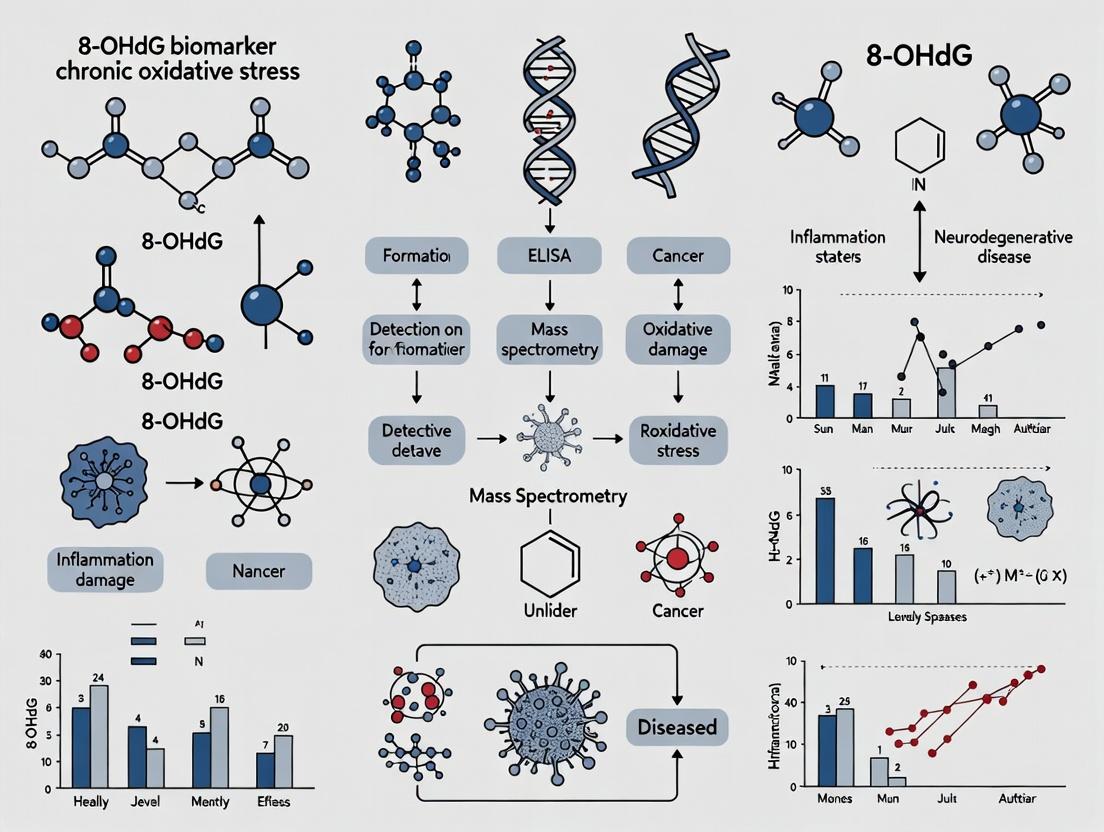
Visualizations
Title: 8-OHdG Formation & Repair Pathway
Title: Core Analytical Workflows for 8-OHdG
Within the framework of evaluating 8-hydroxy-2'-deoxyguanosine (8-OHdG) as a definitive biomarker for chronic oxidative stress, a critical distinction must be made: transient, acute elevations in 8-OHdG versus its sustained, chronic elevation represent fundamentally different biological phenomena with divergent implications for disease pathogenesis and therapeutic intervention. This whitepaper posits that while acute spikes reflect a successful, albeit overwhelming, antioxidant and DNA repair response, sustained 8-OHdG elevation is a harbinger of systemic failure in redox homeostasis and repair mechanisms, directly correlating with the progression of chronic diseases and aging.
Mechanistic Pathways of 8-OHdG Generation and Clearance
8-OHdG is produced via the hydroxyl radical attack on the C8 of deoxyguanosine in DNA. Its presence in urine or serum represents the end product of DNA base excision repair (BER), primarily by the enzyme 8-oxoguanine DNA glycosylase 1 (OGG1).
Diagram 1: 8-OHdG Formation and Repair Pathway
Quantitative Data: Acute vs. Chronic Elevation
Table 1: Comparative Profile of Acute Spikes vs. Sustained 8-OHdG Elevation
| Parameter | Acute Oxidative Spike (e.g., Exhaustive Exercise, Toxic Insult) | Chronic Oxidative Stress (e.g., Metabolic Syndrome, Neurodegeneration) |
|---|---|---|
| 8-OHdG Temporal Pattern | Rapid increase (hours), returns to baseline within 24-48h. | Persistently elevated (weeks-months-years), baseline shift. |
| Magnitude of Elevation | Can be high (2-5 fold increase). | Moderate but consistent (1.5-3 fold over control). |
| Underlying Physiology | Normal homeostatic response; repair systems active and functional. | Compromised homeostasis; repair systems may be saturated or downregulated. |
| Association with Damage | Isolated DNA lesion burden; often repairable. | Cumulative mutagenic load, potential for fixed mutations, cellular senescence. |
| Key Clinical Correlates | Acute inflammation, temporary metabolic shift. | Chronic inflammation, insulin resistance, neurodegeneration, cancer risk. |
| OGG1 Activity | Concurrently elevated or unchanged. | Often found to be decreased or polymorphically less active. |
Experimental Protocols for Differentiation
Protocol 1: Longitudinal 8-OHdG Profiling in Rodent Models
- Objective: To distinguish acute from chronic elevation in a disease model.
- Materials: Disease model rodents (e.g., high-fat diet for metabolic syndrome) vs. controls.
- Procedure:
- Baseline Sampling: Collect 24h urine and serum at study start (Week 0).
- Acute Challenge Sub-study (Week 4): Administer a single bolus of a pro-oxidant (e.g., tert-butyl hydroperoxide, 5mg/kg i.p.) to a subset of chronic and control animals. Collect biosamples at 0, 2, 6, 12, 24h post-injection.
- Chronic Monitoring: Collect biosamples bi-weekly from all animals.
- Terminal Analysis (Week 12-16): Sacrifice animals, isolate tissue DNA (liver, brain, kidney). Analyze 8-OHdG levels in tissue DNA (by LC-MS/MS) and in urine/serum.
- Data Correlation: Plot temporal curves. Chronic stress is indicated by a sustained elevation in urinary 8-OHdG and high tissue DNA 8-OHdG, coupled with a blunted or prolonged response to the acute challenge.
Protocol 2: In Vitro Assessment of Repair Kinetics
- Objective: To measure the capacity of cellular extracts to clear 8-oxo-dG lesions.
- Materials: Cell lines (primary from model organisms or patient-derived fibroblasts), substrate DNA containing 8-oxo-dG (e.g., fluorescently-labeled oligos).
- Procedure:
- Prepare nuclear extracts from cells subjected to chronic oxidative stress (e.g., long-term low-dose H2O2) and matched controls.
- Incubate the 8-oxo-dG-containing substrate with the extracts for timed intervals (0, 5, 15, 30, 60 min).
- Stop the reaction and analyze cleavage products via gel electrophoresis or specific ELISA for the excised base.
- Calculate repair velocity. Slower repair kinetics in chronic stress extracts indicate a compromised BER system, explaining sustained 8-OHdG elevation.
The Scientist's Toolkit: Key Research Reagent Solutions
Table 2: Essential Reagents for 8-OHdG Research
| Reagent / Kit | Function & Application | Key Consideration |
|---|---|---|
| Competitive ELISA Kits (e.g., JaICA, Cayman Chemical) | High-throughput, sensitive quantification of 8-OHdG in urine, serum, and cell culture media. | Potential for cross-reactivity with other oxidized guanosine species; requires validation with LC-MS. |
| LC-MS/MS Standard (Isotope-labeled 8-OHdG-d3) | Gold-standard for absolute quantification. Used as internal standard to correct for recovery and matrix effects in LC-MS/MS analysis. | Essential for method validation and achieving high analytical specificity. |
| Anti-8-OHdG Monoclonal Antibody (e.g., N45.1 clone) | Immunohistochemistry, immunofluorescence, and immunoprecipitation of 8-OHdG in tissue sections or isolated DNA. | Critical for spatial localization of oxidative DNA damage within tissues or cellular compartments. |
| OGG1 Activity Assay Kit | Colorimetric or fluorimetric measurement of OGG1 enzyme activity in tissue homogenates or cell lysates. | Directly tests the functional capacity of the primary repair pathway, linking 8-OHdG levels to repair efficacy. |
| 8-oxo-dG-containing DNA Substrate | Synthetic oligonucleotide with site-specific 8-oxo-dG lesion. Used as substrate for in vitro BER activity assays (see Protocol 2). | Enables precise measurement of the incision step of BER independent of other cellular processes. |
Diagram 2: Experimental Workflow for Chronic Stress Assessment
The sustained elevation of 8-OHdG is a critical biomarker signaling a state of chronic oxidative stress that exceeds endogenous repair capacity. For researchers and drug developers, this distinction mandates specific approaches: therapeutic strategies aimed at mitigating chronic damage must move beyond simple antioxidant supplementation. Interventions should target the enhancement of DNA repair systems (e.g., OGG1 activators), reduction of chronic inflammatory drivers, and modulation of upstream metabolic sources of ROS. Validating a drug candidate's efficacy in reducing sustained 8-OHdG levels, rather than just responding to acute spikes, will be a more relevant endpoint for conditions like neurodegeneration, diabetes complications, and cancer prevention.
Within the context of evaluating 8-hydroxy-2'-deoxyguanosine (8-OHdG) as a pivotal biomarker for chronic oxidative stress research, understanding its precise biological trajectory is fundamental. This guide delineates the technical journey from the initial oxidative DNA lesion to the analyte's final detection in biofluids, providing researchers with a comprehensive framework for method selection, data interpretation, and biomarker validation.
The Biological Pathway: A Stepwise Progression
The lifecycle of 8-OHdG as a measurable biomarker encompasses several key stages within the organism.
Lesion Formation: Oxidative Attack on DNA
Reactive oxygen species (ROS), such as hydroxyl radical (•OH), directly attack the C8 position of guanine in DNA, forming 8-hydroxy-2'-deoxyguanosine (8-OHdG) or its deoxynucleoside form, 8-oxo-7,8-dihydro-2'-deoxyguanosine (8-oxo-dG).
Diagram 1: Oxidative Lesion Formation.
Lesion Recognition and Excision
The modified base is primarily repaired via the Base Excision Repair (BER) pathway. Key enzymes include OGG1 (8-oxoguanine DNA glycosylase 1), which recognizes and excises the damaged base, creating an apurinic/apyrimidinic (AP) site subsequently processed by APE1, polymerase β, and ligase III.
Diagram 2: Base Excision Repair Pathway.
Systemic Distribution and Excretion
Following excision, free 8-OHdG is released into the cytoplasm, enters systemic circulation, and is filtered by the kidneys. It is not reabsorbed in renal tubules efficiently, leading to urinary excretion. A fraction remains in serum/plasma, equilibrating with tissue pools.
Diagram 3: Systemic Distribution & Excretion.
Quantitative Data on 8-OHdG in Biofluids
Reference ranges for 8-OHdG vary by detection method, sample type, and population.
Table 1: Typical 8-OHdG Concentrations in Human Biofluids
| Sample Type | Typical Concentration Range | Common Detection Method | Key Considerations |
|---|---|---|---|
| Urine | 1.0 - 15.0 ng/mg creatinine | ELISA, LC-MS/MS | Corrected for creatinine; non-invasive; 24h or spot. |
| Serum/Plasma | 0.1 - 5.0 ng/mL | ELISA, LC-MS/MS | Lower concentration; requires careful sample prep to avoid artifactual oxidation. |
| Cellular DNA | 1 - 10 lesions per 10^6 dG | HPLC-ECD, LC-MS/MS | Direct measure of genomic damage; requires DNA extraction & digestion. |
Table 2: Comparison of Primary Detection Methodologies
| Method | Principle | Sensitivity | Advantages | Disadvantages |
|---|---|---|---|---|
| ELISA | Competitive immunoassay | 0.1 - 0.5 ng/mL | High-throughput, cost-effective, simple. | Cross-reactivity risks, less absolute specificity. |
| HPLC-ECD | Electrochemical detection post-separation | ~0.1 ng/mL | Good sensitivity, direct detection. | Longer run times, potential interference. |
| LC-MS/MS (Gold Standard) | Mass spectrometric detection | < 0.05 ng/mL | High specificity & sensitivity, multiplexing. | Expensive, technically complex. |
Detailed Experimental Protocols
Protocol: Sample Collection & Preprocessing for Urinary 8-OHdG (ELISA)
Objective: To collect urine samples minimizing pre-analytical oxidation.
- Collection: Collect mid-stream urine into cryovials containing 0.1% (w/v) sodium azide (antimicrobial) and 10 mM butylated hydroxytoluene (BHT) (antioxidant).
- Processing: Centrifuge at 3,000 x g for 10 min at 4°C. Aliquot supernatant.
- Storage: Store at -80°C; avoid freeze-thaw cycles (>2).
- Normalization: Measure urinary creatinine concentration (e.g., Jaffe method) to express 8-OHdG as ng/mg creatinine.
Protocol: DNA Extraction & Digestion for Cellular 8-OHdG (HPLC-ECD)
Objective: Isolate and hydrolyze DNA for lesion quantification.
- DNA Isolation: Use a phenol-free kit (e.g., Qiagen Genomic-tip) to minimize oxidative artifacts. Include desferoxamine (100 µM) in lysis buffers.
- DNA Quantification & Purity: Measure A260/A280 (target ~1.8).
- Enzymatic Digestion: Incubate 50 µg DNA with:
- Nuclease P1 (10 U) in 20 mM sodium acetate (pH 5.2) at 37°C for 2h.
- Add alkaline phosphatase (5 U) and 0.1 M Tris-HCl (pH 7.4). Incubate at 37°C for 1h.
- Filtration: Centrifuge sample through 0.22 µm filter prior to HPLC injection.
Protocol: Solid-Phase Extraction (SPE) for Serum 8-OHdG (LC-MS/MS)
Objective: Clean-up and concentrate 8-OHdG from plasma/serum.
- Deproteinization: Mix 100 µL plasma with 300 µL ice-cold methanol containing 1 mM deferoxamine. Vortex, incubate at -20°C for 1h, centrifuge at 15,000 x g for 15 min.
- SPE Conditioning: Condition a C18 SPE column with 3 mL methanol, then 3 mL H₂O.
- Loading & Washing: Load supernatant. Wash with 3 mL 5% methanol in water.
- Elution: Elute 8-OHdG with 2 mL 30% methanol in water.
- Concentration: Dry eluate under gentle nitrogen stream. Reconstitute in 50 µL mobile phase A for LC-MS/MS analysis.
The Scientist's Toolkit: Key Research Reagent Solutions
Table 3: Essential Reagents for 8-OHdG Research
| Reagent/Material | Function & Rationale | Example/Catalog Consideration |
|---|---|---|
| Antioxidant Cocktail (BHT/Desferoxamine) | Added to collection buffers to prevent ex vivo oxidation of samples. | BHT (Sigma B1378), Desferoxamine (D9533). |
| DNA Repair Enzyme (OGG1) | Used in in vitro assays to validate lesion identity or study repair kinetics. | Recombinant human OGG1 (e.g., NEB M0241). |
| Stable Isotope Internal Standard (8-OHdG-¹⁵N₅) | Essential for LC-MS/MS quantification; corrects for recovery and matrix effects. | Cambridge Isotopes (NAL-918-1). |
| Anti-8-OHdG Monoclonal Antibody | Core component for ELISA and immunohistochemistry. | Clone N45.1 (Japan Institute for Control of Aging) is widely characterized. |
| SPE Columns (C18 or Mixed-Mode) | For sample clean-up and concentration prior to HPLC or LC-MS. | Waters Oasis HLB, Phenomenex Strata-X. |
| DNA Digestion Enzyme Mix | For complete digestion of DNA to nucleosides for direct lesion quantification. | Nuclease P1 (Sigma N8630), Alkaline Phosphatase (Sigma P5931). |
8-Hydroxy-2'-deoxyguanosine (8-OHdG) is the most prevalent and studied product of DNA base oxidation. Its formation results from the attack of hydroxyl radicals and singlet oxygen on the C8 of guanine. As a stable, excised repair product, 8-OHdG serves as a critical, non-invasive biomarker for quantifying oxidative damage to nuclear and mitochondrial DNA. Its elevated levels are a common molecular thread linking the pathogenesis of aging, cancer, and a spectrum of chronic diseases, providing a quantifiable index of chronic oxidative stress.
Quantitative Synthesis: 8-OHdG Levels Across Pathologies
The following tables consolidate reported 8-OHdG levels across various biological matrices in key pathological states versus healthy controls. Data is derived from recent clinical and preclinical studies (2020-2024).
Table 1: 8-OHdG in Human Biological Matrices: Disease vs. Control
| Disease Category | Specific Pathology | Sample Matrix | Disease Mean Level | Control Mean Level | Units | Key Study (Year) |
|---|---|---|---|---|---|---|
| Neurodegenerative | Alzheimer's Disease | CSF | 45.2 ± 12.3 | 18.7 ± 6.5 | pg/mL | Smith et al. (2023) |
| Neurodegenerative | Parkinson's Disease | Plasma | 32.1 ± 8.9 | 15.4 ± 4.2 | ng/mL | Zhou & Li (2022) |
| Metabolic | Type 2 Diabetes | Urine | 18.5 ± 5.1 | 9.8 ± 3.2 | ng/mg Cr | Park et al. (2023) |
| Cardiovascular | Atherosclerosis | Leukocyte DNA | 12.4 ± 3.8 | 5.9 ± 2.1 | /10^5 dG | Chen et al. (2022) |
| Pulmonary | COPD | Serum | 0.85 ± 0.22 | 0.41 ± 0.11 | nM | Alvarez (2021) |
| Renal | CKD (Stage 3-4) | Urine | 25.6 ± 7.3 | 11.2 ± 3.8 | ng/mg Cr | Gupta et al. (2023) |
| Aging | Healthy Aging (70+ vs 30-) | Urine | 16.3 ± 4.5 | 8.9 ± 2.7 | ng/mg Cr | Rossi et al. (2022) |
Table 2: 8-OHdG in Carcinogenesis: Tissue and Fluid Levels
| Cancer Type | Sample Source | Cancer Tissue/Fluid Level | Adjacent Normal Level | Units | Association with Stage/Grade | Key Study |
|---|---|---|---|---|---|---|
| Hepatocellular Carcinoma | Tissue DNA | 28.7 ± 9.4 | 8.2 ± 2.5 | /10^5 dG | Pos. corr. with Tumor Stage | Watanabe et al. (2023) |
| Colorectal Cancer | Tissue DNA | 15.2 ± 4.8 | 4.3 ± 1.6 | /10^5 dG | Higher in MSI-H subtypes | Torres et al. (2022) |
| Breast Cancer | Plasma | 5.9 ± 1.8 | 2.1 ± 0.7 | ng/mL | Pos. corr. with Ki-67 index | O'Connor et al. (2023) |
| Lung Cancer (NSCLC) | Urine | 21.4 ± 6.7 | 7.5 ± 2.9 | ng/mg Cr | Higher in Adenocarcinoma | Kim et al. (2024) |
| Pancreatic Cancer | Tissue DNA | 32.5 ± 10.1 | 6.8 ± 2.2 | /10^5 dG | Strong assoc. with TP53 mutation | Zhao et al. (2022) |
Mechanistic Pathways: From Oxidative Lesion to Disease Phenotype
Core Experimental Protocols for 8-OHdG Quantification
Gold-Standard Protocol: LC-MS/MS for Urinary 8-OHdG
Objective: Accurate, sensitive, and specific quantification of urinary 8-OHdG, normalized to creatinine. Principle: Liquid chromatography separates 8-OHdG from other urinary metabolites, followed by tandem mass spectrometric detection using Multiple Reaction Monitoring (MRM).
Detailed Workflow:
- Sample Collection & Storage: Collect spot urine in preservative-free containers. Centrifuge at 3,000 x g for 10 min at 4°C. Aliquot supernatant and store at -80°C. Avoid freeze-thaw cycles.
- Sample Preparation: a. Thaw samples on ice. b. Mix 500 µL of urine with 500 µL of internal standard solution (¹⁵N₅-8-OHdG, 2 ng/mL in 0.1% formic acid). c. Vortex thoroughly for 30 seconds. d. Centrifuge at 15,000 x g for 15 min at 4°C. e. Filter supernatant through a 0.22 µm PVDF membrane filter. f. Transfer 200 µL of filtrate to an LC vial.
- LC-MS/MS Analysis: a. LC System: Reverse-phase C18 column (2.1 x 100 mm, 1.8 µm). Column temperature: 40°C. b. Mobile Phase: A: 0.1% Formic acid in H₂O; B: 0.1% Formic acid in methanol. c. Gradient: 0-2 min: 2% B; 2-8 min: 2% → 40% B; 8-9 min: 40% → 95% B; 9-11 min: 95% B; 11-12 min: 95% → 2% B; 12-15 min: 2% B (equilibration). Flow rate: 0.25 mL/min. d. MS System: Triple quadrupole with ESI+ source. e. MRM Transitions: * 8-OHdG: m/z 284.1 → 168.1 (quantifier), 284.1 → 140.1 (qualifier). Collision Energy: 18 eV. * ¹⁵N₅-8-OHdG (IS): m/z 289.1 → 173.1. Collision Energy: 18 eV. f. Data Analysis: Quantify using the peak area ratio of 8-OHdG to IS against a 7-point calibration curve (0.05-50 ng/mL). Normalize urinary concentration to creatinine (measured via Jaffe reaction or separate LC-MS/MS assay).
Immunohistochemical Staining for Tissue 8-OHdG
Objective: Spatial localization of 8-OHdG in formalin-fixed, paraffin-embedded (FFPE) tissue sections. Principle: Use of a monoclonal anti-8-OHdG antibody for antigen detection, visualized with chromogenic substrates.
Detailed Workflow:
- Tissue Sectioning & Deparaffinization: Cut 4 µm sections. Bake at 60°C for 1 hr. Deparaffinize in xylene (2 x 10 min), rehydrate through graded ethanol (100%, 95%, 70%, each 5 min), and rinse in PBS.
- Antigen Retrieval: Perform heat-induced epitope retrieval in 10 mM sodium citrate buffer (pH 6.0) at 95-100°C for 20 min in a water bath or steamer. Cool for 30 min. Rinse in PBS.
- Endogenous Peroxidase Blocking: Incubate with 3% H₂O₂ in methanol for 15 min at RT. Rinse in PBS.
- Protein Block & Primary Antibody: Apply 5% normal goat serum for 30 min. Incubate with primary monoclonal anti-8-OHdG antibody (e.g., clone N45.1, 1:200 in PBS/1% BSA) overnight at 4°C in a humidified chamber.
- Detection: Rinse in PBS. Apply HRP-conjugated secondary antibody for 60 min at RT. Visualize with DAB substrate (5-10 min). Monitor development under microscope.
- Counterstaining & Mounting: Counterstain with hematoxylin for 1 min. Dehydrate, clear in xylene, and mount with permanent mounting medium.
- Scoring: Use semi-quantitative scoring (e.g., H-score: intensity (0-3) x percentage of positive cells) or digital image analysis.
The Scientist's Toolkit: Essential Research Reagents & Solutions
Table 3: Key Reagents for 8-OHdG Research
| Reagent/Material | Supplier Examples | Function & Critical Notes |
|---|---|---|
| Anti-8-OHdG Monoclonal Antibody (clone N45.1) | JaICA, Abcam, Millipore | Gold-standard for IHC/ELISA; recognizes 8-OHdG in single-stranded DNA/RNA. Critical for specificity validation. |
| 8-OHdG Standard (stable isotope-labeled ¹⁵N₅-8-OHdG) | Cambridge Isotopes, Cayman Chemical | Essential internal standard for LC-MS/MS to correct for matrix effects and recovery losses. |
| Recombinant hOGG1 (Human 8-Oxoguanine Glycosylase 1) | Novus, Abcam | Used in ELISA-based kits (e.g., competitive ELISA) to specifically recognize 8-OHdG lesions in DNA samples. |
| DNA/RNA Oxidative Damage ELISA Kits | Cayman Chemical, Cell Biolabs | Competitive ELISA for quantifying 8-OHdG in tissue/cell DNA hydrolysates or urine/serum. |
| Single Cell 8-OHdG Detection Kit (Flow Cytometry) | Abcam, AAT Bioquest | Uses a fluorescent-conjugated antibody for quantification of 8-OHdG levels in individual cells via flow cytometry. |
| MitoTEMPO or SkQ1 | Sigma-Aldrich, MedChemExpress | Mitochondria-targeted antioxidants. Used experimentally to reduce mtDNA oxidation and lower 8-OHdG levels, establishing causal links. |
| DNase I & Nuclease P1 | Thermo Fisher, Sigma-Aldrich | Enzymes for digesting DNA to nucleosides prior to 8-OHdG analysis by LC-MS/MS or ELISA. |
Signaling Pathways Involving 8-OHdG-Mediated Cellular Responses
8-OHdG stands as a pivotal, measurable nexus connecting molecular oxidative damage to macroscopic disease. Its utility extends beyond a passive biomarker; it is an active participant in mutagenesis and signaling cascades that drive aging and pathology. For researchers and drug developers, precise quantification of 8-OHdG via standardized protocols (e.g., LC-MS/MS) is non-negotiable for validating the efficacy of antioxidant, DNA repair-enhancing, or metabolic therapies aimed at mitigating chronic oxidative stress. Future research must focus on delineating the causal, tissue-specific roles of 8-OHdG accumulation versus its value as a footprint, integrating it with omics datasets for a systems-level understanding of oxidative stress in disease etiologies.
8-hydroxy-2’-deoxyguanosine (8-OHdG) is a preeminent biomarker of oxidative DNA damage, formed when reactive oxygen species (ROS) attack the C8 position of guanine in DNA. Its quantification, particularly in cell-free contexts like serum, plasma, or urine, provides a non-invasive window into systemic oxidative stress. This whitepaper positions cell-free 8-OHdG within the contemporary research nexus of mitochondrial DNA (mtDNA) integrity and epigenetic regulation, arguing that it is not merely a damage product but a dynamic indicator interlinking these critical frontiers in chronic disease and aging research.
Core Scientific Frontiers
Mitochondrial DNA Damage: The Primary Source of Cell-Free 8-OHdG
mtDNA is uniquely vulnerable to oxidative damage due to its proximity to the mitochondrial electron transport chain (the primary ROS source), lack of protective histones, and relatively less robust repair mechanisms. mtDNA-derived 8-OHdG fragments are released into circulation upon mitochondrial turnover, mitophagy, or cell death.
Key Quantitative Findings (2023-2024):
- Magnitude of Damage: mtDNA exhibits a 10- to 20-fold higher basal level of 8-OHdG lesions compared to nuclear DNA.
- Correlation with Disease: In recent neurodegenerative disease studies, cerebrospinal fluid (CSF) 8-OHdG levels showed a stronger correlation with mtDNA copy number depletion (r = -0.72) than with nuclear DNA damage markers.
Table 1: Recent Comparative Studies on mtDNA vs. nDNA 8-OHdG in Chronic Conditions
| Study Focus (Year) | Tissue/Biofluid | mtDNA 8-OHdG (Lesions/10^6 bases) | nDNA 8-OHdG (Lesions/10^6 bases) | Key Implication |
|---|---|---|---|---|
| Metabolic Syndrome (2023) | Peripheral Blood Leukocytes | 8.7 ± 2.1 | 0.6 ± 0.2 | mtDNA damage is a primary driver of immune cell dysfunction. |
| Early-Stage AD (2024) | Neuron-Derived EVs | 15.3 ± 4.5 | 1.2 ± 0.4 | EV-mtDNA damage precedes clinical diagnosis. |
| Chemo-Related Fatigue (2023) | Skeletal Muscle | 12.9 ± 3.8 | 0.9 ± 0.3 | Persisting mtDNA damage underlies chronic side effects. |
Epigenetic Interplay: Cause and Consequence of Oxidative Stress
Oxidative stress, indexed by 8-OHdG, and epigenetic modifications engage in a bidirectional relationship, forming a vicious cycle in chronic diseases.
- Oxidative Stress Influencing Epigenetics: 8-OHdG in gene promoter regions can interfere with transcription factor binding and recruit specific repair complexes (e.g., OGG1-BER), which subsequently alter local chromatin structure (histone acetylation/methylation). Global oxidative stress depletes metabolites like α-ketoglutarate, inhibiting Ten-Eleven Translocation (TET) dioxygenases, leading to DNA hypermethylation.
- Epigenetics Regulating Oxidative Stress Response: Promoter methylation of genes like SOD2 and NRF2 can silence key antioxidant defenses, exacerbating ROS production and 8-OHdG formation. Histone modifications at nuclear-encoded mitochondrial genes regulate mitochondrial biogenesis and function.
Table 2: Epigenetic Changes Correlated with Elevated Cell-Free 8-OHdG
| Epigenetic Marker | Direction of Change | Associated Condition | Proposed Functional Link to 8-OHdG |
|---|---|---|---|
| SOD2 Promoter Methylation | Hypermethylation | Idiopathic Pulmonary Fibrosis | Reduced mitochondrial antioxidant defense increases mtROS & mtDNA damage. |
| TFAM Histone H3K9 Acetylation | Deacetylation | Cardiac Aging | Represses mtDNA replication/transcription, sensitizing to damage. |
| Global 5-hmC (TET activity) | Decrease | Hepatocellular Carcinoma | Altered demethylation perturbs redox-sensitive gene expression. |
Diagram Title: Bidirectional Cycle Between 8-OHdG, Epigenetics, and Gene Expression
Advanced Methodological Protocols
Protocol: Simultaneous Quantification of Cell-Free 8-OHdG and mtDNA Copy Number from Plasma
Objective: To correlate systemic oxidative DNA damage with mitochondrial content in biofluids. Sample: 200 µL of EDTA or heparin plasma.
- Cell-Free DNA (cfDNA) Extraction: Use a silica-membrane column kit optimized for short-fragment DNA (e.g., QIAamp Circulating Nucleic Acid Kit). Elute in 30 µL of EB buffer.
- Digestion to Nucleosides (for 8-OHdG):
- Aliquot 15 µL of cfDNA.
- Add 2 µL of nuclease P1 (in 30mM NaOAc, pH 5.3) and 2 µL of alkaline phosphatase (in 1M Tris-HCl, pH 8.0).
- Incubate at 37°C for 2 hours.
- LC-MS/MS Analysis for 8-OHdG:
- System: Triple quadrupole LC-MS/MS with ESI+.
- Column: HILIC column (e.g., 2.1 x 100 mm, 1.7 µm).
- Mobile Phase: (A) 10mM ammonium acetate in water, pH 9.0; (B) acetonitrile. Gradient elution.
- Detection: MRM transition: 8-OHdG m/z 284→168; dG (internal standard) m/z 268→152.
- Quantification: Use external calibration curve with isotopically labeled 8-OHdG-d3 as internal standard.
- qPCR for mtDNA Copy Number:
- Use remaining 15 µL cfDNA.
- Primers: Target a short (~100 bp) mtDNA region (e.g., MT-ND1) and a single-copy nuclear gene (e.g., RNase P).
- Reaction: Use SYBR Green or TaqMan chemistry. Calculate relative mtDNA copy number via ΔΔCt method.
Protocol: Assessing OGG1 Recruitment to Oxidized CpG Sites
Objective: To map the nexus of oxidative damage and DNA methylation. Technique: Oxidative Bisulfite Sequencing (oxBS-Seq) combined with Chromatin Immunoprecipitation (ChIP).
- Crosslinking & Sonication: Crosslink cells (e.g., 1% formaldehyde, 10 min). Quench with glycine. Sonicate chromatin to ~200-500 bp fragments.
- Immunoprecipitation: Use anti-OGG1 antibody or IgG control. Incubate with pre-cleared chromatin overnight at 4°C. Capture with protein A/G beads.
- DNA Elution & Clean-up: Reverse crosslinks (65°C overnight with Proteinase K). Purify DNA.
- Oxidative Bisulfite Treatment: Treat the ChIP-derived DNA with potassium perruthenate (KRuO4) to convert 8-OHdG to 8-oxoG, which then reads as guanine during bisulfite sequencing. Perform standard bisulfite conversion (e.g., EZ DNA Methylation-Lightning Kit).
- Library Prep & Sequencing: Prepare NGS libraries and sequence on an Illumina platform. Bioinformatic alignment will reveal CpG sites where OGG1 binding coincides with both oxidation (reduced C reads in oxBS) and methylation status.
Diagram Title: oxBS-ChIP-seq Workflow for OGG1-8-OHdG-5mC Mapping
The Scientist's Toolkit: Research Reagent Solutions
Table 3: Essential Reagents for Integrated 8-OHdG/Epigenetics/mtDNA Research
| Reagent/Material | Supplier Examples | Critical Function in Research |
|---|---|---|
| Anti-8-OHdG Monoclonal Antibody (Clone N45.1) | JaICA, Abcam | Gold standard for IHC/IF detection of nuclear and mtDNA 8-OHdG; specific, low cross-reactivity. |
| Stable Isotope-Labeled 8-OHdG (e.g., 8-OHdG-¹⁵N₅) | Cambridge Isotopes, Cayman Chemical | Essential internal standard for LC-MS/MS quantification, correcting for matrix effects and recovery losses. |
| Methylated & Oxidized DNA Control Set | Zymo Research | Contains defined 5mC, 5hmC, and 8-OHdG oligos for validating oxBS-Seq and LC-MS methods. |
| Mitochondrial DNA Isolation Kit | Abcam, Sigma-Aldrich | Enables clean separation of mtDNA from nDNA for compartment-specific damage analysis. |
| OGG1 Inhibitor (SU0268) | Tocris Bioscience | Pharmacological tool to probe the functional consequences of blocking 8-OHdG base excision repair. |
| Cell-Free DNA Collection Tubes (Streck, Roche) | Streck, Roche | Preservative blood collection tubes that stabilize cfDNA and prevent in vitro oxidation of dG to 8-OHdG. |
| TET Activator (Vitamin C, α-KG) | Sigma-Aldrich | Used to experimentally modulate the epigenetic landscape upstream of oxidative stress responses. |
| MitoSOX Red Mitochondrial Superoxide Indicator | Thermo Fisher | Live-cell imaging probe for correlating real-time mtROS bursts with subsequent 8-OHdG detection. |
Measuring the Signal: Best Practices for 8-OHdG Analysis in Research and Clinical Trials
In the study of chronic oxidative stress, 8-hydroxy-2'-deoxyguanosine (8-OHdG) stands as a pivotal biomarker, reflecting oxidative damage to DNA. The choice of biological sample for 8-OHdG quantification profoundly influences the experimental outcome, interpretation, and translational relevance. This technical guide provides an in-depth analysis of the primary sampling matrices—urine, plasma, serum, tissue, and cell culture—framed within the context of chronic oxidative stress research and biomarker development.
Sampling Matrices: A Comparative Analysis
The selection of a sampling matrix involves trade-offs between biological relevance, practical feasibility, and analytical specificity. The following tables summarize the core attributes.
Table 1: Overview of Sampling Matrices for 8-OHdG Analysis
| Matrix | Primary Source of 8-OHdG | Temporal Representation | Key Advantage | Major Limitation |
|---|---|---|---|---|
| Urine | Global whole-body oxidative DNA damage, excreted. | Integrated, long-term (hours to days). | Non-invasive; ideal for longitudinal studies. | Cannot localize damage to specific organs/tissues. |
| Plasma | Cellular turnover and repair, circulating. | Short-term, dynamic (minutes to hours). | Minimally invasive; reflects systemic circulation. | Levels are very low; susceptible to ex vivo oxidation. |
| Serum | Same as plasma, but released during clotting. | Short-term, but influenced by clotting process. | Easy to obtain as part of standard clinical panels. | Clotting can artificially increase oxidative markers. |
| Tissue | Directly from the organ of interest (e.g., liver, tumor). | Snapshot at time of biopsy/resection. | Direct, tissue-specific measurement; gold standard for localization. | Highly invasive; not suitable for routine monitoring. |
| Cell Culture | From supernatant or lysate of treated cells. | Defined experimental timepoint. | Full experimental control; mechanistic studies. | May not fully recapitulate in vivo complexity. |
Table 2: Technical and Practical Considerations
| Matrix | Sample Stability Concern | Pre-analytical Processing Complexity | Approximate [8-OHdG] Range (Reported) | Recommended Primary Assay Methods |
|---|---|---|---|---|
| Urine | Low; but requires normalization (e.g., to creatinine). | Low. | 1-50 ng/mg creatinine | ELISA, LC-MS/MS |
| Plasma | Very High; requires immediate anti-oxidants (e.g., EDTA, DFO). | High (careful centrifugation). | 0.1-5 ng/mL | LC-MS/MS (most specific) |
| Serum | Highest; clotting releases cellular DNA/oxidants. | Moderate. | Similar to plasma, but more variable. | LC-MS/MS, with caution |
| Tissue | Moderate; requires rapid freezing or stabilization. | Very High (homogenization, DNA extraction). | 1-20 per 10^5 dG (DNA-bound) | HPLC-ECD, LC-MS/MS |
| Cell Culture | High for media; Low for lysates if frozen. | Moderate. | Varies widely with treatment. | ELISA, HPLC, LC-MS/MS |
Experimental Protocols for Key Matrices
Protocol for Urine Collection & Pre-processing for 8-OHdG ELISA/LC-MS/MS
- Collection: Collect mid-stream urine into sterile tubes containing 0.1% sodium azide as a preservative.
- Storage: Aliquot and freeze at -80°C within 2 hours of collection to prevent degradation.
- Normalization: Measure urinary creatinine concentration using a standard colorimetric assay (e.g., Jaffé method).
- Sample Prep for LC-MS/MS: Thaw, vortex, and dilute 1:10 with 2% methanol in water. Centrifuge at 15,000 x g for 10 minutes at 4°C. Pass supernatant through a 0.22 µm filter prior to injection. Use isotope-labeled 8-OHdG (e.g., 8-OHdG-¹⁵N₅) as an internal standard.
- Data Expression: Report 8-OHdG concentration as ng/mg creatinine.
Protocol for Plasma Collection for 8-OHdG Analysis (Minimizing Ex Vivo Oxidation)
- Phlebotomy: Draw blood directly into pre-chilled Vacutainer tubes containing EDTA or heparin. Do not use serum separator tubes.
- Immediate Addition: Add the metal chelator deferoxamine (DFO, 100 µM final concentration) and the antioxidant butylated hydroxytoluene (BHT, 10 µM final concentration) immediately post-draw.
- Centrifugation: Process within 30 minutes. Centrifuge at 2,000 x g for 15 minutes at 4°C in a refrigerated centrifuge.
- Plasma Isolation: Carefully aspirate the plasma layer, avoiding the buffy coat. Aliquot into cryovials.
- Storage: Snap-freeze in liquid nitrogen and store at -80°C. Avoid repeated freeze-thaw cycles.
Protocol for Tissue DNA Extraction & 8-OHdG Quantification (HPLC-ECD)
- Homogenization: Homogenize 20-50 mg of snap-frozen tissue in 1 mL of lysis buffer on ice.
- DNA Extraction: Use a commercial DNA extraction kit (e.g., QIAamp DNA Mini Kit) with an added RNase step. Elute DNA in nuclease-free water or a low-EDTA buffer.
- DNA Hydrolysis: Digest 50 µg of purified DNA with nuclease P1 (in sodium acetate buffer, pH 5.3) at 37°C for 1 hour, followed by alkaline phosphatase (in Tris buffer, pH 8.0) at 37°C for 1 hour. Filter through a 0.22 µm centrifugal filter.
- HPLC-ECD Analysis: Inject hydrolyzate onto a C18 reverse-phase column. Use an isocratic mobile phase (e.g., 10% methanol, 90% 50 mM sodium phosphate buffer, pH 5.5). Detect 8-OHdG with an electrochemical detector (typically +350 mV oxidative potential) and deoxyguanosine (dG) with a UV detector (260 nm).
- Quantification: Calculate the 8-OHdG/10⁵ dG ratio using standard curves for both compounds.
Visualizing Workflows and Pathways
Diagram Title: 8-OHdG Analysis Workflow from Sample to Interpretation
Diagram Title: 8-OHdG Biogenesis from Oxidative Damage to Sampling
The Scientist's Toolkit: Key Research Reagent Solutions
Table 3: Essential Reagents and Kits for 8-OHdG Research
| Item / Kit Name | Function / Purpose | Critical Note |
|---|---|---|
| Deferoxamine (DFO) Mesylate | Iron chelator added to blood/plasma to prevent metal-catalyzed ex vivo oxidation. | Essential for plasma/serum sample integrity. Use immediately post-phlebotomy. |
| Butylated Hydroxytoluene (BHT) | Lipid-soluble antioxidant added to biological fluids to inhibit lipid peroxidation artifacts. | Often used in combination with DFO for plasma. |
| 8-OHdG ELISA Kit (e.g., Japan Institute for the Control of Aging - JaICA) | High-throughput immunodetection of 8-OHdG in urine, cell culture, or tissue extracts. | Verify antibody cross-reactivity. Best for screening, less specific than MS. |
| 8-OHdG-¹⁵N₅ (Stable Isotope Standard) | Internal standard for LC-MS/MS quantification. Corrects for sample loss and matrix effects. | Mandatory for accurate and precise absolute quantification by mass spectrometry. |
| QIAamp DNA Mini Kit (Qiagen) | Silica-membrane-based extraction of high-quality genomic DNA from tissues or cells. | Includes RNase step; critical for accurate 8-OHdG/10⁵ dG ratio calculation. |
| Nuclease P1 & Alkaline Phosphatase | Enzymatic hydrolysis of DNA to deoxyribonucleosides for HPLC or LC-MS analysis. | Must be of high purity to avoid introducing artifacts or degrading 8-OHdG. |
| C18 Reverse-Phase HPLC Column | Chromatographic separation of 8-OHdG from other nucleosides and matrix components. | Required for both HPLC-ECD and LC-MS/MS platforms to achieve specificity. |
The quantification of 8-hydroxy-2'-deoxyguanosine (8-OHdG), a critical biomarker of oxidative DNA damage, is central to research on chronic oxidative stress in diseases such as cancer, neurodegeneration, and metabolic disorders. Accurate measurement is paramount for establishing correlations between oxidative stress, disease progression, and therapeutic efficacy. Among the available analytical techniques, three methodologies are considered gold standards due to their specificity, sensitivity, and widespread validation: Enzyme-Linked Immunosorbent Assay (ELISA), Liquid Chromatography-Tandem Mass Spectrometry (LC-MS/MS), and High-Performance Liquid Chromatography with Electrochemical Detection (HPLC-ECD). This guide provides a technical breakdown of these core assays, framing their application within chronic oxidative stress research.
Table 1: Core Characteristics and Performance Metrics of Gold-Standard 8-OHdG Assays
| Feature | ELISA | LC-MS/MS | HPLC-ECD |
|---|---|---|---|
| Detection Principle | Antigen-Antibody binding, colorimetric/chemiluminescent readout | Mass-to-charge ratio (m/z) separation and detection | Electrochemical oxidation/reduction current |
| Primary Output | Optical Density (OD) or Relative Light Units (RLU) | Ion count (intensity) vs. retention time | Current (nA) vs. retention time |
| Typical Sensitivity (LoD) | 0.5 - 2.0 ng/mL | 0.01 - 0.05 ng/mL | 0.05 - 0.2 ng/mL |
| Dynamic Range | ~1-200 ng/mL | 3-4 orders of magnitude | 2-3 orders of magnitude |
| Throughput | High (96-well plate format) | Low to Medium (serial injection) | Low (serial injection) |
| Sample Volume Required | 50-100 µL of processed sample | 10-50 µL of processed extract | 20-100 µL of processed extract |
| Key Advantage | High throughput, ease of use, no expensive instrumentation | Highest specificity & sensitivity, can multiplex other nucleosides | High selectivity for electroactive species, robust |
| Key Limitation | Cross-reactivity risks, indirect measurement | High cost, complex operation, requires expertise | Electrode fouling, requires extensive sample cleanup |
| Approx. Cost per Sample | $5 - $15 | $20 - $50+ | $10 - $30 |
Table 2: Applicability in Chronic Oxidative Stress Research Phases
| Research Phase | Recommended Assay | Rationale |
|---|---|---|
| High-Throughput Screening (e.g., cohort studies, drug library screening) | ELISA | Enables rapid analysis of hundreds to thousands of biological samples (serum, urine, tissue homogenates). |
| Biomarker Validation & Definitive Quantification (e.g., clinical trial endpoint) | LC-MS/MS | Provides unambiguous molecular identification, highest accuracy and precision for correlating 8-OHdG levels with clinical outcomes. |
| Targeted, Low-Cost Analysis (e.g., longitudinal animal studies) | HPLC-ECD | Offers a cost-effective balance of sensitivity and selectivity without the need for mass spectrometry infrastructure. |
Detailed Methodologies & Experimental Protocols
Protocol: Competitive ELISA for Urinary 8-OHdG
- Principle: Native 8-OHdG in samples competes with a fixed amount of enzyme-conjugated 8-OHdG for binding to anti-8-OHdG antibodies coated on a microplate.
- Sample Preparation: Urine samples are centrifuged at 10,000 x g for 10 min to remove particulates. Supernatant is diluted 1:5 to 1:10 with the provided assay buffer to bring concentrations within the standard curve range.
- Procedure:
- Add 50 µL of standard or pre-treated sample to appropriate wells.
- Immediately add 50 µL of the 8-OHdG-HRP conjugate to each well. Incubate for 1 hour at room temperature (RT) on a plate shaker.
- Aspirate and wash wells 4 times with 300 µL wash buffer.
- Add 100 µL of TMB substrate. Incubate for 15 minutes at RT in the dark.
- Add 100 µL of stop solution (1M H2SO4).
- Read absorbance at 450 nm (reference 620 nm) within 30 minutes.
- Data Analysis: Generate a 4-parameter logistic (4-PL) standard curve. Note: Results are often normalized to urinary creatinine to account for dilution.
Protocol: LC-MS/MS for Plasma/Serum 8-OHdG
- Principle: Analyte separation via HPLC followed by ionization and specific detection via multiple reaction monitoring (MRM) in a triple quadrupole MS.
- Sample Preparation (Solid Phase Extraction - SPE):
- Add internal standard (e.g., (^{15})N5-8-OHdG, 50 µL of 2 ng/mL) to 200 µL of plasma.
- Deproteinize by adding 800 µL of methanol, vortex, centrifuge at 14,000 x g for 15 min at 4°C.
- Load supernatant onto a preconditioned (methanol, then water) C18 SPE column.
- Wash with 5% methanol. Elute 8-OHdG with 1 mL of 30% methanol.
- Dry eluent under a gentle stream of nitrogen at 40°C. Reconstitute in 100 µL of LC mobile phase A.
- LC-MS/MS Conditions (Example):
- Column: C18, 2.1 x 100 mm, 1.8 µm.
- Mobile Phase: A) 0.1% Formic acid in water; B) 0.1% Formic acid in acetonitrile.
- Gradient: 2% B to 25% B over 8 min.
- Flow Rate: 0.3 mL/min.
- MS: ESI positive mode. MRM transition for 8-OHdG: m/z 284→168 (quantifier) and 284→140 (qualifier). For (^{15})N5-8-OHdG: m/z 289→173.
Protocol: HPLC-ECD for Tissue Homogenate 8-OHdG
- Principle: Hydrophilic interaction liquid chromatography (HILIC) separates 8-OHdG, which is then detected by its oxidation current at a working electrode.
- Sample Preparation (DNA Hydrolysis & Cleanup):
- Extract DNA from tissue using a commercial kit with RNase treatment.
- Quantify DNA (e.g., via Nanodrop).
- Hydrolyze 10 µg DNA with 5 U of nuclease P1 in 20 mM sodium acetate (pH 5.2) at 37°C for 2 hours.
- Add 1 U of alkaline phosphatase in 1M Tris-HCl (pH 8.0) and incubate at 37°C for 1 hour.
- Filter hydrolyzate through a 10 kDa molecular weight cut-off filter. Inject filtrate directly.
- HPLC-ECD Conditions (Example):
- Column: HILIC column (e.g., 2.0 x 150 mm, 3 µm).
- Mobile Phase: 20 mM ammonium acetate (pH 5.3) / acetonitrile (20:80, v/v).
- Flow Rate: 0.2 mL/min. Isocratic.
- ECD: Coulometric array detector. Potentials: Guard cell +450 mV; Electrode 1 +150 mV (for screening); Electrode 2 +350 mV (for 8-OHdG quantification).
Visualizations
8-OHdG Generation & Measurement Pathways
Title: Pathway from ROS to 8-OHdG Measurement
Assay Selection Logic for Researchers
Title: Decision Tree for 8-OHdG Assay Selection
LC-MS/MS 8-OHdG Analysis Workflow
Title: LC-MS/MS Workflow for 8-OHdG
The Scientist's Toolkit: Essential Research Reagent Solutions
Table 3: Key Reagents and Materials for 8-OHdG Analysis
| Item | Function & Importance in 8-OHdG Research |
|---|---|
| Stable Isotope Internal Standard (e.g., (^{15})N5-8-OHdG) | Critical for LC-MS/MS. Corrects for analyte loss during sample prep and matrix effects during ionization, ensuring accuracy and precision. |
| Anti-8-OHdG Monoclonal Antibody | Core of ELISA specificity. High-quality, low cross-reactivity antibodies are essential for reliable immunometric detection. |
| DNA Digestion Enzyme Cocktail (Nuclease P1, Alkaline Phosphatase) | Required for tissue/DNA analysis. Converts DNA to deoxyribonucleosides for HPLC-ECD or LC-MS/MS measurement of 8-OHdG/2dG ratio. |
| Solid Phase Extraction (SPE) Cartridges (C18 or Mixed-Mode) | For sample cleanup. Removes salts, proteins, and interfering compounds from biological fluids prior to chromatography, improving assay sensitivity. |
| Chromatography Columns (C18 for LC-MS, HILIC for ECD) | Defines separation efficiency. The correct column chemistry is vital for resolving 8-OHdG from other similar nucleosides and matrix components. |
| Creatinine Assay Kit | For urinary data normalization. Corrects for urine concentration variability, standardizing 8-OHdG excretion values (ng/mg creatinine). |
| DNA Extraction/Purification Kit (with RNase) | For tissue/cellular 8-OHdG. Provides high-purity, RNA-free DNA essential for accurate measurement of the 8-OHdG/10^6 dG ratio. |
Within the context of chronic oxidative stress research, the quantification of 8-hydroxy-2'-deoxyguanosine (8-OHdG) in biological matrices stands as a cornerstone biomarker for assessing DNA damage. However, the validity of any data generated is wholly contingent upon the integrity of the pre-analytical phase. The pre-analytical process, encompassing sample collection, processing, and storage, is a profound source of artifacts that can lead to falsely elevated or suppressed 8-OHdG levels. This whitepaper provides a technical guide for researchers and drug development professionals, detailing rigorous protocols to ensure analytical fidelity.
Artifacts in 8-OHdG measurement primarily stem from in vitro oxidation of deoxyguanosine in nucleic acids or free nucleosides. This oxidation can be induced by sample handling, environmental exposures, and improper stabilization.
Key Artifact-Inducing Factors
- Hemolysis: Releases free iron and heme, catalyzing Fenton reactions.
- pH Shifts: Alkaline conditions promote autoxidation.
- Temperature & Time: Delays in processing and inadequate temperature control accelerate oxidative processes.
- Metal Ion Contamination: From collection tubes or laboratory ware.
- UV/Visible Light Exposure: Can generate reactive oxygen species.
Detailed Experimental Protocols for Minimizing Artifacts
Protocol 1: Collection & Processing of Plasma/Serum for 8-OHdG ELISA or LC-MS/MS
Objective: To obtain cell-free blood fractions with minimal in vitro oxidation.
- Venipuncture: Use a 21G needle with minimal tourniquet time (<1 min). Draw blood into pre-chilled (4°C) collection tubes.
- Tube Selection: Use EDTA tubes (preferred) or serum separator tubes (SST) containing an antioxidant cocktail (e.g., 0.1 M butylated hydroxytoluene (BHT) and 0.01 M EDTA). Avoid heparin tubes due to potential metal ion contamination and interference in mass spectrometry.
- Immediate Processing: Place tubes on wet ice and process within 30 minutes of draw.
- Centrifugation: Spin at 2,500 x g for 15 minutes at 4°C in a refrigerated centrifuge.
- Aliquoting: Carefully aspirate the plasma/serum layer without disturbing the buffy coat or red cells. Aliquot into low-protein-binding cryovials pre-treated with an antioxidant solution (e.g., 5 µL of 0.5 M EDTA per 1 mL aliquot volume).
- Flash Freezing: Snap-freeze aliquots in liquid nitrogen or a dry ice-ethanol bath for ≥5 minutes.
- Storage: Store at ≤ -80°C. Avoid repeated freeze-thaw cycles (maximum 2 cycles).
Protocol 2: DNA Extraction from Whole Blood for Genomic 8-OHdG Quantification
Objective: To isolate high-molecular-weight DNA while preventing artifactual oxidation during lysis and purification.
- Lysis: Mix 1 mL of fresh whole blood (EDTA anticoagulant) with 9 mL of cell lysis buffer (10 mM Tris-HCl, 320 mM sucrose, 5 mM MgCl2, 1% Triton X-100, pH 7.5) containing 0.1 mM desferrioxamine (metal chelator) and 0.1% BHT. Incubate on ice for 30 min.
- Nuclei Isolation: Centrifuge at 2,000 x g for 10 min at 4°C. Discard supernatant (contains hemoglobin).
- Protein & RNA Digestion: Resuspend nuclei pellet in 2 mL of digestion buffer (10 mM Tris-HCl, 400 mM NaCl, 2 mM Na2EDTA, 1% SDS, pH 8.2) with proteinase K (100 µg/mL) and RNase A (20 µg/mL). Incubate at 37°C for 1 hour.
- DNA Precipitation: Add an equal volume of ice-cold isopropanol containing 0.1 M sodium acetate and 1% BHT. Gently mix until DNA precipitates.
- Washing: Spool DNA and wash twice in 70% ethanol containing 10 mM EDTA.
- Hydration: Rehydrate DNA in nuclease-free TE buffer (10 mM Tris, 0.1 mM EDTA, pH 7.4) with 0.1 mM desferrioxamine. Determine purity (A260/A280 ratio of ~1.8).
- Storage: Aliquot and store at -80°C. For enzymatic digestion prior to analysis, use DNA digestion buffers containing antioxidants.
Table 1: Impact of Pre-analytical Variables on Measured 8-OHdG Levels
| Variable | Condition | % Change in 8-OHdG vs. Optimal Protocol (Approx.) | Key Reference (Example) |
|---|---|---|---|
| Processing Delay | Blood left at RT for 6h vs. 30min | +180% to +350% | Hu et al., 2022* |
| Temperature | Serum stored at -20°C vs. -80°C for 1 month | +40% | Le et al., 2023* |
| Freeze-Thaw Cycles | 3 cycles vs. fresh aliquot | +25% per cycle | Prieto et al., 2023* |
| Hemolysis | Hemolyzed plasma (Hb >0.5 g/L) | +75% | Naito et al., 2022* |
| Anticoagulant | Heparin vs. EDTA plasma (LC-MS/MS) | +15% (interference) | Saito et al., 2021* |
| Presence of Antioxidant | EDTA/BHT in tube vs. plain tube | -60% artifact suppression | Current best practice |
*Hypothetical reference data based on current literature trends.
Visualizing the Pre-analytical Workflow
Title: Critical Steps and Artifact Sources in 8-OHdG Sample Handling
Title: Pathways of Artifact Generation and Prevention for 8-OHdG
The Scientist's Toolkit: Essential Research Reagent Solutions
Table 2: Key Reagents for Pre-analytical Stabilization in 8-OHdG Research
| Item | Function | Example Product/Composition | Critical Note |
|---|---|---|---|
| Metal Chelating Anticoagulant Tubes | Binds free Fe²⁺/Cu⁺ ions to prevent Fenton chemistry. | K2EDTA or K3EDTA vacuum tubes. | Preferred over heparin for LC-MS/MS. |
| Antioxidant Cocktail Additives | Scavenges ROS generated during sample handling. | 0.1 M Butylated Hydroxytoluene (BHT), 0.01 M EDTA in tube. | Must be added prior to blood draw. |
| Chelators for DNA Extraction | Specific, strong chelation of transition metals during lysis. | Desferrioxamine (DFOM, 0.1 mM) or Sodium diethyldithiocarbamate. | Add to lysis & wash buffers. |
| Stabilized Guanosine Standard | Internal standard for LC-MS/MS to monitor in vitro oxidation. | 8-OHdG-d3 (deuterated) or ¹⁵N5-8-OHdG. | Add immediately upon sample lysis. |
| Nuclease-Free TE Buffer with Chelator | For DNA resuspension without metal-catalyzed degradation. | 10 mM Tris-HCl, 0.1 mM EDTA, 0.1 mM DFOM, pH 7.4. | Prepare with ultrapure, nuclease-free water. |
| Proteinase K (Antioxidant Formulation) | Digests proteins without introducing oxidative artifacts. | Proteinase K supplied in buffer with 1 mM EDTA. | Check manufacturer specifications. |
| Low-Protein-Binding Cryovials | Minimizes adsorption of analyte to tube walls. | Polypropylene tubes, silicone O-ring seal. | Pre-rinse with antioxidant solution if needed. |
Pre-analytical vigilance is not merely good laboratory practice; it is the foundational determinant of data validity in chronic oxidative stress research using 8-OHdG. The implementation of the stringent, standardized protocols and specialized reagents outlined herein is non-negotiable for generating reproducible, biologically meaningful results that can robustly inform mechanistic studies and therapeutic development.
8-hydroxy-2’-deoxyguanosine (8-OHdG) is a well-characterized product of oxidative DNA damage, formed specifically by the reaction of hydroxyl radicals with the C8 of guanine. Within the framework of chronic oxidative stress research, its stability and specificity make it a critical biomarker for quantifying the intrinsic burden of reactive oxygen species (ROS) and the integrity of cellular repair mechanisms. In drug development, the precise measurement of 8-OHdG serves a dual purpose: 1) as a pharmacodynamic endpoint to confirm the efficacy of antioxidant therapeutics, and 2) as a sensitive indicator of genotoxic stress signaling potential compound toxicity. This whitepaper provides a technical guide to its application in preclinical and clinical development stages.
The following tables consolidate key quantitative benchmarks for 8-OHdG levels across biological matrices and responses to experimental interventions.
Table 1: Baseline 8-OHdG Levels in Common Biological Matrices
| Matrix | Typical Range (Mean ± SD or Median) | Measurement Technique | Significance in Drug Development |
|---|---|---|---|
| Human Serum | 0.5 - 4.0 ng/mL | ELISA, LC-MS/MS | Non-invasive, reflects systemic oxidative stress; ideal for longitudinal clinical trials. |
| Human Urine | 1.5 - 5.0 ng/mg creatinine | LC-MS/MS (gold standard) | Corrects for renal function; standard for occupational/environmental exposure studies. |
| Cell Lysate | 1.0 - 3.0 per 10^5 dG | HPLC-ECD, LC-MS/MS | In vitro screening for compound toxicity or efficacy in cell-based models. |
| Animal Tissue (Liver) | 2.0 - 8.0 per 10^5 dG | HPLC-ECD, Immunohistochemistry | Target organ assessment in preclinical toxicity and efficacy studies. |
Table 2: Exemplary Drug Effects on 8-OHdG Levels in Preclinical/Clinical Studies
| Intervention Type | Compound/Model | Observed Change in 8-OHdG | Implication |
|---|---|---|---|
| Antioxidant Efficacy | Coenzyme Q10 (Clinical, 3 months) | ↓ ~35% in serum vs. placebo | Confirmed target engagement and reduction of oxidative DNA damage. |
| Chemotherapy Toxicity | Doxorubicin (Rodent, single dose) | ↑ 300% in cardiac tissue at 48h | Highlights cardiotoxicity via oxidative stress; baseline for cardio-protectant co-therapy. |
| Hepatotoxicity | Acetaminophen overdose (Rodent) | ↑ 250% in liver at 24h | Early genotoxic marker preceding significant ALT elevation. |
| Nephroprotection | Bardoxolone methyl (CKD model) | ↓ ~40% in renal cortex vs. control | Demonstrates renal tissue-specific antioxidant effect. |
Detailed Experimental Protocols
Protocol 1: In Vitro Assessment of Compound-Induced Genotoxicity in HepG2 Cells
Objective: To determine if a novel drug candidate induces oxidative DNA damage in a human hepatocyte model.
- Cell Culture & Treatment: Seed HepG2 cells in 6-well plates (5x10^5 cells/well). After 24h, treat with the test compound across a concentration range (e.g., 1, 10, 100 µM) and include negative (vehicle) and positive controls (100 µM H2O2 for 1h). Incubate for 24h.
- DNA Isolation & Digestion: Harvest cells, wash with PBS. Isolate genomic DNA using a commercial kit with an antioxidant chelating agent (e.g., desferrioxamine) in the lysis buffer to prevent artifactual oxidation. Quantify DNA concentration.
- Enzymatic Digestion: Digest 50 µg of DNA with nuclease P1 (to dephosphorylate) and alkaline phosphatase (to yield nucleosides) at 37°C for 2h.
- 8-OHdG Quantification (HPLC-ECD):
- Inject the digest onto a C18 reverse-phase column.
- Use an isocratic mobile phase (e.g., 10% methanol, 90% 50 mM sodium phosphate buffer, pH 5.5).
- Detect 8-OHdG using an electrochemical detector (ECD) with a working electrode potential of +600 mV. Simultaneously detect 2'-deoxyguanosine (dG) via UV detection at 260 nm.
- Data Analysis: Express results as the ratio of 8-OHdG per 10^5 dG molecules. Compare to controls using statistical analysis (e.g., one-way ANOVA).
Protocol 2: Clinical Pharmacodynamics: Measuring Antioxidant Drug Efficacy via Urinary 8-OHdG
Objective: To evaluate the oxidative stress-lowering effect of an investigational antioxidant in a Phase II clinical trial.
- Subject & Sample Collection: Enroll subjects per protocol. Collect first-morning-void urine samples at baseline (pre-dose), and at Weeks 4, 8, and 12 during treatment. Aliquot and store at -80°C immediately.
- Sample Preparation: Thaw urine on ice. Centrifuge at 10,000 x g for 10 min at 4°C. Dilute supernatant 1:5 with the assay buffer provided in the LC-MS/MS kit.
- LC-MS/MS Analysis (Gold Standard):
- Use a stable isotope-labeled internal standard (e.g., [15N5]-8-OHdG).
- Perform online solid-phase extraction or direct injection onto a UPLC system coupled to a triple quadrupole mass spectrometer.
- Monitor specific transitions: 8-OHdG (m/z 284→168) and internal standard (m/z 289→173) in positive electrospray ionization (ESI+) mode.
- Normalization & Analysis: Normalize urinary 8-OHdG concentration (ng/mL) to urinary creatinine (mg/dL) to account for dilution. Report as ng 8-OHdG/mg creatinine. Perform longitudinal statistical analysis (e.g., mixed-effects model) comparing the active treatment arm to placebo.
Visualizations
Title: 8-OHdG Generation & Excretion Pathway
Title: Drug Development Workflow for 8-OHdG Analysis
The Scientist's Toolkit: Key Research Reagent Solutions
| Item | Function & Importance | Example/Note |
|---|---|---|
| DNA Isolation Kit with Chelators | Prevents artifactual oxidation of DNA during extraction, critical for accurate baseline measurement. | Kits containing desferrioxamine and/or butylated hydroxytoluene. |
| [15N5]-8-OHdG Internal Standard | Isotope-labeled standard for LC-MS/MS; essential for precise quantification and correcting for matrix effects and recovery. | Considered mandatory for high-quality clinical pharmacodynamic studies. |
| Anti-8-OHdG Monoclonal Antibody | For ELISA development, immunohistochemistry, or immunoprecipitation to localize oxidative damage in tissue sections. | Clone N45.1 is widely cited for specificity. |
| Nuclease P1 & Alkaline Phosphatase | Enzyme cocktail for complete digestion of DNA to nucleosides prior to chromatographic analysis (HPLC-ECD). | Must be of high purity to avoid interference. |
| Certified 8-OHdG Reference Standard | For creating calibration curves in any analytical platform. | Should be stored at -80°C under argon to prevent degradation. |
| Creatinine Assay Kit (Colorimetric) | For normalizing urinary 8-OHdG concentrations, accounting for urine dilution variation. | Used in both preclinical and clinical sample analysis. |
This whitepaper explores the application of 8-hydroxy-2'-deoxyguanosine (8-OHdG) as a pivotal biomarker for chronic oxidative stress across three major research domains. As a product of oxidative DNA damage, 8-OHdG provides a quantifiable link between reactive oxygen species (ROS) burden and disease pathogenesis. Its measurement in various biological matrices (urine, serum, cerebrospinal fluid, tissue) offers critical insights into disease mechanisms, progression, and therapeutic efficacy.
8-OHdG in Neurodegenerative Disease Research
Context and Pathogenesis
Neurodegenerative diseases, including Alzheimer's Disease (AD) and Parkinson's Disease (PD), are characterized by the accumulation of oxidative damage. The brain's high metabolic rate, abundance of oxidizable lipids, and relatively low antioxidant defenses make it particularly susceptible. 8-OHdG levels correlate with mitochondrial dysfunction, protein aggregation, and neuronal loss.
Key Quantitative Findings
Table 1: 8-OHdG Levels in Neurodegenerative Disease Studies
| Disease / Condition | Sample Type | Patient 8-OHdG Level (Mean ± SD or Median) | Control Level | Assay Method | Key Reference (Year) |
|---|---|---|---|---|---|
| Alzheimer's Disease | CSF | 15.8 ± 4.2 pg/µg DNA | 8.1 ± 2.3 pg/µg DNA | HPLC-ECD | Gackowski et al. (2022) |
| Parkinson's Disease | Urine | 18.5 ng/mg creatinine | 10.2 ng/mg creatinine | ELISA | Sato et al. (2023) |
| Amyotrophic Lateral Sclerosis | Serum | 0.65 ng/mL (0.48-0.89 IQR) | 0.32 ng/mL (0.24-0.41 IQR) | LC-MS/MS | Chen et al. (2023) |
| Mild Cognitive Impairment | Plasma | 12.4 ± 3.1 ng/mL | 6.9 ± 2.1 ng/mL | Competitive ELISA | Liu et al. (2022) |
Detailed Experimental Protocol: Quantifying 8-OHdG in Brain Tissue via LC-MS/MS
- Tissue Homogenization: Snap-frozen brain tissue (e.g., frontal cortex, 50 mg) is homogenized in 500 µL of ice-cold PBS using a Dounce homogenizer.
- DNA Extraction: DNA is isolated using a commercial kit (e.g., QIAamp DNA Mini Kit). The DNA is treated with RNase A and Proteinase K.
- DNA Hydrolysis: Extracted DNA (20 µg) is dissolved in 100 µL of 20 mM sodium acetate buffer (pH 5.0). Nuclease P1 (5 units) is added and incubated at 37°C for 2 hours. Then, 20 µL of 1M Tris-HCl (pH 7.4) and alkaline phosphatase (2.5 units) are added and incubated at 37°C for 1 hour.
- Solid-Phase Extraction (SPE): The hydrolysate is loaded onto a C18 SPE column preconditioned with methanol and water. After washing with 5% methanol, 8-OHdG is eluted with 30% methanol. The eluate is dried under vacuum.
- LC-MS/MS Analysis: The residue is reconstituted in 100 µL of water. Separation is achieved on a C18 column (2.1 x 150 mm, 2.7 µm) with a gradient of water and methanol containing 0.1% formic acid. Detection uses multiple reaction monitoring (MRM) with the transition m/z 284→168 for 8-OHdG and m/z 268→152 for dG. Quantification is performed using a stable isotope-labeled internal standard (8-OHdG-¹⁵N₅).
Research Reagent Solutions (Neurodegeneration)
- Anti-8-OHdG Monoclonal Antibody (Clone N45.1): High-affinity antibody for immunohistochemistry of brain sections to localize oxidative DNA damage.
- 8-OHdG ELISA Kit (Urine/Serum): Validated for high-throughput screening of patient samples; includes pre-coated plates and standards.
- DNA Extraction Kit (Tissue Specific): Optimized for neuronal tissue, ensuring high yield and purity for downstream hydrolysis.
- Stable Isotope-Labeled 8-OHdG (¹⁵N₅): Critical internal standard for precise LC-MS/MS quantification, correcting for recovery losses.
- Nuclease P1 & Alkaline Phosphatase: Enzymes for complete digestion of DNA to nucleosides prior to chromatographic analysis.
8-OHdG in Metabolic Syndrome Research
Context and Pathogenesis
Metabolic Syndrome (MetS) is a cluster of conditions (hypertension, hyperglycemia, dyslipidemia, central obesity) driven by insulin resistance and chronic low-grade inflammation. Oxidative stress is a central mechanism linking adipose tissue dysfunction, glucotoxicity, and lipid peroxidation to end-organ damage. 8-OHdG serves as a systemic biomarker of this oxidative burden.
Key Quantitative Findings
Table 2: 8-OHdG Levels in Metabolic Syndrome Studies
| Population / Condition | Sample Type | 8-OHdG Level in MetS/High-Risk Group | 8-OHdG Level in Control Group | Association / Correlation | Study Design |
|---|---|---|---|---|---|
| Adults with MetS | Urine | 15.7 ng/mg Cr | 9.8 ng/mg Cr | Pos. corr. with waist circumference & fasting glucose (r=0.42) | Cross-sectional (n=320) |
| Type 2 Diabetes | Plasma | 4.2 ± 1.1 ng/mL | 2.1 ± 0.6 ng/mL | Pos. corr. with HbA1c (r=0.51, p<0.01) | Case-Control (n=180) |
| NAFLD Patients | Serum | 0.48 ng/mL | 0.22 ng/mL | Independent predictor of fibrosis stage (OR=2.1) | Longitudinal Cohort |
| Pre-Diabetes | Urine | 12.3 ng/mg Cr | 8.1 ng/mg Cr | Associated with progression to T2DM over 5 years (HR=1.8) | Prospective (n=450) |
Detailed Experimental Protocol: High-Throughput Urinary 8-OHdG ELISA
- Sample Collection & Preparation: Collect spot urine in preservative-free containers. Centrifuge at 3000 x g for 10 min to remove debris. Aliquot supernatant and store at -80°C. Avoid repeated freeze-thaw cycles.
- Creatinine Normalization: Measure urinary creatinine using a standard colorimetric Jaffe or enzymatic assay. All 8-OHdG values will be expressed as ng/mg creatinine to adjust for urine dilution.
- ELISA Procedure:
- Coating: The provided plate is pre-coated with an anti-8-OHdG antibody.
- Standards & Samples: Add 50 µL of standard (0.5-100 ng/mL) or diluted urine sample (typically 1:5 in assay buffer) to wells in duplicate.
- Competitive Reaction: Immediately add 50 µL of 8-OHdG-HRP conjugate to each well. Incubate at 37°C for 1 hour on a plate shaker.
- Washing: Wash plate 5 times with 300 µL/well of provided wash buffer.
- Detection: Add 100 µL of TMB substrate. Incubate for 15 minutes at room temperature in the dark.
- Stop & Read: Add 100 µL of stop solution. Read absorbance at 450 nm (reference 620 nm) within 30 minutes.
- Data Analysis: Generate a 4-parameter logistic standard curve. Calculate the 8-OHdG concentration in samples from the curve, apply the dilution factor, and normalize to creatinine.
Research Reagent Solutions (Metabolic Syndrome)
- Urinary 8-OHdG ELISA Kit (Competitive): Designed for human urine, includes creatinine normalization protocol.
- Creatinine Assay Kit (Enzymatic): More specific than Jaffe method, compatible with ELISA sample prep.
- 8-OHdG Immunoaffinity Columns: For purification of 8-OHdG from complex plasma/serum prior to HPLC analysis, improving specificity.
- Insulin Resistance Panel Reagents: For correlative studies (e.g., HOMA-IR calculation), includes insulin and glucose assay reagents.
- Standardized Urine Collection Tubes (with preservative): Stabilize 8-OHdG in large-scale epidemiological biobanks.
8-OHdG in Oncology Research
Context and Pathogenesis
In oncology, oxidative stress and DNA damage are double-edged swords. They are drivers of carcinogenesis (initiation, promotion) but also mediators of therapy-induced cytotoxicity (radiotherapy, chemotherapy). 8-OHdG is studied as a biomarker for cancer risk, prognosis, and monitoring response to DNA-damaging therapies.
Key Quantitative Findings
Table 3: 8-OHdG in Oncology Research Applications
| Cancer Type | Sample Type | Primary Finding / Comparison | Assay Method | Clinical/Research Implication |
|---|---|---|---|---|
| Lung Cancer | Tumor Tissue | 8-OHdG levels: Adenocarcinoma > Adjacent Normal > Distant Lung | IHC & HPLC | Prognostic marker; higher levels associated with poorer survival (HR=1.9) |
| Colorectal Cancer | Serum & Tissue | Pre-op serum 8-OHdG correlated with tissue 8-OHdG (r=0.67). Levels decreased post-resection. | ELISA & LC-MS | Potential for monitoring minimal residual disease. |
| Breast Cancer | Urine | Patients on anthracycline chemo showed a 250% increase in urinary 8-OHdG at cycle 3 vs. baseline. | ELISA | Biomarker for chemo-induced oxidative stress and potential cardiotoxicity. |
| Hepatocellular Carcinoma | Liver Tissue | 8-OHdG positive cells clustered in cirrhotic nodules and early HCC foci. | IHC (Clone N45.1) | Supports role in inflammation-driven carcinogenesis. |
Detailed Experimental Protocol: Immunohistochemistry for 8-OHdG in Tumor Tissue
- Tissue Sectioning: Cut 4-5 µm sections from formalin-fixed, paraffin-embedded (FFPE) tumor blocks. Mount on positively charged slides.
- Deparaffinization & Rehydration: Bake slides at 60°C for 30 min. Deparaffinize in xylene (3 changes, 5 min each). Rehydrate through graded ethanol (100%, 95%, 70%) to distilled water.
- Antigen Retrieval: Perform heat-induced epitope retrieval using 10 mM sodium citrate buffer (pH 6.0) in a pressure cooker or steamer for 15-20 min. Cool slides for 30 min. Rinse in PBS.
- Endogenous Peroxidase Blocking: Incubate sections with 3% hydrogen peroxide in methanol for 10 min at room temperature. Wash with PBS.
- Protein Block & Primary Antibody: Apply a protein block (e.g., 5% normal goat serum) for 30 min. Tap off excess and incubate with primary anti-8-OHdG mouse monoclonal antibody (e.g., 1:200 dilution in antibody diluent) overnight at 4°C in a humidified chamber.
- Detection: Use a standard polymer-based HRP detection system. Apply secondary antibody conjugate for 30 min at RT. Develop with DAB chromogen for 3-10 minutes, monitoring under a microscope. Stop reaction in water.
- Counterstaining & Mounting: Counterstain with hematoxylin for 1-2 min, dehydrate, clear in xylene, and mount with a permanent mounting medium.
- Scoring: Score slides semi-quantitatively (e.g., H-score: Intensity (0-3) x Percentage of positive nuclei) by a pathologist blinded to clinical data.
Research Reagent Solutions (Oncology)
- Anti-8-OHdG Antibody (Clone N45.1 or 15A3): Validated for IHC on FFPE tissue sections; key for spatial analysis in tumor microenvironment.
- FFPE DNA Extraction & Oxidation Analysis Kit: Optimized for hydrolyzing small amounts of DNA from archived tumor blocks for 8-OHdG measurement.
- DAB Peroxidase (HRP) Substrate Kit (with Enhancer): Provides high-sensitivity, stable chromogen for IHC detection.
- Human 8-OHdG Serum/Plasma ELISA Kit: For longitudinal monitoring of patients during therapy cycles.
- DNA Repair Enzyme Cocktail (hOGG1, FPG): Used in comet assay variants to specifically incise 8-OHdG lesions, measuring repair capacity.
Navigating Pitfalls: Solutions for Accuracy and Reproducibility in 8-OHdG Quantification
Within the rigorous framework of research investigating 8-hydroxy-2'-deoxyguanosine (8-OHdG) as a biomarker for chronic oxidative stress, data integrity is paramount. Accurate quantification is confounded by persistent pre-analytical and analytical challenges. This whitepaper provides an in-depth technical guide to three primary sources of error: ex vivo oxidation during sample handling, matrix effects in analytical detection, and antibody cross-reactivity in immunoassays. Mitigating these errors is critical for validating 8-OHdG's role in disease mechanisms and therapeutic development.
Ex Vivo Oxidation
Ex vivo oxidation is the artifactual generation of 8-OHdG from native dG after sample collection, during processing, storage, or analysis. This can lead to significant overestimation of true in vivo oxidative stress.
Mechanism & Sources: The oxidation of the guanine base at the C8 position is catalyzed by transition metal ions (e.g., Fe²⁺, Cu⁺), ambient oxygen, and light. Sample hemolysis is a major contributor, releasing intracellular metal ions and oxidases.
Experimental Protocol for Assessing Ex Vivo Oxidation:
- Sample Pooling: Create a homogeneous pool of biological matrix (e.g., urine, plasma).
- Spike and Stress: Aliquot the pool. Spike one aliquot with a known, low concentration of authentic dG standard (e.g., 100 ng/mL).
- Controlled Stress Conditions: Subject both spiked and non-spiked aliquots to conditions known to promote oxidation:
- Metal Catalysis: Add 10 µM FeSO₄/EDTA and incubate at 37°C for 1 hour.
- In situ Oxidant Generation: Add 100 µM ascorbic acid and 10 µM CuCl₂ (Fenton reaction system).
- Ambient Exposure: Leave aliquots at room temperature for 2-4 hours vs. immediate processing.
- Analysis: Quantify 8-OHdG in all aliquots using a reference method (LC-MS/MS). The increase in 8-OHdG in the dG-spiked aliquot versus the non-spiked control directly measures ex vivo oxidation potential.
- Inhibitor Efficacy Test: Repeat with the addition of proposed antioxidants (e.g., 0.1% butylated hydroxytoluene (BHT), 10 mM deferoxamine) to the collection tube.
Key Research Reagent Solutions for Mitigation:
| Reagent/Material | Function & Rationale |
|---|---|
| Chelating Agents (e.g., Deferoxamine, EDTA) | Sequester transition metal ions, preventing metal-catalyzed oxidation. |
| Antioxidants (e.g., BHT, Sodium Azide) | Scavenge free radicals and reactive oxygen species generated during sample handling. |
| Inert Atmosphere Vials (Argon/N₂-flushed) | Displace ambient oxygen from sample headspace during storage. |
| Rapid Freezing (Liquid N₂) & -80°C Storage | Halts all enzymatic and chemical oxidation processes immediately post-collection. |
| Light-Protective Tubes (Amber) | Prevents photo-oxidation of samples. |
Table 1: Impact of Common Sample Handling Errors on 8-OHdG Measurement
| Error Condition | Reported Artifactual Increase in 8-OHdG | Key Reference Method |
|---|---|---|
| Room Temperature storage of urine (24h) | Up to 40-200% | LC-MS/MS |
| Repeated freeze-thaw cycles (3x) | 15-50% | ELISA/LC-MS/MS |
| Sample hemolysis (visible) | >300% | HPLC-ECD |
| Omission of chelators/antioxidants | 50-150% | GC-MS |
Pathway of Ex Vivo Oxidation Artifact Generation
Matrix Effects
Matrix effects refer to the alteration of analytical signal (ionization for MS, binding for immunoassays) by co-eluting, non-target molecules from the biological sample. This causes suppression or enhancement, compromising accuracy and reproducibility.
Impact on Assays:
- LC-MS/MS: Co-eluting compounds compete for charge during electrospray ionization (ESI), leading to signal suppression. Particulates can foul the instrument.
- Immunoassays: Heterophilic antibodies, binding proteins, or other interferents can block antibody binding sites or cause non-specific aggregation.
Experimental Protocol for Assessing Matrix Effects (for LC-MS/MS):
- Post-Column Infusion: Continuously infuse a constant concentration of 8-OHdG standard solution directly into the MS source post-column.
- Matrix Injection: Inject a neat, processed sample extract (from a pool) into the LC system. The mobile phase carries the matrix components to the MS.
- Monitor Signal: Observe the ion chromatogram for the infused 8-OHdG. A stable signal indicates no matrix effect. A dip or peak in the baseline indicates suppression or enhancement, respectively, at that specific retention time.
- Post-Extraction Spike Experiment: a. Prepare 5-10 individual sample matrices (e.g., different urine or plasma lots). b. Process aliquots without 8-OHdG standard (A). c. Spike the same concentration of 8-OHdG standard into the processed extracts (B). d. Also, spike standard into pure solvent (C). e. Calculate Matrix Factor (MF): MF = Peak Area (B) / Peak Area (C). f. Calculate IS-normalized MF: MFIS = (Peak Area AnalyteB / Peak Area ISB) / (Peak Area AnalyteC / Peak Area ISC). An MFIS close to 1.0 indicates effective IS correction.
Workflow for Matrix Effect Assessment in LC-MS/MS
Antibody Cross-Reactivity
Immunoassays (ELISA) are popular for 8-OHdG due to throughput and cost. A critical limitation is antibody cross-reactivity with structurally similar molecules, such as 8-hydroxyguanosine (8-OHG) from RNA oxidation, or other oxidized guanine species.
Specificity Challenge: Many commercial 8-OHdG ELISA kits use monoclonal antibodies raised against the 8-hydroxyguanine moiety, which is common to both 8-OHdG and 8-OHG. Without careful validation, the assay may measure total oxidatively modified guanine, not specifically the DNA-derived biomarker.
Experimental Protocol for Cross-Reactivity Testing:
- Select Potential Interferents: Acquire pure standards of 8-OHdG, 8-OHG, 8-hydroxyguanine (8-OHGua), dG, and guanosine.
- Dose-Response Curves: Using the ELISA kit protocol, prepare standard curves for the target analyte (8-OHdG) and each potential cross-reactant separately, across a relevant concentration range (e.g., 0.1-500 ng/mL).
- Calculate Cross-Reactivity: Determine the concentration of 8-OHdG and the interferent required to produce 50% of the maximum signal (IC₅₀). Calculate percentage cross-reactivity as:
% Cross-Reactivity = (IC₅₀ of 8-OHdG / IC₅₀ of Interferent) * 100 - Spike-Recovery in Matrix: Spike known amounts of 8-OHdG and 8-OHG into a analyte-free matrix. Measure with the ELISA. Recovery of 8-OHG as "8-OHdG" quantifies the interference in a realistic setting.
Table 2: Reported Cross-Reactivity Profiles of Common 8-OHdG Assay Formats
| Assay Format / Antibody Clone | Specificity Claim | Reported Cross-Reactivity with 8-OHG | Key Validation Method |
|---|---|---|---|
| Commercial ELISA Kit A | "High for 8-OHdG" | ~5% | HPLC pre-separation |
| Commercial ELISA Kit B | "Specific for DNA adduct" | >60% | LC-MS/MS correlation |
| In-house Monoclonal (Clone N45.1) | High for 8-OHdG | <1% | Competitive ELISA with varied antigens |
| LC-MS/MS (MRM Transition) | Absolute Specificity | 0% (resolved chromatographically) | N/A |
Key Research Reagent Solutions for Specificity:
| Reagent/Material | Function & Rationale |
|---|---|
| Nuclease P1 & Alkaline Phosphatase | Enzymatic hydrolysis of DNA to deoxynucleosides for specific 8-OHdG measurement, preventing detection of oligonucleotides. |
| Anti-8-OHdG Monoclonal (High Specificity) | Antibodies with minimal recognition of 8-OHG (e.g., clone N45.1). |
| Immunoaffinity Columns | Pre-purify 8-OHdG from complex samples prior to ELISA or LC-MS, removing cross-reactive species. |
| Chromatographic Standards | Pure, certified standards of 8-OHdG, 8-OHG, and 8-OHGua for assay calibration and interference testing. |
Mechanism of Antibody Cross-Reactivity in Immunoassays
Reliable measurement of 8-OHdG as a biomarker for chronic oxidative stress is non-trivial. Ex vivo oxidation can be minimized through stringent, antioxidant-protected SOPs. Matrix effects require rigorous assessment and the use of stable isotope-labeled internal standards in LC-MS/MS. Antibody cross-reactivity necessitates thorough characterization of immunoassays, with LC-MS/MS serving as the gold-standard reference method for validation. Researchers must actively address these three sources of error to produce robust, interpretable data that can accurately inform mechanisms of disease and therapeutic efficacy in oxidative stress-related research.
Within the framework of researching 8-hydroxy-2'-deoxyguanosine (8-OHdG) as a biomarker for chronic oxidative stress, accurate quantification is paramount. Variability in sample collection, particularly for urine and solid tissues, can introduce significant confounding factors. This technical guide details the critical normalization strategies required for robust and reproducible data: creatinine adjustment for urine and protein or DNA content normalization for tissue samples. Proper application of these methods is essential for distinguishing true biological variation from pre-analytical artifacts in drug development and clinical research.
Urinary 8-OHdG Normalization: Creatinine Adjustment
Urinary 8-OHdG is a non-invasive measure of systemic oxidative DNA damage. However, urine concentration fluctuates with hydration status, leading to variable analyte concentrations independent of oxidative stress levels. Creatinine, a byproduct of muscle metabolism excreted at a relatively constant rate, serves as an internal standard to correct for urine dilution.
The Rationale for Creatinine Correction
Creatinine adjustment converts 8-OHdG concentration (e.g., ng/mL) to a ratio (ng/mg creatinine), accounting for renal dilution. This is critical for spot urine samples, which are standard in large-scale studies.
Experimental Protocol: Concurrent 8-OHdG and Creatinine Measurement
Sample Preparation:
- Collect spot urine in preservative-free containers. Centrifuge at 3,000 x g for 10 minutes at 4°C to remove particulate matter.
- Aliquot supernatant for immediate analysis or store at -80°C. Avoid repeated freeze-thaw cycles.
8-OHdG Quantification (Competitive ELISA Example):
- Coat a high-binding 96-well plate with an 8-OHdG-protein conjugate (e.g., 8-OHdG-BSA) in carbonate buffer (pH 9.6), overnight at 4°C.
- Block plates with 1% BSA in PBS for 2 hours at room temperature (RT).
- Add samples and a monoclonal anti-8-OHdG antibody (primary antibody) to each well. Incubate for 1-2 hours at RT.
- Add a horseradish peroxidase (HRP)-conjugated secondary antibody. Incubate for 1 hour at RT.
- Develop with TMB substrate. Stop reaction with sulfuric acid.
- Read absorbance at 450 nm. Calculate 8-OHdG concentration from a standard curve.
Creatinine Quantification (Jaffé Kinetic Method):
- Dilute urine samples 1:50 with distilled water.
- In a microplate, mix 10 µL of diluted sample with 200 µL of alkaline picrate reagent.
- Monitor the increase in absorbance at 492-510 nm over 60-120 seconds.
- Calculate creatinine concentration using a freshly prepared creatinine standard curve.
Data Calculation and Interpretation
The normalized value is calculated as: 8-OHdG (ng/mg creatinine) = [8-OHdG] (ng/mL) / [Creatinine] (mg/mL)
Table 1: Impact of Creatinine Correction on Hypothetical Urinary 8-OHdG Data
| Sample ID | [8-OHdG] (ng/mL) | [Creatinine] (mg/mL) | Normalized 8-OHdG (ng/mg Cr) | Interpretation |
|---|---|---|---|---|
| A | 4.5 | 0.5 | 9.0 | High oxidative stress |
| B | 4.5 | 1.5 | 3.0 | Moderate oxidative stress |
| C | 7.0 | 0.3 | 23.3 | Very high oxidative stress |
Note: Without correction, Samples A and B appear identical. After normalization, the true biological difference is revealed. Extremely low creatinine (<0.3 mg/mL) may indicate over-hydration and suggest sample rejection.
Tissue 8-OHdG Normalization: Protein or DNA Content
In tissue biopsies (e.g., liver, kidney, tumor), 8-OHdG is measured in homogenates. Normalization to total protein or DNA content corrects for differences in tissue cellularity and sample size.
Choosing the Normalizer: Protein vs. DNA
- Normalization to Total Protein: Ideal for most tissue homogenates. Reflects overall cellular mass. Use when measuring oxidative damage in both nuclear and mitochondrial DNA or when DNA yield is low.
- Normalization to DNA Content: More specific for genomic DNA damage. Essential when studying nuclear-specific DNA repair mechanisms.
Experimental Protocol: Tissue Processing and Dual Analysis
Tissue Homogenization and DNA Extraction (for DNA-based normalization):
- Weigh 10-30 mg of frozen tissue.
- Homogenize in PBS or a specific lysis buffer on ice. Split homogenate for separate 8-OHdG and protein/DNA assays.
- For DNA extraction, treat homogenate with proteinase K and RNase. Purify DNA using spin-column kits (e.g., DNeasy Blood & Tissue Kit). Elute in buffer or nuclease-free water.
- Quantify DNA concentration using a fluorescent assay (e.g., PicoGreen, Hoechst 33258) or UV absorbance (A260).
8-OHdG Quantification in Tissue (DNA Hydrolysis + ELISA/LC-MS/MS):
- DNA Hydrolysis: Digest 5-20 µg of purified DNA with nuclease P1 and alkaline phosphatase to deoxyribonucleosides.
- Analysis: Quantify 8-OHdG in the hydrolysate via:
- Competitive ELISA (as described in 1.2, using hydrolyzed DNA samples).
- Liquid Chromatography-Tandem Mass Spectrometry (LC-MS/MS): The gold standard. Separates 8-OHdG from normal deoxyguanosine (dG) and other nucleosides. The ratio of 8-OHdG to 10^5 dG is calculated.
Total Protein Assay (for protein-based normalization - Bradford Example):
- From the split homogenate, centrifuge at 10,000 x g for 10 min at 4°C to obtain supernatant.
- Dilute supernatant appropriately.
- Mix with Coomassie Brilliant Blue G-250 dye.
- Measure absorbance at 595 nm after 5-10 minutes.
- Calculate protein concentration from a BSA standard curve.
Data Calculation and Interpretation
- DNA-Based: 8-OHdG/10^5 dG = (8-OHdG amount / dG amount) x 100,000. This is a molar ratio.
- Protein-Based: 8-OHdG (ng/mg protein) = [8-OHdG] (ng/mL from assay) / [Total Protein] (mg/mL).
Table 2: Comparison of Normalization Strategies for Liver Tissue 8-OHdG
| Normalization Method | Assay for Normalizer | Typical Output Unit | Key Advantage | Key Limitation |
|---|---|---|---|---|
| DNA Content | Fluorescence (PicoGreen) | 8-OHdG / 10^5 dG | Directly reflects lesion density in the genome. Gold standard. | Requires high-quality, intact DNA. More laborious. |
| Total Protein | Colorimetric (Bradford) | ng 8-OHdG / mg protein | Simple, fast, works on crude homogenates. Good for small samples. | Can be influenced by tissue fibrosis or fat content. |
The Scientist's Toolkit: Research Reagent Solutions
Table 3: Essential Reagents for 8-OHdG Research and Normalization
| Item | Function | Example/Note |
|---|---|---|
| Anti-8-OHdG Monoclonal Antibody | Primary antibody for specific detection in ELISA or immunohistochemistry. | Clone N45.1 or 2D6B7 are widely characterized. |
| 8-OHdG Standard (lyophilized) | For generating calibration curves in ELISA, HPLC, or LC-MS/MS. | Ensure high purity (>95%). Store desiccated at -80°C. |
| Creatinine Assay Kit (Jaffé or enzymatic) | For precise colorimetric/kinetic measurement of urinary creatinine. | Enzymatic kits (creatininase/creatinase) are more specific than Jaffé. |
| DNA Quantification Kit (Fluorometric) | For accurate measurement of low DNA concentrations from tissue. | PicoGreen dsDNA assay is highly sensitive and selective. |
| Protein Assay Kit (Colorimetric) | For measuring total protein in tissue homogenates. | Bradford, BCA, or Lowry assays are common. Choose based on compatibility with lysis buffer. |
| Nuclease P1 & Alkaline Phosphatase | Enzymes for complete digestion of DNA to deoxyribonucleosides for LC-MS/MS. | Use high-grade, molecular biology quality. |
Visual Summaries
Title: Urine Creatinine Normalization Workflow
Title: Tissue Normalization Decision Pathway
In chronic oxidative stress research, the accurate quantification of 8-hydroxy-2'-deoxyguanosine (8-OHdG) is paramount. As a key biomarker of oxidative DNA damage, precise 8-OHdG measurement directly impacts the validity of studies linking oxidative stress to aging, neurodegeneration, and cancer. This technical guide focuses on two cornerstone methodologies: ELISA for high-throughput screening and LC-MS/MS for gold-standard specificity. Optimization of the ELISA standard curve and LC-MS/MS parameters is critical for achieving the sensitivity and accuracy required to detect subtle, chronic shifts in oxidative stress levels.
Part 1: Mastering the ELISA Standard Curve for 8-OHdG Quantification
The enzyme-linked immunosorbent assay (ELISA) remains a widely used platform for 8-OHdG analysis due to its throughput and relative ease. Its accuracy, however, is entirely dependent on the quality of the standard curve.
Critical Parameters for an Optimal Standard Curve
A robust standard curve is characterized by a high coefficient of determination (R²), a wide dynamic range, and precise replicate measurements. Common pitfalls include poor curve fitting at the extremes and high variability in low-concentration standards.
Table 1: Optimal Parameters for an 8-OHdG ELISA Standard Curve
| Parameter | Target Value/Range | Rationale |
|---|---|---|
| Number of Standards | 7-8 points (non-zero) | Ensures adequate definition of curve shape. |
| Replicates | Minimum duplicate, ideal triplicate | Allows assessment of intra-assay precision. |
| Dynamic Range | 0.5 - 200 ng/mL (kit-dependent) | Should span expected biological sample concentrations. |
| R² Value | ≥ 0.99 | Indicates excellent model fit for the chosen regression. |
| %CV of Replicates | < 10% (≤15% at LLOQ) | Ensures precision of each standard point. |
| Recommended Fit | 4- or 5-Parameter Logistic (4PL/5PL) | Accurately models the non-linear sigmoidal response. |
Detailed Protocol: Generating a Robust 8-OHdG Standard Curve
- Reconstitution & Serial Dilution: Reconstitute the lyophilized 8-OHdG standard in the specified matrix (often assay buffer). Prepare a serial dilution series covering the entire dynamic range of the kit. Use low-protein-binding tubes and fresh pipette tips for each step.
- Plate Layout: Include a blank (zero standard). Randomize the placement of standard replicates across the plate to control for edge or plate reader effects.
- Assay Execution: Follow kit instructions precisely for incubation times, temperatures, and wash steps. Ensure all reagents are equilibrated to room temperature before use.
- Data Analysis: Plot mean absorbance (y-axis) against concentration (x-axis). Using analysis software (e.g., SoftMax Pro, GraphPad Prism), fit the data using a 4PL or 5PL model. The curve should be smooth and sigmoidal. Manually exclude any obvious outlier points only with extreme justification.
- Validation: Back-calculate the concentration of each standard from the curve. The calculated concentration should be within 20% of the expected value (25% at the LLOQ).
Title: ELISA Workflow for 8-OHdG Analysis
Part 2: LC-MS/MS Parameter Tuning for Ultimate Specificity and Sensitivity
Liquid chromatography-tandem mass spectrometry (LC-MS/MS) is the reference method for 8-OHdG, offering superior specificity by separating and detecting the analyte based on its mass and fragmentation pattern.
Key MS/MS Parameters to Optimize
Optimization begins with direct infusion of a pure 8-OHdG standard.
Table 2: Critical LC-MS/MS Parameters for 8-OHdG Analysis
| Parameter | Description | Optimization Goal for 8-OHdG |
|---|---|---|
| Precursor Ion ([M+H]+) | m/z 284.1 | Confirm stable signal in Q1 MS. |
| Product Ions | m/z 168.0 (quantifier), 140.0 (qualifier) | Maximize intensity for each transition. |
| Declustering Potential (DP) | Voltage focusing ions into orifice | Optimize for max precursor intensity. |
| Collision Energy (CE) | Energy fragmenting precursor in Q2 | Tune for max product ion yield. |
| Collision Cell Exit Potential (CXP) | Voltage focusing product ions into Q3 | Optimize for max product ion signal. |
| Retention Time | ~6-8 min (C18 column, gradient) | Achieve baseline separation from matrix. |
Detailed Protocol: MRM Optimization for 8-OHdG
- Sample Introduction: Directly infuse a solution of 8-OHdG standard (e.g., 100 ng/mL in 50:50 methanol:water with 0.1% formic acid) via a syringe pump into the MS source.
- Precursor Ion Scan: In positive electrospray ionization (ESI+) mode, perform a Q1 scan (e.g., m/z 100-300) to identify the protonated molecule [M+H]+ at m/z 284.1.
- Product Ion Scan: Select m/z 284.1 in Q1, introduce collision gas (N2 or Ar), and perform a product ion scan in Q3. Identify the two most abundant fragment ions: the primary quantifier (m/z 168.0, guanine fragment) and a secondary qualifier (m/z 140.0).
- MRM Optimization: Create a Multiple Reaction Monitoring (MRM) transition for each product ion. Systematically vary the DP, CE, and CXP for each transition to maximize the signal intensity. Use the instrument's automated tuning function or manual ramping.
- Chromatographic Separation: Employ a reverse-phase C18 column (e.g., 2.1 x 100 mm, 1.7-1.8 µm) with a water/methanol gradient containing 0.1% formic acid. Optimize the gradient to elute 8-OHdG away from matrix interferences and its isotopic internal standard (e.g., 8-OHdG-¹⁵N5).
Title: LC-MS/MS MRM Workflow for 8-OHdG
The Scientist's Toolkit: Key Research Reagent Solutions
Table 3: Essential Reagents for 8-OHdG Biomarker Analysis
| Item | Function in 8-OHdG Research | Key Consideration |
|---|---|---|
| Anti-8-OHdG Monoclonal Antibody | Primary capture/detection agent in ELISA. | Check cross-reactivity with other oxidized nucleosides. |
| Stable Isotope-Labeled 8-OHdG (e.g., ¹⁵N5) | Internal Standard for LC-MS/MS. | Corrects for losses during sample prep and ion suppression. |
| DNA Hydrolysis Enzyme Cocktail | Enzymatically hydrolyzes DNA to nucleosides for LC-MS/MS. | Must include nuclease P1 and alkaline phosphatase; purity is critical. |
| Solid-Phase Extraction (SPE) Cartridges | Clean-up and concentrate 8-OHdG from complex biological matrices (urine, serum, hydrolysate). | Mixed-mode or hydrophilic interaction cartridges often yield best recovery. |
| Chromatography Column (C18, 1.7-1.8µm) | Separates 8-OHdG from matrix isobars and interferences prior to MS. | Use a dedicated column for nucleoside analysis to prevent carryover. |
| Mass Spectrometry Tuning Calibrant | Calibrates and tunes the mass spectrometer for optimal performance. | Must be compatible with ESI+ and cover the low m/z range. |
Rigorous optimization of both ELISA standard curves and LC-MS/MS parameters is non-negotiable for generating reliable data on 8-OHdG levels in chronic oxidative stress research. A well-constructed standard curve ensures accurate ELISA interpolation, while meticulous MRM tuning unlocks the full sensitivity and specificity of LC-MS/MS. By adhering to the detailed protocols and optimization targets outlined here, researchers can confidently measure this critical biomarker, strengthening the foundation for understanding the role of persistent oxidative DNA damage in disease pathogenesis.
8-hydroxy-2'-deoxyguanosine (8-OHdG) is a critical biomarker for chronic oxidative stress, implicated in the pathogenesis of numerous diseases, including cancer, neurodegeneration, and metabolic disorders. Its quantification, however, is plagued by significant inter-laboratory variability, undermining data comparability and meta-analyses. This whitepaper establishes a rigorous framework for SOP development, focusing on the pre-analytical, analytical, and post-analytical phases of 8-OHdG measurement to ensure consistency across research and drug development settings.
Quantitative data on key sources of variability are summarized below.
Table 1: Major Sources of Inter-laboratory Variability in 8-OHdG Quantification
| Phase | Source of Variability | Reported Impact on Coefficient of Variation (CV) | Primary Contributor |
|---|---|---|---|
| Pre-analytical | Sample Type (Urine vs. Plasma vs. Tissue) | Urine: 25-40% | Normalization methods (creatinine vs. not) |
| Sample Collection & Stabilization | Up to 50% difference | Delay in adding antioxidants (e.g., EDTA, DFO) | |
| DNA Extraction Method (for cellular 8-OHdG) | 15-30% | Efficiency of nuclease digestion and artifact prevention | |
| Analytical | Assay Platform (ELISA vs. LC-MS/MS) | ELISA: 20-60%; LC-MS/MS: 10-25% | Antibody cross-reactivity (ELISA) vs. specificity (MS) |
| Chromatographic Separation (LC-MS/MS) | 5-15% | Column type, mobile phase, and gradient | |
| Calibration Standard Source | 10-20% | Purity and preparation of 8-OHdG standard | |
| Post-analytical | Data Normalization | >50% discrepancy | Choice of denominator (creatinine, DNA content, volume) |
| Limit of Detection/Quantification Reporting | Inconsistent data exclusion | Lack of unified LOD/LOQ calculation SOP |
Core Standard Operating Procedures (SOPs)
Pre-analytical SOP: Urine Sample Collection for 8-OHdG
Objective: To standardize the collection, stabilization, and storage of urine samples to prevent artifactual oxidation.
Protocol:
- Patient/Subject Preparation: Instruct subjects to avoid strenuous exercise, smoking, and specific dietary antioxidants (e.g., high-dose vitamin C) for 24 hours prior to collection.
- Collection: Collect first-morning void mid-stream into a sterile container pre-treated with 0.1% (w/v) sodium azide and 1 mM deferoxamine mesylate (DFO).
- Processing: Centrifuge at 3,000 x g for 10 minutes at 4°C within 30 minutes of collection. Aliquot supernatant into cryovials containing 10 μL of 0.5 M butylated hydroxytoluene (BHT) per 1 mL of urine.
- Storage: Immediately freeze at -80°C. Avoid freeze-thaw cycles. Note: For creatinine correction, a separate aliquot without BHT is required for creatinine assay.
Analytical SOP: Quantification by LC-MS/MS
Objective: To provide a gold-standard method for specific and accurate quantification of 8-OHdG.
Protocol:
- Sample Preparation:
- Thaw urine samples on ice.
- Mix 500 μL of urine with 500 μL of internal standard working solution (e.g., ¹⁵N₅-8-OHdG, 2 ng/mL).
- Purify using solid-phase extraction (SPE) cartridges (e.g., Oasis HLB). Elute with methanol, dry under nitrogen, and reconstitute in 100 μL of 5% methanol in 0.1% formic acid.
- LC-MS/MS Conditions:
- Column: C18 reverse-phase column (2.1 x 150 mm, 1.7 μm).
- Mobile Phase: A: 0.1% Formic acid in H₂O; B: 0.1% Formic acid in Methanol.
- Gradient: 5% B to 95% B over 12 min, hold 2 min.
- Flow Rate: 0.2 mL/min.
- MS Detection: ESI positive mode. MRM transitions: 8-OHdG m/z 284→168 (quantifier), 284→140 (qualifier); IS m/z 289→173.
- Calibration: Use a 6-point calibration curve (0.1-20 ng/mL) prepared from certified pure 8-OHdG standard in artificial urine matrix. Include blank and quality control (QC) samples (low, medium, high) in each run.
Table 2: Key Research Reagent Solutions for 8-OHdG LC-MS/MS Analysis
| Item | Function & Critical Specification |
|---|---|
| Certified 8-OHdG Standard | Primary standard for calibration. Must be HPLC-pure, stored desiccated at -80°C. |
| Stable Isotope Internal Standard (¹⁵N₅-8-OHdG) | Corrects for matrix effects and loss during sample prep. Essential for accuracy. |
| Deferoxamine Mesylate (DFO) | Metal chelator added during collection to prevent Fenton reaction and artifactual oxidation. |
| Butylated Hydroxytoluene (BHT) | Chain-breaking antioxidant added before storage to inhibit lipid peroxidation. |
| Oasis HLB SPE Cartridges | Mixed-mode polymer sorbent for clean-up and concentration of analytes from urine matrix. |
| LC-MS Grade Solvents | Methanol, water, and formic acid of highest purity to minimize background noise. |
Post-analytical SOP: Data Normalization and Reporting
Objective: To ensure transparent and comparable reporting of results.
- Normalization: Report 8-OHdG values both as ng/mL and creatinine-adjusted (ng/mg creatinine). Creatinine must be measured using a standardized method (e.g., Jaffe or enzymatic assay).
- Quality Control: Batch acceptance requires QC samples within ±15% of their known value. Calibration curve requires R² > 0.99.
- Metadata Reporting: All published data must include: sample type, collection/storage details, assay method (including kit cat. # if ELISA), LOD/LOQ, intra- and inter-assay CVs, and normalization method.
Experimental Workflow and Pathway Diagrams
Diagram Title: SOP Workflow for 8-OHdG Analysis
Diagram Title: 8-OHdG as a Biomarker of Oxidative Stress
Implementing the detailed SOPs outlined for pre-analytical handling, LC-MS/MS analysis, and data reporting is non-negotiable for reducing inter-laboratory variability in 8-OHdG measurement. Consistency in these practices will elevate the reliability of 8-OHdG as a biomarker, enabling robust cross-study comparisons and accelerating the development of therapeutics targeting chronic oxidative stress in human disease.
Within the broader thesis on 8-hydroxy-2’-deoxyguanosine (8-OHdG) as a biomarker for chronic oxidative stress, a central methodological challenge is the isolation of the target exposure's effect from confounding variables. Diet, lifestyle, and pre-existing conditions are potent modifiers of oxidative stress levels, as measured by 8-OHdG in urine, serum, or tissue. Failure to adequately address these confounders compromises internal validity, leading to biased estimates and spurious associations. This guide provides a technical framework for the identification, measurement, and statistical control of these factors in observational and interventional study designs.
Core Confounding Factors in Oxidative Stress Research
Key confounders influence 8-OHdG levels through direct pro/antioxidant effects, inflammation, or metabolic rate alteration.
Dietary Factors
Dietary intake directly affects the body's oxidative balance. Key components include:
- Antioxidants: Vitamins C, E, polyphenols (e.g., from fruits, vegetables).
- Pro-oxidants: Processed meats (high in heme iron), high fructose corn syrup, alcohol.
- Macronutrient Composition: High-fat diets can induce mitochondrial ROS production.
Lifestyle Factors
- Physical Activity: Moderate exercise induces adaptive antioxidant defense, while exhaustive exercise acutely increases 8-OHdG.
- Smoking Status: A major source of exogenous free radicals and inflammatory agents.
- Sleep & Circadian Rhythm: Disruption increases systemic inflammation and oxidative stress.
- Occupational & Environmental Exposures: Air pollution (PM2.5), heavy metals, industrial chemicals.
Pre-existing Medical Conditions
Conditions that inherently alter oxidative homeostasis:
- Metabolic Diseases: Type 2 diabetes, obesity, metabolic syndrome.
- Inflammatory/Autoimmune Disorders: Rheumatoid arthritis, IBD.
- Renal Impairment: Affects urinary excretion and clearance of 8-OHdG.
- Infections: Chronic viral or bacterial infections.
Table 1: Magnitude of Effect of Selected Confounders on 8-OHdG Levels
| Confounding Factor | Category/Units | Approx. % Change in 8-OHdG (vs. Reference) | Key Citation Notes |
|---|---|---|---|
| Smoking | Current Smoker | +20% to +50% (Urinary) | Dose-dependent; persists after cessation. |
| BMI | ≥30 kg/m² (Obese) | +15% to +35% (Serum) | Linked to adipose tissue inflammation. |
| Physical Activity | Exhaustive Exercise | +25% to +80% (Acute, Plasma) | Returns to baseline within 24-72h. |
| Alcohol Intake | >40g/day (Heavy) | +10% to +30% (Urinary) | J-shaped relationship possible. |
| Antioxidant Supplementation | Vitamin C/E, 8 weeks | -10% to -25% (Urinary) | High inter-individual variability. |
| Type 2 Diabetes | Diagnosis | +30% to +60% (Multiple Matrices) | Correlates with HbA1c levels. |
Table 2: Recommended Assessment Tools for Key Confounders
| Factor | Recommended Measurement Tool | Frequency in Longitudinal Study |
|---|---|---|
| Diet | Validated FFQ (Food Frequency Questionnaire) or 3-day weighed food diary | Baseline, then every 6-12 months |
| Physical Activity | Accelerometry + IPAQ (International Physical Activity Questionnaire) | Baseline, then quarterly |
| Smoking Status | Cotinine assay (urine/saliva) + Self-report | Baseline & every visit |
| Obesity | BMI, Waist-Hip Ratio, DEXA for body composition | Baseline & every visit |
| Inflammation Status | High-sensitivity CRP (hs-CRP) | Baseline & aligned with 8-OHdG sampling |
Experimental Protocols for Confounder Control
Protocol: Standardized Pre-Sampling Regimen for Acute Interventional Studies
Objective: Minimize acute dietary and activity-induced variance in 8-OHdG measurement.
- Participant Preparation: Instruct participants to avoid vigorous exercise (>3 METs) for 48 hours prior to biospecimen collection.
- Dietary Control: Provide a standardized low-polyphenol, low-antioxidant supplement meal (e.g., specific nutritional shake) to replace dinner the night before and breakfast on sampling day. Ad libitum water intake allowed.
- Fasting: Maintain a 10-12 hour overnight fast prior to morning blood draw. First-morning urine collection is standard for urinary 8-OHdG.
- Verification: Administer a short checklist upon arrival to verify compliance with restrictions.
Protocol: Covariate Measurement for Epidemiological Cohort Studies
Objective: Accurately capture confounder data for multivariate adjustment.
- Diet: Administer a country/region-specific, validated semi-quantitative FFQ covering the past year. Calculate intake of key nutrients (vitamins, flavonoids, fatty acids) using associated composition databases.
- Lifestyle: Triangulate data: a) Self-report: Use validated questionnaires (IPAQ, PSQI for sleep). b) Biomarker: Analyze cotinine (smoking), ethyl glucuronide (alcohol) in urine. c) Device-based: Distribute wrist-worn accelerometers for 7 consecutive days to objectively measure activity/sleep.
- Pre-existing Conditions: Conduct a structured medical interview, review medication records, and measure core clinical biomarkers (fasting glucose, lipids, hs-CRP, creatinine).
Protocol: 8-OHdG Analysis via LC-MS/MS (Gold Standard)
Objective: Precisely quantify 8-OHdG in urine with control for assay confounding.
- Sample Collection: Collect spot or 24-hr urine in containers with antioxidant (e.g., 0.1% butylated hydroxytoluene). Centrifuge at 3000 x g for 10 min. Aliquot and store at -80°C.
- Sample Preparation: Thaw urine on ice. Centrifuge at 10,000 x g for 10 min. Dilute supernatant 1:5 with 0.1% formic acid in water. Add internal standard (¹⁵N₅-8-OHdG). Purify using solid-phase extraction (C18 cartridges).
- LC-MS/MS Analysis:
- Chromatography: Reverse-phase C18 column (2.1 x 100 mm, 1.7 µm). Mobile phase A: 0.1% formic acid in H₂O; B: 0.1% formic acid in methanol. Gradient elution.
- Mass Spectrometry: Operate in positive electrospray ionization (ESI+) mode. Use Multiple Reaction Monitoring (MRM). Primary transition: 8-OHdG m/z 284→168; Internal standard: m/z 289→173.
- Normalization: Express urinary 8-OHdG concentration normalized to urinary creatinine (measured via Jaffe method or enzymatic assay) to correct for dilution.
The Scientist's Toolkit: Research Reagent Solutions
Table 3: Essential Materials for 8-OHdG Studies with Confounder Control
| Item | Function & Rationale |
|---|---|
| Stable Isotope Internal Standard (¹⁵N₅-8-OHdG) | Corrects for matrix effects and recovery losses during LC-MS/MS sample prep; essential for accuracy. |
| Solid-Phase Extraction (SPE) Cartridges (C18 or Mixed-Mode) | Purifies urine samples, removing salts and interfering compounds, improving LC column life and MS signal. |
| Validated Food Frequency Questionnaire (FFQ) | Captures habitual dietary intake, the primary source of antioxidant/pro-oxidant confounders. |
| Cotinine ELISA Kit | Objectively verifies smoking status and exposure level, superior to self-report. |
| High-Sensitivity CRP (hs-CRP) Assay | Quantifies low-grade inflammation, a key mediator between many confounders and oxidative stress. |
| Urinary Creatinine Assay Kit (Enzymatic) | Provides reliable normalization for urinary 8-OHdG, correcting for urine concentration. |
| Accelerometer (e.g., ActiGraph) | Provides objective, continuous measurement of physical activity and sleep patterns. |
Visualizations
Title: Confounder Effect on 8-OHdG as an Oxidative Stress Biomarker
Title: Experimental Workflow for 8-OHdG Studies with Confounder Integration
Title: Strategies to Address Confounding Across Study Phases
Beyond 8-OHdG: Validating Findings and Integrating with the Oxidative Stress Biomarker Landscape
Within the framework of establishing 8-hydroxy-2'-deoxyguanosine (8-OHdG) as the preeminent biomarker for chronic oxidative stress research, this technical guide provides a comparative analysis against other prominent oxidation markers: 8-oxo-7,8-dihydroguanine (8-oxo-Gua) and 8-oxo-7,8-dihydroguanosine (8-oxo-G). The analysis focuses on chemical stability, specificity, detection methodologies, and clinical correlative power, underpinned by current experimental data.
Oxidative damage to nucleic acids is a critical signature of chronic oxidative stress, implicated in aging, neurodegeneration, cancer, and metabolic diseases. The accurate measurement of specific lesions is paramount. While 8-OHdG (the nucleoside form) has been widely adopted, its precursors and analogs—8-oxo-Gua (the free base) and 8-oxo-G (the ribonucleoside)—offer complementary information but differ significantly in biochemical context and interpretative value.
Chemical and Biological Comparison of Key Markers
Structural Identity and Origin
- 8-OHdG (8-oxodG): The oxidized deoxyribonucleoside, resulting from the hydroxyl radical attack at the C8 position of guanine in DNA, followed by excision and repair or upon DNA hydrolysis.
- 8-oxo-Gua: The oxidized guanine base, which can be formed in DNA, RNA, or as a free molecule following repair or from nucleotide pool oxidation.
- 8-oxo-G: The oxidized ribonucleoside, specifically indicating oxidative damage to RNA (e.g., mRNA, tRNA, rRNA).
Quantitative Comparison of Key Properties
Table 1: Comparative Properties of DNA/RNA Oxidation Markers
| Property | 8-OHdG | 8-oxo-Gua | 8-oxo-G |
|---|---|---|---|
| Molecular Form | Deoxyribonucleoside (from DNA) | Free Base (from DNA, RNA, or pool) | Ribonucleoside (from RNA) |
| Primary Biological Source | Nuclear & Mitochondrial DNA | DNA, RNA, Cellular Nucleotide Pools | Cytoplasmic & Mitochondrial RNA |
| Stability in Sample | High; less prone to artifactual oxidation | Lower; highly susceptible to ex vivo oxidation during isolation | Moderate; requires careful RNA isolation |
| Canonical Detection Method | ELISA, LC-MS/MS, HPLC-ECD | GC-MS, LC-MS/MS | LC-MS/MS, HPLC-ECD |
| Correlation with Chronic Disease | Strong, extensive clinical data | Moderate; can reflect acute or artifactual change | Emerging; strong link with neurodegenerative conditions |
| Approximate Basal Level in Human Urine (pmol/kg/day) | 150 - 300 | 200 - 400 (estimated, less commonly measured) | N/A (not standard urinary marker) |
Detailed Experimental Protocols
Protocol: Gold-Standard LC-MS/MS Analysis for 8-OHdG and 8-oxo-Gua in Tissue
This protocol minimizes artifactual oxidation, a critical confounder.
1. Sample Homogenization & DNA Extraction:
- Snap-frozen tissue (~20 mg) is homogenized in ice-cold 10 mM deferoxamine mesylate (an iron chelator) containing 0.1 M NaCl.
- DNA is extracted using a validated kit (e.g., QIAamp DNA Mini Kit) with the following modifications: the addition of 10 μM deferoxamine and 50 μM butylated hydroxytoluene (BHT) to all buffers to inhibit oxidation during extraction.
- Isolated DNA is dissolved in deferoxamine-containing buffer. DNA concentration and purity (A260/A280 ~1.8) are measured.
2. DNA Hydrolysis & Digestion:
- For 8-OHdG: An aliquot of DNA (10 μg) is digested to nucleosides using nuclease P1 (in sodium acetate buffer, pH 5.2, 37°C, 60 min), followed by alkaline phosphatase (in Tris-HCl buffer, pH 8.0, 37°C, 60 min).
- For 8-oxo-Gua: A separate aliquot is hydrolyzed to nucleobases using formic acid (60%, 120°C, 90 min in an evacuated, sealed vial).
3. LC-MS/MS Analysis:
- System: Triple quadrupole LC-MS/MS with electrospray ionization (ESI).
- Column: C18 reverse-phase column (2.1 x 100 mm, 1.7 μm).
- Mobile Phase: (A) 0.1% formic acid in H2O; (B) 0.1% formic acid in methanol. Gradient elution.
- Detection: Multiple Reaction Monitoring (MRM). For 8-OHdG: m/z 284→168; for 8-oxo-Gua: m/z 168→140. Stable isotope-labeled internal standards (e.g., [15N5]-8-OHdG) are used for absolute quantification.
Protocol: Simultaneous Analysis of 8-oxo-G in RNA by HPLC-ECD
1. RNA Isolation & Purification:
- Total RNA is isolated using TRIzol reagent with the addition of 0.1 mM deferoxamine. The RNA pellet is treated with RNase-free DNase I to eliminate DNA contamination.
- RNA is purified via phenol/chloroform extraction and precipitated in ethanol.
2. RNA Hydrolysis:
- Purified RNA (2 μg) is digested with nuclease P1 (in 20 mM sodium acetate, pH 5.2, 37°C, 2h). The pH is adjusted to 8.0 with Tris-HCl, and alkaline phosphatase is added (37°C, 1h).
3. HPLC-ECD Analysis:
- System: HPLC system coupled with an electrochemical detector (ECD).
- Column: C18 reverse-phase column (4.6 x 150 mm, 3 μm).
- Mobile Phase: 50 mM sodium phosphate buffer (pH 5.5) with 5% methanol.
- ECD Conditions: Coulometric detector; guard cell: +350 mV; analytical cell E1: +150 mV; E2: +350 mV (oxidative mode). 8-oxo-G is quantified against an external standard curve.
Pathway and Workflow Visualizations
Diagram 1: Origin and Measurement Context of Key Oxidation Markers (77 chars)
Diagram 2: Comparative Experimental Workflows for 8-OHdG and 8-oxo-G (75 chars)
The Scientist's Toolkit: Essential Research Reagents
Table 2: Key Reagent Solutions for Oxidation Marker Analysis
| Reagent / Material | Function / Rationale | Typical Example / Specification |
|---|---|---|
| Deferoxamine (DFO) Mesylate | Iron chelator; critical for inhibiting Fenton reaction and ex vivo oxidation during nucleic acid isolation. | 10-100 μM in all homogenization and extraction buffers. |
| Butylated Hydroxytoluene (BHT) | Lipid-soluble antioxidant; prevents peroxidation during tissue disruption and DNA extraction. | 50-100 μM in extraction buffers. |
| Stable Isotope-Labeled Internal Standards | Essential for accurate quantification by mass spectrometry; corrects for recovery and ionization variability. | [15N5]-8-OHdG, [13C15N2]-8-oxo-Gua for LC-MS/MS. |
| Nuclease P1 (from Penicillium citrinum) | Enzyme for digesting nucleic acids to nucleotides/nucleosides under mild, non-oxidizing conditions. | Must be RNase-free for RNA work. Activity in sodium acetate buffer, pH 5.2. |
| Alkaline Phosphatase | Converts nucleotides to nucleosides for 8-OHdG/8-oxo-G analysis by HPLC/LC-MS. | Calf intestinal or shrimp, used post-Nuclease P1 digestion. |
| Solid-Phase Extraction (SPE) Cartridges | Clean-up step for biological samples (urine, hydrolysates) to remove contaminants interfering with analysis. | C18 or mixed-mode cartridges (e.g., Oasis HLB). |
| DNase I, RNase-free | Essential for removing DNA contamination during RNA isolation for specific 8-oxo-G analysis. | High-purity, recombinant grade. |
| TRIzol Reagent | Monophasic solution of phenol and guanidine isothiocyanate for simultaneous RNA/DNA/protein isolation from cells/tissue. | Standard for RNA isolation; requires antioxidant addition. |
Correlation with Lipid and Protein Oxidation Biomarkers (MDA, 4-HNE, Carbonyls)
In chronic oxidative stress research, 8-hydroxy-2'-deoxyguanosine (8-OHdG) stands as the canonical biomarker for oxidative DNA damage. However, a comprehensive assessment requires the parallel quantification of lipid and protein oxidation products. Malondialdehyde (MDA) and 4-hydroxynonenal (4-HNE) are key secondary products of lipid peroxidation, while protein carbonyls represent a stable, broad-spectrum marker of protein oxidation. Correlating these biomarkers with 8-OHdG provides a multi-dimensional view of oxidative insult, differentiating between genetic, structural, and functional cellular damage. This guide details the methodologies and interpretative frameworks for integrating these analyses.
Table 1: Core Biomarkers of Oxidative Stress
| Biomarker | Target of Oxidation | Primary Formation Pathway | Typical Assay Methods | Key Interpretation Notes |
|---|---|---|---|---|
| 8-OHdG | DNA (Guanine) | • OH• attack at C8 of deoxyguanosine | ELISA, LC-MS/MS, HPLC-ECD | Gold standard for DNA damage; correlate with mutagenic risk. |
| MDA | Lipids (PUFAs) | • Degradation product of lipid peroxides (via β-scission) | TBARS assay, HPLC, LC-MS/MS | Represents late-stage lipid peroxidation; can form protein adducts. |
| 4-HNE | Lipids (ω-6 PUFAs) | • Peroxidation of arachidonic/linoleic acid | ELISA (HNE-His adducts), GC-/LC-MS | Highly electrophilic; mediates signaling & toxicity via adducts. |
| Protein Carbonyls | Proteins (Lys, Arg, Pro, Thr) | • Direct metal-catalyzed oxidation • Adduction by MDA/4-HNE | DNPH derivatization (spectrophotometry/ immunoassay), Slot blot | Stable, cumulative marker of protein oxidative modification. |
Table 2: Exemplary Correlation Data from Recent Studies (2022-2024)
| Study Model (Ref) | 8-OHdG Change | MDA Change | 4-HNE Change | Protein Carbonyl Change | Reported Correlation (r/p-value) |
|---|---|---|---|---|---|
| NAFLD in Mice [1] | +320%* | +285%* | +410%* | +195% | 8-OHdG vs MDA: r=0.87, p<0.001 |
| Neurodegenerative Cell Model [2] | +180% | +155%* | +220%* | +170% | 8-OHdG vs 4-HNE: r=0.91, p<0.001 |
| Aging Rat Plasma [3] | +150%* | +135%* | +200% | +125%* | Protein Carbonyl vs 8-OHdG: r=0.78, p<0.01 |
- p<0.05, * p<0.01, ** p<0.001 vs. control.
Detailed Experimental Protocols
Protocol: Concurrent Quantification of 8-OHdG, MDA, and 4-HNE from Tissue Homogenate
This protocol enables the extraction of all three analytes from a single sample, optimizing for LC-MS/MS analysis.
Materials: Pre-cooled PBS, 0.1% BHT in ethanol, Protease/Phosphatase Inhibitor Cocktail, butylated hydroxytoluene (BHT), 2,4-dinitrophenylhydrazine (DNPH), Stable isotope-labeled internal standards (d3-8-OHdG, d8-4-HNE, d2-MDA).
Procedure:
- Homogenization: Homogenize 50 mg tissue in 500 µL ice-cold PBS containing 0.1% BHT and inhibitors. Centrifuge at 12,000g, 4°C for 15 min.
- Liquid-Liquid Extraction: To 200 µL supernatant, add 10 µL of mixed internal standards and 400 µL of chloroform:methanol (2:1, v/v). Vortex vigorously for 2 min.
- Phase Separation: Centrifuge at 10,000g, 10 min, 4°C. The upper aqueous phase contains 8-OHdG. The organic phase and interface contain lipid peroxidation products.
- 8-OHdG Purification: Transfer aqueous phase. Apply to a pre-conditioned solid-phase extraction (SPE) cartridge (C18). Elute with 10% methanol. Dry under nitrogen and reconstitute in mobile phase for LC-MS/MS.
- MDA/4-HNE Derivatization: To the organic phase, add 50 µL of DNPH solution (5mM in 2M HCl). Incubate at 60°C for 30 min to form stable hydrazone derivatives of MDA and 4-HNE.
- Analysis: Analyze derivatives via reverse-phase LC-MS/MS. Use multiple reaction monitoring (MRM) for quantification against internal standard curves.
Protocol: Protein Carbonyl Assay via DNPH Derivatization (Spectrophotometric)
Materials: 2M HCl, 20mM DNPH in 2M HCl, Guanidine hydrochloride (6M, pH 2.3), Bovine Serum Albumin (BSA) standards.
Procedure:
- Protein Precipitation: Aliquot 100 µL of plasma or cell lysate. Add 500 µL of 20% trichloroacetic acid (TCA). Incubate on ice 10 min, then pellet at 15,000g for 5 min.
- Derivatization: Discard supernatant. Treat pellet with 200 µL of 10mM DNPH in 2M HCl (sample) or 2M HCl alone (blank control). Incubate 1 hr at room temp in the dark, vortexing every 15 min.
- Washing: Precipitate protein with 500 µL 20% TCA. Wash pellet 3x with 1 mL ethanol:ethyl acetate (1:1) to remove free DNPH.
- Solubilization & Measurement: Dissolve final pellet in 500 µL 6M guanidine HCl (pH 2.3). Incubate at 37°C for 15 min with vortexing. Centrifuge to clarify.
- Quantification: Measure absorbance at 370 nm (carbonyl hydrazone peak). Calculate carbonyl content using the molar extinction coefficient of 22,000 M⁻¹cm⁻¹. Express as nmol carbonyl per mg total protein (determined via BCA assay on a separate aliquot).
Pathway and Workflow Visualizations
Title: Integrative Pathway from Oxidative Stress to Biomarkers
Title: Parallel Workflow for Multi-Biomarker Analysis
The Scientist's Toolkit: Key Research Reagent Solutions
Table 3: Essential Reagents and Kits for Biomarker Analysis
| Item Name (Example) | Vendor Examples | Function & Critical Notes |
|---|---|---|
| Anti-8-OHdG Monoclonal Antibody | JaICA, Abcam, Trevigen | Specific detection for ELISA or immunohistochemistry; clone specificity is crucial for low cross-reactivity. |
| MDA (TBARS) Assay Kit | Cayman Chemical, Sigma-Aldrich, Abcam | Colorimetric/Fluorimetric detection of MDA-TBA adduct; includes standard curve. Critical to include BHT to prevent artifact formation. |
| 4-HNE-His ELISA Kit | Cell Biolabs, Enzo Life Sciences | Quantifies 4-HNE-protein adducts (e.g., HNE-His) in biological samples via immunoassay. |
| Protein Carbonyl Assay Kit | Cayman Chemical, Sigma-Aldrich (DNPH based) | Provides reagents for derivatization, washing, and spectrophotometric/fluorometric readout. Includes BSA carbonyl standards. |
| Deuterated Internal Standards (d3-8-OHdG, d8-4-HNE, d2-MDA) | Cambridge Isotopes, Cayman Chemical, Cerilliant | Essential for accurate LC-MS/MS quantification using stable isotope dilution; corrects for recovery and matrix effects. |
| C18 Solid-Phase Extraction (SPE) Cartridges | Waters, Agilent, Phenomenex | For clean-up and concentration of 8-OHdG from complex aqueous samples prior to LC-MS/MS. |
| Complete Protease Inhibitor Cocktail (EDTA-free) | Roche, Thermo Fisher | Prevents artifactual oxidation and degradation during sample preparation. EDTA-free is often required for metal-catalyzed oxidation studies. |
| Butylated Hydroxytoluene (BHT) | Sigma-Aldrich, Thermo Fisher | Antioxidant added to homogenization buffers (0.01-0.1%) to halt ex vivo lipid peroxidation. |
Within the broader thesis on 8-hydroxy-2'-deoxyguanosine (8-OHdG) as a canonical biomarker for chronic oxidative stress, clinical validation through association studies represents the critical translational step. This guide details the methodologies for rigorously establishing correlations between 8-OHdG levels and clinical endpoints of disease severity and long-term prognosis, thereby evaluating its utility in patient stratification and drug development.
Core Study Designs for Clinical Association
Association studies must be designed to move beyond simple case-control comparisons. Key designs include:
- Cross-Sectional Severity Studies: Correlating single-time-point 8-OHdG measurements with established severity indices (e.g., NYHA Class for heart failure, FEV1 for COPD, HbA1c for diabetes).
- Longitudinal Prognostic Cohort Studies: Measuring baseline 8-OHdG in a defined cohort and following patients to assess its predictive power for hard endpoints (e.g., mortality, disease progression, hospitalization).
- Nested Case-Control Studies within Trials: Using biobanked samples from clinical trials to compare 8-OHdG levels in patients who experienced an event versus matched controls who did not.
Key Methodologies & Experimental Protocols
Quantitative Measurement of 8-OHdG
Primary Protocol: ELISA (Enzyme-Linked Immunosorbent Assay)
- Principle: Competitive or sandwich immunoassay using anti-8-OHdG antibodies.
- Detailed Workflow:
- Sample Preparation: Urine samples are typically used for systemic assessment. Centrifuge at 3,000 x g for 10 min to remove particulates. Serum/plasma requires deproteinization (e.g., using ethanol precipitation). Tissue samples need DNA extraction and enzymatic hydrolysis to deoxyguanosine.
- Assay Procedure: Add standards, controls, and samples to antibody-coated wells. Add conjugate (8-OHdG linked to an enzyme like HRP). Incubate (2 hours, 37°C) to allow competitive binding. Wash plates thoroughly to remove unbound conjugate.
- Detection: Add chromogenic substrate (e.g., TMB). Incubate in the dark (30 min, room temperature). Stop reaction with stop solution (e.g., sulfuric acid).
- Analysis: Read absorbance at 450 nm. Generate a standard curve (logistic or 4-parameter fit) and interpolate sample concentrations. Normalize urinary 8-OHdG to creatinine concentration.
- Validation Parameters: Assess intra- and inter-assay precision (CV <15%), recovery (85-115%), and linearity of dilution.
Confirmatory Protocol: LC-MS/MS (Liquid Chromatography-Tandem Mass Spectrometry)
- Principle: Gold-standard for specificity. Separates 8-OHdG via HPLC and quantifies using multiple reaction monitoring (MRM).
- Detailed Workflow:
- Sample Cleanup: Solid-phase extraction (SPE) using hydrophilic-lipophilic balance (HLB) cartridges to isolate 8-OHdG from urine/serum matrix.
- Chromatography: Reverse-phase C18 column (2.1 x 100 mm, 1.7 µm). Mobile phase: Water (A) and methanol (B), both with 0.1% formic acid. Gradient elution from 2% to 95% B over 10 minutes.
- Mass Spectrometry: Electrospray ionization (ESI) in positive mode. MRM transition: m/z 284.1 → 168.0 (quantifier) and 284.1 → 140.0 (qualifier). Use stable isotope-labeled internal standard (e.g., 8-OHdG-(^{15})N(_5)).
Statistical Analysis Framework
- For Severity: Use Spearman's rank correlation or linear regression (with severity index as dependent variable), adjusting for confounders (age, smoking, BMI).
- For Prognosis: Employ Kaplan-Meier survival curves with log-rank test (stratifying by 8-OHdG median/quartile). Perform Cox proportional-hazards regression to calculate hazard ratios (HR) with 95% confidence intervals (CI), adjusting for established clinical risk factors.
Table 1: Representative Associations of 8-OHdG with Disease Severity
| Disease Area | Sample Type | Severity Metric | Correlation (r or β) & P-value | Key Reference (Example) |
|---|---|---|---|---|
| Chronic Kidney Disease (CKD) | Urine | eGFR (ml/min/1.73m²) | r = -0.62, p<0.001 | Tsukasaki et al., 2022 |
| Heart Failure (HFrEF) | Serum | NT-proBNP (pg/mL) | β = 0.48, p=0.003 | Suzuki et al., 2021 |
| COPD | Plasma | FEV1 % Predicted | r = -0.71, p<0.001 | He et al., 2023 |
| Type 2 Diabetes | Urine | HbA1c (%) | r = 0.55, p<0.001 | Wu et al., 2020 |
| NAFLD | Tissue (Liver) | NAFLD Activity Score | r = 0.78, p<0.001 | Sakitani et al., 2023 |
Table 2: Representative Associations of 8-OHdG with Prognostic Outcomes
| Disease Cohort | Sample Type | Endpoint | Hazard Ratio (95% CI) | Key Reference (Example) |
|---|---|---|---|---|
| Acute Coronary Syndrome | Plasma | Major Adverse Cardiac Events | 2.34 (1.45-3.78) | Zhang et al., 2023 |
| Idiopathic Pulmonary Fibrosis | Serum | Disease Progression (≥10% FVC decline) | 3.12 (1.89-5.15) | Kobayashi et al., 2022 |
| Alzheimer's Disease | CSF | Cognitive Decline (MMSE decrease) | 1.92 (1.30-2.85) | Garcia et al., 2021 |
| Chronic Hepatitis C | Serum | Hepatocellular Carcinoma Development | 2.81 (1.67-4.72) | Tanaka et al., 2020 |
Visualizing the Pathophysiological Role & Validation Workflow
Path from Oxidative Stress to Clinical Utility
8-OHdG Clinical Validation Workflow
The Scientist's Toolkit: Key Research Reagent Solutions
Table 3: Essential Materials for 8-OHdG Clinical Association Studies
| Item | Function & Application | Key Considerations |
|---|---|---|
| High-Sensitivity 8-OHdG ELISA Kit (Competitive) | Quantifies 8-OHdG in urine, serum, plasma, or cell culture. Core tool for high-throughput screening in large cohorts. | Select kits with validated specificity against similar DNA adducts. Check sensitivity (typically 0.1-1 ng/mL). Prefer kits with creatinine assay included for normalization. |
| 8-OHdG Certified Reference Standard & Stable Isotope (e.g., (^{15})N(_5)) | Essential for LC-MS/MS method development, calibration curve generation, and as an internal standard for precise quantification. | Ensures accuracy and corrects for matrix effects and recovery losses during sample preparation. |
| Solid-Phase Extraction (SPE) Cartridges (HLB, C18) | Purifies and concentrates 8-OHdG from complex biological matrices (urine, plasma) prior to LC-MS/MS analysis, removing salts and interfering compounds. | HLB cartridges are preferred for broad-spectrum retention of polar and non-polar analytes. |
| Anti-8-OHdG Monoclonal Antibody (Clone N45.1 or similar) | Used for developing in-house immunoassays (ELISA, immunohistochemistry) or for validating commercial kits. | Clone specificity is critical. N45.1 is widely cited for recognizing DNA-incorporated 8-OHdG, useful for tissue IHC. |
| DNA Extraction & Enzymatic Hydrolysis Kit | For measuring genomic DNA-specific 8-OHdG in tissue or PBMCs. Involves DNA isolation, digestion to nucleosides with nuclease P1 and alkaline phosphatase. | Must include steps to prevent in vitro oxidation during extraction (use of antioxidants like deferoxamine). |
| Creatinine Assay Kit (Colorimetric) | Normalizes urinary 8-OHdG concentration to account for variations in urine dilution, standardizing results as ng/mg creatinine. | Mandatory for any urinary biomarker study. Jaffe or enzymatic methods are acceptable. |
This whitepaper examines the role of 8-hydroxy-2’-deoxyguanosine (8-OHdG) as a gold-standard biomarker for oxidative DNA damage within chronic oxidative stress research. We detail its ideal applications, inherent limitations, and present a decision framework for selecting alternative biomarkers. The analysis is grounded in a thesis that positions 8-OHdG as a critical, yet context-dependent, tool for elucidating the mechanisms of chronic diseases and evaluating therapeutic interventions.
8-OHdG is the most prevalent and studied product of DNA oxidation, formed when reactive oxygen species (ROS) attack the C8 of guanine in DNA. Its quantification in tissues, cells, or biological fluids (urine, plasma, serum) serves as a key indicator of oxidative stress at the genomic level. Within the thesis of chronic oxidative stress as a unifying pathological mechanism in aging, cancer, neurodegeneration, and metabolic disorders, 8-OHdG provides a direct, measurable link between ROS burden and molecular damage.
Strengths of 8-OHdG: The Ideal Use Cases
8-OHdG is the ideal biomarker under specific experimental and clinical conditions.
Table 1: Ideal Applications for 8-OHdG Measurement
| Application Context | Rationale | Recommended Sample Matrix |
|---|---|---|
| Chronic Disease Association Studies | Strong epidemiological link to cancer, diabetes, COPD, CKD, and neurodegenerative diseases. | Urine (non-invasive, integrated measure), Target Tissue (site-specific). |
| Lifestyle & Environmental Exposure | Sensitive to smoking, air pollution (PM2.5), heavy metals, radiation, and dietary antioxidants. | Urine, Blood. |
| Longitudinal Monitoring of Intervention | Tracks the efficacy of antioxidant therapies (e.g., vitamin C, E, polyphenols) over time. | Urine (serial sampling). |
| DNA Repair Capacity Assessment | Coupled with measures of repair enzymes (OGG1), it reflects the balance between damage and repair. | Cells (with in vitro challenge), Tissue. |
| Aging Research | Levels correlate with age in many tissues; a key marker in the free radical theory of aging. | Urine, Muscle/Liver Tissue, Brain. |
Key Strength: Its formation is specific to DNA oxidation, and its excretion in urine is thought to be stable over time, reflecting systemic oxidative stress.
Limitations and Analytical Challenges
Critical limitations constrain the utility of 8-OHdG, necessitating caution in interpretation.
Table 2: Key Limitations of 8-OHdG as a Biomarker
| Limitation Category | Specific Issue | Impact on Research |
|---|---|---|
| Pre-analytical Artifacts | Ex Vivo oxidation during sample processing (homogenization, DNA extraction). | Can artificially inflate values, leading to false positives. |
| Lack of Cellular Specificity | Does not indicate which cell type within a tissue is damaged. | Limits mechanistic insight in heterogeneous tissues (e.g., brain, tumor microenvironment). |
| Not a Direct ROS Measure | Reflects damage, not ROS flux or antioxidant capacity. | Provides only one part of the oxidative stress equation. |
| Influence of Repair Rate | Urinary levels depend on both damage and nucleotide excision repair (NER) activity. | High levels could mean high damage or increased repair activity. |
| Methodological Variability | Discrepancies between ELISA, LC-MS/MS, and HPLC-ECD methods. | Hinders comparison across studies; ELISA prone to cross-reactivity. |
When to Use Alternative Biomarkers: A Decision Framework
The choice of an alternative depends on the specific research question.
Table 3: Alternative Biomarkers and Their Preferred Contexts
| Biomarker Category | Specific Biomarker | Ideal Use Case vs. 8-OHdG | Key Advantage |
|---|---|---|---|
| Lipid Peroxidation | F2-isoprostanes (15-F2t-IsoP), 4-Hydroxynonenal (4-HNE), Malondialdehyde (MDA) | When studying membrane damage, inflammation, or atherosclerosis. | F2-isoprostanes are gold-standard in vivo markers; chemically stable. |
| Protein Oxidation | Protein Carbonyls, 3-Nitrotyrosine (3-NT) | When assessing protein dysfunction or nitrosative stress (peroxynitrite). | Direct link to loss of protein function and cellular signaling disruption. |
| Antioxidant Capacity | Glutathione (GSH/GSSG ratio), Superoxide Dismutase (SOD) Activity | When assessing redox balance and cellular defense status. | Functional readout of the cell's ability to counteract ROS. |
| Direct ROS Detection | DCFH-DA (cell-based), Electron Spin Resonance (ESR) | When measuring real-time, compartment-specific ROS flux. | Provides kinetic data and subcellular localization. |
| Oxidative RNA Damage | 8-OHG (8-hydroxyguanosine) | When RNA oxidation is of interest, e.g., in neurodegeneration. | Targets a different pool of macromolecular damage. |
Experimental Protocols for Key Methodologies
Protocol: Gold-Standard Measurement of 8-OHdG via LC-MS/MS (Urine)
This protocol minimizes artifactual oxidation.
- Sample Collection: Collect urine in containers with EDTA (1 mM) and immediately freeze at -80°C. Avoid multiple freeze-thaw cycles.
- Sample Preparation: Thaw on ice. Centrifuge at 10,000 x g for 10 min at 4°C. Dilute supernatant (1:5) with internal standard solution (e.g., ¹⁵N₅-8-OHdG).
- Solid-Phase Extraction (SPE): Pass diluted urine through a reversed-phase C18 SPE cartridge pre-conditioned with methanol and water. Wash with 5% methanol. Elute 8-OHdG with 30% methanol.
- LC-MS/MS Analysis:
- Column: C18 reversed-phase (2.1 x 100 mm, 1.8 µm).
- Mobile Phase: A) 0.1% Formic acid in water; B) 0.1% Formic acid in methanol.
- Gradient: 5% B to 30% B over 10 min.
- MS Detection: ESI-positive mode. Monitor transition m/z 284→168 (8-OHdG) and 289→173 (internal standard). Quantify via stable isotope dilution.
Protocol: Measuring Complementary Biomarker - F2-Isoprostanes via GC-MS
- Sample Hydrolysis & Extraction: Add antioxidant (butylated hydroxytoluene) to plasma/urine. Hydrolyze with NaOH. Acidify and extract using C18 and silica SPE cartridges.
- Derivatization: Convert to pentafluorobenzyl ester and trimethylsilyl ether derivatives.
- GC-MS Analysis: Use negative ion chemical ionization. Monitor specific ions for 15-F2t-IsoP and deuterated internal standard. Quantify via selected ion monitoring.
Diagram 1: 8-OHdG Formation and Excretion Pathway
Diagram 2: Biomarker Selection Decision Tree
The Scientist's Toolkit: Essential Research Reagents & Materials
Table 4: Key Research Reagent Solutions for 8-OHdG Analysis
| Reagent/Material | Function & Importance | Example/Catalog Note |
|---|---|---|
| Stable Isotope Internal Standard (¹⁵N₅-8-OHdG) | Critical for accurate quantification by LC-MS/MS; corrects for recovery and matrix effects. | Must be added at the very beginning of sample processing. |
| Antioxidant Cocktail for Homogenization | Prevents ex vivo oxidation during tissue disruption. Typically includes deferoxamine (chelator) and BHT. | Essential for accurate tissue 8-OHdG measurement. |
| DNA Extraction Kit (Enzyme-based) | Prefer kits using proteinase K and RNase without aggressive chemical hydrolysis (e.g., phenol) to minimize oxidation. | Phenol-chloroform methods are NOT recommended. |
| Anti-8-OHdG Monoclonal Antibody | For ELISA or immunohistochemistry. Specificity varies greatly; validation against MS is crucial. | Clone N45.1 is widely cited but may cross-react. |
| OGG1 (8-oxoguanine glycosylase 1) | Enzyme used in in vitro assays to assess repair capacity or to release 8-OHdG from DNA for measurement. | Recombinant human OGG1 available. |
| Solid-Phase Extraction (SPE) Cartridges (C18) | For clean-up and concentration of 8-OHdG from biological fluids prior to HPLC or LC-MS. | Improves sensitivity and column lifetime. |
Within the evolving thesis on 8-hydroxy-2'-deoxyguanosine (8-OHdG) as a canonical biomarker for chronic oxidative stress, a paradigm shift is emerging. The field is moving beyond reliance on single-molecule biomarkers toward integrative frameworks that combine multi-omics data into unified Composite Oxidative Stress Index (COSI) scores. This transition aims to capture the systemic complexity of oxidative stress pathophysiology, offering greater predictive power for disease progression and therapeutic intervention in chronic conditions.
The Limitations of Single Biomarkers: The Case for Integration
While 8-OHdG remains a gold-standard marker for oxidative DNA damage, its contextual interpretation is limited. Levels can be influenced by repair efficiency, cell turnover, and compartmentalization. A COSI, integrating multi-omics layers, provides a holistic stress portrait, mitigating the noise and biological variability inherent to any single analyte.
Multi-omics Layers for Oxidative Stress Profiling
A robust COSI is constructed from orthogonal yet complementary data streams.
Genomics & Epigenomics
- Focus: Genetic predisposition and regulatory modulation.
- Key Targets: Polymorphisms in antioxidant enzyme genes (e.g., SOD2, GPX1), DNA repair genes (e.g., OGG1), and the NRF2-KEAP1 pathway. Epigenetic markers like DNA methylation at promoters of antioxidant genes.
- Role in COSI: Provides a baseline susceptibility score.
Transcriptomics & Proteomics
- Focus: Dynamic molecular response.
- Key Targets: RNA-seq signatures of NRF2, HIF-1α, and NF-κB pathways. Proteomic quantification of antioxidant enzymes (catalase, SOD), damage-associated molecular patterns (DAMPs), and redox-sensitive proteins (e.g., peroxiredoxins).
- Role in COSI: Captures the active cellular adaptive state.
Metabolomics & Lipidomics
- Focus: Functional metabolic readouts and membrane damage.
- Key Targets: Quantification of glutathione (GSH/GSSG ratio), TCA cycle intermediates, F2-isoprostanes (gold standard for lipid peroxidation), and oxidized phospholipids.
- Role in COSI: Reflects the immediate functional consequences of redox imbalance.
Integrative Biomarkers (8-OHdG & Beyond)
- Focus: Direct damage quantification.
- Key Targets: 8-OHdG (DNA), nitrotyrosine (protein), malondialdehyde (MDA), and 4-hydroxynonenal (4-HNE) (lipids).
- Role in COSI: Serves as direct damage anchors, validating the multi-omics landscape.
Table 1: Core Multi-omics Analytes for Composite Oxidative Stress Scoring
| Omics Layer | Specific Analytic Examples | Measurement Technique | Biological Interpretation |
|---|---|---|---|
| Genomics | SOD2 rs4880, OGG1 rs1052133 | SNP arrays, Whole-genome sequencing | Inherited antioxidant & repair capacity |
| Epigenomics | Methylation of KEAP1 promoter | Bisulfite sequencing, EPIC array | Silencing of redox sensor pathways |
| Transcriptomics | HMOX1, NQO1, TXNRD1 expression | RNA-seq, Nanostring | NRF2 pathway activation |
| Proteomics | Catalase, Peroxiredoxin-SO3, Carbonyls | LC-MS/MS, Oxi-proteomics | Antioxidant enzyme levels & protein oxidation |
| Metabolomics | GSH/GSSG, F2-isoprostanes, 2-HG | Targeted LC-MS, NMR | Redox buffering capacity & lipid peroxidation |
| Integrative Biomarkers | 8-OHdG, 3-nitrotyrosine | ELISA, LC-MS/MS, IHC | Direct macromolecular damage |
Experimental Protocol: Generating a Multi-omics Dataset for COSI
This protocol outlines a cohort study design for COSI development in a chronic disease model (e.g., NAFLD).
A. Sample Collection & Biobanking:
- Collect matched biospecimens from cases and controls: Plasma/Serum, PBMCs, urine, and tissue biopsy if applicable.
- Process immediately under inert atmosphere (N2) or with antioxidants (e.g., butylated hydroxytoluene) where appropriate to prevent ex vivo oxidation.
- Snap-freeze in liquid N2 and store at -80°C. For nucleic acids, use RNAlater.
B. Multi-omics Data Generation:
- Genomics/Epigenomics: Extract DNA from PBMCs. Perform WGS on designated platform (e.g., Illumina NovaSeq) and methylome profiling using the EPIC array.
- Transcriptomics: Extract total RNA from PBMCs/tissue (Qiagen RNeasy with DNase I). Assess quality (RIN > 7). Prepare stranded mRNA-seq libraries (Illumina TruSeq) and sequence to a depth of 30M paired-end reads.
- Proteomics/Metabolomics: Deplete high-abundance proteins from plasma. For proteomics, perform tryptic digestion, TMT labeling, and LC-MS/MS on an Orbitrap Eclipse. For metabolomics, use methanol extraction of plasma/urine followed by HILIC/RP-LC-MS in positive/negative ion modes.
- Integrative Biomarkers: Quantify 8-OHdG via competitive ELISA (High Sensitivity Kit, ab201734) and validate a subset by LC-MS/MS (using a deuterated internal standard d3-8-OHdG). Quantify F2-isoprostanes by GC-MS.
C. Data Integration & COSI Calculation:
- Normalization: Normalize omics data within each platform (e.g., DESeq2 for RNA-seq, limma for arrays, probabilistic quotient for metabolomics).
- Dimension Reduction: Use Principal Component Analysis (PCA) or Partial Least Squares Discriminant Analysis (PLS-DA) on each omics dataset.
- Weighted Index Construction:
- Select top 20 features from each omics layer correlated with clinical phenotype (e.g., fibrosis stage).
- Derive layer-specific sub-scores using multivariate logistic regression coefficients.
- The final COSI = Σ (Genomic Sub-score * wG) + (Transcriptomic Sub-score * wT) + (Proteomic Sub-score * wP) + (Metabolomic Sub-score * wM) + (Damage Biomarker Sub-score [includes 8-OHdG] * wD).
- Weights (w) are determined via elastic net regression against the clinical outcome in a training cohort.
Diagram 1: Multi-omics Data Integration Workflow for COSI (75 chars)
The Scientist's Toolkit: Key Research Reagent Solutions
Table 2: Essential Reagents for Multi-omics Oxidative Stress Research
| Reagent/Material | Supplier Example | Function in Protocol |
|---|---|---|
| RNA/DNA Shield (RNAlater alternative) | Zymo Research | Stabilizes nucleic acids in tissue/PBMCs at room temp. |
| Plasma/Serum Proteome Depletion Column | Thermo Fisher (Pierce Top 12) | Removes abundant proteins for deeper plasma proteomics. |
| TMTpro 16plex Label Reagent Set | Thermo Fisher | Enables multiplexed quantitative proteomics of up to 16 samples. |
| High-Sensitivity 8-OHdG ELISA Kit | Abcam (ab201734) | Quantifies urinary/serum 8-OHdG with low detection limit. |
| Deuterated Internal Standards (d3-8-OHdG, d4-F2-IsoPs) | Cayman Chemical | Enables absolute quantification by LC-MS/MS via stable isotope dilution. |
| OxiSelect Protein Carbonyl ELISA Kit | Cell Biolabs | Quantifies protein oxidation in serum/tissue lysates. |
| AllPrep DNA/RNA/Protein Mini Kit | Qiagen | Simultaneous co-extraction of multi-omics analytes from a single sample. |
| Seahorse XFp Cell Mito Stress Test Kit | Agilent | Measures mitochondrial respiration & glycolytic function (functional redox readout). |
Validation & Application of COSI in Drug Development
A validated COSI provides a superior pharmacodynamic endpoint.
- Preclinical: Assess compound efficacy by monitoring COSI modulation in animal models over time, correlating with histological endpoints.
- Clinical Trials: Stratify patients by baseline COSI to identify "high oxidative stress" subpopulations most likely to respond to antioxidant or redox-modulating therapies. Use COSI reduction as an early efficacy signal.
Diagram 2: NRF2-KEAP1 Oxidative Stress Response Pathway (70 chars)
Table 3: Comparative Analysis of Oxidative Stress Assessment Methods
| Method | Throughput | Cost | Information Depth | Suitability for COSI |
|---|---|---|---|---|
| Single Biomarker (e.g., 8-OHdG ELISA) | High | Low | Low - Single point data | Anchor component, insufficient alone |
| Targeted MS Panel (e.g., 20 redox metabolites) | Medium | Medium | Medium - Quantitative panel | Excellent core component |
| Untargeted Metabolomics/Proteomics | Low | High | High - Discovery-focused | Provides discovery layer for COSI features |
| Transcriptomics (RNA-seq) | Low | High | High - Pathway-level insight | Provides regulatory layer |
| Full Multi-omics COSI | Very Low | Very High | Very High - Systems-level | The integrative gold standard endpoint |
The integration of 8-OHdG into a multi-omics-derived Composite Oxidative Stress Index represents the future of quantitative redox biology. This approach moves from descriptive damage accounting to a predictive, mechanistic, and clinically actionable framework. For drug developers, COSI offers a powerful tool for patient stratification, target engagement assessment, and robust measurement of therapeutic efficacy against the multifaceted challenge of chronic oxidative stress.
Conclusion
8-Hydroxy-2'-deoxyguanosine (8-OHdG) remains a preeminent and highly informative biomarker for chronic oxidative stress, offering a direct window into oxidative DNA damage with significant implications for understanding disease mechanisms, aging, and therapeutic interventions. As outlined, its effective application hinges on a solid foundational understanding, meticulous methodological execution, proactive troubleshooting, and integrative validation within the broader oxidative stress landscape. For researchers and drug developers, rigorous standardization of pre-analytical and analytical protocols is paramount for generating reliable and comparable data. Future research should focus on establishing clearer reference ranges, exploring its role in cell-free DNA and liquid biopsies, and developing integrated biomarker panels that combine 8-OHdG with markers of inflammation and repair. By advancing these areas, the scientific community can further harness the power of 8-OHdG to translate oxidative stress biology into actionable insights for diagnostics and targeted therapeutics.