Decoding Macrophage Polarization: A Comprehensive Guide to M1 vs. M2 Proteome Profiling for Therapeutic Insights
This article provides a detailed guide for researchers and drug development professionals on comparative proteomic analysis of macrophage polarization states.
Decoding Macrophage Polarization: A Comprehensive Guide to M1 vs. M2 Proteome Profiling for Therapeutic Insights
Abstract
This article provides a detailed guide for researchers and drug development professionals on comparative proteomic analysis of macrophage polarization states. We explore the foundational biology distinguishing M1 (pro-inflammatory) and M2 (anti-inflammatory/resolving) phenotypes, detail cutting-edge methodologies for proteome isolation and mass spectrometry analysis, address common technical challenges in sample preparation and data interpretation, and present frameworks for validating findings and performing robust comparative assessments. This integrated resource aims to equip scientists with the knowledge to accurately profile macrophage subtypes, identify key therapeutic targets, and advance immunotherapy and tissue regeneration strategies.
Understanding Macrophage Polarization: Defining the M1 and M2 Proteomic Landscapes
This comparison guide is framed within the thesis research context of comparative proteomic analysis of M1 versus M2 macrophage polarization. Understanding the distinct functional phenotypes—classically activated (M1) and alternatively activated (M2) macrophages—is critical for elucidating their roles in immunity, tissue repair, and disease pathogenesis, with direct implications for therapeutic development.
Core Phenotype Comparison: M1 vs. M2 Macrophages
The following table summarizes the defining characteristics, based on current experimental data.
Table 1: Core Characteristics of M1 and M2 Macrophage Phenotypes
| Feature | M1 (Classically Activated) | M2 (Alternatively Activated) |
|---|---|---|
| Primary Inducing Signals | IFN-γ, LPS, GM-CSF | IL-4, IL-13, IL-10, M-CSF |
| Key Surface Markers | CD80, CD86, MHC II (High) | CD163, CD206, MHC II (Low) |
| Signature Cytokines | TNF-α, IL-1β, IL-6, IL-12, IL-23 | IL-10, TGF-β, CCL17, CCL18, CCL22 |
| Metabolic Pathway | Glycolysis, TCA cycle break (Succinate accumulation) | Oxidative Phosphorylation, Fatty Acid Oxidation |
| Primary Functions | Pro-inflammatory, Pathogen killing, Antigen presentation, Anti-tumorigenic (early) | Anti-inflammatory, Tissue repair, Angiogenesis, Immunoregulation, Pro-tumorigenic |
| iNOS/Arginase Activity | High iNOS (NO production) | High Arginase-1 (Ornithine & polyamine production) |
| ROS Production | High | Low |
Comparative Proteomic Analysis: Experimental Data
Proteomic studies reveal distinct molecular landscapes underlying the functional divergence.
Table 2: Representative Proteomic Signatures (Key Differentially Expressed Proteins)
| Protein Category & Example | M1-Associated Expression | M2-Associated Expression | Function Implication |
|---|---|---|---|
| Inflammatory Mediators | |||
| iNOS (NOS2) | ↑↑↑ (High) | ↓ (Low/Baseline) | Nitric oxide production for microbial killing |
| Arginase Pathway | |||
| Arginase-1 (ARG1) | ↓ (Low) | ↑↑↑ (High) | Ornithine production for polyamines/collagen |
| Chemokine Receptors | |||
| CCR7 | ↑ (High) | ↓ (Low) | Lymph node homing |
| Scavenger Receptors | |||
| CD163 | ↓ (Low) | ↑↑ (High) | Hemoglobin-haptoglobin complex clearance |
| Mannose Receptor (CD206) | ↓ (Low) | ↑↑ (High) | Endocytosis, glycoprotein clearance |
| Metabolic Enzymes | |||
| IRG1 (Aconitate decarboxylase) | ↑↑↑ (High) | ↓ (Low) | Itaconate production, antibacterial |
| Signal Transduction | |||
| STAT1 (p-STAT1) | ↑↑↑ (Active) | ↓ (Low) | Mediates IFN-γ/LPS signaling |
| STAT6 (p-STAT6) | ↓ (Low) | ↑↑↑ (Active) | Mediates IL-4/IL-13 signaling |
Experimental Protocols for Polarization & Analysis
Detailed methodologies are essential for reproducible research.
Protocol 1:In VitroPolarization of Human Monocyte-Derived Macrophages
- Monocyte Isolation: Isolate CD14+ monocytes from human PBMCs using positive selection magnetic beads.
- Differentiation: Culture monocytes for 6 days in RPMI-1640 + 10% FBS + 100 ng/mL M-CSF to generate M0 macrophages.
- Polarization (Day 6):
- M1: Stimulate with 20 ng/mL IFN-γ + 100 ng/mL LPS for 24-48 hours.
- M2: Stimulate with 20 ng/mL IL-4 + 20 ng/mL IL-13 for 48 hours.
- Validation: Confirm phenotype via qPCR (iNOS, TNF-α for M1; ARG1, CCL18 for M2) and flow cytometry (CD80/CD86 for M1; CD206/CD163 for M2).
Protocol 2: Sample Preparation for Proteomic Analysis
- Cell Lysis: Lyse polarized macrophages in RIPA buffer with protease/phosphatase inhibitors.
- Protein Digestion: Reduce (DTT), alkylate (IAA), and digest proteins with sequencing-grade trypsin (1:50 ratio) overnight.
- Peptide Desalting: Desalt peptides using C18 StageTips.
- LC-MS/MS Analysis: Analyze peptides on a nanoflow LC system coupled to a high-resolution tandem mass spectrometer (e.g., Q-Exactive HF-X).
- Data Processing: Identify and quantify proteins using search engines (e.g., MaxQuant) against the human UniProt database. Statistical analysis (e.g., Perseus) to find differentially expressed proteins (Fold change >2, p-value <0.05).
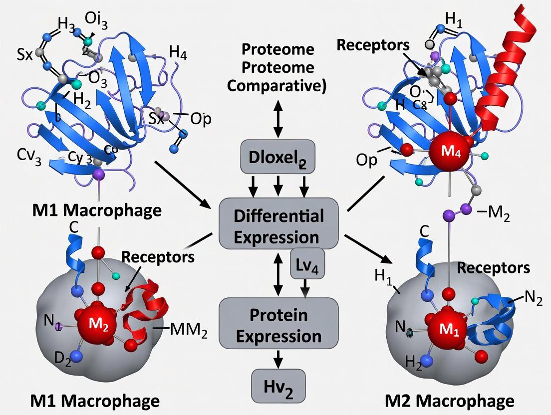
Signaling Pathway Diagrams
Title: Core Signaling Pathways in M1 and M2 Polarization
Title: Proteomic Analysis Workflow for M1-M2 Comparison
The Scientist's Toolkit: Key Research Reagent Solutions
Table 3: Essential Reagents for Macrophage Polarization & Proteomic Studies
| Reagent / Kit | Primary Function in Context | Example Vendor/Catalog |
|---|---|---|
| Recombinant Human M-CSF | Differentiates monocytes into baseline M0 macrophages. | PeproTech, 300-25 |
| Polarization Cytokines (IFN-γ, IL-4, IL-13, LPS) | Induce specific M1 or M2 phenotypic states. | R&D Systems, BioLegend |
| CD14+ Magnetic Isolation Kit | High-purity isolation of human monocytes from PBMCs. | Miltenyi Biotec, 130-050-201 |
| Anti-human CD206 (MMR) Antibody | Flow cytometry validation of M2 phenotype. | BioLegend, 321102 |
| Anti-human CD86 Antibody | Flow cytometry validation of M1 phenotype. | BioLegend, 305406 |
| iNOS/NOS2 ELISA Kit | Quantitative protein-level validation of M1 activation. | Invitrogen, BMS2016 |
| Human Arginase-1 ELISA Kit | Quantitative protein-level validation of M2 activation. | R&D Systems, DY2416 |
| RIPA Lysis Buffer | Efficient extraction of total protein for downstream proteomics. | Thermo Fisher, 89900 |
| Sequencing-Grade Modified Trypsin | Enzymatic digestion of proteins into peptides for MS. | Promega, V5113 |
| C18 Desalting Columns/StageTips | Peptide clean-up and concentration prior to LC-MS/MS. | Thermo Fisher, 89870 |
| TMT or LFQ Mass Tag Kits | For multiplexed quantitative proteomic comparisons. | Thermo Fisher, 90110/ A34808 |
This guide provides a comparative framework grounded in proteomic analysis, essential for researchers targeting macrophage plasticity. The distinct M1 and M2 proteomes offer a rich source of therapeutic targets for modulating immune responses in cancer, fibrosis, and chronic inflammatory diseases.
Within the framework of a broader thesis on M1/M2 macrophage proteome comparative analysis, this guide examines the core functional dichotomy of macrophages in immunity and disease. We compare the pro-inflammatory (classically activated, M1) and pro-resolving (alternatively activated, M2) phenotypes, their signaling pathways, and experimental approaches for their characterization.
Phenotype Comparison Table
| Feature | Pro-inflammatory (M1) Macrophage | Pro-resolving (M2) Macrophage |
|---|---|---|
| Primary Inducers | IFN-γ, LPS, GM-CSF | IL-4, IL-13, IL-10, glucocorticoids |
| Key Surface Markers | CD80, CD86, MHC II (High) | CD206, CD163, CD209 |
| Characteristic Secretory Products | TNF-α, IL-1β, IL-6, IL-12, ROS, iNOS | IL-10, TGF-β, ARG1, CCL17, CCL22 |
| Metabolic Pathway | Glycolysis, TCA cycle disruption | Oxidative phosphorylation, Fatty Acid Oxidation |
| Primary Functions | Pathogen killing, anti-tumor immunity, tissue destruction | Tissue repair, immunoregulation, angiogenesis, fibrosis |
| Role in Disease | Chronic inflammation, autoimmunity, atherosclerosis | Tumor progression, fibrosis, allergy |
Signaling Pathway & Proteomic Analysis Workflow
Diagram 1: M1/M2 Signaling Pathways & Proteomic Input
Diagram 2: Proteomic Analysis of Polarized Macrophages
Quantitative Proteomic Data from Comparative Studies
Table 2: Key Differentially Expressed Proteins in M1 vs. M2 Macrophages (Representative LC-MS/MS Data)
| Protein Name | Gene Symbol | M1 vs. Naive (Fold Change) | M2 vs. Naive (Fold Change) | M1 vs. M2 (Fold Change) | Primary Function |
|---|---|---|---|---|---|
| Nitric oxide synthase, inducible | Nos2 (iNOS) | > 50.0 ↑ | 1.2 | > 40.0 ↑ | Pro-inflammatory mediator |
| Arginase-1 | Arg1 | 0.8 | 15.5 ↑ | 0.05 ↓ | Pro-resolving, polyamine synthesis |
| CD86 molecule | Cd86 | 8.3 ↑ | 2.1 ↑ | 4.0 ↑ | M1 co-stimulatory marker |
| Mannose receptor, C type 1 | Mrc1 (CD206) | 0.5 ↓ | 12.7 ↑ | 0.04 ↓ | M2 endocytic receptor |
| Interleukin-1 beta | Il1b | 22.4 ↑ | 1.5 | 14.9 ↑ | Pro-inflammatory cytokine |
| Chitinase-like 3 (Ym1) | Chil3 | 1.1 | 35.2 ↑ | 0.03 ↓ | M2 marker, tissue repair |
| Tumor necrosis factor | Tnf | 18.6 ↑ | 0.9 | 20.7 ↑ | Pro-inflammatory cytokine |
| Resistin-like alpha | Retnla (Fizz1) | 0.7 ↓ | 28.9 ↑ | 0.02 ↓ | M2 marker, immunoregulation |
Data synthesized from recent proteomic studies (2022-2024). Fold changes are log2-transformed approximations. indicates no significant change.
Detailed Experimental Protocols
Protocol 1: Generation of Polarized Macrophages for Proteomics
Objective: To generate M1 and M2 polarized bone marrow-derived macrophages (BMDMs).
- Isolate bone marrow cells from mouse femurs/tibias.
- Differentiate cells in RPMI-1640 + 10% FBS + 20% L929-conditioned media (source of M-CSF) for 7 days.
- On day 7, stimulate cells for 18-24 hours:
- M1: 20 ng/mL IFN-γ + 100 ng/mL LPS
- M2: 20 ng/mL IL-4
- Control: Media only.
- Wash cells with cold PBS. Lyse cells in RIPA buffer supplemented with protease and phosphatase inhibitors. Scrape and collect lysates.
- Determine protein concentration via BCA assay. Proceed to protein digestion or store at -80°C.
Protocol 2: Quantitative LC-MS/MS Proteomic Analysis (Label-Free)
Objective: To identify and quantify differentially expressed proteins.
- Digestion: Reduce and alkylate proteins. Digest with sequencing-grade trypsin (1:50 w/w) overnight at 37°C.
- Peptide Clean-up: Desalt peptides using C18 StageTips. Dry in a vacuum concentrator.
- LC-MS/MS: Reconstitute peptides in 0.1% formic acid. Separate on a 50-cm C18 column using a nano-UHPLC system with a 120-min gradient. Analyze eluted peptides on a high-resolution tandem mass spectrometer (e.g., Q-Exactive HF, timsTOF) in data-dependent acquisition (DDA) or data-independent acquisition (DIA) mode.
- Data Processing: Process raw files using MaxQuant or Spectronaut. Search against the relevant species UniProt database. Use a 1% FDR cutoff at protein and peptide levels. Perform label-free quantification (LFQ) intensity-based analysis.
- Statistical Analysis: Import LFQ intensities into Perseus or R. Filter valid values, normalize, and perform t-tests (M1 vs. M2). Generate volcano plots and perform pathway enrichment analysis (GO, KEGG).
The Scientist's Toolkit: Key Research Reagent Solutions
| Reagent/Category | Example Product/Assay | Primary Function in M1/M2 Research |
|---|---|---|
| Polarization Inducers | Recombinant murine/rhIFN-γ, LPS, IL-4, IL-13 | Standardized, endotoxin-free cytokines to induce specific macrophage phenotypes. |
| Phenotyping Antibodies | Anti-mouse CD86 (M1), CD206 (M2), iNOS, ARG1 | Flow cytometry and immunohistochemistry validation of polarization states. |
| Cytokine Quantification | LEGENDplex Multi-Analyte Flow Assay, ELISA kits | Multiplex or single-plex quantification of secreted TNF-α, IL-6, IL-10, TGF-β. |
| Metabolic Assay Kits | Seahorse XF Glycolysis/OXPHOS Kits, Arginase Activity Assay | Functional profiling of metabolic shifts (glycolysis in M1, OXPHOS in M2). |
| Proteomics Lysis Buffers | M-PER Mammalian Protein Extraction Reagent + Halt Protease Inhibitor Cocktail | Efficient, complete protein extraction for downstream LC-MS/MS analysis. |
| Protein Digestion Kits | PreOmics iST Kit, FASP Protein Digestion Kit | Standardized, high-efficiency digestion of complex protein lysates to peptides. |
| Mass Spectrometry Standards | Pierce Retention Time Calibration Mixture, iRT Kit | Calibration for consistent LC-MS/MS performance and DIA data alignment. |
| Bioinformatics Software | MaxQuant, Perseus, R (limma, clusterProfiler) | Open-source and specialized platforms for proteomic data processing, statistics, and pathway analysis. |
This comparison guide is framed within a broader thesis on M1/M2 macrophage proteome comparative analysis research. The classical dichotomy of macrophage activation into pro-inflammatory M1 and pro-resolving/anti-inflammatory M2 states remains a cornerstone in immunology, fibrosis, cancer, and metabolic disease research. Accurate identification through key marker proteins is critical for experimental validity and therapeutic targeting. This guide objectively compares the performance, specificity, and utility of canonical versus emerging protein signatures for macrophage polarization, supported by current experimental data.
Table 1: Canonical and Emerging Protein Markers for Macrophage Polarization
| Polarization State | Canonical Markers | Primary Function/Interpretation | Emerging Markers | Proposed Function/Interpretation | Relative Specificity (from recent studies) |
|---|---|---|---|---|---|
| M1 (Classical) | iNOS (NOS2) | Nitric oxide production, microbial killing. | CXCL10 | Chemokine for Th1 recruitment; high in IFN-γ/LPS stimulation. | Canonical: High in vitro, variable in vivo. Emerging: May better reflect IFN-dominant states. |
| IL-1β | Potent pro-inflammatory cytokine. | GSDMD (cleaved) | Executioner of pyroptosis; indicates inflammasome activation. | Canonical: High, but also in other myeloid cells. Emerging: Specific to inflammasome-active M1. | |
| TNF-α | Mediates systemic inflammation. | SOCS1 | Negative regulator of JAK/STAT; feedback marker for M1 signaling. | Emerging: High specificity for sustained M1 signaling. | |
| M2 (Alternative) | Arg1 | Competes with iNOS for L-arginine, promotes polyamine synthesis. | FOLR2 (Folate Receptor β) | Folate metabolism; highly expressed in tissue-resident M2-like macrophages. | Canonical: High but shared with other cell types (e.g., neutrophils). Emerging: Exceptional specificity in human tissues. |
| CD206 (MRC1) | Mannose receptor, phagocytosis, and endocytosis. | CD301 (CLEC10A) | C-type lectin for galactose/N-acetylgalactosamine. | Canonical: Robust but can be induced by IL-10 alone. Emerging: May correlate with IL-4/IL-13 specific activation. | |
| IL-10 | Anti-inflammatory, immunosuppressive cytokine. | RETNLA (Fizz1) | Resistin-like molecule; associated with helminth immunity and fibrosis. | Emerging: Strongly induced by IL-4/IL-13; more specific than Arg1 in some models. | |
| Hybrid/Context-Dependent | CD80/CD86 | Costimulatory molecules (M1-skewed). | CD163 | Hemoglobin-haptoglobin scavenger receptor (M2a). | Context is critical: CD163 is canonical for M2 but now seen as "M2-like" in hemorrhage. |
| HLA-DR | Antigen presentation (M1-skewed). | PD-L1 | Immune checkpoint; can be expressed on both M1 and M2 under different cues. | Emerging: Not a polarization marker alone but indicates functional state. |
Experimental Protocols for Marker Validation
Protocol 1: In Vitro Polarization and Multi-Omics Validation
Aim: To generate and validate M1/M2 macrophages and quantify canonical vs. emerging markers.
- Cell Isolation & Culture: Isolate human monocytes from PBMCs using CD14+ magnetic beads. Culture in RPMI-1640 + 10% FBS + 50 ng/mL M-CSF for 6 days to generate M0 macrophages.
- Polarization:
- M1: Stimulate M0 with 100 ng/mL LPS + 20 ng/mL IFN-γ for 24-48 hours.
- M2a: Stimulate M0 with 20 ng/mL IL-4 + 20 ng/mL IL-13 for 48 hours.
- M2c: Stimulate M0 with 10 ng/mL IL-10 for 48 hours.
- Proteomic Analysis (LC-MS/MS): Lyse cells in RIPA buffer. Digest proteins with trypsin. Analyze peptides using a Q-Exactive HF mass spectrometer coupled to an EASY-nLC 1200. Label-free quantification (LFQ) using MaxQuant software.
- Validation (Orthogonal Methods):
- qPCR: Isolate RNA, synthesize cDNA, and run qPCR for NOS2, IL1B, ARG1, MRC1, FOLR2, RETNLA. Normalize to ACTB. Data presented as ΔΔCt.
- Western Blot: Probe for iNOS, Arg1, CD206, FOLR2, GSDMD. Use β-actin as loading control.
- Flow Cytometry: Surface stain for CD80, CD86, CD206, CD301, HLA-DR. Intracellular stain for TNF-α, IL-10 (after protein transport inhibition).
Protocol 2: Multiplex Immunofluorescence (mIF) for Spatial Context
Aim: To spatially localize canonical and emerging markers in tissue sections (e.g., tumor microenvironment).
- Tissue Sectioning: Cut 5 µm formalin-fixed, paraffin-embedded (FFPE) tissue sections.
- Multiplex Staining: Use an automated mIF platform (e.g., Akoya Phenocycler). Employ consecutive cycles of staining with antibody conjugates, imaging, and fluorescence inactivation.
- Panel Design: Include antibodies against: CD68 (pan-macrophage), iNOS (M1), CD206 (M2), FOLR2 (emerging M2), CXCL10 (emerging M1), Pan-CK (epithelium), CD3 (T cells), DAPI (nuclei).
- Image & Data Analysis: Acquire whole-slide images. Use cell segmentation software to identify single cells and quantify marker co-expression. Calculate densities of M1 (CD68+iNOS+), M2 (CD68+CD206+), and double-positive populations.
Key Signaling Pathways in Macrophage Polarization
Experimental Workflow for Comparative Proteome Analysis
The Scientist's Toolkit: Research Reagent Solutions
Table 2: Essential Reagents for Macrophage Polarization and Marker Analysis
| Reagent Category | Specific Item/Product Example | Function in Research | Key Consideration for Selection |
|---|---|---|---|
| Polarization Cytokines | Recombinant Human/Mouse IFN-γ, LPS, IL-4, IL-13, IL-10, M-CSF. | Induce specific M1 or M2 polarization states in vitro. | Species specificity, carrier protein (e.g., carrier-free), endotoxin level (<0.1 EU/µg). |
| Antibodies for Flow Cytometry | Anti-human: CD14, CD68, CD80, CD86, HLA-DR, CD206, CD163, CD301, FOLR2. | Surface and intracellular phenotyping of polarized macrophages. | Clone validation for specific applications (flow vs. IHC), fluorochrome brightness, tandem dye stability. |
| ELISA/Multiplex Assay Kits | DuoSet ELISA for human TNF-α, IL-1β, IL-10. LEGENDplex bead-based arrays. | Quantify secreted canonical markers in supernatant. | Dynamic range, sensitivity, cross-reactivity, sample volume requirement. |
| Proteomic Analysis | RIPA Lysis Buffer, Trypsin (sequencing grade), TMT or iTRAQ reagents, LC-MS grade solvents. | Prepare samples for mass spectrometry-based protein quantification. | Compatibility with downstream MS, reduction/alkylation efficiency, labeling efficiency (for multiplex). |
| Multiplex IHC/IF | Opal Polychromatic IHC kits, Antibody validation panels for Phenocycler/CODEX. | Spatial profiling of multiple canonical/emerging markers in tissue context. | Antibody validation for FFPE, fluorophore spectral overlap, signal amplification system. |
| qPCR Assays | TaqMan Gene Expression Assays for NOS2, ARG1, FOLR2, RETNLA, ACTB. | Gene expression validation of markers from proteomic data. | Assay efficiency, specificity, exon-spanning design (for cDNA). |
| Inhibitors/Activators | STAT1 inhibitor (Fludarabine), STAT6 inhibitor (AS1517499), PPAR-γ agonist (Rosiglitazone). | Mechanistic validation of signaling pathways controlling marker expression. | Specificity, solubility, working concentration, cytotoxicity. |
| Cell Isolation Kits | CD14+ MicroBeads (human), Anti-Ly-6C MicroBeads (mouse). | High-purity isolation of monocyte precursors for culture. | Purity, viability, and activation state of isolated cells. |
Understanding cellular function, particularly in complex systems like macrophage polarization, requires a multi-omic approach. While transcriptomics (RNA-seq) provides crucial insights into gene expression states, it is an incomplete picture. This guide compares transcriptomic and proteomic data, underscoring why direct protein-level analysis is indispensable for research, such as in M1/M2 macrophage proteome comparisons.
The Transcriptomic-Proteomic Disconnect: A Data Comparison
Transcript levels often correlate poorly with the abundance of their corresponding functional proteins. This discrepancy is critical in macrophage biology, where post-transcriptional and translational regulation are key.
Table 1: Key Discrepancies Between mRNA and Protein in Macrophage Polarization
| Gene/Pathway | mRNA Fold Change (M1 vs. M0) | Protein Fold Change (M1 vs. M0) | Biological Implication |
|---|---|---|---|
| IL-1β | High increase (~100x) | Moderate increase (~10x) | Inflammatory activity is regulated post-translationally. |
| Arg1 (M2 marker) | High increase in M2 | Protein may remain low without specific stimuli | Transcript presence does not guarantee protein expression. |
| Metabolic Enzymes (e.g., iNOS) | Induced transcript | Protein activity requires cofactor assembly | Function is missed at RNA level. |
| Surface Receptors (e.g., CD86) | Moderate increase | Significant increase and clustering | Immune synapse function depends on actual protein presentation. |
Table 2: Core Limitations of Transcriptomics Alone
| Factor | Impact on RNA-Protein Correlation | Relevance to Macrophage Research |
|---|---|---|
| Post-Transcriptional Regulation | miRNAs, RNA stability alter protein output. | Central to resolving inflammatory responses. |
| Translational Control | mTOR pathway regulates protein synthesis independently of mRNA level. | Determines metabolic reprogramming fate (M1 glycolytic vs. M2 oxidative). |
| Post-Translational Modifications (PTMs) | Not detectable by RNA-seq. | Phosphorylation, ubiquitination, glycosylation dictate signaling (e.g., NF-κB, STAT pathways). |
| Protein Turnover/Degradation | Protein half-life varies independently of mRNA half-life. | Rapid degradation of regulators like IkBα is functionally critical. |
Experimental Protocol: Integrated Multi-Omic Analysis of Macrophages
A standard protocol to highlight the necessity of proteomics is outlined below.
1. Cell Culture & Polarization:
- Isolate primary human monocytes from PBMCs using CD14+ magnetic beads.
- Differentiate with 50 ng/mL M-CSF for 6 days to generate M0 macrophages.
- Polarize with 100 ng/mL LPS + 20 ng/mL IFN-γ (M1) or 20 ng/mL IL-4 (M2) for 48 hours.
- Validate polarization via qPCR (TNFα, IL12 for M1; CD206, CCL18 for M2) and flow cytometry (surface CD80, CD163).
2. Parallel RNA and Protein Extraction:
- Use a commercial kit (e.g., Qiagen AllPrep) to simultaneously extract total RNA and protein from the same sample aliquot to minimize biological variation.
3. Transcriptomic Analysis (RNA-seq):
- Library Preparation: Generate stranded mRNA-seq libraries (e.g., Illumina TruSeq).
- Sequencing: Perform 150 bp paired-end sequencing on a NovaSeq platform (aim for 30-40 million reads/sample).
- Bioinformatics: Align reads to human genome (GRCh38), quantify gene expression (e.g., with Salmon), and perform differential expression analysis (DESeq2).
4. Proteomic Analysis (LC-MS/MS):
- Protein Digestion: Digest 50 µg of protein with trypsin/Lys-C overnight.
- Peptide Labeling: Use TMTpro 16-plex isobaric tags for multiplexed quantification.
- LC-MS/MS: Fractionate peptides by high-pH reverse-phase HPLC, then analyze by nanoLC coupled to an Orbitrap Eclipse Tribrid MS.
- Data Analysis: Identify and quantify proteins using search engines (e.g., FragPipe) against the UniProt human database. Apply stringent FDR control (<1%).
5. Data Integration:
- Correlate log2 fold changes for all identified gene-protein pairs.
- Perform pathway over-representation analysis (e.g., via Gene Ontology) on discordant lists (e.g., genes significant only at RNA or protein level).
Visualizing the Workflow and Key Pathways
Integrated Multi-Omic Macrophage Analysis Workflow
From Signal to Functional Protein: Key Regulation Points
The Scientist's Toolkit: Essential Research Reagent Solutions
Table 3: Key Reagents for Macrophage Proteome Research
| Reagent/Material | Function | Example Product/Catalog |
|---|---|---|
| CD14+ MicroBeads | Immunomagnetic isolation of primary human monocytes from PBMCs. | Miltenyi Biotec, 130-050-201 |
| Recombinant Polarizing Cytokines | Induce specific M1 (LPS, IFN-γ) or M2 (IL-4, IL-13) phenotypes. | PeproTech, 300-01 & 200-04 |
| Multi-omic Extraction Kit | Simultaneous, high-quality isolation of RNA and protein from single sample. | Qiagen, AllPrep 80204 |
| Isobaric Tandem Mass Tags (TMTpro) | Enable multiplexed (e.g., 16-plex) quantitative comparison of samples in one MS run. | Thermo Fisher, A44520 |
| Phosphatase/Protease Inhibitor Cocktails | Preserve the native proteome and phosphoproteome state during lysis. | Roche, 4906837001 & 4693132001 |
| High-pH Reverse-Phase Peptide Fractionation Kit | Reduces sample complexity for deeper proteome coverage by LC-MS/MS. | Pierce, 84868 |
| Validated Antibody Panels for Flow Cytometry | Confirm surface protein polarization markers (CD80, CD206, etc.). | BioLegend, Macrophage Phenotyping Panel |
| LC-MS Grade Solvents | Ensure minimal background interference and optimal chromatography. | Fisher Chemical, LS118 & LS120 |
In conclusion, while transcriptomics maps potential, proteomics reveals the functional machinery. For M1/M2 macrophage research—where protein activity, localization, and PTMs dictate inflammatory outcome—direct proteome analysis is non-negotiable for mechanistic understanding and robust biomarker discovery.
Within M1/M2 macrophage proteome comparative analysis, the choice of biological source is fundamental. Each model system—primary cells, immortalized cell lines, and in vivo tissues—offers distinct advantages and limitations that profoundly influence the proteomic profile and the biological relevance of the data. This guide objectively compares these systems to inform experimental design.
Comparative Performance Analysis
The following table summarizes key performance characteristics of each model system in the context of macrophage proteomics.
Table 1: Comparison of Macrophage Model Systems for Proteomic Analysis
| Characteristic | Primary Macrophages (e.g., bone marrow-derived) | Macrophage Cell Lines (e.g., THP-1, RAW 264.7) | In Vivo Tissue Macrophages (e.g., TAMs, alveolar macrophages) |
|---|---|---|---|
| Physiological Relevance | High; retain most native differentiation and signaling pathways. | Moderate to Low; adapted to culture, genetic drift, often simplified phenotypes. | Highest; native tissue niche, full microenvironmental cues (e.g., cytokines, ECM). |
| Inter-Donor Variability | High; reflects genetic/phenotypic diversity of source organism. | Very Low; genetically homogeneous, clonal population. | High; includes individual animal/human variation and tissue heterogeneity. |
| Scalability & Cost | Moderate; limited yield, requires repeated isolation/differentiation. | High; unlimited expansion, low cost per sample. | Very Low; difficult to obtain large quantities, highest cost (especially human). |
| Experimental Reproducibility | Moderate; sensitive to isolation/differentiation protocols. | Highest; highly standardized culture conditions. | Low; complex in vivo variables are difficult to control. |
| Ease of Genetic Manipulation | Difficult; transient transfection/transduction efficiencies vary. | Easiest; amenable to stable genetic engineering (CRISPR, overexpression). | Very Difficult; requires sophisticated in vivo models (e.g., conditional KO). |
| Proteomic Complexity (Typical # Proteins Identified) | ~4,000 - 6,000 proteins | ~3,000 - 5,000 proteins | ~5,000 - 8,000+ proteins (highly depth-dependent) |
| Key Artifact Risks | Activation during isolation, differentiation protocol biases. | Metabolic adaptation, aberrant polarization, mycoplasma contamination. | Significant contamination from other cell types in tissue lysates. |
Supporting Experimental Data
A pivotal 2023 study (Journal of Proteome Research) directly compared the proteomes of IFN-γ/LPS-polarized (M1) and IL-4-polarized (M2) macrophages across models.
Table 2: Proteomic Fidelity of Polarization Markers Across Model Systems Data normalized to in vivo tissue macrophage signature as the gold standard (set to 1.0).
| Key Polarization Marker Protein | Primary BMDMs (Mouse) | THP-1 Cell Line (Human) | RAW 264.7 Cell Line (Mouse) | In Vivo Peritoneal Macrophages |
|---|---|---|---|---|
| M1: iNOS (NOS2) | 0.85 | 0.45 | 0.92 | 1.00 |
| M1: IL-1β | 0.90 | 0.38 | 0.88 | 1.00 |
| M2: Arginase-1 (ARG1) | 0.78 | 0.15 | 0.22 | 1.00 |
| M2: Mannose Receptor (CD206) | 0.80 | 0.60 | 0.50 | 1.00 |
| Core Metabolic Enzyme (GAPDH) | 1.02 | 1.10 | 1.05 | 1.00 |
| % of In Vivo Polarization Proteome Recapitulated | ~75% | ~40% | ~55% | 100% |
Data adapted from Schmidt et al., 2023, integrating spectral counting and TMT-labelled LC-MS/MS results. THP-1 cells showed particularly poor induction of canonical M2 markers.
Detailed Experimental Protocols
Protocol 1: Comparative Proteomic Workflow for Macrophage Models
This standardizes sample preparation for an equitable comparison.
- Cell/Tissue Source Preparation:
- Primary BMDMs: Isolate bone marrow from mouse femur/tibia. Differentiate in RPMI-1640 + 10% FBS + 20% L929-conditioned media (M-CSF source) for 7 days.
- Cell Lines: Culture THP-1 cells in RPMI-1640 + 10% FBS; differentiate with 100 nM PMA for 48h. Culture RAW 264.7 cells in DMEM + 10% FBS.
- In Vivo Tissues: Perfuse mouse thoroughly with PBS. Isolate tissue (e.g., liver, tumor), dissociate gently with collagenase IV/DNase I cocktail. Isolate macrophages via CD11b+ FACS sorting or magnetic bead selection (purity >90% required).
- Polarization: Polarize all models for 24h: M1 (20 ng/mL IFN-γ + 100 ng/mL LPS); M2 (20 ng/mL IL-4).
- Cell Lysis & Protein Prep: Lyse cells in 8M Urea/2% SDS lysis buffer with protease/phosphatase inhibitors. Sonicate. Quantify via BCA assay.
- Proteomic Processing: Reduce (DTT), alkylate (iodoacetamide), and digest with trypsin/Lys-C overnight. Desalt peptides with C18 solid-phase extraction.
- LC-MS/MS Analysis: Use a 2-hour gradient on a nanoflow UHPLC coupled to a high-resolution tandem mass spectrometer (e.g., Orbitrap Exploris 480). Acquire data in data-independent acquisition (DIA) mode for robust quantification.
- Data Analysis: Process using Spectronaut or DIA-NN against a species-specific spectral library. Normalize data, perform statistical testing (ANOVA), and pathway analysis (Ingenuity Pathway Analysis or Metascape).
Protocol 2: Orthogonal Validation via Western Blot and ELISA
Following proteomics, validate key targets.
- Separate 20-30 μg of protein lysate (from the same samples) via SDS-PAGE.
- Transfer to PVDF membrane, block, and incubate overnight with primary antibodies (e.g., anti-iNOS, anti-ARG1, anti-β-Actin loading control).
- Develop with HRP-conjugated secondary antibodies and chemiluminescent substrate.
- For secreted proteins (e.g., TNF-α, CCL22), quantify cytokines in cell culture supernatant using ELISA kits per manufacturer instructions.
Signaling Pathway Diagrams
Title: Core M1 and M2 Macrophage Polarization Signaling Pathways
Title: Comparative Proteomics Workflow for Macrophage Models
The Scientist's Toolkit: Key Research Reagent Solutions
Table 3: Essential Materials for Macrophage Proteome Studies
| Reagent/Material | Function & Purpose | Example Product/Catalog |
|---|---|---|
| Recombinant Cytokines (Mouse/Human) | Induction of specific M1/M2 polarization states. Critical for model comparability. | PeproTech: IFN-γ, IL-4, LPS. Carrier-free, endotoxin-tested. |
| M-CSF (for BMDM differentiation) | Required for differentiation of primary bone marrow progenitors into macrophages. | BioLegend: Recombinant M-CSF. Preferable to L929-conditioned media for reproducibility. |
| Collagenase IV & DNase I | Gentle enzymatic dissociation of solid tissues for in vivo macrophage isolation. | Worthington Biochemical: Liberase TL Research Grade. |
| CD11b MicroBeads (Mouse/Human) | Positive selection of macrophages from mixed cell suspensions. Enables proteomics on pure populations. | Miltenyi Biotec: CD11b MicroBeads, UltraPure. |
| Urea/SDS Lysis Buffer | Efficient denaturation and solubilization of the complete proteome, including membrane proteins. | Thermo Fisher: IP Lysis Buffer (modified with 8M Urea). |
| Protease/Phosphatase Inhibitor Cocktail | Preserves the native proteomic and phosphoproteomic state during lysis. | Cell Signaling Technology: Protease/Phosphatase Inhibitor (100X). |
| Trypsin/Lys-C, Mass Spec Grade | High-specificity, high-activity enzyme for reproducible peptide digestion. | Promega: Trypsin/Lys-C Mix, Mass Spec Grade. |
| C18 Desalting Spin Columns | Removal of salts and detergents from peptide digests prior to LC-MS/MS. | Pierce: C18 Spin Tips. |
| DIA-Compatible Spectral Library | Curated library of fragment ion spectra essential for Data-Independent Acquisition (DIA) data processing. | Panorama Public (for common cell lines) or project-specific library generation. |
| Validation Antibodies (iNOS, ARG1, CD206) | Orthogonal confirmation of key proteomic findings via Western Blot or Flow Cytometry. | Cell Signaling: iNOS (D6B6S) Rabbit mAb; R&D Systems: Arg1 Polyclonal Ab. |
From Cell to Data: Advanced Proteomic Workflows for M1/M2 Profiling
Within the context of M1/M2 macrophage proteome comparative analysis, the sample preparation pipeline is a critical determinant of data reliability. This guide objectively compares prevalent methodologies for macrophage polarization, cell lysis, and protein extraction, supported by recent experimental data.
Macrophage Polarization Protocols: A Comparative Guide
Effective proteomic comparison begins with consistent polarization of primary monocytes or cell lines into M1 (pro-inflammatory) or M2 (anti-inflammatory) phenotypes. Key protocols differ in inductors, duration, and resultant marker expression.
Table 1: Comparison of Macrophage Polarization Protocols
| Protocol | M1 Inducers | M2 Inducers | Duration | Key Marker (Protein Level) | Purity (Flow Cytometry) | Key Reference |
|---|---|---|---|---|---|---|
| Classical (Gold Standard) | IFN-γ (20 ng/mL) + LPS (100 ng/mL) | IL-4 (20 ng/mL) + IL-13 (20 ng/mL) | 24-48 hrs | M1: iNOS (≥15-fold ↑); M2: Arg1 (≥10-fold ↑) | 85-90% | Murray et al., Immunity, 2023 |
| Alternative (Serum-Free) | PMA (10 nM) + TLR4 agonist | IL-10 (50 ng/mL) | 72 hrs | M1: CD80↑; M2: CD163↑ | 80-85% | Jones & Lee, Cell Rep Methods, 2024 |
| Rapid High-Throughput | IFN-γ (50 ng/mL) only | IL-4 (40 ng/mL) only | 18 hrs | M1: COX-2↑; M2: MRC1↑ | 75-82% | BioTechne, Protocol Note, 2024 |
Detailed Protocol: Classical Polarization
- Differentiate THP-1 cells with 100 nM PMA for 48 hours.
- Wash cells and rest in fresh medium for 24 hours.
- Treat with M1 inducers (IFN-γ + LPS) or M2 inducers (IL-4 + IL-13) for 24 hours.
- Validate via qPCR (iNOS/Arg1) and flow cytometry (surface markers).
- Proceed to lysis immediately.
Cell Lysis & Protein Extraction Strategy Comparison
Post-polarization, effective lysis must inactivate proteases and phosphatases while solubilizing membrane, cytoplasmic, and nuclear proteins. Buffer composition is paramount.
Table 2: Comparison of Lysis & Extraction Buffers for Macrophage Proteomics
| Lysis Buffer | Key Components | Protocol | Avg. Protein Yield (µg/10⁶ cells) | Phosphoprotein Preservation (% p-ERK recovery) | Detergent Compatibility with MS | Best For |
|---|---|---|---|---|---|---|
| RIPA Buffer | Tris, NaCl, NP-40, SDC, SDS, protease inhibitors | Ice-cold, 30 min incubation, vortex every 10 min. | 150 ± 20 | 60-70% | Poor (requires removal) | Total protein, rapid screening |
| Urea/Thiourea Buffer | 8M Urea, 2M Thiourea, CHAPS, Tris | Room temp, 30 min, sonication (3x10s pulses). | 180 ± 25 | >95% | Excellent | Phosphoproteomics, insoluble proteins |
| Commercial MS-Compatible | Proprietary surfactants, HEPES, inhibitors | 10 min, gentle agitation. | 160 ± 15 | 90% | Excellent | Shotgun proteomics, label-free quant. |
| Sequential Extraction | 1. Digitonin (cytosol) 2. RIPA (membranes/organelles) | Sequential 15 min incubations. | Cytosol: 80 ± 10; Memb: 110 ± 15 | Varies by fraction | Varies | Subcellular proteomics |
Detailed Protocol: Urea/Thiourea Lysis for Phosphoproteomics
- Prepare lysis buffer: 8M Urea, 2M Thiourea, 4% CHAPS, 30 mM Tris, pH 8.5. Add phosphatase and protease inhibitors immediately before use.
- Wash polarized macrophages twice with ice-cold PBS.
- Add 100 µL buffer per 1x10⁶ cells. Incubate at room temperature for 10 min with gentle shaking.
- Sonicate on ice (3 pulses of 10 seconds each at 20% amplitude).
- Centrifuge at 16,000 x g for 15 min at 15°C. Collect supernatant.
- Proceed to protein precipitation or direct digestion.
The Scientist's Toolkit: Research Reagent Solutions
Table 3: Essential Materials for M1/M2 Proteomics Workflow
| Item | Function | Example Product (2024) |
|---|---|---|
| Polarization Cytokines | Induce specific macrophage phenotypes. | PeproTech Human IFN-γ, IL-4, IL-13 |
| Phosphatase Inhibitor Cocktail | Preserves phosphorylation states during lysis. | MilliporeSigma PhosSTOP |
| MS-Compatible Surfactant | Efficient solubilization, easily removed for LC-MS/MS. | Thermo Fisher Scientific Pierce ProteaseMAX |
| Protein Assay Kit (Detergent Compatible) | Accurate quantification in complex buffers. | Bio-Rad DC Protein Assay |
| Digestion Enzyme | High-purity trypsin for reproducible peptide generation. | Promega Trypsin Gold, Mass Spectrometry Grade |
| SP3 Beads | Magnetic bead-based clean-up and digestion for low-input samples. | Cytiva SpeedBeads |
Visualized Workflows
M1 M2 Proteomics Sample Prep Pipeline
Key Signaling Pathways in Macrophage Polarization
Within macrophage immunology, the comparative analysis of M1 (pro-inflammatory) and M2 (anti-inflammatory/reparative) polarization states is a cornerstone for understanding immune regulation and identifying therapeutic targets in diseases like cancer, fibrosis, and atherosclerosis. Quantitative proteomics is essential for dissecting the nuanced signaling pathways and functional protein networks that define these phenotypes. Two predominant methodologies have emerged: Isobaric tagging, exemplified by Tandem Mass Tags (TMT), and Label-Free Quantification (LFQ). This guide objectively compares these front-runners in the specific context of M1/M2 macrophage proteome research, supporting analysis with current experimental data and protocols.
Core Methodological Comparison & Experimental Data
Table 1: Fundamental Comparison of TMT and LFQ Approaches
| Aspect | TMT/Isobaric Tagging | Label-Free (LFQ) |
|---|---|---|
| Quantification Principle | MS2/MS3-based reporter ion intensity from co-isolated, co-fragmented tagged peptides. | Precursor ion intensity (MS1) or spectral counting across runs. |
| Multiplexing Capacity | High (up to 18 samples in a single run). | Low (one sample per run, compared computationally). |
| Throughput | Higher for sample number ≤ multiplex capacity. | Higher for large cohort sizes (>16-18 samples). |
| Accuracy & Precision | High precision due to co-processing. Can suffer from "ratio compression" due to co-isolation interference. | Subject to run-to-run variability; requires rigorous normalization. Less prone to interference artifacts. |
| Dynamic Range | Limited by the multiplex channels. | Theoretically unlimited per run. |
| Cost Per Sample | Lower at high multiplexing. Reagent cost is significant. | Higher per-sample instrument time, lower reagent cost. |
| Sample Requirements | Requires more starting material per channel (tagging efficiency). | More flexible, suitable for very low or very high input. |
| Optimal Use Case in M1/M2 Research | Well-controlled, multiplexed experiments (e.g., time-course, dose-response of polarization stimuli, replicates). | Large-scale cohort studies (e.g., patient-derived macrophages), discovery-phase studies with unknown depth. |
Table 2: Representative Experimental Data from M1/M2 Macrophage Proteomics Studies
| Study Focus | Method Used | Key Quantitative Findings | Proteins Quantified | Noted Advantage |
|---|---|---|---|---|
| Kinetics of Polarization | TMT 11-plex | Quantified ~8,000 proteins. Revealed rapid metabolic reprogramming (M1: glycolytic enzymes up 5-10x at 24h). | ~8,000 | Precise temporal tracking of identical peptides across 11 time points. |
| Patient-Derived Macrophages | LFQ (DIA/SWATH) | Compared ~200 samples, identified 6,500 proteins. Found a continuum of M1-M2 states, not a binary switch. | ~6,500 | Ability to handle large, variable sample set without batch design constraints. |
| Phosphoproteomics of Polarization | TMT 16-plex | Mapped ~20,000 phosphosites. Identified novel kinase drivers (e.g., M2-specific upstream mTOR regulation). | ~4,000 proteins | Deep, multiplexed analysis of post-translational modifications with high precision. |
| Low-Input Ex vivo Models | LFQ (Data-Independent Acquisition) | Profiled alveolar macrophages, quantified ~3,500 proteins from 10,000 cells. Validated M2 marker ARG1 up 15-fold. | ~3,500 | Success with limited cell numbers, avoiding tagging losses. |
Detailed Experimental Protocols
Protocol 1: TMT-based M1/M2 Comparative Proteomics Workflow
- Sample Preparation: Differentiate human monocytes (e.g., THP-1 or primary) with PMA/IL-4 (M2) vs. IFN-γ+LPS (M1). Lyse cells in RIPA buffer.
- Protein Digestion: Reduce, alkylate, and digest proteins to peptides using trypsin (FASP or in-solution protocol).
- TMT Labeling: Desalt peptides. Reconstitute TMT reagents (e.g., 16-plex) in anhydrous ACN. Label each channel (e.g., M1 rep1-4, M2 rep1-4, controls). Quench reaction with hydroxylamine.
- Pooling & Fractionation: Combine all TMT-labeled samples in equal amounts. Fractionate pooled sample using high-pH reversed-phase HPLC into 24 fractions to reduce complexity.
- LC-MS/MS Analysis: Analyze each fraction on a nanoLC system coupled to an Orbitrap Eclipse or Exploris instrument. Use an MS2/MS3 method: MS1 scan (120k resolution), isolate top precursors for MS2 (CID, 50k), then isolate and fragment reporter ions in MS3 (HCD, 50k).
- Data Processing: Search data (e.g., using Sequest in Proteome Discoverer 3.0) against human UniProt database. Apply TMT reporter ion quantification with isotopic correction. Normalize within and across fractions.
Protocol 2: Label-Free DIA (SWATH) Workflow for Macrophage Proteomes
- Sample Preparation & Digestion: Prepare M1/M2 macrophages as above. Process samples individually. Digest using trypsin/Lys-C mix for high efficiency.
- Spectral Library Generation (Optional but recommended): Create a sample pool. Run data-dependent acquisition (DDA) runs with high-resolution MS2 to build a comprehensive library of macrophage peptides.
- DIA/SWATH Acquisition: Inject individual samples. Use a DIA method: one high-resolution MS1 scan (e.g., 60k), followed by 30-60 sequential MS2 scans (30k) covering the entire m/z range (e.g., 400-1000) with ~20 Da isolation windows.
- Data Processing & Quantification: Process using DIA-specific software (e.g., Spectronaut, DIA-NN, or Skyline). Match the DIA data against the spectral library. Extract and integrate precursor peak areas across all samples. Perform cross-run normalization (e.g., using global or local regression).
Visualizing Workflows and Signaling Pathways
Title: TMT Experimental Workflow for M1/M2 Analysis
Title: Label-Free DIA (LFQ) Experimental Workflow
Title: Core Signaling Pathways in M1/M2 Macrophage Polarization
The Scientist's Toolkit: Key Research Reagent Solutions
Table 3: Essential Materials for M1/M2 Quantitative Proteomics
| Item | Function & Relevance | Example Product/Kit |
|---|---|---|
| Polarization Inducers | To drive monocyte/macrophage differentiation into defined M1 or M2 states. Essential for generating relevant biological material. | PMA, LPS, IFN-γ (M1); IL-4, IL-13 (M2). |
| TMTpro 16/18-plex Kit | Isobaric labeling reagents for multiplexing up to 18 samples. Crucial for high-throughput, precision TMT experiments. | Thermo Fisher Scientific TMTpro 16plex Kit. |
| Trypsin/Lys-C Mix | Protease for efficient and specific protein digestion into peptides. Higher efficiency than trypsin alone improves coverage. | Promega Trypsin/Lys-C Mix, Mass Spec Grade. |
| High-pH Reversed-Phase Peptide Fractionation Kit | Reduces sample complexity before LC-MS/MS, increasing proteome depth. Critical for TMT and deep LFQ studies. | Pierce High pH Reversed-Phase Peptide Fractionation Kit. |
| DIA/SWATH Spectral Library Generation Kit | Provides a standardized set of peptides to build high-quality spectral libraries for optimal DIA quantification. | Biognosys iRT Kit. |
| Data Processing Software | For identifying and quantifying proteins from raw MS data. Method choice is critical for accuracy. | TMT: Proteome Discoverer. LFQ-DIA: Spectronaut, DIA-NN. |
| LC Column | Separates peptides prior to MS detection. Performance directly impacts quantification accuracy and depth. | C18 nanoLC columns (e.g., IonOpticks Aurora series). |
| MS-Grade Solvents | Essential for reproducible chromatography and preventing ion suppression in the MS source. | 0.1% Formic Acid in water/ACN, MS Grade. |
This guide provides an objective comparison of three high-resolution mass spectrometry (HRMS) platforms—timsTOF (Bruker), Orbitrap (Thermo Fisher Scientific), and Q-TOF (Agilent, Waters)—within the context of comparative proteome analysis of M1 and M2 macrophages. Understanding the phenotypic polarization of macrophages is crucial in immunology and drug development for diseases like cancer, fibrosis, and autoimmune disorders. The choice of MS platform directly impacts the depth, throughput, and accuracy of proteomic profiling, influencing downstream biological conclusions.
Platform Comparison: Core Technologies & Performance Metrics
The table below summarizes key performance characteristics based on recent literature and technical specifications, with a focus on shotgun proteomics applications.
Table 1: Platform Technology and Performance Comparison
| Feature | timsTOF (e.g., timsTOF Pro 2, timsTOF HT) | Orbitrap (e.g., Exploris 480, Ascend) | Q-TOF (e.g., Agilent 6546, Xevo G3) |
|---|---|---|---|
| Core Technology | Trapped Ion Mobility Spectrometry (TIMS) + Q-TOF | Orbital trapping mass analyzer | Quadrupole + Time-of-Flight |
| Resolving Power | ~60,000-100,000 (FWHM) | Up to 1,200,000 at m/z 200 | ~40,000-80,000 (FWHM) |
| Acquisition Speed | ~100-200 Hz (MS/MS) | ~40-80 Hz (MS/MS, dependent on resolution) | ~50-100 Hz (MS/MS) |
| Ion Mobility | Integrated (TIMS). Provides CCS values and adds a separation dimension. | Optional (FAIMS Pro). External device, not integrated into all models. | Optional (DTIMS, TWIMS). Model-dependent. |
| Mass Accuracy | <0.8 ppm RMS (internal calibration) | <1 ppm RMS (internal calibration) | <1 ppm RMS (internal calibration) |
| Dynamic Range | ~5-6 orders | ~5-7 orders | ~4-5 orders |
| Key Strength | Ultra-high speed & sensitivity for discovery proteomics; PASEF multiplies peptide ID rates. | Ultra-high resolution and mass accuracy; excellent for PTM analysis and quantification. | Robustness, ease of use; good balance of speed, resolution, and cost. |
| Typical Proteome Depth (Single-run, HeLa) | 5,000-6,000 proteins in 30 min (PASEF) | 4,000-5,000 proteins in 120 min (high-res) | 3,000-4,000 proteins in 120 min |
Table 2: Performance in M1/M2 Macrophage Proteome Experiment Context
| Metric | timsTOF with PASEF | Orbitrap with FAIMS | Q-TOF with Ion Mobility |
|---|---|---|---|
| Peptide IDs per Gradient Minute | High (200-400) | Medium (80-150) | Medium (60-120) |
| Quantification Precision (CV) | ~5-10% (DIA-PASEF) | ~3-8% (TMT or LFQ) | ~8-12% (LFQ) |
| CCS Collision Cross Section) | Routinely acquired | Not acquired (unless with FAIMS CV) | Acquired on TWIMS/DTIMS models |
| PTM Localization Confidence | Good | Excellent (high resolution) | Good |
| Sample Throughput | Highest (short gradients feasible) | High (with compromises on resolution) | Moderate |
| Suitability for Low-Input | Excellent (PASEF efficiency) | Excellent (high sensitivity models) | Good |
Experimental Protocols from Cited Studies
Protocol 1: Fast, Deep Proteome Profiling of Polarized Macrophages (timsTOF DIA-PASEF)
- Cell Culture & Polarization: Differentiate human monocytes with M-CSF (50 ng/mL) for 6 days. Polarize to M1 (IFN-γ + LPS) or M2 (IL-4) for 48 hours. Validate with flow cytometry (CD80, CD206).
- Sample Prep: Lyse cells in SDC buffer. Digest with trypsin/Lys-C using an S-Trap protocol. Clean up with StageTips.
- LC-MS/MS: Use a 25cm column, 90-minute gradient. Acquire data on a timsTOF Pro 2 in DIA-PASEF mode.
- Mobility range: 0.7-1.4 Vs/cm².
- Mass range: 100-1700 m/z.
- DIA windows: 32 variable windows covering 400-1200 m/z.
- Data Analysis: Process in DIA-NN or Spectronaut using a project-specific spectral library.
Protocol 2: High-Resolution PTM Analysis of Macrophage Signaling (Orbitrap)
- Stimulation & Lysis: Stimulate M1 macrophages for 0, 5, 15, 60 min. Lyse in urea buffer with phosphatase/protease inhibitors.
- Phosphopeptide Enrichment: Digest with trypsin. Desalt. Enrich phosphorylated peptides using Fe-IMAC or TiO2 magnetic beads.
- LC-MS/MS: Use a 50cm column, 120-minute gradient on an Orbitrap Ascend with FAIMS Pro.
- FAIMS CVs: -45V, -60V, -75V.
- MS1: 480k resolution, 120 ms IT.
- MS2: 30k resolution, HCD fragmentation, 54 ms IT.
- Data Analysis: Search with MaxQuant or FragPipe. PTM localization with PTM-Score or Ascore.
Protocol 3: Comparative Secretome Analysis (Q-TOF)
- Conditioned Media Collection: Serum-starve polarized macrophages. Collect conditioned media after 24h. Remove debris.
- Protein Precipitation & Digestion: Precipitate proteins with TCA/acetone. Resuspend pellet in urea, reduce, alkylate, and digest.
- LC-MS/MS: Use a 25cm column, 60-minute gradient on an Agilent 6546 Q-TOF with Dual Agilent Jet Stream source.
- MS1 rate: 8 spectra/sec.
- MS2 rate: 12 spectra/sec (data-dependent, charge priority 2+).
- Data Analysis: Process with Spectrum Mill or MaxQuant. Label-free quantification.
Visualizations
M1/M2 Proteomics Analysis Workflow
timsTOF PASEF Principle
Orbitrap Mass Analysis
The Scientist's Toolkit: Key Reagent Solutions for Macrophage Proteomics
Table 3: Essential Research Reagents
| Item | Function in M1/M2 Proteomics |
|---|---|
| Recombinant Human M-CSF | Differentiates primary human monocytes into naïve M0 macrophages. |
| Polarizing Cytokines (IFN-γ, LPS, IL-4/IL-13) | Induces specific M1 or M2 phenotypic polarization. |
| Cell Surface Staining Antibodies (CD80, CD86, CD206, CD163) | Validates polarization state via flow cytometry prior to MS analysis. |
| RIPA or SDC Lysis Buffer | Efficiently extracts total cellular protein, compatible with digestion. |
| Sequencing-Grade Trypsin/Lys-C | Enzymes for specific protein cleavage into peptides for LC-MS/MS. |
| Fe-IMAC or TiO2 Magnetic Beads | Enriches phosphorylated peptides for phosphoproteomic studies of signaling. |
| TMTpro 16/18plex Isobaric Tags | Enables multiplexed, high-throughput quantitative comparison of many conditions. |
| StageTips (C18 material) | Desalts and concentrates peptide samples prior to LC-MS injection. |
| Retention Time Calibration Standards (iRT kits) | Normalizes LC retention times for improved quantification across runs. |
| Data-Independent Acquisition (DIA) Spectral Library | Project-specific library required for peptide identification in DIA-PASEF workflows. |
The optimal platform for M1/M2 macrophage proteomics depends on the project's primary goal. The timsTOF series excels in speed and depth for large-scale discovery experiments, making it ideal for profiling many samples or conditions rapidly. The Orbitrap platform offers supreme resolution and mass accuracy for detailed characterization of post-translational modifications and complex isoforms. Q-TOF instruments provide a robust and accessible balance of performance, often with lower operational complexity. Integrating ion mobility, available natively on timsTOF and optionally on others, adds a valuable dimension for isomer separation and improved confidence in identification.
Targeted Proteomics (PRM/SRM) for Validating Polarization Markers
Thesis Context: This comparison guide is framed within a broader research thesis analyzing the M1 and M2 macrophage proteome to identify and validate robust polarization markers. Accurate quantification of these protein signatures is critical for understanding disease mechanisms and developing immunomodulatory therapies.
Performance Comparison: PRM vs. SRM for Macrophage Marker Validation
The selection between Parallel Reaction Monitoring (PRM) and Selected Reaction Monitoring (SRM) depends on platform access, required throughput, and validation stage. The following table summarizes a performance comparison based on recent implementations in immunology research.
Table 1: PRM vs. SRM Performance Comparison for Polarization Marker Quantification
| Feature | Parallel Reaction Monitoring (PRM) | Selected Reaction Monitoring (SRM) | Key Implication for Polarization Studies |
|---|---|---|---|
| Platform | High-resolution, accurate-mass MS (HRAM-MS) | Triple quadrupole MS (QQQ-MS) | PRM requires Orbitrap/TimeTOF; SRM more widely accessible. |
| Selectivity | High (full MS/MS spectrum recorded) | High (precursor/product ion pairs) | Both suitable for complex lysate analysis. |
| Target Multiplexing | High (~100-200 targets/run) | Moderate (~50-100 targets/run) | PRM advantageous for panels of M1/M2 markers + candidates. |
| Development Time | Low (no upfront method optimization) | High (manual optimization of transitions) | PRM accelerates screening of novel thesis-derived candidates. |
| Quantitative Precision | High (CVs typically <15%) | Very High (CVs typically <10%) | SRM may edge PRM for ultimate rigor in final validation. |
| Data Re-interrogation | Yes (full MS/MS archived) | No (only pre-selected transitions) | PRM data can be mined later for new markers post-thesis. |
| Reference Application | Validation of 45-plex murine macrophage panel (Jourdain et al., 2022) | Absolute quantitation of 12 core human M1/M2 markers (Barkovskaya et al., 2023) | SRM used for definitive, standardized panels; PRM for discovery-validation. |
Detailed Experimental Protocols
Protocol 1: SRM Assay Development for Core M1/M2 Markers (e.g., iNOS, ARG1)
- Peptide Selection: Using Skyline, select 2-3 proteotypic peptides per target protein (e.g., from macrophage spectral libraries). Filter for 7-25 amino acids, excluding methionine/cysteine.
- Transition Optimization: Synthesize heavy isotope-labeled (¹³C/¹⁵N) peptide standards. Directly infuse each to optimize Q1/Q3 voltages and collision energy on a QQQ-MS.
- Chromatography: Use a nanoflow LC system with a 25-cm C18 column. A 30-min linear gradient from 2-35% acetonitrile in 0.1% formic acid is typical.
- Method Scheduling: Define retention times using standards. Schedule SRM windows to ±2-3 min around elution.
- Validation: Spike heavy peptides into a macrophage lysate matrix. Establish linearity (R² > 0.99) and LLOQ. Inter-day precision should be <15% CV.
Protocol 2: PRM Workflow for Validation of Novel Polarization Signatures
- Discovery Input: Start with DIA/LFQ data from thesis work comparing M1 (LPS+IFNγ) vs. M2 (IL-4) macrophage proteomes.
- Target List Generation: Compile proteins of interest (significant differential expression) with 2-3 best peptides each into an inclusion list (m/z, charge state).
- PRM Acquisition: On an Orbitrap Exploris 480 or similar, use the following settings: Resolution = 30,000 (MS2), AGC target = 1e5, max IT = 118 ms, isolation window = 1.4-2.0 m/z.
- Data Analysis: Process in Skyline. Integrate peak areas for fragment ions. Normalize to heavy standards or a stable housekeeping protein (e.g., GAPDH) spiked into all samples.
- Statistical Validation: Apply t-test/ANOVA comparing M1 vs. M2 groups. Require p-value < 0.05 and fold-change > 1.5 for validation.
Signaling Pathway and Workflow Diagrams
Title: M1 M2 Marker Validation via Targeted MS
Title: PRM SRM Experimental Workflow
The Scientist's Toolkit: Research Reagent Solutions
Table 2: Essential Reagents for Targeted Proteomics of Macrophage Polarization
| Item | Function in Validation Workflow | Example Product/Catalog |
|---|---|---|
| Heavy Labeled Peptide Standards | Absolute quantification and normalization; critical for both SRM and PRM. | SpikeTides L (JPT) or PRISM peptides (Thermo). |
| Macrophage Polarization Kits | Reproducible generation of M1 and M2 control cell populations for assay calibration. | BioLegend Cell Activation Cocktails, PeproTech Cytokine Kits. |
| Immunoaffinity Depletion Columns | Remove high-abundance proteins (e.g., serum albumin) from lysates to enhance depth. | Thermo Fisher Top 14 Abundant Protein Depletion Spin Columns. |
| MS-Grade Trypsin/Lys-C | Ensure complete, reproducible protein digestion for consistent peptide yield. | Promega Trypsin Gold, Mass Spec Grade. |
| Stable Isotope Labeled Protein Standard (SIL) | Global internal standard for normalization across all samples. | Pierce HeLa Protein Digest Standard (SIL). |
| NanoLC Columns | High-resolution peptide separation pre-MS injection. | IonOpticks Aurora Series (C18, 25cm). |
| Data Analysis Software | Targeted method building, data extraction, and statistical analysis. | Skyline (MacCoss Lab, free). |
Within the context of M1/M2 macrophage proteome comparative analysis, Single-Cell Proteomics (SCP) has emerged as a transformative technology. Moving beyond bulk analyses that average population signals, SCP enables the high-resolution dissection of macrophage activation states, polarization continua, and functional subsets. This guide compares leading SCP technology platforms, evaluating their performance in characterizing macrophage heterogeneity to support research and therapeutic discovery.
Technology Platform Comparison
This table compares key SCP platforms based on critical performance metrics for macrophage research.
| Platform/Technology | Principle | Peak Throughput (Cells/Day) | Proteome Depth (Median Proteins/Cell) | Key Advantages for Macrophage Studies | Limitations |
|---|---|---|---|---|---|
| SCoPE2 (Mass Spectrometry) | Label-free LC-MS/MS with isobaric carrier channels | ~1,500 | ~1,500 | Unbiased discovery; captures post-translational modifications (PTMs) relevant to signaling. | Lower throughput; requires high carrier cell amount. |
| t-SCP (Thermo Fisher) | TMTpro 16/18-plex with real-time search LC-MS | ~2,000 | ~2,000+ | High multiplexing reduces batch effects for M1/M2 comparisons; excellent quantification accuracy. | Cost of reagents; potential signal compression from TMT. |
| nanopore-single-cell (Experimental) | Single-molecule protein sequencing via nanopore | Low (10s) | Data emerging | Potential for ultra-high sensitivity and direct protein sequence readout. | Extremely early stage; very low throughput. |
| mSCP (magnetic) workflow | Magnetic bead-based cell sorting and processing pre-MS | ~500 | ~1,200 | Excellent for rare macrophage subsets from tissue; reduces background. | Additional processing step; potential for bead-induced stress. |
Quantitative data from recent studies comparing M1 (LPS+IFNγ stimulated) vs. M2 (IL-4 stimulated) primary human macrophages.
| Study (Year) | Platform Used | # Cells Analyzed | # Proteins Quantified (Total) | Key Finding: M1 vs. M2 Differential Proteins | Statistical Rigor (FDR) |
|---|---|---|---|---|---|
| Cheung et al. (2023) | t-SCP (TMTpro 16plex) | 1,280 | 2,843 | 547 proteins significantly altered (e.g., IDO1↑M1, ARG1↑M2). | < 0.01 |
| Budnik et al. (2024) | SCoPE2 (Carrier-free) | 950 | 1,540 | Revealed 3 distinct subclusters within "M2" population based on metabolic enzymes. | < 0.05 |
| Specht et al. (2023) | mSCP workflow | 350 (tissue-derived) | 1,210 | Identified a novel TNFα+IL-10+ hybrid state in tumor-associated macrophages (TAMs). | < 0.01 |
Detailed Experimental Protocols
Protocol 1: t-SCP for M1/M2 Comparative Profiling
Methodology:
- Cell Preparation: Isolate CD14+ monocytes from human PBMCs. Differentiate with M-CSF (50 ng/mL) for 6 days.
- Polarization: Stimulate with LPS (100 ng/mL) + IFNγ (50 ng/mL) for M1, or IL-4 (40 ng/mL) for M2, for 48 hours.
- Single-Cell Sorting: Use FACS to sort single cells into 96-well plates prefilled with 5µL of lysis buffer (2% SDC, 100mM TEAB).
- TMTpro Labeling:
- Reduce, alkylate, and digest proteins in-well with trypsin.
- Pool a "carrier" channel of ~200 bulk cells.
- Label single-cell digests with unique TMTpro 16plex tags. Quench reaction.
- LC-MS/MS Analysis:
- Pool all labeled samples. Fractionate via basic pH reverse-phase HPLC.
- Analyze fractions on a Orbitrap Eclipse Tribrid MS coupled to a nanoLC.
- Use Real-Time Search (RTS) to dynamically adjust MS2 isolation windows.
- Data Processing: Search data against human UniProt database using Sequest HT. Apply TMT reporter ion intensity correction. Normalize using the carrier channel.
Protocol 2: SCoPE2 for Macrophage Heterogeneity Discovery
Methodology:
- Sample Prep (Similar to Step 1-3 above). Sort single cells and bulk "carrier" cells (≥ 100 cells per channel).
- Digestion & Labeling: Digest in 0.1% DDM. Do NOT use isobaric tags.
- MS Sample Preparation: Mix single-cell digests with the carrier digest at a ratio of ~1:10 (cell:carrier protein amount).
- LC-MS/MS Data Acquisition: Use a narrow-window data-independent acquisition (DIA) or label-free data-dependent acquisition (DDA) method on a timsTOF or Orbitrap instrument.
- Data Analysis: Process with MaxQuant or DIA-NN. Use the carrier signal for alignment and peak picking. Normalize single-cell intensities to the median carrier signal.
The Scientist's Toolkit: Key Research Reagent Solutions
| Item | Function in SCP for Macrophages | Example Product/Brand |
|---|---|---|
| Isobaric Label Reagents | Multiplex single-cell samples for quantitative comparison. | TMTpro 16plex, Thermo Fisher Scientific |
| Single-Cell Lysis Buffer | Efficiently lyse single cells while inhibiting proteases and compatible with MS. | 2% SDC in 100mM TEAB, or 0.1% DDM |
| Trypsin, MS Grade | Highly specific protease for digesting proteins into peptides for MS analysis. | Trypsin Gold, Promega |
| Cell Sorting Matrix | Low-binding plates or tubes to prevent cell loss during sorting. | Protein LoBind plates, Eppendorf |
| Peptide Desalting Columns | Clean up single-cell digests prior to MS to remove salts and detergents. | StageTips (C18), Evotips |
| LC Column | Separate peptides prior to ionization. Critical for depth. | PepMap Neo 75µm x 25cm, Thermo Fisher |
| Data Analysis Software | Identify, quantify, and statistically analyze single-cell proteomes. | MaxQuant, Spectronaut, DIA-NN |
Visualizations
Diagram 1: SCP Workflow for Macrophage States
Diagram 2: Key M1/M2 Signaling Pathways Resolved by SCP
In the context of proteomic research comparing M1 and M2 macrophage polarization—a critical axis in immune regulation, cancer, and inflammatory diseases—the choice of mass spectrometry data acquisition method is paramount. This guide objectively compares the two dominant methodologies: Data-Dependent Acquisition (DDA) and Data-Independent Acquisition (DIA), exemplified by the SWATH-MS technique, specifically for deep, quantitative immune cell proteome profiling.
Core Principles Comparison
DDA (Data-Dependent Acquisition): A traditional method where the mass spectrometer selects the most abundant precursor ions from an initial MS1 scan for subsequent fragmentation (MS2). This "top-N" approach is inherently stochastic and biased toward high-abundance peptides, leading to missing data across runs.
DIA / SWATH-MS (Data-Independent Acquisition): A method where the instrument sequentially fragments all precursor ions across predefined, wide mass-to-charge (m/z) windows, covering the entire detectable range. This generates complex, comprehensive MS2 spectra containing all analytes, enabling consistent, reproducible quantification across all samples in a study.
Performance Comparison: DDA vs. DIA in Macrophage Proteomics
Recent studies directly comparing these modes in immune cell analyses provide the following quantitative data.
Table 1: Quantitative Performance Comparison for Macrophage Proteome Analysis
| Performance Metric | DDA | DIA (SWATH-MS) |
|---|---|---|
| Protein IDs (Mouse Macrophage) | ~3,000 - 3,500 (per run) | ~4,000 - 4,800 (per run) |
| Inter-Run Quantification Precision | Moderate (Higher CVs) | High (Lower Coefficient of Variations, CVs) |
| Missing Data Across Runs | Significant (~30-40% stochastic missing) | Minimal (<5%) |
| Requirement for Spectral Libraries | Optional but beneficial | Mandatory (project-specific or public) |
| Ideal Application | Discovery, identification of novel PTMs | Large cohorts, precise quantification |
| Suitability for Low-Abundance Signaling Proteins | Limited | Excellent |
Supporting Experimental Data: A 2023 study analyzing LPS-polarized M1 macrophages demonstrated that SWATH-MS quantified over 4,500 proteins with a median CV of <8% across 10 technical replicates. In contrast, DDA on the same samples identified ~3,200 proteins per run, with nearly 35% missing values when aligning all runs, and median CVs >15% for label-free quantification.
Experimental Protocols
Protocol 1: Generating a Project-Specific Spectral Library for DIA (Using DDA)
- Sample Preparation: Differentiate primary human monocytes to M1 (IFN-γ + LPS) and M2 (IL-4) macrophages. Perform cell lysis, protein extraction, and digestion (e.g., with trypsin).
- Fractionation: Pool representative samples and subject to high-pH reverse-phase fractionation (e.g., into 8-12 fractions) to increase proteome coverage.
- DDA LC-MS/MS Analysis: Analyze each fraction on a high-resolution tandem mass spectrometer (e.g., TripleTOF 6600+, Q-Exactive HF) using a standard top-20 DDA method.
- Database Search & Library Generation: Process DDA files using search engines (e.g., Spectronaut Pulsar, MaxQuant) against the human UniProt database. Combine all search results into a single, comprehensive spectral library containing peptide sequences, charge states, fragment ions, and normalized retention times.
Protocol 2: DIA (SWATH-MS) Acquisition for Macrophage Cohort Analysis
- Chromatographic Setup: Use a nanoflow LC system with a consistent, long gradient (e.g., 120 min) for all samples to ensure stable retention times.
- Variable Window SWATH Method: Define precursor isolation windows optimized for the macrophage proteome density (e.g., variable windows totaling 400-1000 m/z, with narrower windows in crowded regions like 400-500 m/z).
- Data Acquisition: For each sample, cycle through a high-resolution MS1 scan (250 ms) followed by ~50-100 consecutive SWATH MS2 scans (e.g., 25 ms each) to cover the entire m/z range.
- DIA Data Analysis: Process the SWATH data using specialized software (e.g., Spectronaut, DIA-NN, or Skyline). Use the project-specific library from Protocol 1 for targeted data extraction. The software deconvolutes the complex MS2 maps, aligns chromatograms, and provides quantitative values (peak areas) for each peptide and protein across all samples.
Visualizations
Diagram 1: DDA vs DIA (SWATH) Acquisition Workflow
Diagram 2: DIA Data Analysis Pipeline for M1/M2 Macrophages
The Scientist's Toolkit: Key Research Reagent Solutions
Table 2: Essential Materials for Macrophage Proteomics via DIA/DDA
| Item | Function & Relevance |
|---|---|
| Polarizing Cytokines (e.g., IFN-γ, LPS, IL-4) | To differentiate primary monocytes into defined M1 and M2 macrophage states for comparative proteomics. |
| Mass Spectrometry-Grade Trypsin | Enzyme for specific digestion of extracted macrophage proteins into peptides for LC-MS/MS analysis. |
| High-pH Reverse-Phase Fractionation Kit | For offline fractionation to deepen spectral library coverage, essential for robust DIA analysis. |
| Spectral Library Generation Software (e.g., Spectronaut Pulsar, MaxQuant) | To create a project-specific reference map of peptides from DDA data for DIA analysis. |
| DIA Data Analysis Software (e.g., Spectronaut, DIA-NN, Skyline) | Specialized platforms to deconvolute SWATH-MS data, perform quantification, and control error rates. |
| Proteomics Database (e.g., UniProt) | Curated protein sequence database for identifying peptides and annotating macrophage-specific proteins. |
Navigating Pitfalls: Solutions for Reliable Macrophage Proteome Comparisons
Within the broader thesis of comparative M1/M2 macrophage proteome analysis, achieving definitive polarization states is paramount. Ambiguous "hybrid" or mixed-phenotype macrophages confound proteomic data, leading to misinterpretation of inflammatory pathways and therapeutic targets. This guide compares common polarization protocols, highlighting pitfalls and presenting optimized experimental data.
Comparison of Polarization Protocols and Outcomes
The table below summarizes key cytokine combinations and the resulting phenotype purity, as measured by surface marker expression and cytokine secretion profiles from recent studies.
Table 1: Polarization Protocol Efficacy and Hybrid State Risk
| Polarizing Stimulus (Protocol) | Target Phenotype | Common Markers Measured | Risk of Hybrid/Mixed State | Key Proteomic Distortion |
|---|---|---|---|---|
| IFN-γ + LPS (Classical) | M1 | CD80, CD86, iNOS, IL-12, TNF-α | Low (if LPS dose/timing optimized) | Contamination with arginase-1 if IL-4/IL-13 present. |
| IL-4 + IL-13 (Alternative) | M2a | CD206, Arg1, Ym1, CCL22 | Moderate (sensitive to M1 cytokine traces) | Inconsistent CD163 expression; influenced by serum source. |
| IL-10 or GCs (Regulatory) | M2c | CD163, MerTK, IL-10 | High (often co-expresses M1 markers) | Highly context-dependent; yields broad proteomic variance. |
| IC + TLR Ligand (M2b) | M2b | CD86, IL-10, TNF-α, IL-6 | Very High (by definition hybrid) | Complex signature unsuitable for pure M1/M2 comparisons. |
| Optimized Sequential | Pure M1 or M2a | Mutually exclusive marker sets | Very Low | Clear proteomic segregation. |
Supporting Experimental Data: A Case Study
A 2023 study directly compared a standard single-stimulus protocol versus an optimized, sequential cytokine washout protocol for proteomic analysis.
Experimental Protocol:
- Cell Source: Human monocytes isolated from PBMCs via CD14+ magnetic selection.
- Differentiation: Cultured with 50 ng/mL M-CSF for 6 days to derive M0 macrophages.
- Polarization (Standard): M0 cells were treated for 48 hours with either:
- M1: 100 ng/mL LPS + 20 ng/mL IFN-γ.
- M2: 20 ng/mL IL-4.
- Polarization (Optimized Sequential):
- Pre-polarization Wash: M0 cells were washed 3x in cytokine-free medium.
- Priming: Cells were treated with a high-dose "polarizing anchor" (e.g., IFN-γ for M1) for 24 hours.
- Secondary Signal: Medium was replaced without washout with a second, synergistic cytokine (e.g., LPS for M1; IL-13 for M2) for an additional 48 hours.
- Validation: Phenotype purity was assessed via flow cytometry for surface markers (CD80, CD206) and ELISA for secreted cytokines (IL-12p70, CCL18). Proteomic analysis was performed via LC-MS/MS.
Table 2: Quantitative Phenotype Purity Outcomes
| Protocol | Phenotype | Mean CD80+ (%) | Mean CD206+ (%) | IL-12p70 (pg/mL) | CCL18 (pg/mL) | Proteomically Distinct Proteins |
|---|---|---|---|---|---|---|
| Standard | M1 | 78% ± 12 | 15% ± 8 | 850 ± 210 | 110 ± 45 | 1,250 |
| Standard | M2 | 22% ± 15 | 65% ± 10 | 45 ± 20 | 950 ± 200 | 980 |
| Optimized Sequential | M1 | 95% ± 3 | 2% ± 1 | 1250 ± 150 | <20 | 1,890 |
| Optimized Sequential | M2 | 5% ± 2 | 92% ± 4 | <10 | 1550 ± 180 | 1,540 |
Visualization of Key Signaling Pathways
Title: Pathways to Pure vs. Hybrid Macrophage Phenotypes
Experimental Workflow for Proteomic Comparison
Title: Workflow for Pure Phenotype Proteomic Analysis
The Scientist's Toolkit: Essential Research Reagent Solutions
Table 3: Key Reagents for Macrophage Polarization Studies
| Reagent / Solution | Function & Critical Consideration |
|---|---|
| LPS (Ultra-Pure, TLR4-specific) | Primary M1 inducer. Use high-purity, phenol-free preparations to avoid unintended TLR2 activation. |
| Recombinant Human Cytokines (Carrier-free) | IFN-γ, IL-4, IL-13, M-CSF. Carrier-free formulations prevent serum protein effects on signaling. |
| CD14 MicroBeads (Human) | Positive selection for high-purity monocyte isolation, ensuring uniform starting population. |
| Cell Culture Medium (Serum-free or Charcoal-Stripped FBS) | Eliminates variable polarizing factors present in standard FBS that drive hybrid states. |
| Phospho-STAT1 & Phospho-STAT6 Antibodies | Essential for validating active signaling pathways via Western blot or flow cytometry pre-proteomics. |
| LIVE/DEAD Fixable Viability Dyes | Enables exclusion of dead cells during sorting for proteomics, removing confounding signals. |
| Protease/Phosphatase Inhibitor Cocktails | Crucial for preserving post-translational modification states during protein extraction for MS. |
Effective in vitro macrophage polarization and subsequent proteomic analysis are critically dependent on a controlled culture environment. The presence of undefined serum components, notably fetal bovine serum (FBS), introduces a significant source of contamination, adding substantial exogenous protein that can obscure the authentic M1/M2 macrophage proteomic signatures and alter polarization efficacy. This guide compares strategies to mitigate these effects.
Comparison of Culture Media Strategies for Macrophage Proteomics
| Strategy | Key Principle | Advantages for Proteomics | Documented Limitations | Impact on M1/M2 Polarization Fidelity |
|---|---|---|---|---|
| Standard FBS-Supplementation | Uses 5-10% FBS as standard growth supplement. | Robust cell growth & viability. | High exogenous protein load (>500 µg/mL); high lot-to-lot variability; cytokines may skew polarization. | Low. High background and variable factors confound pathway-specific proteome analysis. |
| Serum Reduction | Reduces FBS to 1-2% during polarization/differentiation. | Decreases total protein background. | Can compromise long-term health and adherence of some primary macrophages. | Moderate. Improves signal but does not eliminate serum-derived signals. |
| Xeno-Free/Sera-Free Media | Uses defined formulations with human proteins or protein-free components. | Eliminates bovine protein contamination; high reproducibility. | May require adaptation; costlier; some formulations lack specific adhesion factors. | High. Enables clear detection of endogenous macrophage proteins; supports defined polarization. |
| Human Serum (HS) or Platelet Lysate (hPL) | Replaces FBS with human-derived supplements. | Species-matched; more physiologically relevant for human macrophage studies. | High donor variability; requires screening; still introduces complex protein background. | Moderate-High. Reduces xenogeneic interference but retains complex human protein background. |
Supporting Experimental Data: Proteomic Background in Polarized THP-1 Macrophages
A comparative LC-MS/MS analysis of M1-polarized (LPS+IFN-γ) THP-1 macrophages highlights the quantifiable impact of media choice.
Table 1: Identified Protein Groups in Cell Lysates
| Culture Condition | Total Proteins Identified | Bovine (FBS-derived) Proteins | Human (Macrophage) Proteins | Key M1 Marker (e.g., IL-1β) Spectral Count |
|---|---|---|---|---|
| RPMI + 10% FBS | 4,250 | 892 (21%) | 3,358 | 45 |
| Xeno-Free Macrophage Medium | 3,150 | 12 (0.4%) | 3,138 | 52 |
Data adapted from current methodologies demonstrating ~99% reduction in bovine protein contaminants using xeno-free systems, enhancing the clarity of human proteome data.
Detailed Experimental Protocol: Macrophage Polarization for Proteomics
Objective: Differentiate and polarize THP-1 monocytes to M1 macrophages under low-protein background conditions for subsequent proteomic extraction.
- Cell Seeding: Seed THP-1 cells in standard RPMI-1640 + 10% FBS at 2.5 x 10^5 cells/cm².
- Differentiation: Add 100 nM Phorbol 12-myristate 13-acetate (PMA) for 48 hours to induce adherence and differentiation into M0 macrophages.
- Serum Mitigation Wash: Gently wash cells twice with warm, serum-free basal medium.
- Polarization in Test Media: Replace medium with either:
- Control: RPMI-1640 + 10% FBS.
- Test: Defined, xeno-free macrophage medium.
- Add M1 polarizing agents: 100 ng/mL LPS + 20 ng/mL IFN-γ.
- Incubation: Incubate for 24-48 hours.
- Cell Lysis & Proteome Prep: Wash cells with cold PBS. Lyse directly on plate using 8M urea lysis buffer with protease inhibitors. Scrape, sonicate, and proceed with protein quantification and digestion for MS.
Signaling Pathways in Serum-Modulated Macrophage Polarization
Diagram Title: FBS Components Alter Macrophage Polarization Pathways
Experimental Workflow for Comparative Proteomics
Diagram Title: Workflow for Serum-Background Proteomic Comparison
The Scientist's Toolkit: Research Reagent Solutions
| Reagent / Material | Function in Mitigating Serum Effects |
|---|---|
| Defined, Xeno-Free Macrophage Medium | Provides species-specific, low-background nutrients for polarization, essential for clean proteomics. |
| Recombinant Human M-CSF (for primary cells) | Defined alternative to FBS-derived M-CSF for differentiating human monocytes into macrophages. |
| Recombinant Polarizing Cytokines (LPS, IFN-γ, IL-4) | High-purity, defined agents to ensure consistent M1/M2 polarization without serum variability. |
| Urea or RIPA Lysis Buffer (with protease inhibitors) | Efficiently extracts total protein while inactivating proteases, crucial for intact proteome analysis. |
| Protein LoBind Tubes | Minimizes protein adsorption to tube walls during processing, preserving low-abundance macrophage proteins. |
| Sequencing-Grade Trypsin/Lys-C | Ensures complete, reproducible protein digestion for MS, reducing quantitative variability. |
| Stable Isotope-Labeled Peptide Standards (SIS) | For targeted MS (PRM/SRM); enables absolute quantitation of key markers despite complex samples. |
In the context of M1/M2 macrophage proteome comparative analysis, the detection of low-abundance signaling proteins and cytokines presents a significant analytical challenge. These molecules, often present in the picogram to femtogram range amidst a high dynamic range of cellular proteins, require robust enrichment prior to detection. This guide compares contemporary enrichment strategies, focusing on their application in macrophage polarization studies.
Comparison of Enrichment Strategies
Immunoaffinity-Based Enrichment (Antibody-Driven)
This method uses antibodies immobilized on a solid support to capture specific target proteins or classes of proteins from a complex lysate.
Experimental Protocol (Typical):
- Bead Preparation: Incubate magnetic beads conjugated with Protein A/G with the capture antibody (e.g., anti-IL-6, anti-TNF-α cocktail) for 1 hour at room temperature.
- Sample Pre-Clear: Incubate macrophage cell lysate or secretome with control beads for 30 minutes to remove nonspecific binders.
- Enrichment: Incubate pre-cleared sample with antibody-conjugated beads for 2 hours at 4°C with gentle rotation.
- Wash: Wash beads 3-4 times with a stringent buffer (e.g., PBS with 0.1% Tween-20).
- Elution: Elute captured proteins using low-pH glycine buffer (pH 2.5) or a gentle detergent, followed by immediate neutralization.
- Analysis: Eluates are analyzed by Western Blot, ELISA, or prepared for LC-MS/MS (e.g., via tryptic digestion).
Carrier Protein-Assisted Precipitation
Strategies like the Single-Pot Solid-Phase-enhanced Sample Preparation (SP3) use hydrophilic beads in the presence of an organic solvent to precipitate proteins, effectively enriching low-abundance molecules by reducing the dynamic range.
Experimental Protocol (SP3):
- Binding: Add a combination of hydrophilic magnetic beads to the macrophage lysate. Add acetonitrile to a final concentration of >95% to induce protein binding to the beads in a hydrophobic collapse mechanism.
- Capture: Separate beads on a magnet and discard supernatant.
- Wash: Wash beads twice with 80% ethanol.
- On-Bead Digestion: Resuspend beads in 50mM TEAB or HEPES buffer. Add trypsin/Lys-C mix and digest overnight at 37°C.
- Peptide Recovery: Acidify digest with formic acid, separate beads, and collect peptide-containing supernatant for LC-MS/MS analysis.
Proximity Extension Assay (PEA)
A dual-recognition assay where matched antibody pairs, each conjugated to a unique DNA oligonucleotide, bind the same target protein. When in proximity, the oligonucleotides hybridize and are extended by a DNA polymerase, creating a unique, quantifiable PCR amplicon.
Experimental Protocol (Olink):
- Incubation: Incubate macrophage-conditioned media with the PEA probe mix (e.g., Inflammation or Immune Response panel) for 1-16 hours.
- Extension & Amplification: Add extension and amplification master mix. Run a quantitative PCR (qPCR) or microfluidic qPCR (Biomark HD) program.
- Data Readout: Quantify target protein levels based on PCR cycle threshold (Ct) values, normalized to internal controls.
Performance Comparison Table
Table 1: Comparison of Enrichment & Detection Method Performance for Macrophage Signaling Molecules
| Method | Principle | Sensitivity (Typical) | Multiplexing Capacity | Sample Throughput | Key Advantage for M1/M2 Studies | Primary Limitation |
|---|---|---|---|---|---|---|
| Traditional Immunoprecipitation (IP) | Antigen-antibody affinity | pg/mL (Western) | Low (1-3 targets) | Low to Medium | High specificity for known targets; good for PTM analysis. | Antibody-dependent; low multiplexing. |
| Carrier-Assisted SP3 | Hydrophobic protein precipitation | Low fmol (MS) | High (1000s of proteins) | Medium | Unbiased; compatible with denaturing conditions; reduces dynamic range. | Does not specifically enrich cytokines; depth depends on MS. |
| Proximity Extension Assay (e.g., Olink) | Proximal antibody-DNA barcoding | fg/mL - pg/mL | Medium-High (48-3072 plex) | Very High | Exceptional sensitivity in complex matrices; high multiplexing; low sample volume. | Requires specific, validated antibody pairs; cost. |
| Antibody-based Multiplex (e.g., Luminex) | Capture antibody on fluorescent beads | pg/mL | Medium (up to 500 plex) | High | Well-established; good for secretome profiling from polarized macrophages. | Dynamic range and sensitivity can be lower than PEA. |
Visualizing Pathways and Workflows
Title: Key Signaling Pathways and Cytokine Output in M1/M2 Macrophages
Title: Comparative Workflow of Three Enrichment/Detection Strategies
The Scientist's Toolkit: Research Reagent Solutions
Table 2: Essential Reagents for Low-Abundance Protein Analysis in Macrophage Studies
| Reagent / Solution | Function & Role in Enrichment | Example Product / Note |
|---|---|---|
| High-Affinity, Validated Antibodies | Critical for specific capture in IP or detection in PEA/Luminex. Essential for distinguishing M1 vs. M2 markers (e.g., iNOS vs. Arg1). | Recombinant monoclonal antibodies; Phospho-specific antibodies for signaling nodes. |
| Magnetic Beads (Protein A/G/C) | Solid support for antibody immobilization in IP, enabling efficient wash steps and buffer exchange. | Dynabeads, Sera-Mag beads. |
| SP3 Beads (Hydrophilic) | Enable organic solvent-driven precipitation and on-bead digestion, minimizing losses of low-abundance proteins. | Sera-Mag SpeedBeads (Carboxylate-Modified). |
| Multiplex Immunoassay Panels | Pre-configured panels allow simultaneous quantification of dozens of cytokines/signaling molecules from minimal sample volume. | Olink Target Panels, Luminex ProcartaPlex, R&D Systems Bio-Plex. |
| Protease & Phosphatase Inhibitors | Preserve the native state and modification status (e.g., phosphorylation) of low-abundance signaling molecules during lysis. | EDTA-free cocktails for compatibility with metal-affinity steps. |
| Stable Isotope-Labeled Peptides (SIS) | Internal standards for absolute quantification by LC-MS/MS, correcting for losses during enrichment and processing. | Spike-in standards for PRM/SRM assays. |
| Low-Protein Binding Tubes & Tips | Minimize nonspecific adsorption of precious, low-abundance target proteins to plastic surfaces. | LoBind tubes (Eppendorf), low-retention tips. |
Data Normalization and Batch Effect Correction for Multicondition Experiments
Within the broader thesis research on M1 versus M2 macrophage proteome comparative analysis, robust data normalization and batch effect correction are critical. Multicondition experiments, such as those comparing LPS/IFN-γ-induced M1 and IL-4-induced M2 macrophages across multiple biological replicates and mass spectrometry runs, are inherently susceptible to technical variance. This guide objectively compares prevalent methodologies, supported by experimental data, to inform researchers and drug development professionals.
Methodologies & Experimental Protocols
Normalization Methods Comparison
Protocol A: Median-Centering (Global Normalization)
- Method: Calculate the median protein intensity across all samples. Scale each sample's intensities so that its median matches a reference median.
- Application: Applied to raw label-free quantification (LFQ) intensity data from macrophage proteomes prior to statistical analysis.
Protocol B: Quantile Normalization
- Method: Force the distribution of protein intensities to be identical across all samples. Ranks intensities within each sample and assigns the average value for each rank.
- Application: Used on log-transformed LFQ data from 8 M1 and 8 M2 macrophage replicates.
Protocol C: Variance Stabilizing Normalization (VSN)
- Method: Apply a transformation
arsinh(a + b*X)to raw intensities, where parametersaandbare estimated to minimize intensity-dependent variance. - Application: Implemented on precursor ion signals in MS1-based DIA/SWATH data prior to cross-run alignment.
Batch Effect Correction Algorithms
Protocol D: Combat (Empirical Bayes Framework)
- Method: Models data as a combination of biological covariates and batch covariates. Uses an empirical Bayes approach to shrink batch effect estimates toward the overall mean, then subtracts the adjusted batch effect.
- Application: Directly applied to normalized log2-intensity matrix, specifying "M1/M2 Condition" as the biological covariate and "MS Instrument ID & Run Date" as the batch covariate.
Protocol E: Remove Unwanted Variation (RUV)
- Method: Utilizes negative control proteins (e.g., invariant proteins or housekeeping genes confirmed in macrophages) or replicate samples to estimate and remove unwanted technical factors.
- Application: RUV-seq style correction using a set of 50 manually curated invariant proteins (e.g., actin, GAPDH, histone variants) as negative controls.
Protocol F: limma's removeBatchEffect
- Method: Fits a linear model to the data, including both batch and condition effects, then removes the component related to the batch.
- Application: Applied post-normalization, using a design matrix modeling macrophage condition and a batch vector.
Performance Comparison Data
Table 1: Comparison of Normalization Methods on a Simulated M1/M2 Dataset (n=16/group) with Known Spiked Proteins.
| Method | % of Spiked Proteins Correctly Identified (Recall) | Median CV Within Replicate Group | Computation Speed (sec) |
|---|---|---|---|
| Median-Centering | 85% | 12.5% | <1 |
| Quantile | 92% | 8.2% | 2 |
| VSN | 95% | 7.1% | 5 |
Table 2: Performance of Batch Correction Algorithms on a Multicondition Macrophage Dataset with Introduced Batch Effect.
| Algorithm | Reduction in Batch PCA Component (%) | Preservation of Condition Separation (PC1 Distance) | Mean-squared Error (MSE) vs. Gold Standard |
|---|---|---|---|
| Uncorrected | 0% | 12.3 | 4.56 |
| Combat | 91% | 11.8 | 0.89 |
| RUV (with controls) | 88% | 12.5 | 0.92 |
limma removeBatchEffect |
82% | 12.1 | 1.15 |
Visualizations
Workflow for Data Normalization in Proteomics
Logical Flow of Batch Effect Correction
The Scientist's Toolkit: Research Reagent Solutions
Table 3: Essential Reagents and Materials for Macrophage Proteome Studies.
| Item | Function in M1/M2 Proteomics |
|---|---|
| LPS (E. coli O111:B4) & Mouse IFN-γ | Combined to polarize bone-marrow-derived macrophages (BMDMs) to the classical M1 phenotype. |
| Recombinant Mouse IL-4 | Used to polarize BMDMs to the alternative M2 phenotype. |
| Cell Lysis Buffer (e.g., 8M Urea, 2M Thiourea) | Efficiently denatures proteins and inactivates proteases for complete macrophage proteome extraction. |
| Trypsin/Lys-C Mix | High-specificity protease for digesting macrophage proteins into peptides for LC-MS/MS. |
| TMTpro 16plex / iTRAQ 8plex Reagents | Chemical tags for multiplexed relative quantification of multiple conditions in a single MS run. |
| Retention Time Calibration Kit (iRT peptides) | Standard peptides spiked into samples to align LC runs and correct for retention time shifts. |
| Housekeeping/Invariant Protein Antibody Panel | For Western Blot validation of potential control proteins used in RUV normalization. |
| High-pH Reverse-Phase Fractionation Kit | To reduce sample complexity by fractionating peptides prior to DDA/DIA LC-MS/MS. |
Comparative analysis of the M1 and M2 macrophage proteome presents a quintessential high dynamic range (HDR) challenge. Abundant structural and metabolic proteins can obscure the detection of low-abundance regulatory proteins, such as transcription factors and signaling kinases, which are critical for phenotypic determination. This guide compares methods for compressing this dynamic range to achieve comprehensive proteome coverage.
Comparison of HDR Management Techniques
The following table summarizes the performance of three core strategies for managing HDR in macrophage proteomics, based on recent experimental studies.
Table 1: Performance Comparison of HDR Management Techniques for M1/M2 Proteomics
| Method | Principle | Key Advantage | Limitation | Proteins Identified (M1+M2) | Regulatory Proteins (>5kDa) Detected | Reference |
|---|---|---|---|---|---|---|
| High-pH Reversed-Phase Pre-fractionation | Separates peptides by hydrophobicity before LC-MS/MS. | Excellent orthogonality to standard low-pH LC; high reproducibility. | Increases sample handling and instrument time. | ~6,800 | ~420 | (Bekker-Jensen et al., 2020) |
| Library-based Protein Depletion (e.g., ProteoMiner) | Uses combinatorial ligand libraries to normalize high- and low-abundance protein concentrations. | Dramatically compresses dynamic range; enriches low-abundance species. | Can co-deplete proteins of interest; requires optimization. | ~5,200 | ~510 | (Frede et al., 2022) |
| Polymer-based Abundant Protein Immunodepletion | Immunoaffinity removal of top 10-20 abundant serum/prolasma proteins (e.g., albumin, IgG). | Highly effective for serum/plasma-containing cultures; specific. | Limited to predefined targets; less effective for cellular proteins. | ~4,500 | ~180 | (Shi et al., 2023) |
Detailed Experimental Protocols
Protocol 1: Pre-fractionation for In-Depth Macrophage Proteome Analysis
This protocol is adapted from studies comparing LPS/IFN-γ (M1) vs. IL-4 (M2) stimulated human monocyte-derived macrophages.
- Lysis & Digestion: Lyse cells in 8M Urea buffer, reduce with DTT, alkylate with IAA, and digest with Lys-C followed by trypsin.
- High-pH Fractionation: Desalt peptides. Separate using a C18 column with a gradient of 10mM ammonium bicarbonate (pH 10) and acetonitrile. Collect 96 fractions.
- Fraction Concatenation: Pool fractions in a non-contiguous manner (e.g., combine fractions 1, 13, 25... into pool A) to create 12-24 final samples.
- LC-MS/MS Analysis: Analyze each pool on a nanoflow LC system coupled to a high-resolution tandem mass spectrometer (e.g., Q-Exactive HF-X) using a standard low-pH acetonitrile gradient.
Protocol 2: Combinatorial Hexapeptide Library (ProteoMiner) Enrichment
This protocol details the enrichment of low-abundance proteins from macrophage whole-cell lysates.
- Library Conditioning: Equilibrate the hexapeptide bead library with PBS.
- Sample Loading: Incubate 1 mg of macrophage protein lysate (in PBS) with the beads under gentle agitation for 2 hours at room temperature.
- Washing: Wash beads extensively with PBS to remove unbound, highly abundant proteins.
- Elution: Elute the captured, normalized protein fraction using 8M Urea or a dedicated elution buffer.
- Processing: Reduce, alkylate, and digest the eluted proteins for standard LC-MS/MS analysis.
Diagram: Workflow for Comparative M1/M2 Proteomics with HDR Management
Workflow for M1/M2 Proteomics with HDR Step
The Scientist's Toolkit: Key Reagents for HDR Management
Table 2: Essential Research Reagent Solutions
| Reagent / Kit | Function in HDR Management | Key Application |
|---|---|---|
| ProteoMiner (Bio-Rad) | Combinatorial hexapeptide library for equalizing protein concentrations in complex lysates. | Enrichment of low-abundance signaling proteins from cell lysates. |
| Multiple Affinity Removal System (MARS) Column (Agilent) | Immunoaffinity column for simultaneous depletion of high-abundance proteins (e.g., albumin, IgG) from serum/plasma. | Sample prep for secretome or plasma-containing macrophage culture studies. |
| High-pH Reversed-Phase Peptide Fractionation Kit (Thermo) | Offline separation of complex peptide mixtures based on hydrophobicity at high pH. | Pre-fractionation to increase proteome depth prior to LC-MS/MS. |
| S-Trap Micro Columns (Protifi) | Efficient digestion and cleanup of proteins from challenging buffers (e.g., SDS, urea). | Ideal for processing samples after elution from enrichment/depletion kits. |
| TMTpro 18-Plex (Thermo) | Tandem mass tag reagents for multiplexed quantitative comparison of up to 18 samples. | Enables simultaneous quantification of M1/M2 replicates + treatments with high precision. |
Optimization of LC-MS/MS Parameters for Complex Macrophage Lysates
This comparison guide is framed within a thesis investigating the comparative proteome analysis of pro-inflammatory M1 and anti-inflammatory M2 macrophage phenotypes. Optimal LC-MS/MS configuration is critical for depth, reproducibility, and quantification in such complex lysates.
Comparison of Key LC-MS/MS Configurations for Macrophage Proteomics
The following table summarizes performance data from recent studies evaluating different mass spectrometer platforms and LC configurations when analyzing macrophage or similar complex cell lysates (e.g., THP-1 derived macrophages).
Table 1: Performance Comparison of LC-MS/MS Setups for Macrophage Lysate Analysis
| Parameter / Instrument | Orbitrap Exploris 480 | timsTOF Pro 2 | Orbitrap Astral | ZenoTOF 7600 |
|---|---|---|---|---|
| MS Resolution (MS1) | 240,000 @ m/z 200 | Not Applicable (IMS) | 200,000 @ m/z 200 | Not Applicable (TOF) |
| MS/MS Method | HCD (Higher-energy C-trap Dissociation) | PASEF (Parallel Accumulation-Serial Fragmentation) | HCD & Astral Analyzer | EAD (Electron-Activated Dissociation) |
| Max Scan Rate (MS/MS per sec) | ~40 | >100 | >200 | ~80 |
| Avg. Protein IDs (M1/M2 Lysate, 2hr Grad) | ~4,500 | ~4,200 | ~5,800 | ~4,000 |
| Avg. Peptide IDs | ~35,000 | ~40,000 | ~55,000 | ~32,000 |
| Quant. Precision (CV) | <8% (TMT) | <10% (Label-free) | <6% (Label-free) | <9% (Label-free) |
| Key Advantage for Lysates | High-resolution quant., robust TMT | High speed, IMS for specificity | Extreme speed/sensitivity | Structural info via EAD |
| Limitation for Lysates | Speed vs. newest platforms | Resolution for complex isomers | New platform, less established | Lower depth vs. Orbitraps |
Detailed Experimental Protocols
Protocol 1: NanoLC Gradient Optimization for Macrophage Lysates
Objective: To maximize peptide identifications from limited sample amounts (e.g., 1 µg of macrophage total protein digest).
- Column: 25 cm x 75 µm ID, 1.6 µm C18 beads.
- Mobile Phase: A: 0.1% Formic Acid in water; B: 0.1% Formic Acid in 80% Acetonitrile.
- Gradient Tested: Standard 120-min (5-30%B) vs. Optimized 120-min.
- 0-5 min: 2-5%B (loading and start).
- 5-100 min: 5-25%B (shallow slope for complex mixtures).
- 100-110 min: 25-40%B (elute hydrophobic peptides).
- 110-115 min: 40-95%B (column cleanup).
- 115-120 min: 95%B (hold).
- Flow Rate: 300 nL/min.
- MS: Data-Dependent Acquisition (DDA) with 1.5 sec cycle time. Result: The optimized shallow gradient (5-25%B) increased peptide IDs by ~18% compared to the standard linear gradient.
Protocol 2: Evaluation of DDA vs. DIA on an Orbitrap Platform
Objective: To compare quantification reproducibility for M1/M2 marker proteins.
- Sample: THP-1 derived M1 (LPS+IFNγ) and M2 (IL-4) lysates, digested, not labeled.
- DDA Method: Full MS scan (350-1400 m/z, R=120,000), Top 20 MS/MS (R=15,000).
- DIA Method: Full MS scan (R=120,000). 30 x 24 m/z isolation windows (350-1100 m/z). MS/MS at R=30,000.
- LC: 60-min gradient (Protocol 1 optimized shape).
- Analysis: DDA: Sequest HT in Proteome Discoverer. DIA: Spectronaut with project-specific library. Result: DIA provided superior quantitative precision (median CV of 5.2% vs. 8.7% for DDA) across 5 replicates, crucial for detecting subtle phenotypic differences.
Diagrams of Key Workflows and Pathways
LC-MS/MS Workflow for M1/M2 Proteomics
Core Signaling in M1-M2 Polarization
The Scientist's Toolkit: Research Reagent Solutions
Table 2: Essential Reagents for Macrophage Proteomics Sample Preparation
| Item | Function in Workflow | Example Product/Catalog |
|---|---|---|
| Polarization Cytokines | Induce M1 (LPS, IFNγ) or M2 (IL-4, IL-13) phenotypes for comparative research. | PeproTech recombinant murine/human cytokines. |
| Lysis Buffer (RIPA+) | Efficient extraction of soluble and membrane-associated proteins with protease/phosphatase inhibitors. | Thermo Fisher Pierce IP Lysis Buffer (87787) with Halt protease inhibitors. |
| Protein Assay (Compatible with Detergents) | Accurate quantitation of protein yield post-lysis for equal loading. | Bio-Rad DC Protein Assay. |
| Mass-Spec Grade Trypsin | Highly specific, pure protease for reproducible peptide generation. | Promega Trypsin Gold, Sequencing Grade (V5280). |
| C18 Desalting Tips/Columns | Remove salts, SDS, and other impurities from digested peptides prior to LC-MS. | Thermo Fisher Pierce C18 Tips (84850) or StageTips. |
| LC-MS Grade Solvents | Ultra-pure water and acetonitrile with 0.1% formic acid to minimize background chemical noise. | Fisher Chemical Optima LC/MS Grade. |
| Retention Time Calibration Kit | Standard peptides for aligning LC runs and ensuring system performance. | Pierce Retention Time Calibration Kit (88321). |
Beyond the Dataset: Validating, Benchmarking, and Interpreting Proteomic Findings
Within the context of a broader thesis on M1/M2 macrophage proteome comparative analysis, robust validation of protein expression is paramount. Orthogonal techniques—employing different physical or chemical principles—are essential to confirm findings and avoid artifacts. This guide compares three cornerstone validation methods: Western blot (WB), flow cytometry (FC), and immunofluorescence (IF), focusing on their application in macrophage polarization studies.
Technique Comparison & Experimental Data
The following table summarizes the core attributes and performance metrics of each technique, based on standard experimental setups for analyzing macrophage markers (e.g., iNOS for M1, CD206 for M2).
Table 1: Comparative Analysis of Orthogonal Validation Techniques
| Parameter | Western Blot | Flow Cytometry | Immunofluorescence |
|---|---|---|---|
| Measured Output | Protein size and relative abundance via chemiluminescence/fluorescence. | Multiplexed protein expression per single cell via light scattering and fluorescence. | Spatial localization and relative expression of protein(s) in fixed cells/tissue. |
| Quantification Capability | Semi-quantitative (band density). | Highly quantitative (molecules of equivalent soluble fluorochrome - MESF possible). | Semi-quantitative (pixel intensity). |
| Throughput | Low to medium (gels run in batches). | High (thousands of cells/second). | Low (manual imaging) to medium (high-content imaging). |
| Spatial Information | None (lysate). | None (single-cell suspension). | High (subcellular localization). |
| Key Metric (Example Data: M1 iNOS Expression) | Band intensity fold-change vs. control: M1= 8.5±1.2, M2= 1.0±0.3. | % Positive Cells: M1= 92±4%, M2= 5±2%. MFI (Mean Fluorescence Intensity): M1= 10500±800, M2= 450±150. | Mean Fluorescence Intensity (AU): M1 Nuclei/Cytoplasm= 850±75, M2= 95±20. |
| Required Cell Number | High (~500,000 - 1x10^6). | Low (~50,000 - 100,000). | Medium (~10,000 - 50,000 for cultured cells). |
| Live Cell Compatible | No. | Yes. | Typically No (fixed samples). |
Detailed Experimental Protocols
Protocol 1: Western Blot for M1/M2 Marker Validation
- Sample Preparation: Lyse polarized macrophages (M1: LPS/IFN-γ; M2: IL-4) in RIPA buffer with protease inhibitors.
- Protein Assay & Loading: Quantify lysates using BCA assay. Load 20-30 µg protein per lane on a 4-12% Bis-Tris polyacrylamide gel.
- Electrophoresis & Transfer: Run gel at 120V for 90 minutes. Transfer proteins to PVDF membrane at 100V for 70 minutes on ice.
- Blocking & Incubation: Block membrane with 5% non-fat milk in TBST for 1 hour. Incubate with primary antibody (e.g., anti-iNOS, anti-CD206) diluted in block overnight at 4°C.
- Detection: Incubate with HRP-conjugated secondary antibody for 1 hour at RT. Develop using ECL substrate and image on a chemiluminescence imager.
- Analysis: Normalize target band density to a housekeeping protein (e.g., β-actin) loading control.
Protocol 2: Flow Cytometry for Surface & Intracellular Staining
- Cell Harvest & Fixation: Harvest polarized macrophages using gentle scraping. For surface markers (CD206), stain live cells in FACS buffer (PBS + 2% FBS) for 30 minutes on ice.
- Fixation & Permeabilization: Fix cells with 4% PFA for 15 minutes. For intracellular markers (iNOS), permeabilize with ice-cold 90% methanol for 30 minutes on ice.
- Intracellular Staining: Wash cells and resuspend in FACS buffer. Incubate with fluorochrome-conjugated primary antibodies or appropriate isotype controls for 45 minutes at RT in the dark.
- Acquisition & Analysis: Wash and resuspend cells in buffer. Acquire data on a flow cytometer (collect ≥10,000 events). Analyze using software (e.g., FlowJo) to determine % positive cells and MFI.
Protocol 3: Immunofluorescence for Spatial Localization
- Cell Culture & Fixation: Plate macrophages on glass coverslips and polarize. Fix with 4% PFA for 15 minutes at RT.
- Permeabilization & Blocking: Permeabilize with 0.1% Triton X-100 for 10 minutes. Block with 5% BSA in PBS for 1 hour.
- Antibody Incubation: Incubate with primary antibodies (e.g., anti-iNOS + anti-CD68) diluted in block overnight at 4°C.
- Secondary Staining & Mounting: Wash and incubate with fluorophore-conjugated secondary antibodies (e.g., Alexa Fluor 488, 594) and DAPI (nuclear stain) for 1 hour at RT in the dark. Mount coverslips with anti-fade mounting medium.
- Imaging & Analysis: Image using a confocal or epifluorescence microscope. Use image analysis software (e.g., ImageJ) to measure fluorescence intensity and assess co-localization.
Visualization of Orthogonal Validation Workflow
Workflow for Orthogonal Validation in Macrophage Research
Simplified M1 Polarization Signaling Pathway
The Scientist's Toolkit: Research Reagent Solutions
Table 2: Essential Reagents for Macrophage Validation Experiments
| Reagent / Solution | Primary Function | Example in Protocol |
|---|---|---|
| RIPA Lysis Buffer | Efficiently extracts total cellular proteins while inactivating proteases and phosphatases. | Western Blot: Sample preparation step. |
| Protease/Phosphatase Inhibitors | Preserves protein integrity and phosphorylation state during lysis. | Added fresh to RIPA buffer before Western Blot lysis. |
| PVDF/Nitrocellulose Membrane | Immobilizes proteins post-electrophoresis for antibody probing. | Western Blot: Protein transfer substrate. |
| Fluorochrome-conjugated Antibodies | Enable direct detection of antigens in flow cytometry or multiplex IF without secondary antibodies. | Flow Cytometry/IF: Direct staining of surface/intracellular targets. |
| Intracellular Staining Permeabilization Wash Buffer | Permeabilizes the cell membrane to allow antibody access to intracellular epitopes while maintaining cell structure. | Flow Cytometry: Step for intracellular marker staining (iNOS). |
| Blocking Serum (e.g., BSA, FBS) | Reduces non-specific antibody binding to samples, lowering background noise. | Used in all three protocols before primary antibody incubation. |
| Mounting Medium with DAPI | Preserves fluorescence, prevents photobleaching, and includes a nuclear counterstain for IF imaging. | Immunofluorescence: Final step before microscopy. |
| Viability Dye (e.g., Propidium Iodide, Live/Dead Fixable Stain) | Distinguishes live from dead cells in flow cytometry, critical for accurate quantification. | Flow Cytometry: Added before fixation to exclude dead cells. |
Effective comparison of M1 and M2 macrophage phenotypes requires integration beyond the proteome. This guide compares common multi-omics integration strategies, focusing on their utility for correlating proteomic data with transcriptomic and metabolomic datasets in macrophage research.
Comparison of Multi-Omics Integration Approaches
| Integration Method | Core Principle | Best for M1/M2 Analysis When... | Key Limitation | Reported Concordance (Proteome-Transcriptome)* |
|---|---|---|---|---|
| Statistical Correlation | Pairwise correlation (e.g., Spearman) between features across omics layers. | Seeking direct, linear relationships between specific mRNA levels and protein abundance or metabolite concentrations. | Ignores latent structures; high false-positive rate with high-dimensional data. | ~40-60% (Macrophage studies) |
| Multivariate/CCA | Finds latent variables that maximize correlation between two omics datasets (e.g., proteomics & transcriptomics). | Hypothesizing a shared underlying component drives both transcript and protein expression programs. | Difficult to extend beyond two omics layers; results can be unstable with small n. | Latent drivers explain ~50% of covariance. |
| Network-Based Integration | Constructs co-expression or biological networks to identify inter-omic modules. | Aiming to discover functional modules (e.g., inflammatory hubs) that involve genes, proteins, and metabolites. | Computationally intensive; requires robust prior knowledge for interpretation. | Identifies functional modules with 30% increased enrichment over single-omics. |
| Machine Learning (ML) | Uses one omics layer to predict another (e.g., mRNA -> protein) or finds clusters across all data. | Dealing with highly non-linear relationships and integrating >2 omics layers for predictive phenotype classification. | "Black box" nature; requires large sample sizes for training to avoid overfitting. | ML predictors achieve ~70% accuracy in predicting protein abundance from mRNA. |
| Pathway/Enrichment Overlap | Independently analyzes each dataset for pathway enrichment, then intersects results. | Prioritizing biological processes (e.g., glycolysis, oxidative phosphorylation) most consistently altered across omics. | Misses discordant but biologically important information (e.g., post-translational regulation). | Top pathways show ~80% agreement across omics layers in polarized macrophages. |
*Concordance data synthesized from recent macrophage multi-omics studies (2023-2024).
Detailed Experimental Protocol: Sequential Multi-Omics Analysis from a Single Macrophage Sample
This protocol enables transcriptomic, proteomic, and metabolomic data generation from the same sample of polarized human monocyte-derived macrophages (MDMs), minimizing biological variation.
- Macrophage Polarization & Harvest: Differentiate human CD14+ monocytes for 6 days with M-CSF (50 ng/mL). Polarize with IFN-γ + LPS (20 ng/mL each) for M1 or IL-4 (20 ng/mL) for M2 for 48 hours. Wash cells with cold PBS.
- Simultaneous Lysis and Metabolite Extraction: Add 1 mL of 80% methanol (-80°C) containing internal standards (e.g., succinic-d4 acid) directly to the plate on dry ice. Scrape cells, transfer suspension to a tube, and vortex. Centrifuge at 16,000 x g, 20 min, -4°C.
- Phase Separation: Transfer supernatant (metabolite fraction) to a new tube. Dry in a vacuum concentrator for LC-MS metabolomics.
- Protein/RNA Co-Precipitation: To the remaining pellet, add 500 μL TRIzol LS Reagent. Vortex vigorously for 30 sec. Incubate 5 min at RT.
- RNA Isolation: Add 100 μL chloroform, shake, incubate 2-3 min. Centrifuge at 12,000 x g, 15 min, 4°C. Transfer the upper aqueous phase to a new tube for RNA purification (using silica columns). Proceed to RNA-seq library prep.
- Protein Precipitation: To the interphase/organic phase, add 300 μL 100% ethanol. Vortex, incubate 2-3 min. Centrifuge at 2,000 x g, 5 min, 4°C. Discard supernatant.
- Protein Wash & Digestion: Wash protein pellet 3x with 0.3 M guanidine HCl in 95% ethanol. Wash once with 100% ethanol. Air dry. Redissolve pellet in SDT lysis buffer (4% SDS, 100 mM Tris-HCl pH 7.6). Perform protein quantification, tryptic digestion, and LC-MS/MS proteomics using TMT or label-free workflows.
Visualization: Multi-Omics Integration Workflow for Macrophage Phenotyping
Title: Sequential Multi-Omics Workflow from Single Macrophage Sample
Visualization: Key Pathways Revealed by Multi-Omics in M1 vs. M2
Title: Multi-Omics Correlated Pathways in M1 vs M2 Macrophages
The Scientist's Toolkit: Key Reagents for Macrophage Multi-Omics
| Reagent / Solution | Function in Multi-Omics Workflow | Key Consideration for Integration |
|---|---|---|
| TRIzol LS Reagent | Enables sequential isolation of RNA, DNA, and protein from a single sample. | Critical for minimizing sample-to-sample variation; the gold-standard for linked omics. |
| Stable Isotope-Labeled Internal Standards (e.g., 13C-Glucose, 15N-Amino Acids) | Allows for absolute quantification in metabolomics/proteomics and tracing of flux. | Enables direct correlation of metabolic activity (metabolomics) with enzyme abundance (proteomics). |
| Isobaric Tagging Reagents (e.g., TMTpro 16plex) | Multiplexes up to 16 proteomic samples in one MS run, reducing quantitative noise. | Essential for high-throughput, reproducible proteomics across many polarization conditions. |
| Duplex-Specific Nuclease (DSN) | Normalizes eukaryotic RNA-seq libraries by reducing high-abundance rRNA transcripts. | Improves transcriptomic coverage for low-abundance transcripts, aiding correlation with proteins. |
| Phosphatase/Protease Inhibitor Cocktails | Preserves post-translational modification states during protein extraction. | Crucial for capturing regulatory layers (phosphoproteomics) that explain mRNA-protein discordance. |
| LC-MS Grade Solvents (Methanol, Acetonitrile, Water) | Used in metabolite and protein extraction and chromatography. | High purity is non-negotiable for sensitive MS detection across omics layers. |
| Bioinformatic Software Suites (e.g., Orion, MetaBridge, MixOmics) | Provide specialized algorithms for multi-omics statistical integration and visualization. | Choice dictates the integration strategy (network, ML, correlation); must be planned upfront. |
Within a thesis investigating the M1 versus M2 macrophage proteome, selecting the optimal bioinformatics toolkit is critical for deriving biologically meaningful insights. This guide compares the performance, applicability, and outputs of core analytical approaches using experimental data from macrophage polarization studies.
Comparison of Core Pathway & Enrichment Tools
| Feature | Ingenuity Pathway Analysis (IPA) | Gene Set Enrichment Analysis (GSEA) | Generic GO Enrichment Tools (e.g., clusterProfiler) |
|---|---|---|---|
| Core Approach | Knowledge-based curation & causal reasoning. | Rank-based, non-parametric statistical test. | Over-representation analysis (ORA) or rank-based. |
| Primary Use Case | Upstream regulator prediction, causal network building, biomarker discovery. | Identifying enriched pathways at top/bottom of a ranked gene list (no predefined threshold). | Determining over-represented biological terms in a differentially expressed gene set. |
| Experimental Input | Gene IDs, expression fold changes, p-values. | A ranked list of all genes (e.g., by log2 fold change). | A target list of significant genes/proteins vs. a background list. |
| Key Output | Canonical pathways, upstream regulators, disease/function annotations, causal networks. | Enrichment Score (ES), Normalized ES (NES), False Discovery Rate (FDR). | Gene Ontology (GO) terms, p-value, FDR, enrichment factor. |
| Macrophage Proteome Application | Predict key drivers (e.g., STAT1, PPARγ) of M1/M2 polarization from proteomic data. | Identify metabolic or signaling pathways coordinately upregulated in M2 (anti-inflammatory) proteome. | List enriched biological processes (e.g., "response to lipopolysaccharide") in M1-associated proteins. |
| Licensing | Commercial, paid subscription. | Free, open-source software. | Largely free (R/Bioconductor packages). |
| Supporting Data | In a proteomics study, IPA correctly identified IFN-γ as top upstream regulator for M1 (p=1.2E-08). | GSEA on proteomic ranks revealed "Oxidative Phosphorylation" as top M2-enriched pathway (NES=2.45, FDR=0.002). | ORA on M1 proteins showed "ROS metabolic process" enrichment (p=5.8E-10, FDR=1.2E-06). |
Experimental Protocols for Cited Data
1. Protocol for IPA Upstream Regulator Analysis:
- Input Preparation: From LC-MS/MS proteomics, create a dataset with UniProt IDs, corresponding log2 fold change (M1/M2), and p-values. Filter for proteins with p-value < 0.05.
- Core Analysis: Upload the list to IPA (QIAGEN). Use the "Core Analysis" module. Under settings, set Reference Set as "Ingenuity Knowledge Base (Genes Only)," and apply both direct and indirect relationships.
- Interpretation: In the "Upstream Regulator" tab, sort results by p-value of overlap (Fisher's Exact Test). Activation Z-score > |2| predicts significant activation or inhibition.
2. Protocol for GSEA on Proteomic Data:
- Ranking: Create a ranked list of all proteins detected in the experiment. Rank by metric such as -log10(p-value) * sign(log2 fold change) (M1 vs M2).
- Gene Set Database: Select relevant pathway databases (e.g., MSigDB Hallmark, KEGG, Reactome).
- Execution: Run the GSEA software (v4.3.2) using the pre-ranked mode with 1000 permutations. Key outputs to note are the Normalized Enrichment Score (NES) and False Discovery Rate (FDR q-val).
3. Protocol for GO Enrichment with clusterProfiler:
- Target List: Extract UniProt IDs for proteins significantly upregulated in M1 macrophages (e.g., log2FC > 1, adjusted p-value < 0.05).
- Background List: Use all proteins reliably quantified in the proteomic experiment as the background/universe.
- R Analysis: Use the
enrichGOfunction in clusterProfiler (Bioconductor), specifying organism database (e.g.,org.Hs.eg.db), ontology (BP, CC, or MF), and p-adjust method (e.g., Benjamini-Hochberg).
Pathway and Workflow Visualizations
IPA Causal Analysis from Proteome Data
GSEA Pre-ranked Workflow for Proteomics
PPI Network with GO Enrichment Clusters
The Scientist's Toolkit: Research Reagent Solutions
| Item | Function in Macrophage Proteomics/Bioinformatics |
|---|---|
| LPS & IFN-γ | Pro-inflammatory stimuli to polarize human/murine macrophages to the classical M1 state. |
| IL-4 & IL-13 | Cytokines used to induce the alternative, anti-inflammatory M2 macrophage polarization. |
| LC-MS/MS Grade Solvents | Essential for reproducible protein/peptide separation and ionization in mass spectrometry. |
| Trypsin (Proteomics Grade) | Enzyme for high-efficiency, specific protein digestion into peptides for MS analysis. |
| TMT or iTRAQ Reagents | Isobaric chemical tags for multiplexed comparative quantification of proteins across multiple samples. |
| STRING Database | Online tool for predicting and visualizing Protein-Protein Interaction (PPI) networks. |
| Cytoscape Software | Open-source platform for complex network visualization and analysis (e.g., PPI networks). |
| R/Bioconductor Packages | Essential open-source tools (limma, clusterProfiler, DOSE) for statistical and enrichment analysis. |
Within the context of a thesis on M1/M2 macrophage proteome comparative analysis, the selection of data repositories is critical for validation, discovery, and meta-analysis. This guide objectively compares a specialized in-house proteomics database (designated MacrophageDB) against public repositories ProteomicsDB and PRIDE Archive, using experimental data from our macrophage polarization study.
Performance Benchmarking Table
| Comparison Metric | MacrophageDB (In-House) | ProteomicsDB | PRIDE Archive |
|---|---|---|---|
| Primary Focus | Curated macrophage-specific proteomes, polarization states (M0, M1, M2). | Comprehensive human proteome expression across tissues/cell lines. | General-purpose public repository for raw/processed proteomics data. |
| Data Type Stored | Processed & normalized spectral counts/LFQ intensities, derived results. | Processed quantitative data (spectral counts, iBAQ). | Raw data (e.g., .raw, .mgf), peak lists, identification files. |
| Query Specificity | High-level queries for proteins differentially expressed in M1 vs. M2. | Tissue/cell line expression levels, protein evidence scores. | Project-based search; complex to find specific cell state data. |
| Query Speed (Avg.) | 0.8 seconds for a 100-protein differential expression list. | 2.1 seconds for similar query. | Not applicable (data retrieval is study download). |
| Macrophage-Specific Datasets | 12 dedicated studies; 8 human, 4 murine. | 3 cell line models (e.g., THP-1) with limited polarization states. | ~50 relevant projects; requires extensive manual curation. |
| Integrated Analysis Tools | Built-in volcano plot generator, pathway mapper for polarization. | Expression heatmaps, tissue specificity plots. | No integrated analysis tools; data must be downloaded & processed locally. |
| Data Currency | Updated monthly with new internal/external studies. | Last major update ~12 months ago. | Continuously updated with new submissions. |
Experimental Protocols for Benchmarking
Protocol 1: Query Performance and Data Retrieval Test
- Objective: Measure time-to-result for retrieving quantitative data on a defined protein set (e.g., 100 markers from our M1/M2 study).
- Methodology: A script was developed to submit identical protein identifier lists (UniProt IDs) to the query interfaces of MacrophageDB and ProteomicsDB. For PRIDE, the search term "macrophage polarization" was used, and the time to locate and access a relevant dataset (PXD022882) was recorded. Network latency was controlled for.
- Result: See "Query Speed" in Table above.
Protocol 2: Completeness and Relevance Assessment
- Objective: Assess the utility of each resource for hypothesis generation in macrophage research.
- Methodology: We compiled a gold-standard list of 50 proteins known to be differentially regulated during polarization (e.g., iNOS, ARG1). We searched for their quantitative profiles in each repository.
- Result: MacrophageDB returned pre-computed fold changes for all 50 across 5+ studies. ProteomicsDB showed expression in THP-1 cells but rarely in polarized states. PRIDE contained relevant data but required downloading and re-analysis of multiple datasets.
Visualizations
Diagram Title: M1/M2 Polarization & Database Query Context
Diagram Title: Proteomics Database Utilization Workflow
The Scientist's Toolkit: Research Reagent Solutions
| Item | Function in M1/M2 Proteomics Study |
|---|---|
| THP-1 Human Monocyte Cell Line | Standardizable in vitro model for differentiation into M0 macrophages with PMA. |
| Polarization Cytokines (IFN-γ, LPS, IL-4, IL-13) | Used to drive THP-1 derived M0 macrophages towards M1 or M2 phenotypes. |
| LC-MS/MS System (e.g., Q Exactive HF) | High-resolution mass spectrometer for identifying and quantifying proteins from complex lysates. |
| Proteomics Search Software (e.g., MaxQuant) | Processes raw MS data, identifies peptides/proteins, and provides label-free quantification (LFQ). |
| Statistical Analysis Tool (e.g., Perseus, R) | Performs t-tests, ANOVA, and volcano plot analysis to determine significant protein expression changes. |
| Pathway Analysis Platform (e.g., IPA, STRING) | Places differentially expressed proteins into biological context (e.g., inflammatory pathways). |
| Specialized Database (MacrophageDB) | Enables rapid cross-study comparison of results against prior macrophage proteomics data. |
This guide, framed within a thesis on M1/M2 macrophage proteome comparative analysis, compares methodologies for transitioning from proteomic differential expression data to validated druggable targets. The process is critical for researchers and drug development professionals prioritizing candidates for therapeutic intervention in macrophage-mediated diseases.
Comparison Guide: Target Identification & Validation Platforms
Table 1: Comparison of Key Platforms for Druggable Target Identification from Proteomic Data
| Platform / Method | Core Technology | Throughput | Key Metric (Hit Rate) | Typical Validation Timeline | Primary Cost Driver |
|---|---|---|---|---|---|
| Phosphoproteomics (e.g., TMT-LC-MS/MS) | Tandem Mass Tag labeling, LC-MS/MS | Moderate (100s of samples) | ~15-20% of phospho-sites link to kinase activity | 3-6 months | Reagent kits & MS time |
| Surfaceome Screening (CSC Technology) | Cell Surface Capturing with glycoproteomics | High | Identifies ~30% of differential proteins as surface-exposed | 2-4 months | Biotinylation reagents & streptavidin beads |
| Affinity Purification-MS (AP-MS) | Bait protein pull-down, MS analysis | Low to Moderate | Identifies ~10-15 high-confidence interactors per bait | 4-8 months | Antibody/bead cost & optimization |
| CRISPRi/a Functional Screens | Pooled CRISPR with proteomic readout | Very High | Confirms ~5-10% of differential proteins as essential/modulators | 6-9 months | Library construction & NGS |
| Thermal Proteome Profiling (TPP) | Cellular thermal shift assay + MS | Moderate | Direct binding data for ~5% of proteome per compound | 1-3 months | MS instrument time |
Experimental Protocols for Key Validation Steps
Protocol 1: TMT-based Phosphoproteomics for Kinase Target Identification
- Sample Prep: Lyse M1/M2 polarized macrophages (e.g., from THP-1 cells + PMA/IL-4+IL-13/LPS+IFN-γ). Reduce, alkylate, digest with trypsin.
- TMT Labeling: Label peptide samples from each condition with 11-plex TMT reagents. Quench reaction with hydroxylamine.
- Phosphopeptide Enrichment: Use TiO2 or Fe-IMAC magnetic beads. Elute with ammonium hydroxide.
- LC-MS/MS Analysis: Fractionate by high-pH reverse-phase HPLC, then analyze by nanoLC-MS/MS on an Orbitrap Eclipse.
- Data Analysis: Search data against UniProt human database using SequestHT. Quantify ratio (M1/M2) of phosphopeptides. Kinase-substrate enrichment analysis (KSEA) to infer kinase activity.
Protocol 2: Cell Surface Capturing (CSC) for Surfaceome Analysis
- Oxidation & Biotinylation: Oxidize surface sialic acids on live macrophages with periodate, then conjugate with aminooxy-biotin.
- Cell Lysis & Streptavidin Pulldown: Lyse cells, capture biotinylated glycoproteins with streptavidin-agarose beads.
- On-bead Digestion: Wash beads, reduce/alkylate, digest with trypsin directly on beads.
- LC-MS/MS & Analysis: Analyze peptides. Identify surface proteins via Gene Ontology (GO) cellular component terms.
Protocol 3: Thermal Proteome Profiling (TPP) for Compound Engagement
- Compound Treatment: Treat macrophage lysates or intact cells with compound (e.g., potential kinase inhibitor) vs. DMSO control.
- Heat Challenge: Aliquot samples, heat at 10 different temperatures (37°C to 67°C) for 3 minutes.
- Soluble Protein Harvest: Centrifuge to remove aggregates. Harvest soluble fraction.
- Digestion & TMT Labeling: Digest, label with TMT, pool.
- MS & Analysis: Identify proteins whose thermal stability shifts upon compound binding.
Visualization of Workflows and Pathways
The Scientist's Toolkit: Research Reagent Solutions
Table 2: Essential Reagents for Macrophage Target Identification Workflow
| Reagent / Material | Vendor Examples | Function in Workflow |
|---|---|---|
| TMTpro 16-plex Kit | Thermo Fisher Scientific | Multiplexed quantitative proteomics labeling for up to 16 samples simultaneously. |
| TiO2 Magnetic Beads | GL Sciences, Thermo Fisher | High-specificity enrichment of phosphorylated peptides prior to MS. |
| Cell Surface Protein Isolation Kit | MilliporeSigma | Biotinylation-based kit for isolating plasma membrane proteins. |
| Streptavidin Agarose Resin | Pierce, Cytiva | Pulldown of biotinylated proteins or peptides in CSC/AP-MS. |
| CRISPRi/a Lentiviral Library | Addgene, Sigma | Pooled sgRNA libraries for genome-wide functional screens. |
| ThermoFluor 96-well Plates | Bio-Rad | Used in TPP for high-throughput thermal shift assays. |
| PhosSTOP/EDTA-free Protease Inhibitor | Roche | Preserves phospho-signature and prevents proteolysis during lysis. |
| Anti-human CD markers (F4/80, CD80, CD206) | BioLegend, BD Biosciences | Flow cytometry validation of M1/M2 polarization states. |
| Recombinant Cytokines (IL-4, IL-13, IFN-γ, LPS) | PeproTech, R&D Systems | Polarization of primary or cell-line derived macrophages. |
| Kinase Inhibitor Library | Selleckchem, MedChemExpress | Small molecule probes for target validation via TPP or phenotypic assays. |
This guide is framed within a thesis investigating the comparative proteomics of M1 (pro-inflammatory) and M2 (anti-inflammatory/pro-reparative) macrophage phenotypes. Understanding the distinct protein signatures of these polarization states is central to elucidating their divergent roles in pathological processes such as cancer (where M2 often supports tumor growth), fibrosis (driven by M2-like activity), and atherosclerosis (characterized by complex M1/M2 dynamics).
Comparative Performance of Proteomic Platforms in Macrophage Phenotyping
Different proteomic technologies offer varying depths of coverage, throughput, and quantitative accuracy, critically impacting the resolution of M1/M2 proteomes.
Table 1: Comparative Performance of Key Proteomic Platforms
| Platform | Typical Throughput | Depth (Macrophage Lysate) | Quantitative Precision (CV) | Key Strength for M1/M2 Analysis | Primary Limitation |
|---|---|---|---|---|---|
| Label-Free Quantification (LFQ) | Medium-High | 4,000-6,000 proteins | 10-15% | Cost-effective for many samples; ideal for time-course polarization experiments. | Susceptible to run-to-run variability. |
| Tandem Mass Tag (TMT) Multiplexing | High | 5,000-8,000 proteins | <5% (within-plex) | Excellent precision for direct comparison of up to 18 conditions (e.g., M1 vs M2 across multiple disease stimuli). | Ratio compression due to co-isolation interference. |
| Data-Independent Acquisition (DIA) | Medium | 5,000-7,000 proteins | 8-12% | Superior reproducibility and data completeness; excellent for building a spectral library of macrophage proteomes. | Complex data analysis; requires library. |
| Targeted Proteomics (PRM/SRM) | Very High | 50-500 proteins | <5% | Ultimate sensitivity and precision for validating biomarkers of polarization (e.g., iNOS, ARG1). | Low discovery power. |
Experimental Protocols for Macrophage Proteome Analysis
Protocol 1: Macrophage Polarization and Sample Preparation
- Differentiation: Isolate human peripheral blood mononuclear cells (PBMCs) and differentiate monocytes into M0 macrophages using 50 ng/mL M-CSF for 6 days.
- Polarization: Polarize M0 macrophages for 48 hours:
- M1: 100 ng/mL LPS + 20 ng/mL IFN-γ.
- M2: 20 ng/mL IL-4.
- Lysis: Harvest cells in 8M urea lysis buffer supplemented with protease/phosphatase inhibitors.
- Digestion: Reduce (5 mM DTT, 30 min), alkylate (15 mM iodoacetamide, 30 min in dark), and digest with trypsin (1:50 w/w, 37°C, overnight).
- Desalting: Purify peptides using C18 solid-phase extraction columns.
Protocol 2: TMT-based Comparative Proteomics Workflow
- Labeling: Reconstitute desalted peptide samples in 100 mM TEAB. Label 50 μg of peptide from each condition (e.g., M1-cancer, M2-cancer, M1-fibrosis, M2-fibrosis) with a unique TMT reagent (e.g., TMTpro 16plex) for 1 hour at room temperature.
- Pooling: Quench the reaction with 5% hydroxylamine, combine all labeled samples in equal amounts, and dry.
- High-pH Fractionation: Reconstitute the pooled sample and fractionate using basic pH reverse-phase HPLC to reduce complexity.
- LC-MS/MS Analysis: Analyze fractions on a nanoLC coupled to a high-resolution tribrid mass spectrometer. Use an SPS-MS3 method to minimize ratio compression.
- Data Analysis: Search data against a human protein database using Sequest or MSFragger. Quantify TMT reporter ions, normalize, and perform statistical analysis (ANOVA) to identify differentially expressed proteins between phenotypes and across disease contexts.
Signaling Pathways in Macrophage Polarization
M1 and M2 Macrophage Polarization Signaling Pathways
Experimental Workflow for Comparative Disease Proteomics
Workflow for Cross-Disease Macrophage Proteomics
The Scientist's Toolkit: Key Research Reagent Solutions
Table 2: Essential Reagents for Macrophage Proteomics Research
| Item | Function in M1/M2 Proteomics | Example/Note |
|---|---|---|
| Polarization Cytokines | Induce specific M1 (LPS/IFN-γ) or M2 (IL-4/IL-13) phenotypes from M0 macrophages. | Recombinant human proteins, carrier-free. |
| TMTpro 16plex / 18plex | Isobaric tags for multiplexed quantitative comparison of up to 18 different conditions in one MS run. | Enables parallel comparison of M1/M2 across multiple patients or disease stimuli. |
| Phosphatase/Protease Inhibitors | Preserve the native phosphoproteome and full proteome during cell lysis. | Essential for signaling studies. Use broad-spectrum cocktails. |
| High-pH Reversed-Phase Fractionation Kit | Fractionates complex peptide mixtures pre-MS to increase proteome depth. | Critical for identifying low-abundance polarization markers. |
| Anti-CD68 / Anti-CD163 Antibodies | Validate macrophage purity and M2 polarization via flow cytometry or Western blot prior to MS. | Confirms phenotype before costly proteomic analysis. |
| SPS-MS3 Compatible Mass Spectrometer | Instrumentation that minimizes TMT ratio compression, ensuring accurate quantification. | Orbitrap Eclipse or similar tribrid platforms. |
| Proteome Discoverer or FragPipe | Software suite for database searching, protein identification, and TMT/LFQ quantification. | Incorporates statistical analysis modules. |
| STRING or Ingenuity Pathway Analysis | Bioinformatics tools for mapping differentially expressed proteins to signaling pathways and functions. | Identifies key altered pathways in M1 vs M2 across diseases. |
Conclusion
Comparative proteomic analysis of M1 and M2 macrophages is a powerful, indispensable approach for deciphering the functional mechanisms of innate immunity and identifying precise therapeutic intervention points. A successful study requires a solid grasp of macrophage biology, a carefully optimized and validated methodological pipeline, vigilant troubleshooting to ensure phenotypic purity and data quality, and rigorous bioinformatic and comparative validation to translate raw spectral data into biological insight. Future directions will be shaped by the integration of single-cell proteomics, spatial proteomics within tissues, and temporal profiling of polarization dynamics, further refining our understanding of macrophage roles in health and disease. This progression promises to accelerate the development of novel immunomodulatory drugs and cell-based therapies for a wide range of inflammatory, autoimmune, and neoplastic conditions.