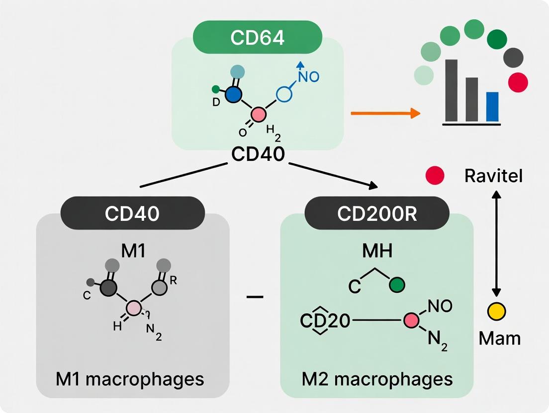
Decoding Macrophage Polarization: A Flow Cytometry Guide to M1/M2 Markers CD64, CD40 & CD200R
This comprehensive guide provides researchers, scientists, and drug development professionals with a detailed protocol and critical analysis for characterizing macrophage polarization states (M1 and M2) using flow cytometry.
Decoding Macrophage Polarization: A Flow Cytometry Guide to M1/M2 Markers CD64, CD40 & CD200R
Abstract
This comprehensive guide provides researchers, scientists, and drug development professionals with a detailed protocol and critical analysis for characterizing macrophage polarization states (M1 and M2) using flow cytometry. Focusing on the key surface markers CD64 (FcγRI), CD40, and CD200R, we explore their foundational biology, present optimized multi-color panel methodologies, address common troubleshooting scenarios, and validate their specificity against classical markers. The article serves as a practical resource for accurately identifying functional macrophage subsets in immunology, oncology, and inflammation research.
Understanding Macrophage Markers: The Biological Roles of CD64, CD40, and CD200R in M1/M2 Polarization
Within the broader thesis on validating CD64, CD40, and CD200R as discriminatory markers for M1/M2 macrophage subtyping via flow cytometry, this guide details the core plasticity paradigm. Macrophage functional polarization into classical (M1) or alternative (M2) activation states is a cornerstone of immunology and therapeutic development. Precise identification is critical for correlating phenotype with disease outcomes and drug mechanisms.
The Polarization Paradigm: M1 vs. M2
Macrophage activation is a spectrum, but the M1/M2 framework describes two functionally antagonistic extremes.
M1 (Classical Activation):
- Inducers: IFN-γ, LPS, GM-CSF.
- Key Functions: Pro-inflammatory responses, microbial killing, antitumor activity, ROS/RNS production.
- Typical Secretome: High IL-1β, IL-6, IL-12, TNF-α, iNOS.
M2 (Alternative Activation):
- Subtypes: M2a (IL-4/IL-13), M2b (Immune Complexes + TLR/IL-1R ligands), M2c (IL-10, glucocorticoids).
- Key Functions: Anti-inflammatory responses, tissue repair, angiogenesis, immunoregulation, tumor progression.
- Typical Secretome: High IL-10, TGF-β, CCL17, CCL22, Arginase-1.
Quantitative Marker Comparison
The following table consolidates core surface and intracellular markers, including those central to our thesis research (CD64, CD40, CD200R).
Table 1: Core Markers for M1 and M2 Macrophage Polarization
| Marker Category | Marker | M1 Expression | M2 Expression | Key Function/Note |
|---|---|---|---|---|
| Surface Receptors | CD64 (FcγRI) | High (Constitutive) | Moderate/High | High-affinity IgG receptor; Thesis anchor marker. |
| CD40 | High (Inducible) | Low | Co-stimulatory; drives pro-inflammatory response. | |
| CD200R | Low | High (M2a, M2c) | Inhibitory receptor; dampens inflammation. | |
| CD80/CD86 | High | Low | Co-stimulatory molecules for T cell activation. | |
| CD163 | Low | High (M2c) | Hemoglobin scavenger receptor. | |
| CD206 (MMR) | Low | High (M2a) | Mannose receptor; endocytosis. | |
| Intracellular/Secreted | iNOS (NOS2) | High | Very Low | M1-defining enzyme; produces NO. |
| Arginase-1 (ARG1) | Very Low | High (M2a) | M2a-defining enzyme; competes with iNOS for arginine. | |
| Cytokines | IL-12high, IL-23, TNF-α | IL-10high, TGF-βhigh | Functional readouts. |
Experimental Protocol:In VitroPolarization & Flow Cytometry
This protocol is foundational for generating cells for thesis validation experiments.
A. Human Monocyte-Derived Macrophage Polarization
- Isolation: Isolate CD14+ monocytes from PBMCs using magnetic beads.
- Differentiation: Culture monocytes for 6 days in RPMI-1640 + 10% FBS + 50 ng/mL M-CSF.
- Polarization (24-48 hr):
- M1: Add 20 ng/mL IFN-γ + 100 ng/mL LPS.
- M2a: Add 20 ng/mL IL-4 + 20 ng/mL IL-13.
- M2c: Add 20 ng/mL IL-10.
- Harvest: Use cell dissociation buffer.
B. Flow Cytometry Panel for Surface Staining (Example)
- Prepare single-cell suspension.
- Viability Stain: Use a fixable viability dye (e.g., Zombie NIR) for 20 min on ice.
- Fc Block: Incubate with human Fc block (10 min, RT).
- Surface Antibody Cocktail: Incubate for 30 min in the dark at 4°C.
- Panel Suggestion: CD64-BV421, CD40-PE, CD200R-APC, CD86-PE/Cy7, CD206-FITC, HLA-DR-PerCP/Cy5.5.
- Wash twice and fix (e.g., 4% PFA for 15 min).
- For Intracellular Staining (iNOS/Arginase-1): Permeabilize (0.5% saponin), then incubate with intracellular antibodies for 45 min at 4°C.
- Acquire data on a flow cytometer (e.g., BD Fortessa) with appropriate compensation controls. Analyze using FlowJo.
Diagram 1: In vitro macrophage polarization and staining workflow.
Key Signaling Pathways
Diagram 2: Core M1 polarization signaling pathways.
Diagram 3: Core M2a polarization signaling pathway.
The Scientist's Toolkit: Research Reagent Solutions
Table 2: Essential Reagents for Macrophage Plasticity Research
| Reagent Category | Specific Example | Function in Research |
|---|---|---|
| Polarization Cytokines | Recombinant Human IFN-γ, LPS (E. coli), IL-4, IL-13, IL-10, M-CSF | Induce specific M1 or M2 polarization states in vitro. |
| Flow Cytometry Antibodies | Anti-human CD64, CD40, CD200R, CD206, CD86, HLA-DR; iNOS, Arginase-1 | Surface and intracellular phenotyping of polarized populations. |
| Cell Isolation Kits | CD14+ MicroBeads (human), Pan Monocyte Isolation Kit (mouse) | Isolation of primary monocytes for differentiation. |
| Viability & Fixation Dyes | Fixable Viability Dye eFluor 780, Zombie NIR, BD Cytofix/Cytoperm | Distinguish live/dead cells and enable intracellular staining. |
| Critical Assay Kits | Nitric Oxide (NO) Assay Kit, Arginase Activity Assay Kit, ELISA for IL-12/IL-10 | Functional validation of polarization state. |
| Signaling Inhibitors | STAT6 Inhibitor (AS1517499), NF-κB Inhibitor (BAY 11-7082) | Mechanistic studies to validate pathway involvement. |
Within the comprehensive framework of CD64, CD40, and CD200R as discriminative markers for human macrophage polarization (M1 vs. M2), CD64 stands out with dual significance. As the high-affinity receptor for monomeric IgG (FcγRI), it serves as a robust pan-macrophage marker, distinguishing macrophages from dendritic cells and monocytes. Concurrently, its elevated expression is strongly associated with classical (M1) activation induced by IFN-γ and LPS, positioning it as a critical metric in flow cytometry-based immunophenotyping for research and therapeutic development.
Biology and Signaling Pathways of CD64
CD64 (FcγRI, gene name FCGR1A) is a 72 kDa transmembrane glycoprotein belonging to the immunoglobulin superfamily. Its high affinity for IgG (Ka ~10^8–10^9 M^-1) allows it to bind monomeric IgG at steady state. The receptor lacks intrinsic signaling capability but non-covalently associates with the FcR γ-chain homodimer, which contains an immunoreceptor tyrosine-based activation motif (ITAM).
Diagram: CD64 (FcγRI) Signaling and M1 Association
Quantitative Expression Data Across Cell Types and States
The utility of CD64 as a discriminative marker is grounded in its distinct expression patterns across myeloid lineages and activation states, as quantified by flow cytometry (Mean Fluorescence Intensity, MFI) and quantitative PCR.
Table 1: CD64 Expression Across Human Myeloid Cells & Activation States
| Cell Type / Condition | CD64 Surface MFI (Relative) | CD64 mRNA (FCGR1A) Fold Change | Key Differential Markers |
|---|---|---|---|
| Resting Monocytes | High (10,000-15,000) | Baseline (1x) | CD14++ CD16- |
| M1 Macrophages(IFN-γ + LPS) | Very High (25,000-40,000) | 8x - 12x | CD64++, CD40++, CD80/86+, CD200R- |
| M2a Macrophages(IL-4/IL-13) | Low/Moderate (5,000-8,000) | 0.5x - 1x | CD64+, CD200R++, CD163++, CD206+ |
| M2c Macrophages(IL-10) | Moderate (7,000-10,000) | 1x - 2x | CD64+, CD163++, CD200R+ |
| Classical DCs (cDCs) | Negative/Low (<1,000) | <0.1x | CD11c++, HLA-DR++, CD64- |
| Plasmacytoid DCs (pDCs) | Negative (<500) | <0.05x | CD123++, BDCA-2+, CD64- |
| Neutrophils | Inducible (Low to High)* | Inducible | CD66b+, CD16+, CD64 inducible by G-CSF |
*Neutrophils upregulate CD64 markedly during infection or inflammation, a key clinical biomarker.
Critical Experimental Protocols
Flow Cytometry Panel for Human Macrophage Phenotyping
This protocol details a 10-color panel to discriminate macrophage subsets using CD64 as an anchor.
Staining Protocol:
- Cell Preparation: Generate macrophages from human PBMC-derived monocytes using 7-day culture with 50 ng/mL M-CSF. Polarize with 20 ng/mL IFN-γ + 100 ng/mL LPS (M1) or 20 ng/mL IL-4 (M2a) for 48 hours. Harvest with non-enzymatic cell dissociation buffer.
- Viability Stain: Resuspend 1x10^6 cells in 100 µL PBS. Add a viability dye (e.g., Zombie NIR, 1:1000). Incubate 15 min at RT in the dark. Wash with FACS buffer (PBS + 2% FBS + 1 mM EDTA).
- FC Block: Incubate cells with Human TruStain FcX (Fc Receptor Blocking Solution) for 10 min on ice.
- Surface Staining: Add the antibody cocktail (see Toolkit Table 1) in 100 µL FACS buffer. Incubate 30 min on ice in the dark. Wash twice.
- Fixation: Fix cells in 4% PFA for 15 min at RT (optional, depending on sorting needs). Wash and resuspend in FACS buffer.
- Acquisition: Acquire on a flow cytometer capable of detecting 10 colors (e.g., 3-laser Aurora). Collect ≥50,000 events per sample.
- Gating Strategy: Gate single cells → live cells → CD14+/CD68+ macrophages → Analyze CD64 vs. CD200R. M1: CD64hi CD200Rlo/neg. M2a: CD64low/mod CD200Rhi.
Diagram: Macrophage Phenotyping Flow Gating Strategy
CD64 Internalization & Function Assay
This protocol measures receptor functionality via ligand-induced internalization.
Protocol:
- Differentiate and polarize macrophages as in 4.1.
- Chill cells on ice. Stain surface CD64 with a conjugated antibody (e.g., anti-CD64-BV421) at saturating concentration for 30 min on ice. Do not fix.
- Split cells into two tubes. Wash one tube (Time 0 control) and keep on ice.
- To the other tube, add pre-warmed media containing a cross-linking agent (e.g., F(ab')2 anti-mouse Ig, 10 µg/mL) to trigger CD64 clustering and internalization. Incubate at 37°C for 15, 30, and 60 min.
- Immediately stop internalization by transferring tubes to ice and adding ice-cold FACS buffer.
- Wash cells and analyze by flow cytometry. Compare MFI at each time point to the Time 0 control. Calculate % internalization = [1 - (MFIt / MFIt0)] x 100.
- Expected Outcome: M1 macrophages typically show more rapid and extensive CD64 internalization due to higher baseline expression and active signaling machinery.
The Scientist's Toolkit: Research Reagent Solutions
Table 2: Essential Reagents for CD64/Macrophage Research
| Reagent Category | Specific Item/Clone (Example) | Function & Application Notes |
|---|---|---|
| Anti-Human CD64 Antibodies | Clone 10.1 (BV421, PE, APC) | Gold-standard clone for flow cytometry. High affinity, specific for FcγRI. |
| Polarization Cytokines | Recombinant Human IFN-γ, LPS, IL-4, M-CSF | Induce classical (M1) and alternative (M2a) polarization in monocyte-derived macrophages. |
| Validation Antibodies | Anti-CD14 (Clone M5E2), Anti-CD68, Anti-CD163, Anti-CD200R (OX-108) | Define lineage and polarization state in multi-parameter panels. |
| Fc Receptor Block | Human TruStain FcX (Fc Block) | Critical for reducing non-specific antibody binding to CD64 and other FcγRs. |
| Intracellular Staining Kit | Foxp3/Transcription Factor Staining Buffer Set | For co-staining with transcription factors (e.g., STAT1, IRF5 for M1) post-surface staining. |
| Cross-linking Reagent | F(ab')2 Goat Anti-Mouse IgG | To cross-link bound anti-CD64 primary antibody and trigger controlled receptor internalization. |
| qPCR Assays | TaqMan Gene Expression Assays: FCGR1A, NOS2, ARG1, CD40, CD200R | Quantify mRNA expression of CD64 and polarization markers. |
| Functional Assay Kits | Phospho-STAT1 (Tyr701) ELISA Kit | Confirm upstream M1-polarizing signaling activity. |
Data Interpretation and Integration with CD40 & CD200R
The diagnostic power of CD64 is maximized when integrated into a multi-marker panel. In the context of the broader thesis:
- CD64 serves as the pan-macrophage and M1-skewing marker.
- CD40, a co-stimulatory molecule, is co-upregulated with CD64 on M1 macrophages, amplifying pro-inflammatory responses.
- CD200R, an inhibitory receptor, is highly expressed on M2 subsets and is negatively correlated with CD64 expression.
A combined analysis using CD64 and CD200R provides a robust, two-dimensional axis for identifying and quantifying the M1/M2 balance in complex samples like tumor infiltrates or atherosclerotic plaques.
CD40, a member of the tumor necrosis factor receptor (TNFR) superfamily, is a critical costimulatory molecule expressed on antigen-presenting cells (APCs) such as macrophages, dendritic cells, and B cells. Its engagement with CD40 ligand (CD40L, CD154) on T cells provides a potent signal that drives pro-inflammatory immune responses. This guide examines CD40's role in polarizing macrophages toward an M1-like, classically activated phenotype, characterized by the production of inflammatory cytokines (e.g., IL-12, TNF-α), high antigen presentation capacity, and microbicidal activity. This discussion is framed within a broader thesis investigating the utility of surface markers, including CD64, CD40, and CD200R, for delineating macrophage subsets (M1 vs. M2) via flow cytometry in research and therapeutic contexts.
CD40 Signaling and Pro-Inflammatory Pathways
CD40 lacks intrinsic enzymatic activity and relies on TNFR-associated factor (TRAF) adapter proteins to transduce signals. Ligation of CD40 leads to recruitment of TRAFs (primarily TRAF2, TRAF3, TRAF5, TRAF6), initiating downstream cascades including NF-κB, MAPK (p38, JNK, ERK), and PI3K pathways. This results in the transcriptional upregulation of genes central to M1 macrophage function.
Diagram 1: Core CD40 Signaling to M1 Genes.
Association with M1-Like Phenotypes: Key Evidence
Activation of CD40 on macrophages reinforces the M1 polarization program initiated by IFN-γ and LPS. It synergizes with TLR signaling, amplifies NF-κB activity, and sustains the expression of M1-associated surface markers and cytokines.
Table 1: Impact of CD40 Signaling on Macrophage M1 Polarization Markers
| Marker/Cytokine | Change with CD40 Engagement (vs. Control) | Functional Consequence in M1 Phenotype |
|---|---|---|
| Surface CD40 | Upregulated (2-5 fold increase in MFI) | Enhances sensitivity to CD40L+ T cell help |
| HLA-DR | Increased (1.5-3 fold) | Enhanced antigen presentation |
| CD80/CD86 | Markedly increased (3-10 fold) | Elevated T cell costimulatory capacity |
| IL-12p70 | Potently induced (50-500 pg/mL)* | Drives Th1 polarization |
| TNF-α | Synergistic release with TLRs (10-100 ng/mL)* | Mediates inflammation & cytotoxicity |
| iNOS (NOS2) | Upregulated mRNA & protein (2-8 fold) | Nitric oxide production for microbicidal activity |
| IL-1β, IL-6 | Increased secretion | Pro-inflammatory cytokine storm |
Secreted amounts are cell/system dependent (e.g., human monocyte-derived macrophages, mouse peritoneal macrophages).
Experimental Protocols for Assessing CD40 in Macrophage Polarization
Protocol:In VitroGeneration and CD40-Mediated Polarization of Human M1 Macrophages
Objective: To generate M1-polarized macrophages from human monocytes and assess the additive/synergistic effect of CD40 engagement on the M1 phenotype.
Materials: See "Scientist's Toolkit" below. Procedure:
- Monocyte Isolation: Isolate CD14+ monocytes from PBMCs using positive selection (magnetic beads) or adherence. Culture in RPMI-1640 + 10% FBS + 1% Pen/Strep + 25 ng/mL M-CSF for 6 days to differentiate into M0 macrophages.
- Polarization & Stimulation: On day 6, replace medium. Set up conditions:
- M0: Medium only.
- M1: Add 20 ng/mL IFN-γ + 100 ng/mL LPS (E. coli 055:B5).
- M1 + CD40: Add IFN-γ/LPS and stimulate with soluble recombinant CD40L (1-2 µg/mL) + enhancer (e.g., cross-linking antibody or protein polymerizer).
- CD40 Only: Add CD40L/enhancer only. Incubate for 24-48 hours.
- Analysis:
- Flow Cytometry: Harvest cells with gentle scraping. Stain for surface markers: CD40-PE, CD64-FITC, CD200R-APC, HLA-DR-PerCP, CD86-BV421. Include isotype controls. Acquire on a flow cytometer and analyze MFI and population frequency.
- Cytokine Measurement: Collect supernatant. Quantify IL-12p70, TNF-α, IL-10 via ELISA or multiplex bead array.
- Functional Assay: Co-culture stimulated macrophages with autologous CFSE-labeled T cells and recall antigen. Measure T cell proliferation (CFSE dilution) and IFN-γ production.
Diagram 2: M1 Polarization and CD40 Stimulation Workflow.
Protocol: Multicolor Flow Cytometry Panel for M1/M2 Discrimination
Objective: To immunophenotype macrophages, distinguishing M1 (CD40hi, CD64+, CD200Rlo) from M2 (CD40lo, CD64+, CD200Rhi) subsets.
Staining Procedure:
- Harvest & Wash: Harvest cells, wash with PBS, and count.
- Viability Staining: Resuspend cell pellet in PBS containing a fixable viability dye (e.g., Zombie NIR) for 15 min at RT in the dark. Wash with FACS buffer (PBS + 2% FBS + 0.1% NaN2).
- FC Block (Optional): Incubate with human Fc receptor blocking reagent for 10 min on ice.
- Surface Staining: Add predetermined titers of fluorescent antibody cocktail directly. Typical panel: CD40-PE, CD64-FITC, CD200R-APC, HLA-DR-PerCP-Cy5.5, CD14-BV510, CD86-BV421. Vortex gently, incubate 30 min on ice in the dark.
- Wash & Fix: Wash twice with cold FACS buffer. Fix cells in 1-4% PFA for 20 min on ice. Wash once and resuspend in FACS buffer for acquisition.
- Acquisition & Analysis: Acquire on a ≥3-laser flow cytometer. Compensate using single-stained controls. Gate on single cells (FSC-A vs FSC-H), viable cells, CD14+ macrophages, then analyze marker expression.
The Scientist's Toolkit: Essential Research Reagents
Table 2: Key Reagent Solutions for CD40/Macrophage Research
| Reagent | Example Product/Catalog # | Function in Experiment |
|---|---|---|
| Recombinant Human/Mouse CD40L | Soluble trimeric protein, often with enhancer | The primary agonist to stimulate CD40 signaling in vitro. |
| Anti-Human CD40 (Agonistic Ab) | Clone 5C3, G28.5 | Used as an alternative to CD40L to cross-link and activate CD40. |
| M-CSF (CSF-1) | Recombinant protein | Differentiates monocytes into baseline M0 macrophages. |
| IFN-γ & LPS (E. coli) | Recombinant proteins, purified TLR4 agonist | Standard cocktail to induce classical M1 polarization. |
| Fluorochrome-conjugated Antibodies | Anti-CD40, CD64, CD200R, HLA-DR, CD86, CD14 | Essential for immunophenotyping by flow cytometry. |
| Fixable Viability Dye | Zombie Dye, LIVE/DEAD | Distinguishes live from dead cells during flow analysis. |
| Cytokine Detection Kit | ELISA for IL-12p70, TNF-α, IL-10; Luminex panels | Quantifies functional secretory output of macrophages. |
| Fc Receptor Blocking Reagent | Human TruStain FcX | Reduces nonspecific antibody binding via FcγRs. |
Data Interpretation and Therapeutic Context
Table 3: Comparative Profile of Key Macrophage Markers in Polarization States
| Marker | M1-Like (IFN-γ+LPS+CD40L) | M2-Like (IL-4/IL-13) | Notes for Flow Gating |
|---|---|---|---|
| CD40 | High (MFI: 10³-10⁴) | Low/Moderate (MFI: 10²-10³) | Definitive marker for activation. |
| CD64 (FcγRI) | High | High | Pan-macrophage marker; not polarizing. |
| CD200R | Low/Negative | High | Strong inhibitory receptor; key for M2 ID. |
| HLA-DR | Very High | Moderate | Antigen presentation capacity. |
| CD86 | Very High | Moderate | Costimulatory function. |
| Chemokine Receptor | CCR7, CXCR3 | CCR2, CXCR4 | Migration patterns. |
The robust pro-inflammatory role of CD40 makes it a double-edged sword: a target for agonism in cancer immunotherapy (e.g., to boost APCs) and for antagonism in autoimmune and chronic inflammatory diseases. Integrating CD40 analysis with other markers (CD64 for lineage, CD200R for M2 bias) in flow cytometry panels provides a powerful tool for deconvoluting macrophage heterogeneity in disease tissues and evaluating therapeutic efficacy.
CD200 receptor (CD200R) is a type I transmembrane glycoprotein belonging to the immunoglobulin superfamily. It functions as an inhibitory immune checkpoint, delivering suppressive signals upon binding to its ligand, CD200. In macrophage biology, CD200R signaling is a critical regulatory pathway associated with the anti-inflammatory, pro-tissue repair, and immunoregulatory functions characteristic of M2-like regulatory macrophages. This places CD200R alongside other markers (e.g., CD64 for FcγRI, CD40 for activation) as a key identifier within the comprehensive flow cytometric profiling of macrophage polarization states (M1 vs. M2). Targeting this axis holds significant therapeutic potential in cancer, autoimmunity, and inflammatory diseases.
CD200R Signaling and Biological Function
The CD200-CD200R interaction initiates a potent immunosuppressive signal within myeloid cells.
Pathway: Ligation of CD200R leads to the phosphorylation of immunoreceptor tyrosine-based inhibitory motifs (ITIMs) in its cytoplasmic tail. This recruits adaptor proteins Dok1 and Dok2, which subsequently activate RasGAP, leading to the inhibition of Ras/MAPK pathways. This cascade ultimately suppresses pro-inflammatory cytokine production (e.g., TNF-α, IL-12) and promotes an alternative activation phenotype.
Diagram: CD200R Inhibitory Signaling Pathway
Flow Cytometric Identification of CD200R+ M2-like Macrophages
A multi-parametric panel is essential for accurately identifying CD200R-expressing regulatory macrophages within heterogeneous cell populations.
Key Marker Panel
Table 1: Core Markers for Macrophage Phenotyping via Flow Cytometry
| Marker | Expression Profile | Primary Function | Association |
|---|---|---|---|
| CD200R | Constitutively expressed; highly upregulated on M2c/M2-reg. | Inhibitory checkpoint receptor; transduces anti-inflammatory signals. | Key M2-like Reg. |
| CD64 (FcγRI) | High on monocytes/macrophages; further upregulated by IFN-γ. | High-affinity IgG receptor; mediates ADCP, pro-inflammatory signaling. | Pan-Macrophage / M1 |
| CD40 | Inducible; generally higher on M1, but also on activated M2. | Costimulatory molecule; promotes activation, antigen presentation. | Activation (M1 bias) |
| CD80/CD86 | Inducible; typically higher on M1 macrophages. | Costimulatory ligands for CD28/CTLA-4; promote T cell activation. | M1 |
| CD163 | Shed/scavenger receptor; highly expressed on M2a/M2c. | Hemoglobin-haptoglobin scavenger; anti-inflammatory. | M2 |
| CD206 (MMR) | Mannose receptor; highly expressed on M2a. | Endocytic pattern recognition receptor; phagocytosis. | M2a |
| HLA-DR | Constitutively expressed; modulated by cytokines. | Antigen presentation (MHC Class II). | Activation |
| MerTK | Tyrosine kinase; expressed on M2c and tissue-resident. | Efferocytosis, resolution of inflammation. | M2-like Reg. |
Standardized Staining Protocol
Protocol: Surface Staining for Macrophage Phenotyping
- Cell Preparation: Isolate PBMCs or tissue-derived single-cell suspensions. For tissues, use gentle enzymatic digestion (e.g., collagenase IV/DNase I). Include a viability dye (e.g., Zombie NIR) in all steps.
- Fc Receptor Block: Resuspend ~1x10^6 cells in 100µL FACS buffer (PBS + 2% FBS + 1mM EDTA). Add human Fc block (e.g., TruStain FcX) or purified anti-CD16/CD32 for mouse cells. Incubate for 10 min on ice.
- Surface Antibody Staining: Add pre-titrated antibody cocktail directly without washing. Example Panel: anti-human CD45-BV785, CD14-BV605, CD64-APC, CD200R-PE, CD40-PerCP-Cy5.5, CD163-BV421, HLA-DR-FITC. Vortex gently and incubate for 30 min in the dark at 4°C.
- Wash & Fix: Wash cells twice with 2mL cold FACS buffer. Centrifuge at 300-400 x g for 5 min. Resuspend in 200-300µL of 1-2% PFA or commercial fixation buffer for 20 min at 4°C if immediate acquisition is not possible.
- Acquisition: Acquire on a flow cytometer equipped with appropriate lasers and filters. Collect a minimum of 100,000 events in the live single-cell gate.
- Gating Strategy: See workflow diagram below.
Diagram: Flow Cytometry Gating Strategy for M2-like Macs
The Scientist's Toolkit: Essential Research Reagents
Table 2: Key Reagent Solutions for CD200R/Macrophage Research
| Reagent | Example Product (Supplier) | Function in Experiment |
|---|---|---|
| Anti-human CD200R mAb | Clone OX108 (BioLegend), Clone 380525 (R&D Systems) | Primary identifier for CD200R protein in flow cytometry, microscopy, or functional blockade. |
| Recombinant CD200-Fc | Human CD200 / OX2 Fc Chimera (R&D Systems) | Ligand for activating CD200R signaling in in vitro functional assays (M2 polarization). |
| Fc Receptor Block | Human TruStain FcX (BioLegend), anti-mouse CD16/32 (Tonbo) | Reduces non-specific antibody binding, critical for clear surface marker detection. |
| Multicolor Flow Cytometry Antibody Panel | Anti-CD64, CD40, CD200R, CD163, CD206, HLA-DR (Various) | Simultaneous phenotyping of macrophage activation states. |
| M1/M2 Polarizing Cytokines | IFN-γ + LPS (M1); IL-4, IL-10, or Glucocorticoids (M2) (PeproTech) | Generation of control M1 and M2 macrophage populations for assay validation. |
| Phospho-Dok1/Dok2 Antibody | Phospho-Dok1 (Tyr362) (Cell Signaling Tech) | Detection of CD200R pathway activation via western blot or phospho-flow. |
| Collagenase/DNase I | Collagenase IV, DNase I (Worthington, Sigma) | Enzymatic digestion of solid tissues for macrophage isolation. |
Recent Quantitative Findings & Clinical Relevance
Table 3: Recent Quantitative Data on CD200R Expression and Function
| Study Context | Key Finding (Quantitative) | Method Used | Implication |
|---|---|---|---|
| Tumor-Associated Macrophages (TAMs) | CD200R+ TAMs in ovarian cancer correlated with high IL-10 (≥2-fold), low IL-12, and poor patient survival (HR: 1.8). | Flow Cytometry, IHC, RNA-seq | CD200R defines an immunosuppressive TAM subset; a therapeutic target. |
| M2c Polarization | IL-10 treatment upregulated CD200R expression by ~4.5-fold compared to M0, higher than M2a (IL-4; ~2.1-fold). | In vitro polarization, qPCR, MFI by Flow Cytometry | CD200R is a hallmark of regulatory M2c macrophages. |
| Autoimmunity (RA) | Synovial fluid macrophages from RA patients showed 60% higher CD200R MFI vs. osteoarthritis controls. Functional assays showed reduced TNF-α upon CD200 ligation. | Ex vivo Flow Cytometry, Cytokine ELISA | The CD200R pathway is present but potentially dysfunctional or overridden in chronic inflammation. |
| Biomarker in Sepsis | Persistently high CD200R expression on monocyte-derived macrophages predicted immune paralysis and secondary infection (AUC = 0.82). | Longitudinal Flow Cytometry | CD200R serves as a potential biomarker for immunosuppressive states. |
Advanced Experimental Protocols
Protocol:In VitroGeneration of CD200R+ M2c Macrophages
Objective: Generate and validate human M2c regulatory macrophages.
- Monocyte Isolation: Isolate CD14+ monocytes from PBMCs using positive magnetic selection.
- M0 Differentiation: Culture monocytes (1x10^6/mL) in RPMI-1640 + 10% FBS + 50ng/mL M-CSF for 6 days.
- M2c Polarization: On day 6, stimulate M0 macrophages with 20ng/mL IL-10 + 50ng/mL recombinant CD200-Fc (to engage CD200R) for 48 hours. Control: M0 media only.
- Validation:
- Flow Cytometry: Harvest cells and stain for CD200R, CD163, CD86, HLA-DR. Expected: High CD200R, high CD163, low CD86.
- Functional Assay: Re-stimulate with 100ng/mL LPS for 24h. Collect supernatant. Measure TNF-α (low) and IL-10 (high) via ELISA.
Protocol: CD200R Signaling Assay (Phospho-Flow)
Objective: Measure proximal signaling events after CD200R engagement.
- Cell Stimulation: Aliquot 5x10^5 THP-1-derived macrophages or primary macrophages per tube. Stimulate with 2µg/mL CD200-Fc or control human IgG-Fc for 0, 5, 15, and 30 minutes at 37°C.
- Fixation & Permeabilization: Immediately add pre-warmed 4% PFA, incubate 10 min at 37°C. Wash, then resuspend in 100% ice-cold methanol for 10 min on ice to permeabilize.
- Intracellular Staining: Wash with FACS buffer, stain with anti-phospho-Dok1 (Tyr362) antibody conjugated to PE for 1h at RT.
- Acquisition & Analysis: Acquire on a flow cytometer. Analyze the geometric MFI of the p-Dok1 channel over time to visualize signaling kinetics.
The classical M1/M2 macrophage paradigm, defined by markers such as CD80/CD86 (M1) and CD206/CD163 (M2), provides a foundational but oversimplified view of macrophage heterogeneity. In the context of comparative biology and complex disease states, this binary classification is insufficient. Emerging markers like CD64 (FcγRI), CD40, and CD200R offer nuanced, complementary data that reveal activation trajectories, functional states, and regulatory pathways not captured by classical indicators. This technical guide details their integrative use in flow cytometry, supporting a broader thesis on deciphering macrophage plasticity in research and drug development.
Core Marker Functions and Complementarity
The complementarity of these markers lies in their biological roles. CD64 is a high-affinity IgG receptor integral to phagocytosis and immune complex clearance, often upregulated by IFN-γ but also by certain anti-inflammatory stimuli, bridging early activation and resolution phases. CD40, a potent co-stimulatory molecule from the TNF receptor superfamily, drives pro-inflammatory cytokine production and antigen presentation, but its expression and signaling are tightly regulated. CD200R, an inhibitory receptor, delivers potent immunosuppressive signals upon binding to its ligand CD200, directly countering M1-like activation.
Table 1: Functional Comparison of Macrophage Markers
| Marker | Primary Function | Classical Association | Complementary Insight Provided |
|---|---|---|---|
| CD64 (FcγRI) | High-affinity phagocytic receptor for IgG | M1 (IFN-γ inducible) | Identifies activated macrophages regardless of polarization; marker of immune complex-driven activation. |
| CD40 | Co-stimulation, T cell priming, cytokine storm potential | M1 (LPS/IFN-γ inducible) | Measures activation potential and immunostimulatory capacity; key for checkpoint-targeted therapies. |
| CD200R | Inhibitory signaling, suppression of inflammation | M2 (IL-10/GC inducible) | Identifies regulatory, resolution-phase macrophages; indicates active suppression of M1 responses. |
| CD80/CD86 | Co-stimulation (B7 family), T cell activation | Canonical M1 | Standard for pro-inflammatory, antigen-presenting capacity. |
| CD206 (MMR) | Endocytic receptor, phagocytosis of glycoproteins | Canonical M2 | Standard for alternative activation, tissue remodeling. |
| CD163 | Hemoglobin-haptoglobin scavenger receptor | Canonical M2 | Standard for anti-inflammatory, hemoglobin clearance. |
Integrated Flow Cytometry Panel Design and Gating Strategy
A comprehensive panel should capture the continuum of states. A suggested 8-color panel for human macrophages: CD45 (BV510), CD64 (FITC), CD40 (PE), CD200R (PE-Cy7), CD80 (APC), CD86 (APC-R700), CD206 (BV605), CD163 (BV421). Live/Dead fixable dye is mandatory.
Diagram 1: Sequential Gating Strategy for Macrophage Analysis
Title: Macrophage Gating and Phenotype Identification Workflow
Detailed Experimental Protocol: Polychromatic Flow Cytometry
A. Macrophage Generation & Stimulation
- Isolate PBMCs from human blood via density gradient centrifugation (Ficoll-Paque).
- Adherence-purify monocytes (2-hour adherence in serum-free RPMI).
- Differentiate monocytes into macrophages (M0) with 100 ng/mL M-CSF for 6-7 days.
- Stimulate M0 macrophages for 24-48 hours:
- M1: 100 ng/mL LPS + 50 ng/mL IFN-γ.
- M2: 20 ng/mL IL-4 + 20 ng/mL IL-13.
- Test Condition: e.g., 10 ng/mL Dexamethasone + 10 µg/mL Immune Complexes.
B. Cell Staining for Flow Cytometry
- Harvest cells with gentle scraping in PBS + 2mM EDTA.
- Fc Block: Incubate with human Fc receptor block (e.g., Human TruStain FcX) for 10 min on ice.
- Surface Staining: Add antibody cocktail (pre-titrated) in Brilliant Stain Buffer. Vortex, incubate 30 min in the dark at 4°C.
- Wash twice with FACS Buffer (PBS + 2% FBS + 1mM EDTA).
- Viability Stain: Resuspend in fixable viability dye (e.g., Zombie NIR) in PBS for 15 min at RT in dark.
- Wash once, then fix cells with 2% PFA for 15 min at 4°C. Wash, resuspend in FACS Buffer.
- Acquire data on a flow cytometer equipped with appropriate lasers (e.g., 3-laser, 16-detector configuration). Collect ≥50,000 events in the live macrophage gate.
C. Data Analysis
- Use compensation beads for single-color controls to build a spectral unmixing matrix.
- Apply the sequential gating strategy (Diagram 1).
- Use dimensionality reduction (t-SNE, UMAP) on concatenated files from all conditions to visualize marker co-expression patterns.
- Quantify population frequencies and calculate Mean Fluorescence Intensity (MFI) ratios (e.g., CD64 MFI / CD200R MFI) for comparative analysis.
Table 2: Example Quantitative Data Output from Integrated Analysis
| Stimulation | % CD80+ CD86+ | % CD206+ CD163+ | % CD64+ CD40+ | % CD64+ CD200R+ | CD64 MFI (x10³) | Notes |
|---|---|---|---|---|---|---|
| M-CSF (M0) | 5.2 ± 1.1 | 18.5 ± 3.2 | 12.3 ± 2.4 | 8.7 ± 1.8 | 4.1 ± 0.9 | Baseline state. |
| LPS+IFN-γ (M1) | 89.7 ± 5.6 | 3.1 ± 0.8 | 95.2 ± 4.1 | 2.5 ± 0.7 | 22.5 ± 3.4 | High CD40, low CD200R. |
| IL-4+IL-13 (M2) | 3.8 ± 0.9 | 85.4 ± 6.7 | 25.4 ± 4.3 | 15.6 ± 3.2 | 8.9 ± 1.5 | Moderate CD64. |
| Dex + ICs | 10.3 ± 2.5 | 65.8 ± 7.1 | 78.9 ± 6.5 | 55.2 ± 8.3 | 15.6 ± 2.8 | Hybrid phenotype: High CD64 & CD200R. |
Signaling Pathway Context
The integrative role of these markers is best understood within their signaling networks. CD40 and CD200R often act in opposition, fine-tuning macrophage responses.
Diagram 2: CD40 and CD200R Signaling Crosstalk in Macrophages
Title: CD40 Pro-inflammatory vs. CD200R Anti-inflammatory Signaling
The Scientist's Toolkit: Essential Research Reagents
Table 3: Key Reagent Solutions for Integrated Macrophage Phenotyping
| Reagent Category | Specific Example | Function in Experiment |
|---|---|---|
| Cytokines/Growth Factors | Recombinant Human M-CSF, IFN-γ, IL-4, IL-13 | Generation of M0, M1, and M2-polarized macrophages. |
| Polarization Inducers | Ultrapure LPS, Dexamethasone | Standard M1 polarization; inducer of regulatory/anti-inflammatory states. |
| Immune Complexes | Heat-aggregated IgG or OVA-anti-OVA complexes | To stimulate Fc receptor (CD64)-mediated activation pathways. |
| Flow Cytometry Antibodies | Anti-human CD64, CD40, CD200R, CD80, CD86, CD206, CD163 | Primary detection tools for surface marker expression. |
| Fc Block | Human TruStain FcX (anti-CD16/32) | Reduces non-specific antibody binding via Fc receptors. |
| Viability Stain | Fixable Viability Dye eFluor 780 or Zombie NIR | Distinguishes live from dead cells during analysis. |
| Staining Buffer | Brilliant Stain Buffer (BSA) | Prevents fluorochrome polymer aggregation, improving stain index. |
| Dimensionality Reduction Software | FlowJo Plugins (t-SNE, UMAP) or CITRUS | For high-dimensional, unbiased analysis of macrophage subsets. |
The integration of CD64, CD40, and CD200R with classical M1/M2 markers transforms flow cytometry from a descriptive tool into a dynamic analytical platform. It captures hybrid states, regulatory checkpoints, and functional potential, moving beyond a static binary model. For drug developers, this approach is critical for identifying novel immunomodulatory targets (e.g., CD40 agonists, CD200R antagonists) and defining pharmacodynamic biomarkers in oncology, autoimmunity, and fibrosis, where macrophage plasticity is a central disease mechanism.
From Theory to Practice: Designing and Executing a CD64/CD40/CD200R Flow Cytometry Panel
Within the context of CD64, CD40, CD200R, M1, and M2 macrophage marker research, meticulous flow cytometry panel design is paramount. This technical guide details core strategies for assembling high-parameter panels that ensure robust discrimination of macrophage phenotypes in human and mouse samples, focusing on fluorochrome selection, spillover management, and logical gating hierarchies to yield reliable, publication-quality data.
Fluorochrome Selection Strategy
Selection is guided by antigen density, instrument configuration, and spectral overlap. The core principle is to pair bright fluorochromes with low-density antigens and dim fluorochromes with high-density antigens.
Table 1: Recommended Fluorochrome Pairing for Key Macrophage Markers
| Marker | Phenotype Association | Antigen Density | Recommended Fluorochromes (Bright) | Recommended Fluorochromes (Dim) |
|---|---|---|---|---|
| CD64 | Pan-macrophage, M1-skewed | High | BV421, BV605 | FITC, PE-Cy5 |
| CD40 | M1 (Activation) | Medium-Low | PE, APC | BV510, PerCP-Cy5.5 |
| CD200R | M2 (Immunoregulatory) | Low | APC-R700, PE-Cy7 | BV650, Alexa Fluor 700 |
| CD80 / CD86 | M1 (Activation) | Low-Medium | PE-Dazzle594, BV711 | PerCP-eFluor710 |
| CD163 / CD206 | M2 (Alternative) | Medium | APC, Spark NIR 685 | PE-Cy5, BV750 |
| HLA-DR / MHC II | Antigen Presentation | High | BV786, APC-Cy7 | FITC, PE |
Experimental Protocol 2.1: Antigen Density Titration
- Conjugate Titration: For each antibody-fluorochrome conjugate, perform a titration experiment using positive control cells (e.g., LPS/IFN-γ stimulated PBMCs for M1 markers).
- Staining: Prepare a series of antibody dilutions (e.g., 0.06 µg/test to 1.0 µg/test). Stain cells following standard protocols.
- Analysis: Acquire data and plot Median Fluorescence Intensity (MFI) against antibody concentration. The optimal concentration is at the plateau just before the saturation point, providing maximal signal-to-noise.
- Validation: Confirm staining index (SI = [MFIpositive – MFInegative] / [2 × SD_negative]) is >5 for low-density markers.
Spillover Management and Compensation
Spectral spillover is quantified via the Spillover Spreading Matrix (SSM). Effective management requires pre-panel calculation and post-acquisition compensation.
Table 2: Example Spillover Spreading Matrix (SSM) for a 7-Color Panel
| Fluorochrome | Laser/Detector | BV421 | PE | PE-Cy7 | APC | APC-Cy7 | BV786 | FITC |
|---|---|---|---|---|---|---|---|---|
| BV421 | 405/450-50 | -- | 0.02 | 0.01 | 0.00 | 0.00 | 0.00 | 0.15 |
| PE | 561/582-15 | 0.00 | -- | 0.35 | 0.03 | 0.01 | 0.00 | 0.02 |
| PE-Cy7 | 561/780-60 | 0.00 | 0.01 | -- | 0.00 | 0.02 | 0.00 | 0.00 |
| APC | 640/660-20 | 0.00 | 0.00 | 0.01 | -- | 0.45 | 0.04 | 0.00 |
| APC-Cy7 | 640/780-60 | 0.00 | 0.00 | 0.02 | 0.01 | -- | 0.01 | 0.00 |
| BV786 | 405/780-60 | 0.00 | 0.00 | 0.12 | 0.05 | 0.01 | -- | 0.00 |
| FITC | 488/525-50 | 0.05 | 0.25 | 0.00 | 0.00 | 0.00 | 0.00 | -- |
Experimental Protocol 3.1: Single-Color Control Preparation for Compensation
- Materials: Use compensation beads (e.g., UltraComp eBeads) or intensely stained cells.
- Staining: Prepare one tube for each fluorochrome in the panel. Add the relevant antibody-conjugate to a separate bead/cell aliquot at the titrated optimal concentration.
- Acquisition: Run each single-color control on the cytometer using the same voltage settings as the experimental panel.
- Software Calculation: Use flow cytometry software to calculate the compensation matrix automatically from the single-color files. Manually verify by reviewing compensated controls.
Diagram: Spectral Overlap and Compensation Logic
Title: Flow Cytometry Spillover and Compensation Pathway
Gating Hierarchy for Macrophage Phenotyping
A hierarchical gating strategy is critical to accurately identify and subset macrophage populations from heterogeneous samples.
Experimental Protocol 4.1: Sequential Gating for Tissue-Derived Macrophages
- Singlets: Plot FSC-A vs. FSC-H to gate on single cells, excluding aggregates.
- Live/Dead Discrimination: Gate on live cells using a viability dye (e.g., Zombie NIR, APC-Cy7 channel).
- Lineage Exclusion: Gate out T-cells (CD3+), B-cells (CD19+), and NK cells (CD56+) from human samples (or CD45R+, NK1.1+ for mouse).
- Myeloid Lineage: Gate on CD45+ CD11b+ (human/mouse) or CD45+ HLA-DR+ (human) cells.
- Macrophage Identification: Gate on CD64+ cells from the myeloid parent.
- M1/M2 Subsetting: From CD64+ macrophages:
- M1-like: Gate on CD40hi CD200Rlo/-.
- M2-like: Gate on CD200Rhi CD40lo.
- Further Resolution: Analyze expression of CD80/CD86 (M1) and CD163/CD206 (M2) within the subsets defined in step 6.
Diagram: Macrophage Gating Hierarchy Workflow
Title: Sequential Gating Strategy for M1/M2 Macrophages
The Scientist's Toolkit: Research Reagent Solutions
Table 3: Essential Reagents for Macrophage Flow Cytometry
| Reagent / Material | Function & Rationale | Example Product(s) |
|---|---|---|
| High-Quality Antibody Conjugates | Specific detection of low-density markers (e.g., CD200R). Critical for panel success. | BioLegend Brilliant Violet, Thermo Fisher Super Bright, BD Horizon BUV |
| Cell Viability Stain | Exclusion of dead cells to reduce non-specific antibody binding and autofluorescence. | Zombie dyes, LIVE/DEAD Fixable stains, 7-AAD |
| Fc Receptor Blocking Reagent | Blocks non-specific antibody binding via Fcγ receptors, highly expressed on macrophages. | Human/Mouse Fc Block (CD16/32), purified IgG, serum |
| Cell Activation Cocktails | Positive controls for M1 (CD40, CD80) and M2 (CD200R, CD206) marker expression. | LPS + IFN-γ, IL-4 + IL-13 |
| Compensation Beads | Consistent, bright particles for generating accurate single-color compensation controls. | UltraComp eBeads, ArC Reactive Beads |
| Cell Dissociation Reagent (Tissue) | Isolate viable macrophages from solid tissue with minimal surface epitope damage. | GentleMACS, Liberase TL, collagenase IV |
| Intracellular Fix/Perm Buffer | For staining of intracellular/secreted M2 markers (e.g., Arginase-1). | Foxp3/Transcription Factor Staining Buffer Set |
| Standardized Validation Cells | Cell lines or frozen PBMCs for consistent panel validation across experiments. | THP-1 (human), RAW 264.7 (mouse), commercial PBMCs |
This guide details optimized protocols for the preparation and immunophenotypic analysis of macrophages from tissues and culture, specifically for the detection of M1/M2 polarization markers (CD64, CD40, CD200R) via flow cytometry. This work is integral to a thesis investigating macrophage heterogeneity in inflammatory diseases and cancer immunotherapy, where precise discrimination of functional subsets via surface markers is critical for biomarker discovery and therapeutic target validation.
Sample Preparation Protocols
Preparation of Cultured Human Monocyte-Derived Macrophages (hMDMs)
Principle: Isolate monocytes from peripheral blood, differentiate into macrophages, and polarize with specific cytokines to induce M1 or M2 phenotypes.
Detailed Protocol:
- Peripheral Blood Mononuclear Cell (PBMC) Isolation: Collect human blood in heparinized tubes. Dilute blood 1:1 with PBS. Layer 35 mL of diluted blood over 15 mL of Ficoll-Paque PLUS in a 50 mL tube. Centrifuge at 400 × g for 30-35 minutes at 20°C with no brake. Carefully aspirate the PBMC layer at the interface.
- Monocyte Isolation: Wash PBMCs twice in PBS + 2% FBS. Resuspend cells in MACS buffer. Isolate CD14⁺ monocytes using positive selection with anti-CD14 microbeads and an LS column per manufacturer's instructions.
- Differentiation: Seed CD14⁺ monocytes at 0.5-1 × 10⁶ cells/mL in complete RPMI-1640 (10% FBS, 1% Pen/Strep, 2mM L-Glutamine) supplemented with 50 ng/mL human M-CSF. Culture for 6-7 days, with medium refreshment on day 3 or 4.
- Polarization: On day 6-7, stimulate macrophages to induce polarization for 24-48 hours.
- M1: 100 ng/mL LPS + 20 ng/mL IFN-γ.
- M2: 20 ng/mL IL-4.
Tissue Dissociation for Resident Macrophage Isolation (Murine Spleen/Peritoneal Cavity)
Principle: Mechanically and enzymatically digest solid tissues to create a single-cell suspension while preserving surface epitopes.
Detailed Protocol:
- Spleen: Place spleen in a 70 µm cell strainer set in a 6 cm dish with 3 mL of digestion medium (RPMI, 1 mg/mL Collagenase D, 50 µg/mL DNase I). Mash tissue with plunger. Incubate at 37°C for 20 min. Add 5 mL of cold FACS buffer (PBS, 2% FBS, 2mM EDTA) and strain through a 70 µm filter. Centrifuge at 500 × g for 5 min at 4°C. Lyse red blood cells using ACK lysis buffer for 3 min on ice.
- Peritoneal Lavage: Euthanize mouse, make small ventral incision. Inject 5-10 mL of cold lavage buffer (PBS, 2% FBS, 10 U/mL heparin) into the peritoneal cavity. Gently massage abdomen. Aspirate fluid using a syringe with a 21G needle. Centrifuge cells at 500 × g for 5 min at 4°C.
Staining Protocol for Flow Cytometry
Principle: Use a multi-color antibody panel to distinguish macrophages (CD64⁺) and their polarization state via CD40 (M1-associated) and CD200R (M2-associated).
Detailed Protocol:
- Cell Counting & Viability: Count cells using a hemocytometer with Trypan Blue or an automated cell counter. Aim for 1-5 × 10⁶ cells per staining panel.
- Fc Receptor Block: Resuspend cell pellet in 100 µL of FACS buffer containing a purified anti-mouse CD16/32 (FcγIII/II Receptor) antibody (1:100) for mouse cells, or human Fc Block for human cells. Incubate for 15 minutes on ice.
- Surface Staining: Add the antibody cocktail directly to the blocking mixture without washing. Use titrated antibodies in a total volume of 100 µL.
- Typical Panel: CD64-BV421, F4/80-FITC (mouse)/CD11b-BV510 (human), CD40-PE, CD200R-PerCP-Cy5.5, CD45-APC, Live/Dead Fixable Aqua (viability dye). Incubate for 30 minutes in the dark on ice.
- Wash & Fix: Wash cells twice with 2 mL cold FACS buffer. Centrifuge at 500 × g for 5 min at 4°C. Resuspend cells in 200-300 µL of FACS buffer. Pass through a 35 µm cell strainer cap into a FACS tube. Optionally, fix cells in 1-2% PFA for 15 min on ice if not acquiring immediately.
Flow Cytometry Acquisition & Gating Strategy
Instrument Setup: Calibrate cytometer daily using CST beads. Adjust PMT voltages using unstained and single-color compensation controls. Create a compensation matrix. Gating Hierarchy:
- Singlets: Plot FSC-A vs FSC-H to exclude doublets.
- Live Cells: Gate on viability dye-negative population.
- Leukocytes: Gate on CD45⁺ cells.
- Macrophages: For mouse: CD64⁺ F4/80⁺. For human: CD64⁺ CD11b⁺.
- Phenotyping: Analyze expression of CD40 (M1-skewing) and CD200R (M2-skewing) on the macrophage population.
Data Presentation
Table 1: Expected Marker Expression on Polarized Human Macrophages
| Macrophage Subset | Polarizing Signal | CD64 | CD40 | CD200R |
|---|---|---|---|---|
| M0 (Unpolarized) | M-CSF only | High | Low/- | Low |
| M1-like | LPS + IFN-γ | High | High | Low |
| M2-like | IL-4 | High | Low | High |
Table 2: Key Research Reagent Solutions
| Reagent | Function | Example/Catalog # |
|---|---|---|
| Ficoll-Paque PLUS | Density gradient medium for PBMC isolation | Cytiva, 17144002 |
| Collagenase D | Enzymatic tissue digestion for cell isolation | Roche, 11088858001 |
| Human/M-CSF | Differentiation factor for human macrophages | PeproTech, 300-25 |
| LPS & IFN-γ | Cytokines for M1 polarization | Sigma, L4391 & PeproTech, 300-02 |
| IL-4 | Cytokine for M2 polarization | PeproTech, 200-04 |
| CD16/32 Block (α-FcR) | Prevents non-specific antibody binding | BioLegend, 101302 |
| Live/Dead Fixable Dye | Viability discrimination | Thermo Fisher, L34957 |
| Fluorochrome-conjugated Antibodies | Direct immunolabeling of surface targets | See panel above (BioLegend, BD) |
| Flow Cytometry Compensation Beads | Instrument calibration & compensation | BD, 552843 |
Visualized Workflows & Pathways
Title: Sample Preparation & Staining Workflow (76 chars)
Title: Signaling Pathways Driving M1 & M2 Marker Expression (77 chars)
In high-parameter flow cytometry research, particularly in the nuanced characterization of macrophage subsets via markers like CD64, CD40, CD200R, and M1/M2 signatures, rigorous controls are non-negotiable. The accurate identification of M1 (pro-inflammatory) and M2 (anti-inflammatory/resolving) phenotypes hinges on precise fluorescence measurements. Without essential controls, spectral overlap (spillover) and non-specific antibody binding can lead to false-positive interpretations and incorrect population gating, compromising data integrity. This guide details the implementation and critical role of unstained, Fluorescence Minus One (FMO), and isotype controls within the specific context of CD64/CD40/CD200R/M1/M2 macrophage immunophenotyping.
The Role of Each Control in Macrophage Phenotyping
| Control Type | Primary Purpose | Key Application in Macrophage Studies | Common Pitfall if Omitted |
|---|---|---|---|
| Unstained | Determine autofluorescence & instrument noise. | Baseline for myeloid cells, which often have high autofluorescence. | Overestimation of dim marker expression (e.g., CD200R). |
| FMO | Define correct gate boundaries for positive populations. | Crucial for setting gates on co-expressed markers (e.g., CD64+CD40+ M1) and dim markers. | False positive events: Incorrect classification of M1 vs. M2 subsets. |
| Isotype | Assess non-specific, Fc receptor-mediated antibody binding. | Important for markers prone to Fc binding on macrophages (e.g., CD16/32). | Misinterpretation of low-level positive staining as specific signal. |
Summary of Quantitative Impact of Controls: A survey of recent literature indicates that improper gating due to omitted FMO controls can lead to a median overestimation of positive populations by 15-30% in polychromatic panels (>8 colors). For macrophage markers, the effect is most pronounced for dim markers like CD200R.
Table 1: Representative Impact of FMO Controls on Macrophage Gating Decisions
| Marker of Interest | Typical Panel Context | Median False-Positive Rate Without FMO | Recommended Control |
|---|---|---|---|
| CD40 (M1-associated) | CD64+ HLA-DR+ macrophages | 12-25% | FMO for CD40 |
| CD200R (M2-associated) | CD64+ CD163+ macrophages | 20-35% | FMO for CD200R |
| CD206 (M2-associated) | Complex 10+ color panel | 15-30% | FMO for CD206 |
Detailed Experimental Protocols
3.1. Sample Preparation & Staining Protocol for Human Monocyte-Derived Macrophages
- Cell Source: Human peripheral blood mononuclear cells (PBMCs) isolated via density gradient centrifugation. Differentiate monocytes with 50 ng/mL M-CSF for 6 days to generate M0 macrophages.
- Stimulation: Polarize with 100 ng/mL LPS + 20 ng/mL IFN-γ (M1) or 20 ng/mL IL-4 (M2) for 48 hours.
- Antibody Panel Example: CD45-BV510 (viability), CD64-PE-Cy7, HLA-DR-PerCP-Cy5.5, CD40-APC, CD200R-PE, CD163-BV421, CD206-FITC.
- Staining Procedure:
- Harvest cells, wash with PBS.
- Fc Block: Incubate with human Fc receptor blocking reagent for 10 minutes on ice.
- Viability Stain: Incubate with fixable viability dye for 15 minutes in PBS.
- Surface Stain: Wash, then incubate with antibody cocktail for 30 minutes in the dark at 4°C.
- Wash twice with cold FACS buffer.
- Fix cells with 1-2% PFA (optional) and resuspend in FACS buffer for acquisition.
3.2. Preparation of Control Tubes
- Unstained Control: Cells processed identically but with no antibodies added.
- FMO Controls: Prepare one tube for each fluorochrome in the panel. The tube contains all antibodies except the one conjugated to the fluorochrome of interest. Example: For "FMO-CD200R-PE," include all antibodies except the anti-CD200R-PE.
- Isotype Controls: For each antibody clone, use a tube where the specific antibody is replaced by an irrelevant antibody of the same isotype and conjugated to the same fluorochrome, at the same concentration. Critical: Use in conjunction with Fc block.
Data Acquisition & Gating Strategy
Acquire all control and experimental samples using identical instrument settings on a flow cytometer calibrated with compensation beads. Apply a standardized gating hierarchy.
Title: Flow Gating Strategy for M1/M2 Macrophages with FMO.
The Scientist's Toolkit: Research Reagent Solutions
Table 2: Essential Materials for Controlled Macrophage Flow Cytometry
| Item | Function & Rationale | Example/Note |
|---|---|---|
| High-Quality, Titrated Antibodies | Ensure specific, bright staining with minimal lot-to-lot variance. Critical for dim markers like CD200R. | Clone specificity matters (e.g., CD64 clone 10.1). |
| Human/Mouse Fc Receptor Block | Reduces non-specific antibody binding to FcγRs abundantly expressed on macrophages. | Essential before surface staining. |
| UltraComp eBeads / Compensation Beads | Generate single-color controls for accurate spectral spillover compensation. | Must be used for each experiment. |
| Fixable Viability Dye | Distinguish live/dead cells. Dead cells cause nonspecific binding. | Use a dye compatible with your laser/filter setup. |
| Pre-Mixed FMO Control Tubes | Commercial kits can save time and reduce pipetting errors for complex panels. | Available for common human/mouse panels. |
| Matched Isotype Controls | Irrelevant antibodies matched to the specific clone's isotype and fluorochrome. | Must be used at the same concentration as the primary Ab. |
Pathway & Experimental Workflow Visualization
Title: From Stimulus to Detection: Role of Controls.
Accurate phenotyping of human macrophage subsets (classically activated M1 and alternatively activated M2) via surface markers like CD64, CD40, CD200R, and others is critical for immunological research and therapeutic development. The reliability of this data is fundamentally dependent on the initial data acquisition phase on the flow cytometer. This guide details the technical best practices for optimizing cytometer configuration, photomultiplier tube (PMT) voltage, and threshold settings to ensure high-resolution, reproducible detection of these often low-density and co-expressed markers.
Core Principles of Signal Optimization
The goal of voltage and threshold optimization is to maximize the Signal-to-Noise Ratio (SNR) and the Stain Index (SI) for each parameter.
- Stain Index (SI) = (Median Positive – Median Negative) / (2 × SD of Negative) A higher SI indicates better separation between positive and negative populations.
Systematic Optimization Protocol
Pre-Acquisition: Cytometer Configuration & Calibration
Daily QC Protocol:
- Run standardized calibration beads (e.g., CS&T, Rainbow, or similar).
- Record laser delays, PMT voltages, and %CV for each fluorescence channel.
- Adjust PMT voltages to target values established during initial instrument characterization to maintain day-to-day consistency.
- For macrophage markers, verify the alignment of the violet (405nm) and red (640nm) lasers, crucial for markers like CD200R and CD204 often detected with BV421 or APC conjugates.
Determining Optimal PMT Voltage
Experimental Method: Voltage Titration
- Prepare a single stained control for each fluorochrome-conjugated antibody in your panel (e.g., CD64-FITC, CD40-PE, CD200R-APC).
- Use unstained and fluorescence-minus-one (FMO) controls for the same sample.
- Acquire the single-stained sample at a series of PMT voltages (e.g., 300V, 400V, 500V, 600V).
- For each voltage, record the median fluorescence intensity (MFI) of the positive population and the MFI & standard deviation (SD) of the negative population.
- Calculate the Stain Index (SI) for each voltage.
- The optimal voltage is typically found on the linear part of the voltage vs. MFI curve, before the SI plateaus or declines.
Table 1: Example Voltage Titration Data for CD64-FITC on Human Monocytes
| PMT Voltage (V) | Pos. MFI | Neg. MFI | Neg. SD | Stain Index |
|---|---|---|---|---|
| 350 | 8,500 | 520 | 28 | 142.5 |
| 400 | 18,200 | 850 | 35 | 248.2 |
| 450 | 35,000 | 1,500 | 55 | 304.5 |
| 500 | 58,000 | 2,900 | 120 | 229.6 |
| 550 | 78,000 | 5,100 | 280 | 130.2 |
Optimal Voltage: ~450V (Highest SI)
Setting Appropriate Thresholds
For macrophage analysis, which often involves rare subsets or low-density markers, a dual-threshold strategy is recommended:
- Primary Threshold (FSC or SSC): Set on FSC-A to exclude small debris and platelets. Adjust so that all intact cells are included.
- Secondary Threshold (Fluorescence): Consider a minimal threshold on a pan-myeloid marker (e.g., CD11b or CD14) when isolating macrophages from heterogeneous tissues. This must be validated against an unstained control to ensure true populations are not excluded.
Application to a CD64/CD40/CD200R Panel
Validated Experimental Workflow:
- Sample Prep: Isolate PBMCs or tissue-derived cells. Stimulate with LPS/IFN-γ (M1) or IL-4/IL-13 (M2) for 24-48h as required.
- Staining: Fc block, then stain with viability dye and surface antibody cocktail. Include FMO controls for CD40 and CD200R due to potential continuum of expression.
- Instrument Setup: Apply pre-determined optimal voltages from single-stain titrations. Set threshold on FSC-A.
- Acquisition: Acquire ≥100,000 events in the live cell gate. Use low flow rate (≤14 µL/min) for optimal sensitivity.
- Monitoring: Track time vs. event rate to identify clogging. Verify scale during acquisition; adjust voltage if populations are compressed at axis extremes.
Diagram Title: Flow Cytometry Workflow for Macrophage Phenotyping
The Scientist's Toolkit: Key Research Reagent Solutions
Table 2: Essential Materials for Macrophage Flow Cytometry
| Item | Function in Experiment | Example Product/Catalog |
|---|---|---|
| Fluorochrome-Conjugated Antibodies | Detection of specific surface markers. Critical for panel design. | Anti-human CD64-FITC, CD40-PE, CD200R-APC, CD80-BV421, CD206-PerCP-Cy5.5 |
| Cell Stimulation Cocktails | Polarize monocytes/macrophages to M1 or M2 states for assay validation. | LPS + IFN-γ (M1); IL-4 + IL-13 (M2) |
| Fc Receptor Blocking Reagent | Reduce non-specific antibody binding, crucial for CD64 (an FcγR) staining. | Human TruStain FcX or purified human IgG |
| Viability Dye | Exclude dead cells to improve accuracy of marker expression analysis. | Fixable Viability Dye eFluor 506 or Zombie NIR |
| Compensation Beads | Generate single-color controls for accurate spectral unmixing. | Anti-Mouse/Rat Ig κ/Negative Control Compensation Particles Set |
| Standardized Calibration Beads | Daily QC and performance tracking of cytometer sensitivity and alignment. | CS&T Beads, Rainbow Calibration Particles |
| Cell Isolation Kits | Enrich target populations from complex tissues (e.g., tumors, synovium). | Human Monocyte Isolation Kit (negative selection) |
| Flow Cytometry Staining Buffer | Provide optimal pH and protein content for surface staining steps. | PBS with 2% FBS and 0.09% NaN3 |
Table 3: Target Performance Metrics for Optimal Acquisition
| Parameter | Target Value/Range | Purpose & Rationale |
|---|---|---|
| Laser %CV (via beads) | < 3% for 8-peak beads | Indicates laser stability and alignment. |
| PMT Voltage (Typical Range) | 350V - 650V | Fluorochrome- and instrument-dependent. Set via titration. |
| Stain Index (SI) for Key Marker | > 3 (Minimum), Aim > 5 | Ensures clear resolution of dim populations (e.g., CD200R on M2). |
| Event Rate during Acquisition | 200 - 1,000 events/sec | Maintains sample stream stability and reduces coincidence. |
| Threshold (FSC-A on human cells) | 10,000 - 30,000 (linear scale) | Excludes sub-cellular debris while retaining small cells. |
This whitepaper details the application of a high-parameter flow cytometry panel targeting CD64, CD40, CD200R, and M1/M2 markers to dissect macrophage heterogeneity. This work is framed within a broader thesis that posits: Precise, functional stratification of macrophage populations via combinatorial surface and intracellular marker analysis is critical for elucidating their dichotomous roles in disease progression and for identifying novel therapeutic targets. This panel is specifically applied to complex in vivo and in vitro models of the tumor microenvironment (TME), autoimmune inflammation, and fibrotic disease.
Panel Design and Quantitative Marker Profiles
The core 10-color panel is designed for simultaneous identification of macrophage lineage, activation state, and functional propensity.
| Marker | Conjugate | Primary Biological Function & Interpretation | Typical Expression in Models (Median Fluorescence Intensity Range) |
|---|---|---|---|
| CD64 (FcγRI) | BV421 | High-affinity IgG receptor; constitutively expressed on monocytes/macrophages. Key for definitive macrophage gating vs. dendritic cells. | High (TME: 10⁴–10⁵, Autoimmune: 10⁴–10⁵, Fibrosis: 10⁴–10⁵) |
| CD40 | PE | Co-stimulatory molecule; indicates pro-inflammatory, immunostimulatory (M1-like) activation and antigen-presentation capacity. | Variable (TME: Low/Med, Autoimmune: High, Fibrosis: Med) |
| CD200R | APC | Inhibitory receptor; transduces immunosuppressive signals, associated with anti-inflammatory, reparative (M2-like) functions. | Variable (TME: High, Autoimmune: Low, Fibrosis: High) |
| CD86 | BV510 | Co-stimulatory molecule; marker for classical activation (M1). | Variable (TME: Low, Autoimmune: High, Fibrosis: Low/Med) |
| CD206 (MMR) | PE-Cy7 | Mannose receptor; hallmark marker for alternative activation (M2). | Variable (TME: High, Autoimmune: Low, Fibrosis: High) |
| CD11b | PerCP-Cy5.5 | Integrin; myeloid cell adhesion and migration. Pan-myeloid marker. | High (All Models: 10⁴–10⁵) |
| F4/80 | FITC | EGF-family transmembrane protein; mature murine tissue-resident macrophages. | High (Tissue-Specific) |
| Ly-6C | Alexa Fluor 700 | Monocyte differentiation marker; inflammatory monocytes (Ly-6Chi) vs. patrolling (Ly-6Clo). | Variable (Autoimmune: Ly-6Chi↑) |
| MHC II (I-A/I-E) | BV605 | Antigen presentation complex; required for T-cell activation, often downregulated in suppressive TME. | Variable (TME: Low, Autoimmune: High) |
| Live/Dead | Fixable Viability Dye eFluor 780 | Cell viability discrimination. | N/A |
Experimental Protocols
1. Sample Processing from Murine Disease Models
- Tumor Model: Harvest solid tumors (e.g., MC38 colon carcinoma) at ~1.5 cm³. Mechanically dissociate using a GentleMACS dissociator with the appropriate enzyme cocktail (e.g., Tumor Dissociation Kit, mouse). Generate a single-cell suspension and lyse RBCs.
- Autoimmune Model: Inflamed tissue from a model like experimental autoimmune encephalomyelitis (EAE) or collagen-induced arthritis (CIA). Perfuse mice with PBS prior to organ harvest. Inflamed spinal cord (EAE) or synovial tissue is digested with a neural or multi-tissue dissociation kit, respectively.
- Fibrosis Model: Liver from carbon tetrachloride (CCl₄) model or lung from bleomycin model. Perfuse liver via portal vein with PBS. Tissues are minced and digested with collagenase IV/DNase I.
- For all: Filter suspension through a 70-µm strainer, wash with FACS buffer (PBS + 2% FBS + 1mM EDTA), and count.
2. Staining Protocol for Surface Markers
- Resuspend up to 10⁷ cells in 100 µL of FACS buffer.
- Add Fc Block (anti-CD16/32) at 1 µg/10⁶ cells. Incubate on ice for 10 minutes.
- Add titrated antibody cocktail for surface markers (CD64, CD200R, CD40, CD86, CD206, CD11b, F4/80, Ly-6C, MHC II). Vortex gently and incubate for 30 minutes in the dark at 4°C.
- Wash twice with 2 mL FACS buffer, centrifuging at 400 x g for 5 min.
- Resuspend in fixation buffer (e.g., 4% PFA) for 15 min at 4°C if intracellular staining is not required. Wash once and resuspend in FACS buffer for acquisition.
3. Intracellular Staining (for iNOS, Arg1, Cytokines)
- After surface staining, fix cells with IC Fixation Buffer (e.g., Foxp3/Transcription Factor Staining Buffer Set) for 30-60 min at 4°C.
- Wash twice with 1X Permeabilization Buffer from the same kit.
- Resuspend cell pellet in 100 µL Permeabilization Buffer containing titrated antibodies against intracellular targets (e.g., iNOS, Arg1). Incubate for 30-60 min at 4°C in the dark.
- Wash twice with Permeabilization Buffer, then resuspend in FACS buffer for acquisition on a flow cytometer capable of detecting 10+ colors (e.g., 3-laser Aurora or LS RFortessa).
Signaling Pathways in Macrophage Polarization
Macrophage Polarization via CD40 and CD200R Signaling
Experimental Workflow for Disease Model Analysis
Macrophage Phenotyping Workflow in Disease Models
The Scientist's Toolkit: Essential Research Reagents
| Reagent/Material | Supplier Examples | Function in Protocol |
|---|---|---|
| Tumor Dissociation Kit, mouse | Miltenyi Biotec | Enzymatic blend for gentle, effective solid tumor dissociation into single-cell suspensions. |
| Collagenase IV / DNase I | Sigma-Aldrich, Worthington | For dissociation of fibrotic tissues (liver, lung) and inflamed synovium. |
| Fc Block (anti-mouse CD16/32) | BioLegend, Tonbo Biosciences | Blocks non-specific antibody binding to Fc receptors, critical for clean macrophage staining. |
| Fluorochrome-Conjugated Antibodies | BioLegend, BD Biosciences, Thermo Fisher | Pre-titrated antibodies for the core panel and intracellular targets (iNOS, Arg1). |
| Foxp3/Transcription Factor Staining Buffer Set | Thermo Fisher | Optimized buffers for fixation and permeabilization for intracellular antigen staining. |
| Fixable Viability Dye eFluor 780 | Thermo Fisher | Distinguishes live from dead cells, improving data quality by excluding non-viable events. |
| Precision Cell Strainers (70 µm) | Falcon, pluriSelect | Removes cell clumps and tissue debris to prevent cytometer clogging. |
| 10-Color+ Flow Cytometer | Cytek Aurora, BD Symphony | High-parameter analyzer necessary for resolving complex macrophage subsets simultaneously. |
| Flow Cytometry Analysis Software | FlowJo, FCS Express | For advanced data visualization, gating, and population quantification. |
Solving Common Challenges: Expert Tips for Optimizing Macrophage Marker Staining and Analysis
In the phenotypic and functional analysis of macrophages—including the delineation of subsets via markers like CD64 (FcγRI), CD40, CD200R, and M1/M2 signatures—flow cytometry is indispensable. However, reliable data is often compromised by poor signal-to-noise ratios. This guide, framed within a thesis on macrophage immunobiology, addresses three core technical challenges: Low Antigen Density, Suboptimal Antibody Titration, and Fc Receptor-Mediated Non-Specific Binding. Mastering these aspects is critical for drug development professionals aiming to accurately quantify target expression and assess therapeutic modulation.
Table 1: Summary of Key Troubleshooting Parameters & Quantitative Benchmarks
| Challenge | Typical Impact on MFI/Detection | Recommended Solution | Key Quantitative Metric |
|---|---|---|---|
| Low Antigen Density | Signal < 10² MFI, poor separation from isotype. | Use high-photon-yield fluorophores (e.g., PE, BV421), amplify signal. | Staining Index > 3 is desirable for low-density targets. |
| Improper Antibody Titration | High background or saturated signal, increased NSB, wasted reagent. | Perform full titration for each new antibody lot. | Optimal concentration: 80-90% of saturation MFI, with minimal background. |
| Fc Receptor Blockade | False-positive staining, particularly in CD64+ macrophages. | Use purified anti-CD16/32 or species-specific serum. | Post-blockade, isotype control MFI should drop by ≥50% for FcR+ cells. |
| Photomultiplier Tube (PMT) Voltage | Signal suboptimal or off-scale. | Set voltage using unstained and single-color controls. | Target: Place negative population in first log decade, positive peak on-scale. |
| Fluorophore Selection | Poor resolution, spillover spreading. | Match bright fluorophores to low-density antigens. | Reference: Brightness Index (PE=1.0, FITC=0.3, APC=0.8, BV421=1.2). |
Experimental Protocols
Protocol: Fc Receptor Blockade for Murine Macrophages
- Objective: To prevent non-specific antibody binding via FcγRs (especially CD16/32 and CD64).
- Reagents: Purified anti-mouse CD16/32 antibody (clone 2.4G2), FACS Buffer (PBS + 2% FBS + 0.1% NaN₃).
- Procedure:
- Prepare a single-cell suspension from tissue (e.g., peritoneal lavage, tumor digest) or culture.
- Wash cells twice with cold FACS Buffer.
- Resuspend cell pellet (up to 1x10⁷ cells) in 100µL FACS Buffer containing 1µg of anti-CD16/32 antibody.
- Incubate on ice for 10-15 minutes.
- Without washing, proceed directly to surface antibody staining cocktail addition.
Protocol: Serial Antibody Titration
- Objective: To determine the antibody concentration that provides optimal specific signal with minimal background.
- Reagents: Antibody of interest, Isotype control, FACS Buffer.
- Procedure:
- Prepare a 2x stock solution of the antibody at the manufacturer's recommended concentration.
- Perform a series of 1:2 dilutions in FACS Buffer to generate at least 5 concentrations (e.g., 1x, 0.5x, 0.25x, 0.125x, 0.0625x).
- Aliquot a constant number of cells (e.g., 2.5x10⁵) into separate tubes for each dilution, including an unstained control.
- Add 50µL of each antibody dilution to the respective cell pellet. Incubate in the dark (on ice for surface markers, per fixation protocol for intracellular).
- Wash, acquire on flow cytometer, and analyze. Plot MFI of the specific antibody and its isotype control against concentration. The optimal point is where the specific signal curve begins to plateau, while the isotype signal remains low.
Visualizing the Workflow & Biology
Title: Troubleshooting Workflow for Macrophage Flow Cytometry
Title: Mechanism of Fc Receptor Blockade in Macrophages
The Scientist's Toolkit
Table 2: Essential Research Reagent Solutions for Macrophage Flow Cytometry
| Reagent/Material | Function & Rationale | Example/Note |
|---|---|---|
| Purified anti-CD16/32 | Blocks mouse FcγRII/III to prevent non-specific antibody binding. Critical for myeloid cells. | Clone 2.4G2; use before surface staining. |
| Human FcR Blocking Reagent | Blocks human Fc receptors; often a mix of purified antibodies. | Essential for human PBMC or tumor-infiltrating macrophage analysis. |
| Brilliant Violet & PE-Cyanine Dyes | High photon-yield fluorophores for detecting low-density antigens (e.g., CD200R). | BV421, BV711, PE-Cy7, PE-Cy5.5. |
| Pre-titrated Antibody Panels | Validated, spillover-optimized panels save time and ensure reproducibility. | Commercial M1/M2 (CD64, CD40, CD206, CD163) panels. |
| Compensation Beads | Antibody-capture beads for generating accurate compensation matrices. | Required for multicolor experiments >3 colors. |
| Viability Dye | Distinguishes live cells from dead cells to exclude nonspecific staining. | Fixable viability dyes (e.g., Zombie NIR) are compatible with fixation. |
| Cell Stimulation Cocktail | For inducing cytokine production (e.g., TNF-α, IL-10) for functional M1/M2 profiling. | Used with protein transport inhibitors (brefeldin A). |
| Transcription Factor Buffer Set | Permeabilizes nuclear membrane for staining of intracellular proteins (e.g., pSTATs). | Required for markers like RORγt or FoxP3 in some subsets. |
1. Introduction and Context
The accurate identification of macrophage polarization states (e.g., M1 via CD64, M2 via CD200R) using flow cytometry is a cornerstone of immunology and drug development research. However, macrophage intrinsic properties—high phagocytic activity, granularity, and abundant intracellular vesicles—generate significant autofluorescence, complicating the detection of low-abundance surface markers like CD40. This autofluorescence elevates background, obscures dim positive populations, and reduces assay sensitivity. Within the context of a thesis investigating CD64, CD40, and CD200R expression dynamics, effective management of this background is not merely an optimization step but a prerequisite for generating reliable, publishable data.
2. Sources of Autofluorescence in Macrophages
Primary contributors include:
- Flavin adenine dinucleotide (FAD/FMN) and nicotinamide adenine dinucleotide (NAD(P)H): Emit in the 450-550 nm range (blue-green).
- Lipofuscin: A broad-spectrum emitter accumulating in lysosomes with age/cell activation.
- Extracellular matrix proteins (e.g., collagen, elastin) in tissue-derived samples.
3. Practical Reduction Strategies: A Multi-Faceted Approach
3.1. Sample Preparation & Pre-Analysis
| Strategy | Mechanism | Practical Protocol | Key Consideration |
|---|---|---|---|
| Serum Starvation | Reduces internalization of fluorescent serum components. | Culture macrophages in serum-free/low-serum media for 2-4 hours pre-harvest. | Can subtly alter activation state; include appropriate controls. |
| Quenching Agents | Chemical reduction of fluorescent molecules. | Post-fixation, incubate cells with 0.1M Glycine in PBS or 1mg/ml Sodium Borohydride (fresh) for 10-30 min on ice. | Borohydride is light-sensitive and can damage some epitopes. |
| Photobleaching | Light-mediated oxidation of fluorophores. | Expose fixed cell suspension to intense broad-spectrum light (e.g., fluorescent desk lamp) for 30-60 min on ice. | Use only on fixed cells; efficiency varies. |
3.2. Flow Cytometry Hardware & Setup
| Strategy | Mechanism | Implementation |
|---|---|---|
| Laser & Filter Selection | Avoids autofluorescence excitation/collection. | Use fluorophores excited by >600nm lasers (e.g., APC, Alexa Fluor 700) where autofluorescence is minimal. |
| Detector Optimization | Maximizes signal-to-noise. | Use a "Fluorescence Minus One (FMO)" control to set voltage thresholds, not an unstained control. |
| Spectral Unmixing | Computationally separates overlapping signals. | Utilize full-spectrum or spectral flow cytometers; acquire a single-stained control for each fluorophore. |
3.3. Panel Design & Experimental Strategy
| Strategy | Rationale | Application Example |
|---|---|---|
| Assign Bright Fluorophores to Dim Markers | Prioritizes detection sensitivity. | Assign Brilliant Violet 421 (high brightness) to low-expression CD40, not to high-expression CD64. |
| Use Tandem Dyes Cautiously | Reduces spillover spreading error. | Prefer brilliant polymer dyes over conventional tandems for markers requiring high precision. |
| Inclusion of a Dump/Discard Channel | Gates out autofluorescent debris/dead cells. | Use a viability dye (e.g., Zombie NIR) combined with lineage exclusion markers. |
4. Experimental Protocol: Integrated Workflow for High-Fidelity Macrophage Immunophenotyping
Protocol: Reduced-Background Staining for CD64, CD40, and CD200R on Bone Marrow-Derived Macrophages (BMDMs).
Materials:
- Differentiated BMDMs (M0, M1-polarized with LPS/IFN-γ, M2-polarized with IL-4).
- Staining Buffer: PBS + 2% FBS + 1mM EDTA.
- Fc Block: Anti-CD16/32 antibody.
- Antibody Cocktail: Brilliant Violet 421 anti-mouse CD40, PE/Cyanine7 anti-mouse CD64, APC anti-mouse CD200R, FITC anti-mouse F4/80 (for primary gating).
- Viability Dye: Zombie NIR Fixable Viability Kit.
- Fixation Buffer: 4% Paraformaldehyde (PFA).
- Quenching Solution: 0.1M Glycine in PBS.
Procedure:
- Harvest & Wash: Gently detach BMDMs. Wash cells twice with cold PBS to remove media components.
- Viability Staining: Resuspend cell pellet in 100µl PBS. Add 1µl Zombie NIR dye, incubate 15 min in dark at RT. Wash with 2ml staining buffer.
- Fc Block: Resuspend pellet in 100µl staining buffer with Fc block (1:100 dilution). Incubate 10 min on ice.
- Surface Staining: Add directly titrated antibody cocktail. Vortex gently. Incubate 30 min on ice, protected from light.
- Wash: Wash cells twice with 2ml cold staining buffer.
- Fixation: Resuspend in 250µl 4% PFA. Incubate 15 min at RT in dark.
- Quenching: Add 1ml 0.1M Glycine solution. Centrifuge, aspirate supernatant.
- Resuspension & Acquisition: Resuspend in 300-500µl staining buffer. Pass through a 35µm cell strainer. Acquire on a flow cytometer within 24 hours.
- Controls: Prepare single-stained compensation beads, FMO controls (for each marker), and an unstained control.
5. Visualizing the Strategy Workflow
Integrated Workflow for Low-Background Macrophage Staining
6. The Scientist's Toolkit: Key Research Reagent Solutions
| Item | Function & Rationale |
|---|---|
| Zombie NIR Fixable Viability Dye | A near-IR reactive dye for dead cell exclusion. Excitation at 633nm/emission >780nm places it outside main autofluorescence spectrum, reducing compensation spread into critical channels (e.g., PE, APC). |
| Brilliant Violet 421 (BV421) Conjugates | A bright, polymer-based fluorophore ideal for detecting low-expression markers like CD40. Its sharp emission peak improves separation from autofluorescence compared to broad-spectrum dyes like FITC. |
| Anti-CD16/32 (Fc Block) | Prevents nonspecific, Fc receptor-mediated antibody binding to macrophages, a major source of false-positive signal and high background. Essential for clean M1/M2 marker detection. |
| APC/Cyanine7 Tandem Dye | Allows CD64 (high expression) to be detected in a longer-wavelength channel, minimizing spillover into detectors used for dimmer markers. Note: Tandems can be sensitive to fixation. |
| Sodium Borohydride (Fresh) | A potent reducing agent that chemically quenches aldehyde-induced fluorescence from PFA fixation and some intrinsic autofluorescence (e.g., from lipofuscin). |
| Spectral Flow Cytometry Reference Controls | For spectral unmixing, single-stained controls (cells or beads) for every fluorophore are non-negotiable. They are the raw data for the unmixing algorithm to separate signals. |
7. Data Analysis: Gating Strategy to Isolate True Positive Signals
Sequential Gating to Minimize Background Interference
8. Conclusion
Effectively managing autofluorescence in macrophage flow cytometry requires a systematic approach integrating pre-analytical quenching, intelligent panel design (prioritizing long-wavelength fluorophores for key markers like CD200R and CD40), and rigorous gating using FMO controls. By implementing these strategies, researchers can significantly improve the resolution and reliability of CD64, CD40, and CD200R data, providing a solid experimental foundation for robust M1/M2 discrimination and advancing their broader thesis aims.
The canonical M1/M2 macrophage dichotomy, defined by classical (e.g., IFN-γ/LPS) and alternative (e.g., IL-4/IL-13) activation, is a critical framework in immunology, cancer, and fibrosis research. However, this binary model is an oversimplification. In vivo and in complex disease models, macrophages exist within a spectrum of activation states, exhibiting substantial phenotypic and functional heterogeneity. This presents a significant analytical challenge in flow cytometry. Relying on one or two markers (e.g., CD86 for M1, CD206 for M2) leads to poorly resolved, overlapping populations and obscures biologically relevant subsets.
This whitepaper, framed within a broader thesis on CD64, CD40, CD200R in M1/M2 macrophage research, details advanced gating strategies. We posit that the simultaneous, high-dimensional analysis of surface markers, including CD64 (FcγRI, constitutive on macrophages), CD40 (a potent M1-associated co-stimulatory molecule), and CD200R (an inhibitory receptor associated with M2-like regulation), in combination with traditional markers, is essential for deconvoluting the continuous M1/M2 spectrum into discrete, functionally distinct subsets.
Core Marker Panel and Quantitative Expression Profiles
A comprehensive panel must include lineage confirmation, M1-skewing, M2-skewing, and regulatory markers. The following table summarizes key markers and their expression profiles.
Table 1: Core Macrophage Marker Panel for Spectrum Resolution
| Marker | Alternative Name | Primary Association | Expression Profile (Relative Mean Fluorescence Intensity) | Key Function/Role |
|---|---|---|---|---|
| CD64 | FcγRI | Pan-Macrophage | High (Constitutive) | High-affinity IgG receptor; validates macrophage lineage. |
| CD40 | TNFRSF5 | M1 Spectrum | Low (Resting) → Very High (M1) | Co-stimulatory molecule; promotes pro-inflammatory cytokine production. |
| CD200R | OX2R | M2/Regulatory Spectrum | Variable → High (M2-Regulatory) | Immunoregulatory receptor; transduces anti-inflammatory signals. |
| CD86 | B7-2 | M1 Spectrum | Moderate → High (M1) | Co-stimulatory ligand for CD28; antigen presentation. |
| CD206 | MRC1 | M2 Spectrum | Low → High (M2) | Mannose receptor; phagocytosis and endocytosis. |
| CD163 | Scavenger Receptor | M2 Spectrum (Heme) | Low → High (M2) | Hemoglobin-haptoglobin scavenger receptor; anti-inflammatory. |
| HLA-DR | MHC Class II | Antigen Presentation | High (M1) > Moderate (M2) | Presents antigen to CD4+ T cells; often heightened in M1. |
| CD11b | Integrin αM | Myeloid Lineage | High | Adhesion, migration, and phagocytosis. |
| Ly-6C | -- | Monocyte/Subset | High (Inflammatory Monocytes) | Distinguishes monocyte-derived macrophage subsets in mice. |
Experimental Protocol: High-Dimensional Flow Cytometry for Macrophage Subsetting
A. Sample Preparation & Stimulation
- Cells: Human monocyte-derived macrophages (MDMs) from PBMCs or murine peritoneal/bone marrow-derived macrophages.
- Polarization: (1) M1: 20 ng/mL IFN-γ for 24h, followed by 100 ng/mL LPS for final 4-6h. (2) M2: 20 ng/mL IL-4 for 48h. (3) M0: Unstimulated controls. Include complex conditioning (e.g., tumor-conditioned media) for disease-relevant spectra.
- Harvesting: Use gentle cell dissociation buffer. Avoid trypsin, which cleaves surface proteins like CD200R.
B. Staining Protocol for Surface Markers
- Fc Block: Incubate cells with Human/Mouse Fc Receptor Blocking Solution for 10-15 minutes on ice.
- Viability Stain: Add a fixable viability dye (e.g., Zombie NIR) in PBS for 15 minutes in the dark.
- Surface Staining: Prepare antibody cocktail in Brilliant Stain Buffer. Use titrated, pre-conjugated antibodies against CD64, CD40, CD200R, CD86, CD206, CD163, HLA-DR, CD11b, and lineage exclusion markers (CD3, CD19, CD56). Incubate for 30 minutes in the dark at 4°C.
- Wash & Fix: Wash twice with cold FACS Buffer. Fix cells with 1-2% PFA for 15 minutes. Acquire on a flow cytometer capable of detecting 12+ colors within 24-48 hours.
C. Advanced Gating Strategy: Sequential Boolean Gating The key is moving beyond one-dimensional plots.
- Singlets → Live Cells → Lineage (CD3/19/56) Neg → CD11b+ → CD64+ (to select pure macrophages).
- High-Dimensional Analysis: Use t-Distributed Stochastic Neighbor Embedding (t-SNE) or Uniform Manifold Approximation and Projection (UMAP) on all macrophage-gated events (CD64+) using the panel of functional markers (CD40, CD200R, CD86, CD206, etc.) to visualize the spectrum.
- Identification of Subsets: Apply FlowSOM or PhenoGraph clustering on the high-dimensional data to objectively identify clusters.
- Manual Gating Validation: Based on clustering, define subsets using Boolean combinations:
- Subset 1 (M1-High): CD40++ CD86+ HLA-DR++ CD200R-.
- Subset 2 (M2-High): CD200R++ CD206++ CD163+ CD40-.
- Subset 3 (Hybrid/Intermediate): CD200R+ CD40+ CD86+ CD206+. This is the critical, often missed population.
- Subset 4 (Regulatory M2): CD200R+++ CD163++ HLA-DRlow.
Visualization of Analytical Workflow and Signaling Axes
Title: Workflow for Resolving Macrophage Heterogeneity
Title: CD40 vs CD200R Signaling Axes in Polarization
The Scientist's Toolkit: Essential Research Reagents
Table 2: Key Reagent Solutions for Advanced Macrophage Gating
| Reagent Category | Specific Example/Product | Function in the Protocol |
|---|---|---|
| Fc Receptor Block | Human TruStain FcX; Mouse Anti-CD16/32 | Blocks non-specific antibody binding via Fc receptors, critical for clear CD64/200R signals. |
| Viability Dye | Zombie NIR Fixable Viability Kit | Distinguishes live from dead cells, excluding artifacts from dead cell uptake. |
| Brilliant Stain Buffer | BD Horizon Brilliant Stain Buffer | Mitigates fluorophore interaction and quenching in high-parameter panels with BV/YG dyes. |
| High-Parameter Antibodies | Recombinant anti-human CD200R (Clone OX108) | Essential for detecting low-abundance receptors; recombinants reduce lot variation. |
| Cytokines for Polarization | Premium-grade recombinant human/mouse IFN-γ, IL-4, IL-13, LPS | Ensures specific and potent polarization to generate reference M1/M2 spectra. |
| Cell Dissociation Reagent | Enzyme-free, PBS-based dissociation buffer | Preserves sensitive epitopes (e.g., CD200R) during harvesting from adherent culture. |
| Clustering Software | FlowJo (with Plugins), R (cytolibrary), Python (Scanpy) | Enables t-SNE/UMAP visualization and computational clustering (FlowSOM) for subset discovery. |
This technical guide addresses critical optimization strategies for flow cytometric analysis of macrophages in challenging sample types, specifically within the broader thesis research on CD64, CD40, and CD200R markers for distinguishing M1 and M2 macrophage polarization states. These difficult samples—fixed tissue sections, archived cell preparations, and complex co-culture systems—present unique hurdles in antigen preservation, cell integrity, and signal specificity, directly impacting the accuracy of phenotypic identification using these key surface receptors.
Strategies for Fixed Tissue Sections
Challenge: Cross-linking fixatives (e.g., formalin) create methylene bridges that mask epitopes, particularly detrimental for conformational epitopes of CD40 and CD200R. This necessitates robust antigen retrieval.
Key Protocol: Heat-Induced Epitope Retrieval (HIER) for Flow Cytometry from Tissue Digests
- Tissue Digestion: Following fixation, mince 1 cm³ of tissue and incubate with a digestion cocktail (e.g., 2 mg/mL Collagenase IV, 0.1 mg/mL DNase I in RPMI) at 37°C for 45-60 minutes with agitation. Quench with 10% FBS.
- Cell Isolation: Pass digest through a 70 µm strainer, wash with PBS, and perform density gradient centrifugation (e.g., Percoll) to isolate mononuclear cells.
- Antigen Retrieval: Resuspend cell pellet in 1-2 mL of retrieval buffer (Tris-EDTA, pH 9.0, or Citrate buffer, pH 6.0). Heat in a water bath at 95-100°C for 20 minutes. Cool rapidly on ice for 15 minutes.
- Staining: Wash cells twice with FACS buffer. Proceed with Fc receptor blocking and standard antibody staining for CD64, CD40, CD200R, and lineage markers (CD11b, CD14, CD68).
Data Summary:
Table 1: Impact of Antigen Retrieval Buffer on Marker MFI Recovery in Formalin-Fixed Tissue
| Macrophage Marker | No Retrieval (MFI) | Citrate Buffer pH 6.0 (MFI) | Tris-EDTA pH 9.0 (MFI) | Optimal Buffer |
|---|---|---|---|---|
| CD64 (FcγRI) | 520 ± 45 | 2,150 ± 210 | 3,980 ± 320 | Tris-EDTA pH 9.0 |
| CD40 | 310 ± 32 | 1,890 ± 185 | 4,250 ± 410 | Tris-EDTA pH 9.0 |
| CD200R | 85 ± 15 | 780 ± 95 | 1,540 ± 135 | Tris-EDTA pH 9.0 |
| CD68 (Control) | 8,950 ± 650 | 9,200 ± 720 | 8,870 ± 690 | Both |
Optimization for Archived Cells (Cryopreserved)
Challenge: Ice crystal formation during freeze-thaw damages cell membranes and can lead to loss of low-density antigens like CD200R, while increasing non-specific antibody binding.
Key Protocol: Viability-Staining and Dead Cell Exclusion for Archived Samples
- Controlled Thawing: Rapidly thaw cryovial in a 37°C water bath until a small ice crystal remains. Immediately transfer cells to 10 mL of pre-warmed thawing medium (RPMI + 50% FBS + 10 U/mL DNase I).
- Wash and Filter: Centrifuge at 300 x g for 5 minutes. Resuspend in complete medium and filter through a 40 µm strainer.
- Viability Staining: Use a fixable viability dye (e.g., Zombie NIR) at a 1:1000 dilution in PBS for 15 minutes at room temperature in the dark. Wash twice.
- Fc Block and Surface Staining: Incubate with human Fc block (10 min). Stain with a pre-titrated antibody cocktail against CD45, CD11b, CD14, CD64, CD40, and CD200R for 30 minutes at 4°C.
- Fixation and Analysis: Fix with 1-2% PFA for 10 minutes. Analyze immediately, ensuring gating excludes aggregates (FSC-H vs FSC-A) and viability dye-positive dead cells.
Data Summary:
Table 2: Effect of Freeze-Thaw Cycle on Macrophage Marker Expression (MFI ± SD)
| Sample Condition | CD64 MFI | CD40 MFI | CD200R MFI | Viability (%) |
|---|---|---|---|---|
| Freshly Isolated | 12,500 ± 950 | 8,300 ± 620 | 3,200 ± 290 | 98.5 ± 0.5 |
| Cryopreserved (1 cycle) | 11,800 ± 870 | 7,950 ± 580 | 2,150 ± 210 | 92.3 ± 1.8 |
| Cryopreserved (2 cycles) | 10,500 ± 920 | 6,800 ± 550 | 1,050 ± 175 | 85.7 ± 3.1 |
Strategies for Co-culture Systems
Challenge: Cell-cell adhesion and shared secretion of cytokines in co-cultures (e.g., macrophages with tumor spheroids) cause high background, cell doublets, and phagocytic uptake of fluorescent probes.
Key Protocol: Disassociation and Selective Staining for Macrophages in Co-culture
- Gentle Disassociation: Use enzyme-free, EDTA-based dissociation buffer (e.g., 5-10 mM EDTA in PBS) at 37°C for 5-10 minutes with gentle pipetting to preserve surface markers.
- Myeloid Cell Enrichment (Optional): For highly heterogeneous cultures, use a negative selection magnetic bead kit (e.g., deplete CD3/CD19/CD56 cells) to enrich for myeloid lineage before staining.
- Intracellular Cytokine Staining (for Polarization): Stimulate co-culture with LPS/IFN-γ (M1) or IL-4 (M2) for 4-6 hours with a protein transport inhibitor (e.g., Brefeldin A). After surface staining (CD64/CD40/CD200R), fix, permeabilize (using saponin-based buffer), and stain intracellularly for TNF-α (M1) or CCL22 (M2).
- Doublet Discrimination: Critically use FSC-A vs FSC-H and SSC-A vs SSC-H gating to exclude cell aggregates during flow analysis.
Data Summary:
Table 3: Marker Profile of Macrophages Co-cultured with Tumor Cells (72 hours)
| Co-culture Condition | CD64 MFI | CD40 MFI | CD200R MFI | % TNF-α+ (M1) | % CCL22+ (M2) |
|---|---|---|---|---|---|
| Macrophages Alone | 15,200 ± 1100 | 9,500 ± 800 | 4,100 ± 350 | 68 ± 5 | 12 ± 3 |
| + Tumor Cell Line A | 14,800 ± 1050 | 3,200 ± 400 | 9,800 ± 850 | 15 ± 4 | 72 ± 6 |
| + Tumor Cell Line B | 16,100 ± 1200 | 8,100 ± 700 | 5,200 ± 450 | 55 ± 6 | 28 ± 5 |
The Scientist's Toolkit: Research Reagent Solutions
Table 4: Essential Reagents for Macrophage Marker Analysis in Difficult Samples
| Reagent / Material | Function / Purpose |
|---|---|
| Tris-EDTA Buffer (pH 9.0) | High-pH antigen retrieval buffer; crucial for unmasking CD40 and CD200R epitopes in fixed tissue. |
| Collagenase IV / DNase I Cocktail | Enzymatic digestion of fixed tissues to liberate single cells for flow cytometry. |
| Fixable Viability Dye (e.g., Zombie NIR) | Distinguishes live from dead cells in archived samples; fixable for later intracellular staining. |
| Recombinant Human Fc Block (CD16/32) | Blocks non-specific antibody binding via Fc receptors, critical for low-abundance markers like CD200R. |
| Protein Transport Inhibitor (Brefeldin A) | Inhibits cytokine secretion, allowing intracellular accumulation for M1/M2 polarization staining (TNF-α, CCL22). |
| Saponin-Based Permeabilization Buffer | Permeabilizes cell membranes post-fixation for intracellular staining without destroying light scatter properties. |
| EDTA-Based, Enzyme-Free Cell Dissociation Buffer | Gently dissociates adherent cells in co-cultures while preserving sensitive surface epitopes. |
| Magnetic Bead Myeloid Enrichment Kit | Negative selection kit to deplete non-myeloid cells (T, B, NK), enriching macrophages from complex co-cultures. |
| Pre-Titrated Antibody Cocktails | Optimized, ready-to-use mixes for CD64, CD40, CD200R, CD11b, CD14, CD68 ensure consistent staining across difficult sample preparations. |
Visualizations
Diagram Title: Optimization Workflow for Difficult Sample Types
Diagram Title: M1/M2 Markers & Associated Signaling
Within the broader thesis on delineating M1 and M2 macrophage activation states via flow cytometry, surface markers CD64, CD40, and CD200R offer high potential for precise immunophenotyping. However, their interpretation is fraught with nuance. This guide details common pitfalls in data interpretation arising from biological complexity, reagent issues, and methodological variability, providing robust experimental frameworks to ensure accurate conclusions in research and drug development.
Key Marker Biology and Misinterpretation Risks
CD64 (FcγRI): A high-affinity IgG receptor canonically associated with pro-inflammatory M1 macrophages. The primary pitfall is assuming its expression is exclusive to M1 states. It is constitutively expressed on monocytes and can be further upregulated by anti-inflammatory stimuli like IL-10 and glucocorticoids, leading to potential misclassification.
CD40: A costimulatory molecule, strongly induced by IFN-γ (M1 driver). The risk lies in interpreting CD40 as a stable M1 marker. Its expression is transient and highly activation-dependent; it can also be expressed on some M2 subsets upon alternative activation, confounding clear M1/M2 segregation.
CD200R: An inhibitory receptor, often linked to M2-like, regulatory functions. The critical misinterpretation is equating CD200R positivity solely with an M2 phenotype. It is also expressed on myeloid-derived suppressor cells (MDSCs) and can be present on M1 cells in certain tolerogenic or exhaustion contexts.
Table 1: Expression Profiles and Key Confounders of CD64, CD40, and CD200R
| Marker | Canonical Association | Key Upregulating Signal(s) | Primary Pitfall / Confounder | Reported Expression Range* (MFI or % Positive) |
|---|---|---|---|---|
| CD64 (FcγRI) | M1 / Pro-inflammatory | IFN-γ, LPS, TNF-α | Upregulation by IL-10/GCs; High constitutive monocyte expression | Monocytes: 90-100% (High MFI)M1 (IFN-γ+LPS): 95-100% (Very High MFI)M2 (IL-4/IL-13): 70-90% (Mod-High MFI) |
| CD40 | M1 / Immunostimulatory | IFN-γ, CD40L, TLR agonists | Transient expression; Inducible on some M2 subsets | Resting: 5-20% (Low MFI)M1 (IFN-γ): 60-85% (High MFI)M2 (IL-4): 10-40% (Low-Mod MFI) |
| CD200R | M2 / Immunoregulatory | IL-4, IL-10, Glucocorticoids | Expression on MDSCs; Context-dependent M1 expression | Resting: 10-30% (Low MFI)M1 (IFN-γ): 5-25% (Low MFI)M2 (IL-4): 50-75% (Mod-High MFI) |
*Ranges synthesized from recent literature; actual values are system-dependent.
Detailed Experimental Protocols
Protocol 1: Primary Human Monocyte-Derived Macrophage Differentiation & Polarization
- Isolation: Isolate CD14+ monocytes from PBMCs using positive magnetic selection.
- Differentiation: Culture monocytes for 6 days in RPMI-1640 + 10% FBS + 100 ng/mL M-CSF.
- Polarization: On day 6, polarize for 48 hours:
- M0: Media only.
- M1: 100 ng/mL IFN-γ + 100 ng/mL LPS.
- M2: 40 ng/mL IL-4 + 20 ng/mL IL-13.
- Harvest: Detach cells using enzyme-free dissociation buffer.
- Staining: Proceed to Protocol 2.
Protocol 2: High-Parameter Flow Cytometry Staining for CD64, CD40, CD200R
- Viability & FC Block: Resuspend 0.5-1x10^6 cells in PBS. Stain with viability dye (e.g., Zombie NIR) for 15 min. Wash. Incubate with human Fc receptor blocking solution for 10 min.
- Surface Staining: Add titrated antibody cocktail (see Toolkit) in Brilliant Stain Buffer. Incubate 30 min at 4°C in the dark. Wash twice.
- Fixation: Fix cells in 2% PFA for 15 min. Wash. Resuspend in staining buffer.
- Acquisition: Acquire on a ≥3-laser flow cytometer. Use standardized voltage settings from daily QC beads.
- Controls: Include FMO (Fluorescence Minus One) for each marker and isotype/biological negative controls.
Protocol 3: Data Analysis Gating Strategy
- Singlets: Use FSC-A vs. FSC-H to gate single cells.
- Live Cells: Gate on viability dye-negative population.
- Macrophage Gate: Use high side-scatter (SSC-A) and CD11b/CD68 positivity.
- Marker Analysis: Use FMO controls to set negative gates for CD64, CD40, and CD200R. Report both % positive and Median Fluorescence Intensity (MFI).
- Contextual Gating: Analyze markers in combination (e.g., CD64+CD200R+ "hybrid" population).
Signaling Pathway and Workflow Visualizations
Title: Signaling Inputs Regulating Marker Expression
Title: Macrophage Flow Cytometry Experimental Workflow
The Scientist's Toolkit: Research Reagent Solutions
Table 2: Essential Reagents and Materials for Reliable Macrophage Immunophenotyping
| Item / Reagent | Function / Role | Key Consideration to Avoid Pitfalls |
|---|---|---|
| Recombinant Human M-CSF | Drives monocyte-to-macrophage differentiation. | Use consistently across experiments; concentration affects basal marker expression. |
| Polarization Cytokines (e.g., IFN-γ, IL-4, IL-13) | Induce defined M1 or M2 phenotypes. | Verify biological activity via dose-response; endotoxin-free grade is critical. |
| Fluorochrome-Conjugated Anti-Human CD64 Antibody | Detects FcγRI surface expression. | Clone 10.1 recommended; avoid clones sensitive to blocking or internalization. |
| Fluorochrome-Conjugated Anti-Human CD40 Antibody | Detects CD40 (TNFRSF5) expression. | Clone 5C3 is standard; titrate carefully due to variable density. |
| Fluorochrome-Conjugated Anti-Human CD200R Antibody | Detects inhibitory receptor CD200R1. | Clone OX-108; ensure specificity for CD200R1 vs. other family members. |
| Brilliant Stain Buffer | Mitigates fluorochrome polymer aggregation (e.g., Brilliant Violet dyes). | Essential for polychromatic panels to prevent spectral artifacts and false positives. |
| Ultra-LEAF Purified Antibodies | Low-endotoxin, azide-free format for functional studies (e.g., CD40L). | Required when including stimulating/blocking antibodies in polarization cultures. |
| Human Fc Receptor Blocking Solution | Reduces non-specific antibody binding via Fc receptors. | Critical before surface staining, especially for CD64. |
| Compensation Beads (Anti-Mouse/Rat Ig κ) | Generate single-color controls for spectral compensation. | Must be used with the same antibody clones as the experimental stain. |
| Viability Dye (e.g., Zombie NIR) | Distinguishes live from dead cells. | Dead cells cause non-specific binding; must be used prior to fixation. |
Benchmarking Marker Specificity: Validating CD64/CD40/CD200R Against Functional Assays and Transcriptomics
1. Introduction
This technical guide addresses the pivotal challenge in macrophage immunology: correlating surface phenotype, as identified by flow cytometry, with functional cytokine output. In the context of CD64, CD40, and CD200R as discriminative surface markers for M1/M2 macrophage research, this document provides a framework for linking these markers to canonical functional profiles, primarily TNF-α (pro-inflammatory) and IL-10 (anti-inflammatory). Accurate correlation is essential for drug development targeting specific macrophage subsets in disease.
2. Surface Marker to Functional Profile: A Reference Table
The following table summarizes established correlations based on current literature, serving as a foundational reference. Note that context (stimulus, tissue, disease state) can modulate these relationships.
Table 1: Correlation of Key Surface Markers with Functional Cytokine Profiles in Human Macrophages
| Surface Marker | Common Association | Correlated Cytokine Profile | Functional Implication |
|---|---|---|---|
| CD64 (FcγRI) | M1 (Classical Activation) | High TNF-α, IL-6, IL-12; Low IL-10 | Pro-inflammatory, host defense, immune activation. |
| CD40 | M1 / Immuno-activation | High TNF-α, IL-12; Potentiates IL-23 | T-cell priming and activation, strong inflammatory response. |
| CD200R | M2 (Alternative Activation) | High IL-10, TGF-β; Low TNF-α, IL-12 | Resolution of inflammation, immunoregulation, tissue repair. |
| CD206 (MMR) | M2 (Various subtypes) | High IL-10, CCL17, CCL22; Variable TNF-α | Endocytic clearance, tissue remodeling, immunomodulation. |
| CD86 | M1 / Antigen Presentation | Context-dependent: Often with TNF-α/IL-12 | Co-stimulation for T-cell activation. |
| CD163 | M2 (Heme scavenger) | High IL-10; Low IL-12 | Anti-inflammatory, hemoglobin clearance. |
3. Detailed Experimental Protocol: Surface & Intracellular Cytokine Staining for Correlation
This protocol details a simultaneous assessment of surface markers and intracellular cytokines from a single sample using flow cytometry.
- Objective: To quantify the co-expression of surface markers (CD64, CD40, CD200R) and intracellular cytokines (TNF-α, IL-10) in human monocyte-derived macrophages (MDMs).
- Key Reagents & Solutions:
- Cell Stimulation Cocktail: PMA (Phorbol 12-myristate 13-acetate), Ionomycin, and Brefeldin A (protein transport inhibitor).
- Fluorochrome-conjugated Antibodies: Anti-CD64, CD40, CD200R, TNF-α, IL-10, and viability dye.
- Fixation/Permeabilization Buffer: Commercial kit (e.g., Foxp3/Transcription Factor Staining Buffer Set).
- Flow Cytometry Staining Buffer: PBS with 2% FBS and 2mM EDTA.
- Procedure:
- Cell Preparation & Stimulation: Differentiate MDMs from PBMCs for 5-7 days with GM-CSF (for M1-like) or M-CSF (for M2-like). Harvest cells and plate at 1x10^6 cells/mL in a 24-well plate.
- Stimulation: Add cell stimulation cocktail containing Brefeldin A to culture. Incubate for 4-6 hours at 37°C, 5% CO₂. Include an unstimulated control with Brefeldin A only.
- Harvest & Surface Stain: Harvest cells, wash, and resuspend in staining buffer. Stain with surface marker antibodies (CD64, CD40, CD200R) and viability dye for 30 minutes at 4°C in the dark.
- Fixation & Permeabilization: Wash cells, then fix and permeabilize using the commercial buffer set according to manufacturer's instructions.
- Intracellular Cytokine Stain: Wash with permeabilization buffer, then stain with anti-TNF-α and anti-IL-10 antibodies for 30 minutes at 4°C in the dark.
- Acquisition & Analysis: Wash, resuspend in buffer, and acquire data on a flow cytometer capable of detecting 5+ colors. Analyze using software (e.g., FlowJo, FCS Express). Gate on live, single cells, then analyze dual parameter plots (e.g., CD64 vs. TNF-α, CD200R vs. IL-10) or use Boolean gating to identify complex populations.
4. The Scientist's Toolkit: Key Research Reagents
Table 2: Essential Reagents for Macrophage Phenotype-Function Correlation Studies
| Reagent/Category | Example/Function | Purpose in Experiment |
|---|---|---|
| Polarizing Cytokines | GM-CSF (for M1 bias), M-CSF (for M2 bias), IL-4/IL-13 (M2a), IL-10/TGF-β (M2c) | Drives differentiation of monocytes into specific macrophage subsets with distinct surface phenotypes. |
| Activation/Stimulation Agents | LPS + IFN-γ (M1), Immune Complexes + LPS (M1), IL-4/IL-13 (M2a) | Provides specific stimuli to elicit functional cytokine responses linked to phenotype. |
| Protein Transport Inhibitors | Brefeldin A, Monensin | Blocks cytokine secretion, allowing intracellular accumulation for detection by flow cytometry. |
| Fluorochrome-conjugated Antibodies | Anti-human CD64, CD40, CD200R, CD206, TNF-α, IL-10 | Direct detection and quantification of surface and intracellular targets. Requires careful panel design for spectral overlap. |
| Fixation/Permeabilization Buffers | Commercial kits (e.g., BD Cytofix/Cytoperm, Foxp3 buffer sets) | Preserves cell structure and allows intracellular antibody access while maintaining light scatter properties. |
| Viability Dye | Propidium Iodide (PI), 7-AAD, Live/Dead fixable dyes | Excludes dead cells from analysis, improving accuracy of marker and cytokine quantification. |
| Flow Cytometry Compensation Beads | Anti-mouse/rat/hamster Ig κ/negative control compensation particles | Critical for setting accurate spectral compensation in multicolor panels. |
5. Visualizing Signaling Pathways and Experimental Workflow
Diagram 1: Signaling from Surface Marker to Cytokine Production (Max Width: 760px)
Diagram 2: Flow Cytometry Gating Strategy for Correlation (Max Width: 760px)
6. Advanced Considerations and Data Interpretation
Correlation does not always imply causation. CD40 engagement can directly signal for TNF-α production, while CD200R engagement directly inhibits pro-inflammatory pathways. However, markers like CD163 are more indicative of a regulatory state. Multidimensional analysis (t-SNE, UMAP, or SPADE) of high-parameter flow cytometry data is increasingly used to identify novel phenotype-function clusters beyond the classical M1/M2 dichotomy. For drug development, the correlation between a target (e.g., CD40) and a specific, disease-relevant cytokine profile (e.g., TNF-α) validates the target and provides a pharmacodynamic biomarker for clinical trials.
Within the broader thesis on the identification and functional characterization of human macrophage polarization states using the surface marker combination CD64 (FcγRI), CD40, and CD200R via flow cytometry, validation with orthogonal methods is paramount. The flow cytometry panel (e.g., CD64+CD40hi for M1-like and CD64+CD200Rhi for M2-like) provides high-throughput, single-cell surface protein data. However, confirming these phenotypic assignments requires correlating them with functional, transcriptional, and spatial data. This guide details the integration of qPCR (for iNOS/NOS2 and Arg1 mRNA) and Immunofluorescence (IF) to validate and deepen the findings from the core flow cytometry research.
Orthogonal Method 1: Quantitative PCR (qPCR) for Functional Transcripts
Experimental Protocol:
- Cell Sorting & Lysis: Following the flow cytometry assay for CD64/CD40/CD200R, sort pure populations (e.g., CD64+CD40hi and CD64+CD200Rhi) into RNA-stabilizing buffer. A minimum of 10,000 cells per population is recommended.
- RNA Extraction: Use a column-based or magnetic bead kit suitable for low cell numbers. Include an on-column DNase I digestion step.
- cDNA Synthesis: Use a high-efficiency reverse transcriptase with oligo(dT) and/or random primers.
- qPCR Reaction:
- Primers: Use validated primer sets.
- iNOS (NOS2): Forward: 5'-CAGCTGGGCTGTACAAACCTT-3', Reverse: 5'-CATTGGAAGTGAAGCGTTTCG-3' (Product: ~150 bp).
- Arg1: Forward: 5'-GTGGAAACTTGCATGGACAAC-3', Reverse: 5'-ATCACCTTGCCAATCCCCAG-3' (Product: ~110 bp).
- Housekeeping: HPRT1 or GAPDH.
- Master Mix: Use a SYBR Green or probe-based chemistry.
- Cycling: Standard 2-step protocol (95°C denaturation, 60°C annealing/extension for 40 cycles).
- Primers: Use validated primer sets.
- Analysis: Calculate ΔΔCt values relative to the housekeeping gene and a control sample (e.g., unpolarized M0 macrophages).
Data Presentation:
Table 1: Representative qPCR Validation of Sorted Macrophage Populations
| Sorted Population (via Flow) | Mean ΔCt iNOS (vs. HPRT1) | Mean ΔCt Arg1 (vs. HPRT1) | Fold Change iNOS (vs. M0) | Fold Change Arg1 (vs. M0) | Inference |
|---|---|---|---|---|---|
| M0 (Unpolarized) | 12.5 ± 0.3 | 9.8 ± 0.4 | 1.0 | 1.0 | Baseline |
| CD64+ CD40hi (M1-like) | 7.2 ± 0.5 | 13.1 ± 0.6 | ~36.5x ↑ | ~11x ↓ | High iNOS, low Arg1 validates M1 profile. |
| CD64+ CD200Rhi (M2-like) | 15.8 ± 0.4 | 5.1 ± 0.3 | ~11x ↓ | ~27.9x ↑ | High Arg1, low iNOS validates M2 profile. |
Orthogonal Method 2: Immunofluorescence (IF) for Spatial Protein Localization
Experimental Protocol:
- Cell Culture & Stimulation: Differentiate macrophages on glass coverslips. Stimulate for M1 (IFN-γ + LPS) and M2 (IL-4/IL-13) polarization.
- Fixation & Permeabilization: Fix with 4% PFA for 15 min. Permeabilize with 0.1% Triton X-100 for intracellular targets (iNOS, Arg1).
- Immunostaining:
- Blocking: 1-2 hours in 5% BSA/1% serum.
- Primary Antibodies (Overnight, 4°C):
- Surface/Validation: Mouse anti-CD40 (M1 correlate) OR Rabbit anti-CD200R (M2 correlate).
- Functional: Chicken anti-iNOS OR Goat anti-Arg1.
- Pan-macrophage: Rabbit anti-Iba1 or anti-CD68 (if needed).
- Secondary Antibodies (1 hour, RT): Use highly cross-adsorbed antibodies with distinct fluorophores (e.g., Alexa Fluor 488, 568, 647).
- Imaging & Analysis: Acquire images using a confocal microscope. Perform quantitative analysis (Mean Fluorescence Intensity, MFI) for co-localization (Manders' coefficient) between surface markers and functional proteins.
Data Presentation:
Table 2: Immunofluorescence Co-localization Analysis
| Polarizing Signal | Target 1 (Surface) | Target 2 (Intracellular) | Mean Fluorescence Intensity (MFI) Target 2 | Manders' Overlap Coefficient (M1 vs M2) | Validation Outcome |
|---|---|---|---|---|---|
| IFN-γ + LPS (M1) | CD40 | iNOS | High | High (>0.7) | CD40+ cells show high iNOS protein. |
| IFN-γ + LPS (M1) | CD200R | iNOS | Low | Low (<0.3) | CD200R- cells are not iNOS+. |
| IL-4 (M2) | CD200R | Arg1 | High | High (>0.7) | CD200R+ cells show high Arg1 protein. |
| IL-4 (M2) | CD40 | Arg1 | Low | Low (<0.3) | CD40- cells are not Arg1+. |
The Scientist's Toolkit: Research Reagent Solutions
Table 3: Essential Reagents for Integrated Macrophage Validation
| Item | Function in Validation | Example/Note |
|---|---|---|
| Fluorescence-Activated Cell Sorter (FACS) | Isolation of pure CD64+CD40hi and CD64+CD200Rhi populations for downstream qPCR. | Requires a sorter with at least 3 lasers. |
| High-Efficiency RNA Kit | Extraction of intact RNA from low cell numbers (e.g., 10k-50k cells) sorted from flow cytometry. | e.g., Column-based silica-membrane kits. |
| TaqMan or SYBR qPCR Assays | Quantitative measurement of NOS2 (iNOS) and Arg1 mRNA expression levels. | TaqMan probes offer higher specificity. |
| Validated Antibody Panels | Multi-color flow cytometry (surface) and multiplex immunofluorescence (surface + intracellular). | Critical: Verify clone compatibility for human vs. mouse and staining applications. |
| Confocal Microscope | High-resolution imaging to visualize co-localization of surface markers (CD40/CD200R) with functional proteins (iNOS/Arg1). | Enables z-stacking and 3D reconstruction. |
| Image Analysis Software | Quantification of fluorescence intensity (MFI) and co-localization coefficients from IF images. | e.g., ImageJ (Fiji), Imaris, or CellProfiler. |
Visualizing the Integrated Validation Workflow
Diagram Title: Integrated Orthogonal Validation Workflow
Signaling Pathways for Target Gene Regulation
Diagram Title: iNOS and Arg1 Gene Regulation Pathways
This technical guide provides an in-depth analysis of emerging CD64/CD40/CD200R flow cytometry panels against the classical M1/M2 macrophage polarization paradigm. Framed within a broader thesis on macrophage immunophenotyping, we present quantitative data, experimental protocols, and essential resources for researchers and drug development professionals.
Macrophage functional states are traditionally classified using surface markers and cytokines indicative of pro-inflammatory (M1) or anti-inflammatory/reparative (M2) activation. The classical M1/M2 dichotomy, while foundational, is increasingly viewed as an oversimplification of a continuous spectrum of activation states. Emerging panels focusing on markers like CD64 (FcγRI), CD40, and CD200R offer a more nuanced and potentially more reliable method for characterizing human macrophage subsets in health and disease, particularly in translational research.
Quantitative Comparison of Marker Panels
Table 1: Core Marker Comparison
| Marker | Traditional Panel Association | Expression/Function | CD64/CD40/CD200R Panel Role | Key Reference (Sample Type) |
|---|---|---|---|---|
| CD64 (FcγRI) | M1-skewed | High-affinity IgG Fc receptor; upregulated by IFN-γ. | Primary segregator; identifies mature, inflammatory-prone monocytes/macrophages. | (Human PBMCs, Sepsis) |
| CD40 | M1 (Activation) | TNF receptor superfamily; costimulatory signal, enhances antigen presentation. | Activation state indicator; correlates with immune responsiveness. | (Human Monocyte-Derived Macrophages) |
| CD200R | M2-skewed | Immunoregulatory receptor; transduces anti-inflammatory signals. | Regulatory state indicator; identifies macrophages with inhibited inflammatory responses. | (Human Tumor-Associated Macrophages) |
| CD80/CD86 | M1 | Costimulatory molecules for T-cell activation. | Not primary in this panel; used for secondary validation. | N/A |
| CD163 | M2 | Hemoglobin scavenger receptor; induced by IL-10. | Not primary in this panel; alternative anti-inflammatory marker. | N/A |
| CD206 (MMR) | M2 | Mannose receptor; phagocytosis and endocytosis. | Not primary in this panel; alternative activation marker. | N/A |
Table 2: Performance Metrics in Disease Models (Representative Data)
| Disease Context | Panel Used | Key Finding (Quantitative) | Advantage Over M1/M2 |
|---|---|---|---|
| Sepsis | CD64/CD40/CD200R | CD64+CD40+ subset increased 5.8-fold vs. controls (p<0.001); superior diagnostic accuracy (AUC=0.94) for infection. | Better distinguishes infection from non-infestion inflammation than CD86/CD206. |
| Rheumatoid Arthritis | CD64/CD40/CD200R vs. CD80/CD163 | CD200R- synovial macrophages showed 3.2x higher TNF-α production upon restimulation. | Better functional correlation with cytokine production. |
| Solid Tumors | CD64/CD40/CD200R | CD64+CD200R+ TAMs associated with poor prognosis (HR=2.1); gradient not captured by M1/M2. | Identifies hybrid or transitional states within the TAM continuum. |
Experimental Protocols
Protocol 1: Staining and Flow Cytometry for CD64/CD40/CD200R Panel on Human PBMCs
Materials: Fresh or cryopreserved human PBMCs, Ficoll-Paque, flow cytometry staining buffer (PBS + 2% FBS + 0.1% NaN2), Fc receptor blocking reagent (human IgG). Antibodies:
- FITC anti-human CD64 (Clone 10.1)
- PE anti-human CD40 (Clone 5C3)
- APC anti-human CD200R (Clone OX-108)
- PerCP-Cy5.5 anti-human CD14 (Clone M5E2)
- Live/Dead fixable viability dye (e.g., Aqua Dead Cell Stain).
Procedure:
- Cell Preparation: Isolate PBMCs using density gradient centrifugation. Wash twice and resuspend at 10^7 cells/mL in staining buffer.
- Viability Staining: Incubate cells with viability dye for 20 min at 4°C in the dark. Wash.
- Fc Block: Incubate cells with Fc block (10 µg/mL human IgG) for 10 min on ice.
- Surface Staining: Add antibody cocktail (optimized titers) directly to cells. Vortex gently and incubate for 30 min at 4°C in the dark.
- Wash & Fix: Wash cells twice with staining buffer. Fix with 1-2% paraformaldehyde (PFA) for 15 min at 4°C. Wash and resuspend in buffer for acquisition.
- Acquisition: Acquire data on a flow cytometer (e.g., BD FACSymphony) within 24 hours. Collect ≥100,000 events in the live, single-cell gate.
- Gating Strategy: Gate lymphocytes/monocytes by FSC-A/SSC-A > single cells (FSC-H/FSC-A) > live cells > CD14+ monocytes > analyze CD64, CD40, CD200R expression.
Protocol 2: In Vitro Macrophage Polarization & Panel Validation
Materials: Human monocytic cell line (THP-1) or purified CD14+ monocytes, PMA (Phorbol 12-myristate 13-acetate), IFN-γ & LPS (for M1 polarization), IL-4 & IL-13 (for M2 polarization). Procedure:
- Differentiation: Treat THP-1 cells with 100 nM PMA for 48 hours to differentiate into adherent macrophages. For primary cells, culture monocytes with 50 ng/mL M-CSF for 6 days.
- Polarization:
- M1: Stimulate with 20 ng/mL IFN-γ + 100 ng/mL LPS for 24-48 hours.
- M2: Stimulate with 20 ng/mL IL-4 + 20 ng/mL IL-13 for 24-48 hours.
- Harvest: Scrape adherent cells gently. Wash and proceed with staining as in Protocol 1.
- Analysis: Compare expression profiles of CD64/CD40/CD200R versus traditional markers (e.g., CD80, CD86 for M1; CD163, CD206 for M2) using flow cytometry. Use median fluorescence intensity (MFI) ratios for comparison.
Signaling Pathways and Logic
The Scientist's Toolkit: Research Reagent Solutions
Table 3: Essential Materials for Panel Implementation
| Item | Supplier Examples | Function in Experiment |
|---|---|---|
| Anti-human CD64 (FITC) | BioLegend (Clone 10.1), BD Biosciences | Primary marker for inflammatory monocyte/macrophage identification. |
| Anti-human CD40 (PE) | BioLegend (Clone 5C3), Thermo Fisher | Marker for activation state and antigen-presentation potential. |
| Anti-human CD200R (APC) | BioLegend (Clone OX-108), R&D Systems | Marker for immunoregulatory, anti-inflammatory phenotype. |
| Anti-human CD14 (PerCP-Cy5.5) | BD Biosciences, BioLegend | Lineage marker to gate classical monocytes. |
| LIVE/DEAD Fixable Viability Dye | Thermo Fisher, BioLegend | Distinguishes live cells from dead cells, critical for data accuracy. |
| Fc Receptor Blocking Solution (Human) | Miltenyi Biotec, BioLegend | Blocks nonspecific antibody binding via Fc receptors. |
| Cell Staining Buffer (with Azide) | Tonbo Biosciences, BioLegend | Provides optimal pH and protein background for surface staining. |
| Flow Cytometer with 4+ Colors | BD FACSymphony, Beckman CytoFLEX | Instrument capable of detecting multiple fluorochromes simultaneously. |
| Flow Cytometry Analysis Software | FlowJo, FCS Express | For data visualization, gating, and statistical analysis of population subsets. |
This technical guide is framed within a broader thesis investigating the nuanced roles of macrophage polarization states (classically activated M1 and alternatively activated M2) in disease pathogenesis and therapeutic response. Central to this research is the validation of surface markers—specifically CD64, CD40, and CD200R—via flow cytometry to accurately delineate M1-like and M2-like phenotypes in complex in vivo research models. This document provides an in-depth analysis of marker validation case studies in two critical disease contexts: sepsis and solid tumors.
Core Marker Biology and Rationale
The selection of CD64 (FcγRI), CD40, and CD200R is based on their differential expression and functional relevance:
- CD64 (FcγRI): A high-affinity IgG receptor constitutively expressed on monocytes and macrophages. It is strongly upregulated by IFN-γ (an M1 stimulus) and serves as a robust marker for pro-inflammatory, M1-polarized macrophages.
- CD40: A co-stimulatory molecule belonging to the TNF receptor superfamily. Its expression is enhanced on M1 macrophages. Ligation by CD40L on T cells drives macrophage activation towards pro-inflammatory and immunostimulatory functions, critical in anti-tumor immunity and sepsis hyperinflammation.
- CD200R (OX2R): An inhibitory receptor that, upon binding its ligand CD200, transduces anti-inflammatory signals. It is more commonly associated with M2-like, regulatory, or tissue-reparative macrophage phenotypes. Its expression can be induced by IL-4/IL-13.
Case Study 1: Sepsis Model (Murine CLP)
Experimental Context
Sepsis involves a dysregulated immune response to infection, characterized by an initial hyperinflammatory phase (often M1-dominant) followed by a protracted immunosuppressive phase (associated with M2 polarization). The cecal ligation and puncture (CLP) model is the gold standard for polymicrobial sepsis.
Validation Protocol
Objective: To temporally profile macrophage polarization in peritoneal and splenic compartments post-CLP. Sample Collection: Peritoneal lavage and spleen at 6h (early), 24h (peak), and 72h (late) post-CLP. Sham-operated animals serve as controls. Flow Cytometry Panel:
- Lineage Exclusion: CD3, CD19, Ly6G.
- Macrophage Identification: F4/80 (high), CD11b (high).
- Phenotype Markers: CD64 (M1), CD200R (M2), CD40 (M1/activation). MHC-II and CD86 are included as additional activation markers.
- Viability Dye: Zombie NIR.
- Intracellular Staining (Optional): For IL-1β/TNF-α (M1) or Arg1 (M2) post-fixation/permeabilization. Gating Strategy: Live → Single cells → Lin- → F4/80+ CD11b+ → Analysis of CD64, CD200R, CD40 expression (MFI and % positive).
Key Quantitative Findings
Table 1: Macrophage Marker Dynamics in Murine CLP Sepsis
| Time Point | Compartment | CD64 (MFI, Δ vs Sham) | CD200R (% Positive) | CD40 (MFI) | Inferred Phenotype Shift |
|---|---|---|---|---|---|
| 6 hours | Peritoneal Cavity | +300% | 15% | +250% | Strong M1 Skew |
| 24 hours | Peritoneal Cavity | +180% | 35% | +150% | Mixed M1/M2 |
| 72 hours | Spleen | +50% | 65% | No change | Dominant M2 Skew |
| Sham | Both | Baseline | 10% | Baseline | Homeostatic |
Interpretation
Data validate CD64 and CD40 as markers of early inflammatory response. A marked increase in CD200R+ macrophages in the spleen at 72h correlates with the onset of immunoparalysis, highlighting its utility as a marker for immunosuppressive M2-like phenotypes in late sepsis.
Case Study 2: Solid Tumor Model (Murine MC38)
Experimental Context
Tumor-associated macrophages (TAMs) are predominantly skewed towards an M2-like, pro-tumorigenic phenotype that supports angiogenesis, matrix remodeling, and immunosuppression. The MC38 colorectal adenocarcinoma model is used to study TAM biology and immunotherapy.
Validation Protocol
Objective: To characterize TAM subsets in untreated tumors versus tumors treated with an immune checkpoint inhibitor (anti-PD-1). Sample Processing: Tumors harvested at ~100-150 mm³, digested with collagenase IV/DNase I. Myeloid-derived suppressor cells (MDSCs) excluded (Ly6G+, Ly6Chigh). Flow Cytometry Panel:
- TAM Identification: CD11b+, F4/80+, MHC-II (variable), Ly6C (low/neg).
- Phenotype Markers: CD64, CD200R, CD40. Crucial Addition: CD206 (mannose receptor) as a canonical M2 marker.
- Functional Marker: PD-L1. Gating Strategy: Live → Single cells → CD11b+ → Ly6G- Ly6Clow → F4/80+ → Analysis of CD64/hi CD200R/hi CD40/hi populations.
Key Quantitative Findings
Table 2: TAM Phenotype in MC38 Tumors ± Anti-PD-1 Therapy
| Tumor Group | CD64hi (% of TAMs) | CD200Rhi (% of TAMs) | CD40hi (% of TAMs) | CD206hi (% of TAMs) | PD-L1 (MFI) |
|---|---|---|---|---|---|
| Untreated | 20% ± 5 | 75% ± 8 | 15% ± 4 | 80% ± 7 | High |
| Anti-PD-1 Treated | 45% ± 10 | 50% ± 9 | 40% ± 8 | 55% ± 10 | Moderate |
Interpretation
In untreated tumors, TAMs are overwhelmingly CD200Rhi CD64low, aligning with an M2-like state. Successful anti-PD-1 therapy drives a significant shift, increasing the proportion of CD64hi and CD40hi TAMs, indicating a re-education towards an M1-like, immunostimulatory phenotype. CD200R remains a stable marker of the residual pro-tumorigenic TAM subset.
Experimental Protocols
Protocol 1: Peritoneal Macrophage Isolation for Sepsis Time-Course.
- Euthanize mouse and aseptically expose the abdominal wall.
- Inject 10 mL of ice-cold PBS + 2% FBS + 1mM EDTA into the peritoneal cavity.
- Gently massage the abdomen for 1 minute.
- Aspirate the lavage fluid (~8-9 mL recovery).
- Centrifuge at 400 x g for 5 min at 4°C. Lyse RBCs if present. Proceed to staining.
Protocol 2: Tumor-Infiltrating Leukocyte Preparation.
- Weigh and mince harvested tumor with a scalpel.
- Digest in RPMI containing 1 mg/mL Collagenase IV, 50 µg/mL DNase I for 30-45 min at 37°C with agitation.
- Pass through a 70 µm strainer. Centrifuge at 500 x g for 5 min.
- Resuspend in 30-40% Percoll (or equivalent) and centrifuge at 700 x g for 20 min (no brake) to separate leukocytes from debris.
- Wash cells twice with FACS buffer before antibody staining.
Protocol 3: Surface Stain Flow Cytometry.
- Count cells and aliquot 1-2 x 10^6 cells per tube.
- Block Fc receptors with anti-CD16/32 antibody (1 µg/10^6 cells) for 15 min on ice.
- Add pre-titrated antibody cocktail in Brilliant Stain Buffer. Incubate for 30 min in the dark at 4°C.
- Wash twice with 2 mL FACS buffer.
- Resuspend in fixation buffer (e.g., 1% PFA) or proceed directly to acquisition on a flow cytometer equipped with appropriate lasers and filters for your fluorochromes.
Signaling and Experimental Workflow Diagrams
Title: Signaling Pathways Driving Macrophage Polarization in Sepsis
Title: Workflow for Tumor Myeloid Cell Isolation and Analysis
Title: Hierarchical Gating Strategy for Macrophage Phenotyping
The Scientist's Toolkit: Research Reagent Solutions
Table 3: Essential Reagents for Macrophage Phenotyping Studies
| Item | Function & Specification | Example Vendor/Cat # (Representative) |
|---|---|---|
| Anti-mouse CD64 (FcγRI) | Primary marker for M1-like macrophages. Clone X54-5/7.1, recommended for flow cytometry. | BioLegend, 139306 |
| Anti-mouse CD200R | Primary marker for M2-like/regulatory macrophages. Clone OX110, detects the receptor. | Invitrogen, 12-5201-82 |
| Anti-mouse CD40 | Co-stimulatory marker for activated/M1 macrophages. Clone 3/23. | BD Biosciences, 553787 |
| Anti-mouse F4/80 | Pan-macrophage marker. Clone BM8. | Tonbo Biosciences, 50-4801 |
| Anti-mouse CD11b | Integrin for myeloid cell identification. Clone M1/70. | Many vendors |
| Collagenase Type IV | Enzyme for gentle dissociation of solid tumors. | Worthington, LS004188 |
| DNase I | Prevents cell clumping by digesting extracellular DNA. | Sigma, D4513 |
| Percoll / Lympholyte-M | Density gradient medium for leukocyte enrichment from single-cell suspensions. | Cytiva, 17089109 |
| Fc Block (α-CD16/32) | Prevents non-specific antibody binding via Fc receptors. Clone 2.4G2. | BD Biosciences, 553142 |
| Zombie NIR Viability Dye | Fixed-cell compatible dye to exclude dead cells. | BioLegend, 423105 |
| Brilliant Stain Buffer | Mitigates fluorochrome polymer interaction (e.g., for Brilliant Violet dyes). | BD Biosciences, 566385 |
The translation of macrophage polarization research, centered on markers like CD64, CD40, CD200R, and the M1/M2 paradigm, from single-laboratory discoveries to validated clinical biomarkers is critically dependent on inter-laboratory reproducibility. Multi-center studies, essential for robust clinical validation, are frequently undermined by technical variability in flow cytometry protocols. This whitepaper provides a technical guide for standardizing macrophage immunophenotyping across sites, focusing on pre-analytical, analytical, and data analysis phases to ensure data fidelity and accelerate clinical translation.
Quantitative data from recent inter-laboratory comparisons and proficiency testing programs highlight key variability sources.
Table 1: Primary Sources of Inter-Laboratory Variability in Macrophage Marker Quantification
| Source Category | Specific Variable | Reported Impact on MFI/CV | Recommended Mitigation |
|---|---|---|---|
| Pre-analytical | Tissue dissociation method (enzymatic vs. mechanical) | CD64 MFI CV: 25-40% | Standardize enzyme cocktail (e.g., gentleMACS) & time. |
| Cell resting time post-isolation | CD40 expression CV: >30% | Mandate 6-hour rest in serum-free media. | |
| Cryopreservation vs. fresh analysis | CD200R+ population CV: 35% | Prefer fresh; if frozen, use standardized freeze medium. | |
| Analytical | Antibody clone and vendor | M2 (CD206) % CV: 22-50% | Centralize reagent sourcing, validate clone specificity. |
| Fluorochrome brightness & spillover | Compensation errors increase CV by 15-25% | Use tandem dye QC checks, centralized spillover matrix. | |
| Gating strategy (manual vs. automated) | M1/M2 ratio CV: >60% | Implement automated, standardized gating algorithms. | |
| Instrumental | Flow cytometer make/model & laser configuration | MFI CV across 3 instruments: 20-35% | Regular calibration with standardized beads (e.g., CS&T). |
| Daily performance verification | Drift in MFI over 1 week: up to 18% | Daily QC with 8-peak rainbow beads for laser alignment. |
Standardized Experimental Protocols
Core Protocol: Standardized Human Monocyte-Derived Macrophage (MDM) Generation & Staining
- Monocyte Isolation: Isolate PBMCs via density gradient centrifugation (Ficoll-Paque PLUS, 400 x g, 30 min, room temp, no brake). Isolate CD14+ monocytes using positive selection (anti-CD14 microbeads, LS column). Yield should be recorded (target >90% purity).
- Macrophage Differentiation: Culture 1x10^6 cells/mL in RPMI-1640 + 10% heat-inactivated FBS + 1% Pen/Strep + 50 ng/mL recombinant human M-CSF for 6 days.
- Polarization (Day 6): For M1: Add 100 ng/mL LPS + 20 ng/mL IFN-γ for 48 hours. For M2: Add 20 ng/mL IL-4 for 48 hours.
- Harvesting & Staining:
- Use enzyme-free cell dissociation buffer (10 min, 37°C).
- Wash twice in cold PBS + 0.5% BSA + 2mM EDTA (Staining Buffer).
- Viability Stain: Use fixable viability dye eFluor 780 (1:1000 in PBS, 20 min, 4°C, dark).
- Surface Staining: Resuspend in 100µL staining buffer with pre-titrated antibody cocktail. Standardized Cocktail: CD45-BV510 (HI30), CD14-PerCP-Cy5.5 (M5E2), CD64-FITC (10.1), CD40-PE (5C3), CD200R-APC (OX-108), CD206-PE-Cy7 (15-2). Incubate 30 min, 4°C, dark.
- Wash twice, fix in 1.6% paraformaldehyde (20 min, 4°C). Acquire on flow cytometer within 24 hours.
Mandatory Instrument Quality Control Protocol
- Daily: Run standardized 8-peak rainbow calibration particles. Record Mean Fluorescence Intensity (MFI) for each peak. Accept run if all peaks are resolved and channel CVs are <3%.
- Weekly: Perform full CS&T or equivalent calibration for spectral compensation and laser delay.
- Per Experiment: Include unstained, single-stained (compensation) controls, and fluorescence-minus-one (FMO) controls for each marker.
Data Analysis & Gating Standardization
The largest source of variability is often subjective gating. The following workflow must be adopted across all sites.
Diagram 1: Standardized Gating Workflow for Macrophage Phenotyping
Key Signaling Pathways in Macrophage Polarization
Understanding the pathways behind marker expression is critical for interpreting data.
Diagram 2: Signaling Pathways Driving M1/M2 Marker Expression
The Scientist's Toolkit: Essential Research Reagent Solutions
Table 2: Key Reagents for Standardized Macrophage Flow Cytometry
| Reagent Category | Specific Product/Clone | Function & Rationale for Standardization |
|---|---|---|
| Cell Isolation | Ficoll-Paque PLUS (Cytiva) | Standardized density medium for consistent PBMC yield and viability. |
| CD14 MicroBeads, human (Miltenyi) | Positive selection ensures high-purity monocyte population for differentiation. | |
| Differentiation | Recombinant Human M-CSF (PeproTech) | Gold-standard cytokine for generating M0 macrophages; use same source/batch. |
| Polarization | Ultrapure LPS (InvivoGen) & rhIFN-γ (PeproTech) | High-purity, low-endotoxin agonists for reproducible M1 polarization. |
| Recombinant Human IL-4 (PeproTech) | Key cytokine for M2 polarization; batch-to-batch consistency is critical. | |
| Flow Antibodies | CD64 Clone 10.1 (BioLegend) | High-affinity clone specific for FcγRI, well-established for M1 identification. |
| CD200R Clone OX-108 (BioLegend) | Reliable clone for detecting the inhibitory CD200 receptor on M2 cells. | |
| CD206 Clone 15-2 (BioLegend) | Recognizes the mannose receptor, a canonical M2 marker. | |
| Viability Stain | Fixable Viability Dye eFluor 780 (Invitrogen) | Near-IR dye minimizes spectral overlap with common fluorochromes. |
| Calibration | BD CS&T Beads (BD Biosciences) or CytoFLEX Daily QC Fluorospheres (Beckman) | Instrument-specific particles for performance tracking and standardization. |
| Data Analysis | FlowJo LLC (with Plugins) or FCS Express | Software capable of implementing shared workspace templates and batch analysis. |
Achieving inter-laboratory reproducibility for CD64/CD40/CD200R-based macrophage phenotyping demands a rigid, end-to-end standardized workflow. This includes centralized reagent procurement, meticulous protocol harmonization, mandatory instrument QC, and the adoption of automated, algorithm-driven analysis. Multi-center consortia must establish a central core lab for ongoing proficiency testing and data audit. Only through such rigorous standardization can flow cytometric assessment of macrophage polarization transition from a research tool to a reliable biomarker for clinical trials in immunology, oncology, and beyond.
Conclusion
The strategic use of CD64, CD40, and CD200R in flow cytometry panels offers a refined and functionally relevant approach to dissecting macrophage polarization beyond the classical M1/M2 dichotomy. This guide synthesizes the journey from foundational biology through robust methodology, troubleshooting, and rigorous validation, empowering researchers to generate reliable, high-dimensional data. As macrophage-targeted therapies advance, these markers will be crucial for patient stratification and monitoring treatment efficacy. Future directions will involve integrating these surface markers with single-cell transcriptomics and spatial biology to unravel the full complexity of macrophage heterogeneity in health and disease, paving the way for more precise immunomodulatory interventions.