Decoding the Epigenetic Clockwork: DNA Methylation Signatures as Biomarkers and Mechanisms of Chronic Inflammation
This article provides a comprehensive review of DNA methylation in chronic inflammation, addressing four core intents for a research and drug development audience.
Decoding the Epigenetic Clockwork: DNA Methylation Signatures as Biomarkers and Mechanisms of Chronic Inflammation
Abstract
This article provides a comprehensive review of DNA methylation in chronic inflammation, addressing four core intents for a research and drug development audience. First, it establishes the foundational role of DNA methylation as a key regulator of inflammatory gene expression and a recorder of inflammatory exposure. Second, it details current methodologies for profiling methylation signatures and their translation into diagnostic, prognostic, and therapeutic applications. Third, it addresses common challenges in analysis, data interpretation, and study design, offering optimization strategies for robust research. Finally, it examines validation frameworks, compares methylation signatures with other biomarker classes, and discusses their integration into clinical and pharmaceutical pipelines. The synthesis aims to equip researchers with the knowledge to leverage epigenetic signatures for advancing precision medicine in inflammatory diseases.
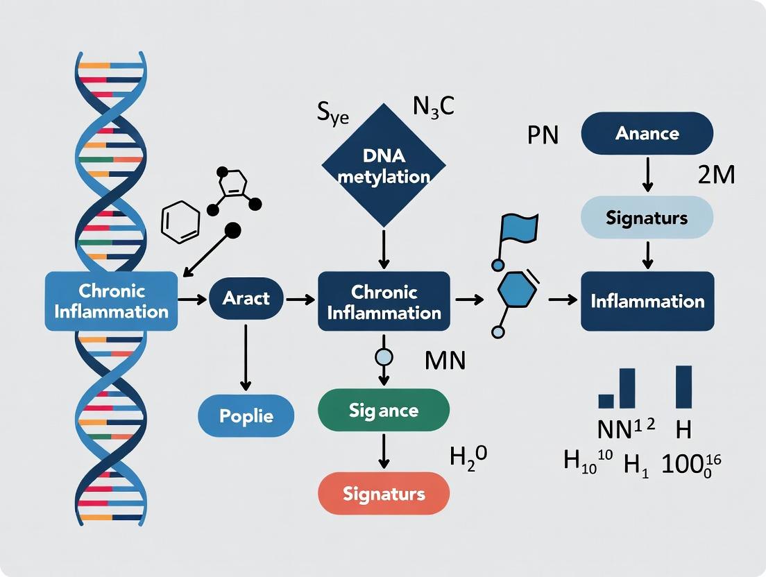
The Epigenetic Nexus: How DNA Methylation Regulates and Reflects Chronic Inflammation
Within the broader thesis on DNA methylation signatures for chronic inflammation research, a precise delineation of epigenetic regulation across inflammatory gene pathways is paramount. Chronic inflammation, a driver of numerous pathologies, is underpinned by a persistent imbalance in immune signaling. This whitepaper provides an in-depth technical guide to the core DNA methylation patterns that differentially regulate pro-inflammatory (e.g., NF-κB, JAK-STAT) and anti-inflammatory (e.g., IL-10, TGF-β) signaling pathways. Defining this landscape is critical for identifying novel diagnostic biomarkers and therapeutic targets for inflammatory diseases.
Key Methylation Patterns in Inflammatory Pathways
DNA methylation at CpG islands in promoter regions typically silences gene expression, while methylation in gene bodies or enhancer regions can have variable effects. The patterns summarized below are derived from recent studies on immune cells (e.g., monocytes, macrophages, T cells) in chronic inflammatory states.
Table 1: Core Methylation Patterns in Pro-inflammatory Pathways
| Gene/Pathway Element | Gene Symbol | Typical Methylation State in Inflammation | Functional Consequence | Key Experimental System (Cell Type) |
|---|---|---|---|---|
| Tumour Necrosis Factor Alpha | TNF | Hypomethylation at promoter | Sustained high expression, cytokine storm | Human Monocytes (M1 Macrophages) |
| Interleukin 6 | IL6 | Hypomethylation at enhancer regions | Enhanced IL-6 production | Peripheral Blood Mononuclear Cells (PBMCs) |
| Interleukin 1 Beta | IL1B | Hypomethylation in promoter & conserved non-coding sequence | Primed for rapid transcription | Monocyte-derived Macrophages |
| Cyclooxygenase-2 | PTGS2 | Hypomethylation at proximal promoter | Elevated prostaglandin synthesis | Synovial Fibroblasts (Rheumatoid Arthritis) |
| Toll-like Receptor 4 | TLR4 | Hypomethylation at specific CpG sites | Hyper-responsiveness to LPS | Monocytes in Sepsis |
Table 2: Core Methylation Patterns in Anti-inflammatory & Resolution Pathways
| Gene/Pathway Element | Gene Symbol | Typical Methylation State in Inflammation | Functional Consequence | Key Experimental System (Cell Type) |
|---|---|---|---|---|
| Interleukin 10 | IL10 | Hypermethylation at promoter & intronic regulatory regions | Suppressed expression, impaired resolution | Regulatory T Cells (Tregs) in IBD |
| Transforming Growth Factor Beta 1 | TGFB1 | Hypermethylation at promoter | Reduced TGF-β1 production, loss of immunosuppression | Tumor-Associated Macrophages |
| Suppressor of Cytokine Signaling 3 | SOCS3 | Hypermethylation at promoter | Sustained JAK-STAT signaling due to lack of feedback inhibition | Asthma Airway Epithelial Cells |
| Arginase 1 | ARG1 | Hypermethylation at promoter | Impaired alternative (M2) macrophage activation | Macrophages in Atherosclerosis |
| Forkhead Box P3 | FOXP3 | Hypermethylation at TSDR (Treg-specific demethylated region) | Loss of Treg stability and function | Tregs in Autoimmunity |
Detailed Experimental Protocols
Protocol 1: Genome-wide Methylation Analysis of Inflammatory Cell Subsets via Whole-Genome Bisulfite Sequencing (WGBS)
- Objective: To map methylation patterns at single-base resolution in sorted immune cell populations.
- Cell Sorting: Isolate target cells (e.g., CD14+ monocytes, CD4+CD25+FOXP3+ Tregs) from patient/control PBMCs using FACS. Purity >98% is critical.
- DNA Extraction & Quality Control: Use a phenol-chloroform or column-based method. Assess integrity via Bioanalyzer (RIN/DIN >7.0).
- Bisulfite Conversion: Treat 100ng-1μg genomic DNA using the EZ DNA Methylation-Lightning Kit (Zymo Research). Convert unmethylated cytosines to uracil.
- Library Preparation & Sequencing: Construct WGBS libraries using a post-bisulfite adapter tagging method (e.g., Accel-NGS Methyl-Seq DNA Library Kit). Sequence on an Illumina NovaSeq platform to achieve >30x coverage.
- Bioinformatics Analysis: Align reads to a bisulfite-converted reference genome (e.g., using Bismark). Call differentially methylated regions (DMRs) between conditions using DSS or methylKit.
Protocol 2: Targeted Methylation Analysis of Specific Loci via Pyrosequencing
- Objective: To quantitatively validate methylation levels at specific CpG sites within a gene of interest (e.g., IL10 promoter).
- Bisulfite Conversion: As in Protocol 1.
- PCR Amplification: Design primers (one biotinylated) flanking the target CpG island. Perform PCR on converted DNA.
- Pyrosequencing: Bind the biotinylated PCR product to streptavidin Sepharose beads. Denature, wash, and anneal the sequencing primer. Analyze on a Pyrosequencing instrument (e.g., Qiagen PyroMark Q96). The percentage of C vs. T at each CpG is quantified as % methylation.
- Validation: Include fully methylated and unmethylated control DNA in each run. Analyze samples in triplicate.
Signaling Pathway and Workflow Visualizations
Title: Methylation Impact on Inflammatory Gene Expression
Title: Methylation Analysis Experimental Workflow
The Scientist's Toolkit: Key Research Reagent Solutions
Table 3: Essential Reagents for DNA Methylation Studies in Inflammation
| Reagent / Kit | Primary Function | Key Application in Protocol |
|---|---|---|
| EpiQuick Total RNA Extraction Kit | Isolates high-quality, DNA-free total RNA. | Required for correlating methylation patterns with gene expression (qRT-PCR) from the same sample. |
| MethylCode Bisulfite Conversion Kit | Efficiently converts unmethylated cytosine to uracil. | Core step for both WGBS and targeted pyrosequencing; high conversion efficiency (>99.5%) is critical. |
| Illumina Infinium MethylationEPIC 850K BeadChip | Interrogates >850,000 CpG sites genome-wide. | Cost-effective discovery platform for profiling methylation in large patient cohorts. |
| PyroMark PCR Kit (Qiagen) | Optimized for amplification of bisulfite-converted DNA with high fidelity. | Preparation of templates for precise pyrosequencing of target loci. |
| Methylated & Unmethylated Human Control DNA (Zymo) | Provides 100% and 0% methylation benchmarks. | Essential controls for bisulfite conversion efficiency and pyrosequencing calibration. |
| Anti-5-methylcytosine Antibody (Clone 33D3) | Immunoprecipitates methylated DNA fragments. | Used for MeDIP-seq, an alternative method for enriching methylated genomic regions. |
| M.SssI CpG Methyltransferase | Catalyzes the in vitro methylation of all CpG sites. | Generation of fully methylated positive control DNA for assay development. |
This whitepaper is framed within the broader thesis that specific DNA methylation signatures are both biomarkers and functional regulators of chronic inflammation. A central mechanistic pillar of this thesis is the bidirectional epigenetic dysregulation observed in immune cells: the targeted hypermethylation and silencing of key immune-suppressive genes, concurrent with the hypomethylation and aberrant activation of pro-inflammatory inflammasome components. This imbalance creates a self-reinforcing inflammatory loop, perpetuating tissue damage and disease progression in conditions such as rheumatoid arthritis, systemic lupus erythematosus, and inflammatory bowel disease. Understanding this precise epigenetic code is critical for developing targeted diagnostics and therapies.
Core Mechanistic Pathways
Hypermethylation of Immune Suppressor Genes
Promoter hypermethylation mediated by DNA methyltransferases (DNMTs) leads to the transcriptional silencing of genes critical for maintaining immune tolerance and limiting inflammation.
Key Targets:
- SOCS1 (Suppressor of Cytokine Signaling 1): Silencing leads to unregulated JAK/STAT signaling and enhanced cytokine production.
- SHP-1 (PTPN6): A protein tyrosine phosphatase; its loss results in hyperactive lymphocyte and myeloid cell signaling.
- FOXP3: The master regulator of Treg function; hypermethylation impairs regulatory T cell development and function.
- PTEN: Loss allows for hyperactive PI3K/AKT signaling, promoting cell survival and inflammatory responses.
Hypomethylation of Inflammasome Components
Global or locus-specific DNA hypomethylation, potentially driven by reduced DNMT activity or increased TET-mediated demethylation, results in the overexpression of proteins central to inflammasome assembly and activation.
Key Targets:
- NLRP3: The sensor component of the NLRP3 inflammasome.
- CASP1 (Caspase-1): The effector protease that cleaves pro-IL-1β and pro-IL-18.
- IL1B & IL18: Genes encoding the potent pro-inflammatory cytokines.
- ASC (PYCARD): The adaptor protein facilitating inflammasome assembly.
Table 1: Key Methylation Changes in Chronic Inflammatory Diseases
| Target Gene | Epigenetic Change | Disease Context (Example) | Average % Methylation Change (vs. Control) | Associated Functional Outcome |
|---|---|---|---|---|
| SOCS1 Promoter | Hypermethylation | Rheumatoid Arthritis Synovium | +25-40% | Reduced SOCS1 mRNA, p-STAT3 increase |
| FOXP3 TSDR | Hypermethylation | SLE CD4+ T cells | +15-30% | Impaired Treg suppressive function |
| NLRP3 Intron 1 | Hypomethylation | IBD Monocytes | -20-35% | 3-5 fold increase in NLRP3 mRNA |
| IL1B Enhancer | Hypomethylation | Psoriasis Skin Lesions | -30-45% | Elevated IL-1β protein secretion |
| SHP-1 Promoter | Hypermethylation | ALCL Lymphoma | +50-70% | Hyperphosphorylation of Src kinases |
Table 2: Correlation of Methylation Status with Clinical Parameters
| Biomarker (Methylation) | Disease | Correlation Coefficient (r) with CRP | Correlation with Disease Activity Index |
|---|---|---|---|
| SOCS1 Promoter Methylation | RA | +0.65 | DAS28: +0.71 |
| NLRP3 Hypomethylation | Crohn's | +0.58 | CDAI: +0.62 |
| FOXP3 TSDR Methylation | SLE | +0.52 | SLEDAI: +0.59 |
Detailed Experimental Protocols
Protocol: Genome-Wide Methylation Analysis (Infinium EPIC Array)
Objective: To identify differentially methylated regions (DMRs) associated with inflammasome genes and immune suppressors.
- Bisulfite Conversion: Isolate genomic DNA from PBMCs or sorted immune cells. Treat 500ng DNA with the EZ DNA Methylation-Lightning Kit, converting unmethylated cytosines to uracil.
- Whole-Genome Amplification & Enzymatic Fragmentation: Amplify converted DNA followed by enzymatic fragmentation.
- Array Hybridization: Apply the fragmented, bisulfite-converted DNA to the Illumina Infinium MethylationEPIC BeadChip. Hybridize at 48°C for 16-24 hours.
- Single-Base Extension & Staining: Perform a single nucleotide primer extension with fluorescently labeled nucleotides.
- Imaging & Analysis: Scan the BeadChip using an iScan system. Process intensity data using
minfi(R/Bioconductor). Normalize using SWAN or NOOB. DMRs are identified withDMRcate(adjusted p-value < 0.05, delta beta > |0.1|).
Protocol: Targeted Bisulfite Sequencing (Pyrosequencing)
Objective: Quantitative validation of candidate DMRs at single-CpG resolution.
- PCR Amplification: Design primers for the promoter region of interest (e.g., SOCS1, NLRP3). Perform PCR on bisulfite-converted DNA using HotStarTaq Plus.
- Pyrosequencing Preparation: Immobilize the biotinylated PCR product on Streptavidin Sepharose HP beads. Denature and wash to obtain a single-stranded template.
- Sequencing & Quantification: Load the template into a Pyrosequencer (e.g., Qiagen PyroMark Q96). Dispense sequential nucleotides; quantify light emission (pyrograms) proportional to incorporation. Calculate percent methylation for each CpG using PyroMark Q96 software.
Protocol:In VitroFunctional Validation (DNMT Inhibition)
Objective: To establish causality between methylation status and gene expression/function.
- Cell Culture & Treatment: Culture THP-1 monocytes or primary patient-derived macrophages. Treat with 5-aza-2'-deoxycytidine (Decitabine) at 1µM for 72 hours, refreshing media/drug every 24 hours. Include DMSO vehicle control.
- Post-Treatment Analysis:
- DNA Extraction & Pyrosequencing: Isolate DNA to confirm demethylation at target loci.
- RNA Extraction & qRT-PCR: Isolate RNA, synthesize cDNA, and perform qPCR for SOCS1, NLRP3, IL1B using SYBR Green. Normalize to GAPDH.
- Functional Assay: Differentiate THP-1 cells with PMA, then prime with LPS (100ng/mL, 4h) and activate with nigericin (5µM, 1h). Measure IL-1β in supernatant by ELISA.
Pathway & Workflow Diagrams
Diagram 1: Epigenetic Regulation of the Inflammatory Balance
Diagram 2: Experimental Workflow for Methylation-Function Analysis
The Scientist's Toolkit: Research Reagent Solutions
Table 3: Essential Reagents for DNA Methylation & Inflammation Research
| Reagent / Kit Name | Vendor (Example) | Primary Function in This Context |
|---|---|---|
| EpiTect Fast DNA Bisulfite Kit | Qiagen | Efficient conversion of unmethylated cytosines for downstream methylation analysis. |
| Infinium MethylationEPIC Kit | Illumina | Genome-wide profiling of >850,000 CpG sites, covering enhancers and gene bodies. |
| PyroMark PCR Kit | Qiagen | Provides optimized reagents for high-fidelity amplification of bisulfite-converted DNA for pyrosequencing. |
| Zymo DNA Clean & Concentrator | Zymo Research | Reliable purification and concentration of bisulfite-converted DNA. |
| Active Motif Methylated DNA IP (MeDIP) Kit | Active Motif | Enrichment for methylated DNA sequences using an anti-5mC antibody for sequencing. |
| Decitabine (5-aza-2'-dC) | Sigma-Aldrich | DNMT inhibitor used for in vitro demethylation experiments to establish causality. |
| TET1/2/3 Recombinant Proteins | Active Motif | For in vitro demethylation assays to study enzymatic activity on target sequences. |
| Human IL-1β ELISA Kit | R&D Systems | Quantification of mature IL-1β protein secreted upon inflammasome activation. |
| Nigericin | InvivoGen | Potent K+ ionophore used as a standard NLRP3 inflammasome activator in cellular models. |
| Methylation-Specific PCR (MSP) Primers | Custom (Eurofins) | For rapid, qualitative assessment of methylation status at specific promoter regions. |
The search for reliable, stable DNA methylation signatures as biomarkers and mechanistic drivers of chronic inflammation is a cornerstone of modern translational research. A central, unresolved question within this thesis is the nature of the relationship between epigenetic reprogramming and inflammatory signaling. This whitepaper interrogates the "Methylation-Inflammation Feedback Loop" (MIFL), examining whether specific methylation events are primary causes of dysregulated inflammation, downstream consequences of cytokine exposure, or components of a self-sustaining, perpetuating cycle that maintains chronic disease states. Resolving this causality is critical for drug development, as it dictates whether epigenetic modifiers, anti-inflammatory biologics, or combination therapies hold the most promise.
Core Mechanisms: Pathways and Molecular Players
The MIFL operates through bidirectional crosstalk between epigenetic machinery and inflammatory signal transduction.
Inflammation-Driven Methylation Changes
Pro-inflammatory cytokines (e.g., TNF-α, IL-1β, IL-6) and pathogen-associated molecular patterns (PAMPs) can alter the expression and activity of DNA methyltransferases (DNMTs) and Ten-Eleven Translocation (TET) dioxygenases, leading to genome-wide and gene-specific methylation changes.
Methylation-Mediated Regulation of Inflammation
Conversely, methylation changes at promoters, enhancers, and transposable elements directly silence anti-inflammatory genes (e.g., PPARG, KLF4) or derepress pro-inflammatory genes (e.g., S100A8, MMP9), amplifying the inflammatory response.
Table 1: Key Inflammatory Mediators that Modulate Epigenetic Enzymes
| Mediator | Target Enzyme | Effect on Activity/Expression | Functional Outcome |
|---|---|---|---|
| IL-6 (via STAT3) | DNMT1 | Upregulation | Global hypermethylation |
| TNF-α (via NF-κB) | TET2 | Downregulation | Reduced hydroxymethylation |
| Reactive Oxygen Species | TET1/2/3 | Oxidative Inactivation | CpG island hypermethylation |
| LPS (TLR4 agonist) | DNMT3B | Upregulation | Silencing of immune regulators |
Experimental Evidence & Methodological Guide
Protocol: Establishing Causal DirectionIn Vitro
To dissect whether methylation changes are cause or consequence, researchers employ timed pharmacological and genetic interventions.
A. Inflammation-to-Methylation Protocol:
- Cell Stimulation: Treat primary human macrophages or relevant cell lines with a pro-inflammatory stimulus (e.g., 100 ng/mL LPS, 20 ng/mL TNF-α) for durations ranging from 2h (acute) to 72h (chronic).
- DNA/RNA Extraction: Harvest cells at multiple timepoints. Use parallel samples for RNA-seq and DNA extraction for bisulfite sequencing.
- Epigenetic Analysis: Perform Whole Genome Bisulfite Sequencing (WGBS) or Reduced Representation Bisulfite Sequencing (RRBS). Compare methylation profiles (Δβ > 0.2, FDR < 0.05) against unstimulated controls.
- Integration: Overlap differentially methylated regions (DMRs) with chromatin accessibility (ATAC-seq) and transcriptomic data to identify primary regulatory events.
B. Methylation-to-Inflammation Protocol:
- Epigenetic Perturbation: Use a DNMT inhibitor (5-Aza-2’-deoxycytidine, 1μM for 72h with medium change every 24h) or CRISPR-dCas9-TET1/DNMT3A constructs to target specific loci in naive cells.
- Challenge Assay: After washout/establishment of edits, challenge cells with a sub-optimal inflammatory stimulus (e.g., low-dose LPS, 1 ng/mL).
- Readout: Quantify cytokine output (ELISA/MSD assay for IL-6, TNF-α) and phospho-protein signaling (Western blot for p-NF-κB, p-STAT3). Compare to untreated, challenged controls.
Key Quantitative Findings from Recent Studies
Table 2: Selected Recent Findings on the MIFL in Chronic Diseases
| Disease Context | Key Methylation Change | Associated Inflammatory Pathway | Proposed Role in Loop | Citation (Year) |
|---|---|---|---|---|
| Rheumatoid Arthritis | Hypomethylation of S100A8 enhancer (Δβ = -0.35) | IL-17/NF-κB, amplifies neutrophil recruitment | Perpetuating Cycle | Müller et al. (2023) |
| Ulcerative Colitis | Hypermethylation of PPARG promoter (Δβ = +0.28) | TNF-α driven; loss of anti-inflammatory response | Consequence & Perpetuator | Calderón et al. (2024) |
| Alzheimer’s Disease (Microglia) | Hyper-methylation of TMEM119 (Δβ = +0.41) | IFN-I signature, impaired phagocytosis | Early Cause | Sierksma et al. (2023) |
| Atherosclerosis | Hypomethylation of MMP9 in monocytes (Δβ = -0.22) | NLRP3 inflammasome activation | Amplifying Consequence | Kim et al. (2024) |
The Scientist's Toolkit: Essential Research Reagents & Solutions
Table 3: Key Reagent Solutions for MIFL Research
| Reagent / Material | Supplier Examples | Function in MIFL Research |
|---|---|---|
| Ultra-Pure LPS (TLR4 agonist) | InvivoGen, Sigma-Aldrich | Standardized inducer of robust inflammatory signaling for "inflammation-to-methylation" studies. |
| Recombinant Human Cytokines (TNF-α, IL-6, IL-1β) | PeproTech, R&D Systems | For precise, dose- and time-dependent cellular stimulation to model chronic exposure. |
| DNA Methyltransferase Inhibitors (5-Aza-dC, DAC) | Cayman Chemical, Selleckchem | Pharmacological demethylation agents to test the "methylation-to-inflammation" axis. |
| TET Activators (Vitamin C, DMOG) | Sigma-Aldrich, Tocris | Compounds used to enhance active demethylation pathways and assess anti-inflammatory effects. |
| CRISPR-dCas9-TET1/DNMT3A Fusion Systems | Addgene (Plasmids) | For locus-specific, targeted epigenetic editing to establish direct causality at specific genes. |
| Methylation-Sensitive & -Specific PCR Kits (qMSP) | Qiagen (EpiTect), Zymo Research | Quantitative assessment of methylation changes at candidate gene regions. |
| Infinium MethylationEPIC v2.0 BeadChip | Illumina | Genome-wide profiling of >935,000 CpG sites for discovery-phase methylation signature identification. |
| Cell-Free DNA Methylation Isolation Kits (for liquid biopsy) | Norgen Biotek, MagMAX | Enables analysis of inflammation-associated methylation signatures from patient plasma/serum. |
The prevailing evidence from recent studies supports the model of a perpetuating cycle. Initial inflammatory insults (from infection, injury, or autoimmunity) trigger specific epigenetic reprogramming, which in turn entrenches a hyper-responsive or refractory state in immune and stromal cells. This creates a stable "memory" of inflammation, making the system prone to relapse and chronicity. For drug development, this implies that combination therapies targeting both the inflammatory pathway (e.g., cytokine blockers) and the epigenetic machinery (e.g., selective DNMT inhibitors) may be necessary to break the cycle effectively, especially in established disease. The ongoing challenge is to delineate tissue- and cell-type-specific methylation signatures that are drivers versus passengers in this loop, providing precise targets for next-generation epigenetic immunotherapies.
This whitepaper, framed within a broader thesis on DNA methylation signatures for chronic inflammation research, details established epigenetic linkages between specific cytosine-phosphate-guanine (CpG) dinucleotide methylation loci and major autoimmune diseases. The systematic identification of these signatures provides a mechanistic bridge between genetic risk, environmental triggers, and dysregulated immune responses, offering biomarkers for diagnosis, stratification, and novel therapeutic targeting.
Established Disease-Specific Methylation Loci
The following table summarizes key, replicated CpG sites and genes associated with altered DNA methylation in Rheumatoid Arthritis (RA), Inflammatory Bowel Disease (IBD), and Systemic Lupus Erythematosus (SLE).
Table 1: Key Established Methylation Loci in Autoimmune Diseases
| Disease | Gene/Locus | CpG Site/Region | Methylation Change (vs. Control) | Functional Implication & Associated Pathway |
|---|---|---|---|---|
| Rheumatoid Arthritis (RA) | FOXP3 | TSDR (Treg-specific demethylated region) | Hypomethylation in specific subsets | Increased Treg stability/function; Immune tolerance. |
| IL6R | cg00574958 (intronic) | Hypomethylation | Increased IL-6 receptor signaling; JAK/STAT pathway. | |
| CXCL12 | Promoter region | Hypermethylation | Reduced SDF-1 expression; Altered leukocyte migration. | |
| Inflammatory Bowel Disease (IBD) | IRF5 | Multiple promoter CpGs | Hypomethylation | Increased IRF5 expression; Enhanced Type I IFN response. |
| TNF | cg10782316 (upstream) | Hypomethylation (in active disease) | Increased TNF-α production; Pro-inflammatory cytokine signaling. | |
| SFRP2 | Promoter region | Hypermethylation | Wnt/β-catenin pathway dysregulation; Epithelial repair impaired. | |
| Systemic Lupus Erythematosus (SLE) | IFIT1, IFI44L | Multiple CpGs across gene bodies | Hypomethylation | Interferon signature activation; Antiviral response mimicry. |
| CD40LG | CG island on X chromosome | Hypomethylation (in female T cells) | CD40L overexpression; B-cell over-activation & autoantibody production. | |
| ITGAL (CD11a) | Promoter region | Hypomethylation | Increased LFA-1 expression; Enhanced lymphocyte adhesion & activation. |
Experimental Protocols for Signature Identification & Validation
Protocol 1: Genome-wide Methylation Profiling (Discovery Phase)
- Objective: To identify differentially methylated positions (DMPs) and regions (DMRs) between case and control cohorts.
- Methodology (Bisulfite Conversion + Microarray):
- Sample Preparation: Isolate genomic DNA from target cells (e.g., CD4+ T cells, whole blood) using a column-based kit. Quantify DNA and assess purity (A260/A280 ~1.8).
- Bisulfite Conversion: Treat 500 ng DNA with sodium bisulfite using the EZ DNA Methylation Kit (Zymo Research). This converts unmethylated cytosines to uracil, while methylated cytosines remain unchanged.
- Microarray Hybridization: Amplify converted DNA and fragment. Hybridize to an Illumina Infinium MethylationEPIC BeadChip (~850,000 CpG sites). Fluorescent staining detects methylation status at each probe.
- Data Acquisition & Preprocessing: Scan array with an iScan system. Process intensity data (IDAT files) in R/Bioconductor using
minfi. Perform normalization (e.g., SWAN), background correction, and probe filtering (remove cross-reactive and SNP-associated probes). - Differential Analysis: Use linear modeling with empirical Bayesian moderation (
limmapackage) to test for methylation differences (M-values) between groups, adjusting for covariates (age, sex, cell composition via Houseman method). Significant DMPs: FDR-adjusted p-value <0.05, |Δβ| > 0.1.
Protocol 2: Targeted Bisulfite Pyrosequencing (Validation Phase)
- Objective: To quantitatively validate DMPs from discovery in an independent cohort.
- Methodology:
- PCR Primer Design: Design primers (using PyroMark Assay Design SW) flanking the target CpG(s). One primer is biotinylated for strand separation.
- Bisulfite PCR: Amplify 20 ng of bisulfite-converted DNA (from Protocol 1, step 2) in a 25 µL reaction. Use HotStarTaq Plus DNA Polymerase (Qiagen) with cycling: 95°C for 5 min; 45 cycles of (95°C 30s, Ta°C 30s, 72°C 30s); 72°C final extension 5 min.
- Pyrosequencing: Bind biotinylated PCR product to Streptavidin Sepharose HP beads. Wash, denature with NaOH, and anneal sequencing primer to the single-stranded template. Perform sequencing-by-synthesis on a PyroMark Q96 MD system using the PyroMark Gold Q96 CDT reagent kit. Nucleotide dispensation order is predetermined by sequence context.
- Quantitative Analysis: Software (PyroMark Q96) generates pyrograms and calculates percent methylation at each CpG site per sample. Compare mean methylation between groups using t-tests or Mann-Whitney U tests.
Visualizing Key Signaling Pathways Impacted by Methylation
Diagram 1: Epigenetic Dysregulation in Autoimmunity (76 chars)
Diagram 2: IL6R Hypomethylation Activates JAK-STAT (60 chars)
The Scientist's Toolkit: Essential Research Reagent Solutions
Table 2: Key Reagents for Methylation Signature Research
| Reagent/Material | Supplier Example | Critical Function in Protocol |
|---|---|---|
| Genomic DNA Isolation Kit (Blood/Cells) | Qiagen (QIAamp DNA Blood Mini Kit), Zymo Research (Quick-DNA Miniprep Kit) | High-quality, protein/RNase-free DNA extraction for bisulfite conversion. |
| Bisulfite Conversion Kit | Zymo Research (EZ DNA Methylation Kit), Qiagen (EpiTect Fast DNA Bisulfite Kit) | Standardized conversion of unmethylated C to U, preserving methylated C. Critical for downstream accuracy. |
| Infinium MethylationEPIC BeadChip | Illumina | Genome-wide microarray for profiling ~850,000 CpG sites. Gold standard for discovery. |
| PyroMark PCR Kit (with Bisulfite Converted DNA) | Qiagen (PyroMark PCR Kit) | Optimized polymerase for robust amplification of bisulfite-treated, GC-poor templates. |
| PyroMark Q96 Reagent Kit | Qiagen (PyroMark Gold Q96 CDT Reagents) | Contains enzymes, substrate, and nucleotides for precise sequencing-by-synthesis. |
| DNA Methylation Standards (0%, 100%) | New England Biolabs (Human Methylated & Non-methylated DNA Set) | Controls for bisulfite conversion efficiency and pyrosequencing assay calibration. |
| Cell Separation Kits (e.g., CD4+ T cell isolation) | Miltenyi Biotec (MACS MicroBeads), STEMCELL Technologies | Isolation of specific immune cell populations to reduce methylation heterogeneity from mixed samples. |
R/Bioconductor Packages (minfi, limma, DMRcate) |
Open Source | Essential software suites for raw data processing, normalization, and statistical analysis of methylation arrays. |
Inflammaging, a portmanteau of inflammation and aging, describes the chronic, low-grade, systemic inflammatory state that characterizes aging in the absence of overt infection. This phenomenon is a significant risk factor for morbidity and mortality in the elderly, contributing to the pathogenesis of age-related diseases such as atherosclerosis, type 2 diabetes, Alzheimer's disease, and sarcopenia. The central thesis of contemporary research posits that persistent dysregulation of the immune system is underpinned by durable epigenetic reprogramming, with DNA methylation (DNAm) emerging as a primary molecular ledger of this process. This whitepaper, framed within a broader thesis on DNA methylation signatures for chronic inflammation research, provides an in-depth technical guide to the current understanding, methodologies, and translational implications of epigenetics in inflammaging.
Epigenetic Foundations of Inflammaging
Aging is associated with two broad, antagonistic epigenetic phenomena: global hypomethylation of intergenic and repetitive regions, and locus-specific hypermethylation, often at gene promoter-associated CpG islands. Inflammaging is intricately linked to these shifts. Key mechanistic insights include:
- Immunosenescence and Epigenetic Drift: The functional decline of adaptive immunity (immunosenescence) is accompanied by cumulative, stochastic changes in DNAm, altering the transcriptional landscape of immune cell progenitors and mature cells.
- Cellular Senescence and the Senescence-Associated Secretory Phenotype (SASP): Senescent cells accumulate with age and secrete a pro-inflammatory cocktail (SASP). The establishment and maintenance of senescence are epigenetically controlled. DNAm patterns, including at the p16INK4a (CDKN2A) promoter, serve as both a marker and a mediator of this state.
- Trained Immunity and Innate Immune Memory: Epigenetic reprogramming of innate immune cells (e.g., monocytes, macrophages) following an initial stimulus can lead to a long-term hyper-responsive phenotype, a process called trained immunity. This is driven by metabolic shifts and changes in histone modifications and DNAm, potentially contributing to a chronic inflammatory baseline.
- Mitochondrial Dysfunction and Epigenetic Cross-talk: Age-related mitochondrial decline leads to increased reactive oxygen species (ROS), which can inhibit DNA methyltransferase activity and cause TET enzyme-mediated DNA demethylation, directly linking metabolic stress to epigenetic change.
Key DNA Methylation Signatures in Inflammaging
Research identifies specific DNAm patterns associated with inflammaging, both as biomarkers of biological age/phenotype and as potential mechanistic drivers.
Table 1: Established DNA Methylation Clocks and Inflammatory Correlates
| Clock/Signature Name | Core Genes/Probes | Association with Inflammatory Markers | Primary Utility |
|---|---|---|---|
| Horvath's Multi-Tissue Clock | 353 CpG sites (e.g., ELOVL2, FHL2, PENK) | Correlates with IL-6, TNF-α, CRP levels. Acceleration seen in chronic inflammatory diseases. | Estimator of biological age across most tissues. |
| Hannum's Blood Clock | 71 CpG sites (e.g., ASPA, ITGA2B, NHLRC1) | Strongly associated with CRP and cell counts (neutrophil-lymphocyte ratio). | Phenotypic age estimator, optimized for blood. |
| DNAm PhenoAge (Levine) | 513 CpG sites | Incorporates clinical chemistry markers (incl. CRP, albumin). Predicts mortality, morbidity, inflammation. | Estimator of mortality risk & phenotypic age. |
| GrimAge | 1030 CpG proxies for plasma proteins (e.g., GDF-15, PAI-1) & smoking | Strongest predictor of mortality. Components like PAI-1 are inflammation-sensitive. | Estimator of mortality risk & disease burden. |
| Inflammaging-Specific Signatures | CpGs in SERPINA12, JAK/STAT pathway genes, ALOX12 | Derived from cohorts stratified by IL-6/CRP levels. Show enrichment in immune response pathways. | Specific biomarker for inflammatory aging status. |
Table 2: Hypermethylated Genes in Key Inflammaging Pathways
| Pathway | Example Genes | Proposed Functional Consequence |
|---|---|---|
| Immune Regulation | FOXP3 (T-reg cells), SIRT1 | Loss of immune tolerance, reduced anti-inflammatory response. |
| Cellular Senescence | CDKN2A (p16), CDKN2B (p15) | Stabilization of senescence, persistent SASP secretion. |
| Metabolic Regulation | PPARG, GLUT4 (SLC2A4) | Insulin resistance, metabolic dysfunction. |
| Antioxidant Defense | SOD2, GPX3 | Increased oxidative stress, NF-κB activation. |
Experimental Protocols for Investigating DNA Methylation in Inflammaging
Genome-Wide DNA Methylation Profiling (Illumina EPIC Array)
Objective: To identify differentially methylated positions (DMPs) and regions (DMRs) associated with inflammaging phenotypes. Protocol Summary:
- Sample Preparation: Isolate genomic DNA from target tissue (typically peripheral blood mononuclear cells - PBMCs, sorted immune cell populations, or tissue biopsies). Quantity and assess quality (A260/A280 ~1.8, A260/A230 >2.0, intact on gel).
- Bisulfite Conversion: Treat 500 ng of DNA using the EZ DNA Methylation Kit (Zymo Research). This converts unmethylated cytosine to uracil, while methylated cytosine remains unchanged.
- Whole-Genome Amplification & Enzymatic Fragmentation: Amplify converted DNA and fragment it enzymatically.
- Array Hybridization & Scanning: Apply fragmented DNA to the Illumina Infinium MethylationEPIC BeadChip, which interrogates >850,000 CpG sites. After primer extension, fluorescent staining, and scanning, intensity data (IDAT files) is generated.
- Bioinformatic Analysis:
- Preprocessing: Use
minfi(R/Bioconductor) for background correction, dye-bias equalization (Noob), and probe filtering (remove cross-reactive, SNP-containing probes). - Normalization: Perform functional normalization or SWAN.
- Differential Methylation: Use
DSSorlimmato identify DMPs/DMRs between groups (e.g., high vs. low inflammatory markers, young vs. old). Adjust for covariates (cell composition, batch, sex). Significant sites: FDR-adjusted p-value <0.05, delta-beta >|0.05|. - Interpretation: Annotate to genes; perform pathway enrichment (GO, KEGG); overlap with known epigenetic clocks.
- Preprocessing: Use
Cell-Type Deconvolution Using Reference Methylomes
Objective: To estimate immune cell composition from bulk tissue DNAm data, critical for confounder adjustment and understanding immune system remodeling. Protocol Summary:
- Select Reference Dataset: Use a validated reference matrix of cell-type-specific DNAm signatures (e.g., the Reinius reference for PBMCs: Granulocytes, Monocytes, B-cells, CD4+ T, CD8+ T, NK).
- Deconvolution Analysis: Apply a computational tool like
Houseman's method(implemented inminfi),EpiDISH, orCETSto the preprocessed beta-value matrix from the EPIC array. - Output & Validation: The tool outputs estimated proportions of each cell type per sample. Validate by comparing with proportions from flow cytometry on a subset of samples (if available). Use estimated proportions as covariates in differential methylation analysis.
Targeted Bisulfite Pyrosequencing for Validation
Objective: To quantitatively validate DMPs identified in genome-wide screens in an independent cohort. Protocol Summary:
- Primer Design: Design PCR primers (using PyroMark Assay Design SW) to amplify a ~100-300 bp region surrounding the target CpG(s). One primer is biotinylated.
- PCR & Pyrosequencing: Perform PCR on bisulfite-converted DNA. Bind biotinylated PCR product to Streptavidin Sepharose HP beads. Denature and wash to obtain single-stranded template. Anneal sequencing primer. Perform pyrosequencing on a PyroMark Q48 or Q96 system. The dispensation order of nucleotides determines the sequence context.
- Quantification: Software (PyroMark Q48 Autoprep) calculates the percentage methylation at each CpG site based on the ratio of T (converted from unmethylated C) to C (methylated C) signal peaks.
The Scientist's Toolkit: Essential Research Reagents & Solutions
Table 3: Key Research Reagent Solutions for Inflammaging Epigenetics
| Category | Product/Kit Example | Function in Research |
|---|---|---|
| DNA Isolation | QIAamp DNA Blood Maxi Kit (Qiagen), PureLink Genomic DNA Kits (Thermo Fisher) | High-quality, high-molecular-weight genomic DNA extraction from blood/tissue for bisulfite conversion. |
| Bisulfite Conversion | EZ DNA Methylation Kit (Zymo Research), EpiTect Fast DNA Bisulfite Kit (Qiagen) | Converts unmethylated cytosines to uracil for downstream methylation-specific analysis. Gold standard pre-processing step. |
| Genome-Wide Array | Infinium MethylationEPIC BeadChip Kit (Illumina) | Interrogates >850,000 CpGs genome-wide. The standard tool for discovery-phase methylation profiling. |
| Targeted Methylation | PyroMark PCR & Q48 Advanced Reagents (Qiagen), Agena EpiTYPER | Enables quantitative, high-throughput validation of candidate CpG sites with high accuracy. |
| Cell Separation | Ficoll-Paque PLUS (Cytiva), EasySep Human PBMC Isolation Kit (STEMCELL) | Isolation of PBMCs or specific immune cell subsets from whole blood for cell-type-specific analysis. |
| Senescence Detection | SA-β-Galactosidase Staining Kit (Cell Signaling), C12FDG Probe (Invitrogen) | Detects senescent cells (a source of SASP) in tissue or culture, a key inflammaging phenotype. |
| Cytokine Quantification | Luminex Multiplex Assays (R&D Systems), ELLA Automated Immunoassay (ProteinSimple) | Measures panels of inflammatory cytokines (IL-6, TNF-α, IL-1β, CRP) to stratify subjects by inflammaging status. |
| DNMT/TET Modulators | 5-Aza-2′-deoxycytidine (DNMT inhibitor), Vitamin C (TET co-factor) | Tool compounds to manipulate the DNA methylation machinery in in vitro or ex vivo models to test causality. |
Translational Implications and Future Directions
The deciphering of inflammaging's epigenetic signature has profound implications:
- Biomarker Development: DNAm clocks (PhenoAge, GrimAge) are superior predictors of healthspan and mortality than chronological age. Inflammaging-specific signatures may identify individuals at high risk for specific age-related pathologies.
- Target Identification: DMRs in genes regulating immune checkpoints, SASP, or metabolic pathways reveal novel therapeutic targets for "molecular rejuvenation."
- Intervention Testing: Epigenetic clocks provide a quantitative, sensitive endpoint for clinical trials of interventions aimed at mitigating inflammaging (e.g., senolytics, mTOR inhibitors, lifestyle interventions).
The future lies in moving from correlation to causation using single-cell multi-omics, longitudinal studies, and epigenetic editing (CRISPR-dCas9-DNMT/TET) in relevant in vivo models to formally test the role of specific methylation events in driving the inflammaging phenotype. Integrating methylation data with other omics layers will be essential for constructing a complete, causal model of age-related chronic inflammation.
From Lab to Clinic: Profiling Techniques and Translational Applications of Inflammatory Methylation Marks
This technical guide compares key methodologies for DNA methylation analysis, framed within a thesis investigating epigenetic signatures of chronic inflammation. Identifying stable, tissue-specific methylation biomarkers is critical for understanding disease pathogenesis, stratifying patient populations, and developing novel therapeutics. The choice between genome-wide discovery (EWAS) and targeted validation (Pyrosequencing, MS-HRM) forms the cornerstone of a robust epigenetic research pipeline.
Core Methodology Comparison
Table 1: High-Level Comparison of DNA Methylation Analysis Methods
| Feature | Genome-wide (EWAS e.g., Array/Sequencing) | Pyrosequencing | Methylation-Specific High-Resolution Melting (MS-HRM) |
|---|---|---|---|
| Scope | Hypothesis-free, genome-wide (~850K to >28M CpGs) | Targeted, single to few CpG sites (<10 per assay). | Targeted, region-specific (amplicon-based, ~50-300bp). |
| Resolution | Single CpG (arrays) or base-pair (seq). | Quantitative, single CpG resolution. | Semi-quantitative, provides methylation range of the amplicon. |
| Throughput | High (100s-1000s of samples). | Low to medium. | Medium to high (plate-based). |
| Cost per Sample | High ($200-$1000+). | Low to moderate. | Very low. |
| DNA Input | Moderate to High (50-250ng). | Low (10-20ng). | Very Low (1-10ng). |
| Primary Application | Discovery, biomarker screening. | Validation, absolute quantification. | Screening, mutation detection, pre-validation. |
| Quantitative Precision | High (array), Variable (seq). | Excellent (precision ~±2-5%). | Moderate (distinguishes 0%, 50%, 100% or ranges). |
| Best For (Inflammation Thesis) | Unbiased discovery of novel methylation loci associated with inflammatory status. | Precise validation of candidate loci in large cohorts; longitudinal monitoring. | Rapid, cost-effective screening of candidate regions across many samples. |
Table 2: Typical Performance Metrics (Recent Data)
| Method | Sensitivity | Reproducibility (CV) | Dynamic Range | Turnaround Time (post-PCR) |
|---|---|---|---|---|
| EWAS (Array) | Detects >5% Δβ* | <5% | 0-1 (β-value) | Days (hybridization, scan) |
| Pyrosequencing | ~5% methylated allele | 2-8% | 0-100% methylation | 1-2 hours |
| MS-HRM | ~1-10% methylated allele | 5-15% (inter-sample) | Distinct melting profiles | 10-30 minutes |
*Δβ: Difference in beta-value (methylation proportion).
Detailed Experimental Protocols
Protocol A: Infinium MethylationEPIC BeadChip Array (EWAS) Workflow
- Bisulfite Conversion: Treat 250-500ng genomic DNA using the EZ DNA Methylation Kit (Zymo Research). Incubate: 98°C for 10 min, 64°C for 2.5 hours.
- Whole-Genome Amplification & Enzymatic Fragmentation: Converted DNA is amplified, fragmented, and precipitated.
- Array Hybridization: Resuspend sample in hybridization buffer, denature at 95°C, and load onto the BeadChip. Hybridize at 48°C for 16-24 hours.
- Single-Base Extension & Staining: The chip undergoes extension with labeled nucleotides, followed by immunohistochemical staining.
- Imaging & Data Extraction: Scan the array using an iScan system. Extract intensity data (IDAT files) for downstream bioinformatic analysis (e.g., using
minfiorSeSAMepackages in R).
Protocol B: Pyrosequencing for Targeted CpG Quantification
- Bisulfite Conversion: Treat 20ng DNA as in Protocol A.
- PCR Amplification: Design primers (one biotinylated) for a ~100-200bp region. Perform PCR with hot-start Taq polymerase. Verify amplicon on agarose gel.
- Template Preparation: Bind 10-20µL biotinylated PCR product to Streptavidin Sepharose HP beads. Denature with NaOH and wash to isolate the single-stranded template.
- Primer Annealing: Anneal sequencing primer (0.3µM) to the template at 80°C for 2 minutes.
- Pyrosequencing Run: Load template into a PyroMark Q96/48 instrument. Dispense nucleotides (A, C, G, T) sequentially. Measure light emission (via luciferase) upon incorporation. Data analysis using PyroMark Q96 software generates % methylation per CpG.
Protocol C: Methylation-Specific High-Resolution Melting (MS-HRM)
- Bisulfite Conversion: As above.
- PCR with Saturating Dye: Amplify the target region with primers designed to flank CpG sites (do not cover CpGs themselves). Use a master mix containing a saturating DNA dye (e.g., EvaGreen, LCGreen Plus).
- High-Resolution Melting: Run on a dedicated HRM instrument (e.g., LightCycler 480, QuantStudio 5). Post-PCR, heat from 65°C to 95°C with high data acquisition (0.02°C/step). The dye fluorescence decreases as double-stranded DNA denatures.
- Analysis: Software (e.g., LightCycler 480 Gene Scanning) normalizes and shifts melting curves. Compare sample curve shapes to standards (0%, 10%, 50%, 100% methylated controls) to estimate methylation level.
Visualizations
Title: DNA Methylation Analysis Method Decision Workflow
Title: Chronic Inflammation to DNA Methylation Alterations
The Scientist's Toolkit: Research Reagent Solutions
Table 3: Essential Materials for DNA Methylation Analysis
| Item | Function & Rationale |
|---|---|
| EZ DNA Methylation Kit (Zymo Research) | Gold-standard bisulfite conversion. Efficiently converts unmethylated cytosine to uracil while preserving methylated cytosine. Critical for all downstream methods. |
| Infinium MethylationEPIC BeadChip (Illumina) | Genome-wide array interrogating >850,000 CpG sites. Provides broad coverage of regulatory regions relevant to inflammation (enhancers, promoters, gene bodies). |
| PyroMark PCR Kit (Qiagen) | Optimized for bisulfite-converted DNA. Includes HotStarTaq DNA Polymerase and dNTPs, ensuring specific amplification of converted templates for Pyrosequencing. |
| PyroMark Q96/48 Instrument & Cartridges | Integrated system for sequencing-by-synthesis. Contains enzymes (DNA polymerase, ATP sulfurylase, luciferase) and substrate (APS, luciferin) for quantitative light emission. |
| EvaGreen or LCGreen Plus Dye (Biotium) | Saturating fluorescent dyes for MS-HRM. Bind double-stranded DNA without inhibiting PCR and produce high-fidelity melting curves. |
| Methylated & Unmethylated Human Control DNA (e.g., MilliporeSigma) | Essential for creating standard curves in Pyrosequencing and MS-HRM, and for controlling bisulfite conversion efficiency in EWAS. |
| High-Quality DNA Isolation Kit (e.g., QIAamp, MagMAX) | Consistent yield of high-molecular-weight, protein-free genomic DNA is fundamental for reproducible bisulfite conversion and PCR. |
In chronic inflammation research, DNA methylation signatures provide a powerful lens to understand long-term immune dysregulation, disease mechanisms, and therapeutic targets. The biological interpretation and translational potential of these signatures are fundamentally constrained by the choice of biospecimen. This guide details the technical considerations for three primary sources—peripheral blood, solid tissue, and liquid biopsies—within the context of detecting and validating methylation biomarkers for chronic inflammatory diseases.
Blood: The Hub of Systemic Profiling and Deconvolution
Peripheral blood is a minimally invasive source reflecting systemic immune status. However, it is a heterogeneous mixture of cell types, each with a unique methylome. Analyzing bulk blood DNA yields a confounded signal, making cell-type deconvolution essential.
Core Principle: Deconvolution algorithms use reference methylation signatures of purified cell types to estimate their proportions in a mixed sample.
Key Experimental Protocol: Reference-Based Deconvolution using Methylation Microarrays
- Reference Panel Creation: Isolate pure leukocyte populations (e.g., CD4+ T-cells, CD8+ T-cells, B-cells, NK cells, Monocytes, Granulocytes) from healthy donors using fluorescence-activated cell sorting (FACS) or magnetic-activated cell sorting (MACS).
- DNA Extraction & Bisulfite Conversion: Extract genomic DNA and treat with sodium bisulfite to convert unmethylated cytosines to uracil.
- Microarray Hybridization: Hybridize converted DNA to a genome-wide methylation array (e.g., Illumina EPIC).
- Bioinformatic Analysis: Identify differentially methylated CpG sites (DMPs) that are uniquely methylated/unmethylated in each cell type to create a reference matrix.
- Deconvolution of Study Samples: Process bulk blood samples from a cohort through steps 2-3. Use a computational tool (e.g., MethylCIBERSORT, EpiDISH, minfi) to estimate cellular proportions by regressing the bulk methylation profile against the reference matrix.
Quantitative Comparison of Common Deconvolution Algorithms
| Algorithm/Tool | Basis/Method | Key Input | Output | Strengths for Inflammation Research |
|---|---|---|---|---|
| EpiDISH | Robust partial correlations | Reference Methylome Matrix | Cell type proportions | Handges noisy data well; includes a "tissue" component. |
| MethylCIBERSORT | Support Vector Regression | Signature Matrix (top DMPs) | Cell type proportions | High accuracy with well-defined leukocyte references. |
| Houseman (minfi) | Constrained projection | Pre-defined library of 600 CpGs | CD4+/CD8+/B/NK/Mono/Gran | Fast, standardized for historical Illumina 450k data. |
| CIBERSORTx | Deconvolution using support vector regression | Signature Matrix | Cell type proportions & imputed profiles | Can impute cell-type-specific gene expression. |
Diagram: Blood Methylation Deconvolution Workflow
Tissue: The Gold Standard for Localized Inflammation
Solid tissue biopsies (e.g., synovium in rheumatoid arthritis, gut in IBD, skin in psoriasis) provide direct access to the site of pathology, capturing cell-type-specific methylation changes in stromal and immune cells at the lesion.
Key Experimental Protocol: Laser Capture Microdissection (LCM) for Tissue-Specific Profiling
- Tissue Preparation: Snap-freeze biopsy tissue in OCT. Cryosection (5-10 µm) and mount on membrane slides. Perform rapid H&E or immunofluorescence staining to identify regions of interest (ROI).
- Microdissection: Use a LCM system to precisely isolate the ROI (e.g., inflammatory infiltrate, glandular epithelium) under microscopic visualization.
- DNA Extraction: Digest the captured cells with proteinase K in a microcentrifuge tube. Extract DNA using a column-based micro-kit optimized for low inputs.
- Whole-Genome Bisulfite Sequencing (WGBS) or Array: Perform bisulfite conversion. For high-quality DNA (>50 ng), use WGBS for base-resolution methylome. For lower inputs or larger cohorts, use amplification-based methods (e.g., Enhanced Reduced Representation Bisulfite Sequencing (ERRBS)) or targeted bisulfite sequencing panels.
Liquid Biopsies: Capturing Circulating Epigenetic Signals
Liquid biopsies, primarily cell-free DNA (cfDNA), offer a dynamic, non-invasive window into systemic and tissue-specific processes. In inflammation, cfDNA methylation can signal immune cell death and tissue turnover.
Key Experimental Protocol: cfMethylation Sequencing for Inflammation Monitoring
- Plasma Collection & cfDNA Isolation: Collect blood in EDTA or Streck tubes. Process within 2 hours: double centrifugation (e.g., 1600xg, 10min; then 16000xg, 10min) to obtain platelet-poor plasma. Isolve cfDNA using a silica-membrane kit (e.g., QIAamp Circulating Nucleic Acid Kit). Quantify with fluorometry.
- Library Preparation & Bisulfite Conversion: Use a dedicated low-input bisulfite sequencing kit (e.g., Swift Biosciences Accel-NGS Methyl-Seq or Twist NGS Methylation Detection System). This involves end-repair, adapter ligation, bisulfite conversion, and PCR amplification.
- Sequencing & Bioinformatic Analysis: Perform shallow whole-genome bisulfite sequencing (sWGBS, ~5-10x) or targeted sequencing. Map reads to bisulfite-converted reference genome. Use tools like MethAtlas or deconvolution models to trace the tissue-of-origin of cfDNA fragments, identifying organs under immune attack.
Diagram: cfDNA Methylation Analysis Workflow
The Scientist's Toolkit: Essential Research Reagents & Materials
| Item | Function & Relevance |
|---|---|
| PAXgene Blood DNA Tubes | Stabilizes cellular composition and genomic DNA in whole blood for up to 7 days at room temp, crucial for reproducible blood methylomics. |
| MACS/FACS Separation Kits | Magnetic or fluorescent antibody-based kits for isolating pure leukocyte populations to build high-quality deconvolution reference panels. |
| Illumina Infinium MethylationEPIC v2.0 Kit | Industry-standard microarray for cost-effective, high-throughput profiling of ~935,000 CpG sites across the genome. |
| Zymo Research EZ DNA Methylation-Lightning Kit | Rapid sodium bisulfite conversion kit (<90 min) for minimal DNA degradation, critical for low-input samples like LCM or cfDNA. |
| Arcturus LCM System & Caps | Precision microdissection system for isolating specific cell populations from heterogeneous tissue sections for pure methylation signatures. |
| QIAamp Circulating Nucleic Acid Kit | Optimized silica-membrane columns for high-yield, consistent isolation of short-fragment cfDNA from plasma/serum. |
| Swift Accel-NGS Methyl-Seq Kit | Enzymatic conversion-based library prep for bisulfite sequencing, reduces DNA loss vs. chemical conversion, ideal for limited cfDNA. |
| MethylCIBERSORT / EpiDISH (R Packages) | Key bioinformatics tools for deconvolving bulk methylation data into constituent cell-type proportions. |
The study of DNA methylation signatures has emerged as a cornerstone of chronic inflammation research. The dynamic nature of DNA methylation provides a molecular record of immune system activity and dysregulation, offering unprecedented insights into disease pathophysiology. Within this broader thesis, the development of methylation-based biomarkers—specifically, methylation clocks—represents a transformative approach to quantifying disease activity and predicting flares in complex inflammatory conditions such as rheumatoid arthritis (RA), systemic lupus erythematosus (SLE), and inflammatory bowel disease (IBD). Unlike chronological age estimators, these disease-specific clocks are trained on clinical activity indices, aiming to provide objective, quantifiable, and repeatable measures of inflammatory burden directly from accessible tissues like peripheral blood.
Core Principles: From Epigenetic Marks to Predictive Models
DNA methylation, the addition of a methyl group to a cytosine nucleotide in a CpG dinucleotide context, is a key regulator of gene expression. In chronic inflammation, widespread epigenetic reprogramming occurs in immune cells. Disease activity methylation clocks are built by identifying CpG sites whose methylation status correlates strongly with clinical disease activity scores (e.g., DAS28-ESR for RA, SLEDAI for SLE). These clocks are distinct from aging clocks, which track chronological or biological age, as they are calibrated to a clinical phenotype of inflammation.
The predictive power for flares relies on longitudinal sampling. Methylation patterns that precede a clinical flare by weeks or months can be identified, suggesting an epigenetic "priming" state. Key cell-type-specific methylation changes in CD4+ T cells, monocytes, and neutrophils are often central to these signatures, underscoring the importance of accounting for cellular heterogeneity in analysis.
Current Quantitative Data Landscape
Table 1: Performance Metrics of Published Disease Activity Methylation Clocks
| Disease | Clock Name / Key CpGs | Training Cohort (n) | Validation Cohort (n) | Correlation with Clinical Index (r/p) | Key Tissue | Reference (Year) |
|---|---|---|---|---|---|---|
| Rheumatoid Arthritis | RA-MRS (Methylation Risk Score) | ~300 (EIRA) | ~100 (ACPA+ at-risk) | r=0.72 with DAS28-CRP | Whole Blood | Plant et al. (2022) |
| Systemic Lupus Erythematosus | SLE Disease Activity Clock | 189 (DISCOVER) | 58 (VALIDATE) | r=0.65 with SLEDAI-2K | Peripheral Blood Mononuclear Cells (PBMCs) | Li et al. (2023) |
| Inflammatory Bowel Disease | IBD Inflammation Score | 228 (Crohn's disease) | 97 (UC/CD cohort) | AUC=0.89 for active vs. remission | Whole Blood | Somineni et al. (2021) |
| Juvenile Idiopathic Arthritis | JIA Activity Predictor | 112 (polyarticular JIA) | 45 (independent set) | r=0.68 with JADAS-71 | Whole Blood | ... |
Table 2: Flare Prediction Performance from Longitudinal Studies
| Disease | Study Design | Lead Time (Prior to Flare) | Predictive AUC / Sensitivity/Specificity | Top Predictive Cell Type | Epigenetic Pathway Enrichment |
|---|---|---|---|---|---|
| SLE | Monthly sampling over 12 months | 2-3 months | AUC 0.78-0.85 | CD8+ T cells | IFN signaling, T cell receptor signaling |
| Ulcerative Colitis | Bi-weekly to monthly sampling | 1-2 months | Sensitivity 82%, Specificity 75% | Neutrophils | IL-17, JAK-STAT signaling |
| RA (ACPA+ at-risk) | Baseline sampling, follow-up over 24 months | 6-12 months | Hazard Ratio 3.2 for high vs. low score | Naive B cells | B cell activation, NF-κB signaling |
Detailed Experimental Protocols
Protocol A: Building a Disease Activity Methylation Clock
Objective: To construct a supervised machine learning model predicting a continuous clinical activity score from genome-wide methylation data.
Step 1: Cohort Selection & Phenotyping
- Recruit a well-characterized patient cohort (minimum n=150, larger for heterogeneous diseases).
- Collect detailed clinical activity indices (e.g., DAS28, SLEDAI) concurrently with biospecimen draw.
- Key Material: Standardized clinical assessment forms, SOPs for biospecimen (whole blood, PBMCs) collection in EDTA or citrate tubes, PAXgene Blood DNA tubes.
Step 2: DNA Extraction & Methylation Profiling
- Extract high-quality DNA using kits optimized for bisulfite conversion (e.g., Qiagen DNeasy Blood & Tissue Kit).
- Perform bisulfite conversion using the EZ DNA Methylation Kit (Zymo Research).
- Perform genome-wide methylation profiling using the Illumina Infinium EPIC v2.0 BeadChip (>= 930,000 CpG sites).
- Quality Control (QC): Check bisulfite conversion efficiency (>99%), sample call rate (>98%), probe detection p-value (<10^-16).
Step 3: Bioinformatic Preprocessing
- Process raw
.idatfiles usingminfiorSeSAMein R. - Perform normalization (e.g., Noob, BMIQ), batch correction (ComBat), and removal of cross-reactive and polymorphic probes.
- Perform cell-type deconvolution using reference-based methods (e.g.,
EpiDISH,minfi's Houseman method) to estimate proportions of immune cell subsets.
Step 4: Feature Selection & Model Training
- Split data into discovery (70%) and hold-out test (30%) sets.
- In discovery set, perform an elastic net regression (via
glmnet) with the clinical score as outcome and all QC-passed CpG sites as features. Use 10-fold cross-validation to select hyperparameters (alpha, lambda) that minimize mean squared error. - The model selects a parsimonious set of CpGs (typically 50-500) with non-zero coefficients, forming the "clock."
Step 5: Validation & Deployment
- Apply the trained model to the held-out test set and independent validation cohorts.
- Assess performance via Pearson correlation (r) between predicted and observed scores, and R^2.
- Deploy the final model as a linear weighted sum:
Predicted Score = β0 + (β1 * Methylation_Value_CpG1) + (β2 * Methylation_Value_CpG2) + ....
Protocol B: Longitudinal Analysis for Flare Prediction
Objective: To identify methylation signatures that predict future disease flares.
Step 1: Study Design & Sampling
- Establish a longitudinal cohort with frequent, fixed-interval sampling (e.g., monthly) over 1-2 years, regardless of clinical state.
- Define a standardized, objective flare criterion (e.g., increase in clinical index > X points, need for treatment escalation).
- Annotate each sample as "pre-flare," "flare," or "stable."
Step 2: Differential Methylation Analysis
- For each patient, align samples on a timeline relative to the flare event (T0).
- Use linear mixed models (e.g., in
limmaorlme4) to identify CpGs where methylation change over time differs significantly between patients who flare and those who remain stable. Account for within-patient correlation.
Step 3: Building a Classifier
- Use machine learning (e.g., Random Forest, LASSO logistic regression) on samples from a defined "at-risk" period (e.g., 1-3 months pre-flare) versus stable remission samples to build a binary classifier.
- Optimize the lead time by testing different pre-flare windows.
- Validate using leave-one-patient-out cross-validation or an independent cohort.
Step 4: Mechanistic Validation
- Sort immune cell subsets by FACS from independent samples based on classifier risk score.
- Perform functional assays (e.g., cytokine production, phospho-flow) on high-risk vs. low-risk cells to link epigenetic state to cellular phenotype.
Visualizations
Title: Methylation Clock Development Pipeline
Title: Epigenetic Priming Mechanism for Flares
The Scientist's Toolkit: Research Reagent Solutions
Table 3: Essential Materials for Methylation Biomarker Research
| Item & Example Product | Function in Workflow | Critical Notes |
|---|---|---|
| PAXgene Blood DNA Tubes (Qiagen) | Stabilizes nucleic acids in whole blood at collection, preserving methylation patterns. | Essential for multi-center studies; prevents in vitro methylation changes during shipping. |
| EZ DNA Methylation Kit (Zymo Research) | Gold-standard bisulfite conversion of unmethylated cytosines to uracil. | High conversion efficiency (>99%) is critical for data quality. |
| Infinium EPIC v2.0 BeadChip (Illumina) | Genome-wide interrogation of >930,000 CpG sites covering enhancers, gene bodies, promoters. | Latest version improves coverage of regulatory regions. Requires Illumina iScan system. |
Methylation QC & Analysis Software: minfi (R/Bioconductor), SeSAMe (R/Python) |
Primary pipelines for raw .idat file import, normalization, QC, and differential analysis. |
SeSAMe reduces technical artifacts and improves precision. |
Cell Type Deconvolution Reference: EpiDISH (R), Flow Sorted Methylomes |
Estimates proportions of immune cell subsets from bulk tissue data, correcting for heterogeneity. | Crucial for interpreting blood-based clocks. Public references (e.g., Reinius cohort) available. |
Elastic Net Regression: glmnet (R) |
Performs feature selection (CpGs) and builds the linear predictive model simultaneously. | Prevents overfitting; ideal for p >> n problems (many CpGs, few samples). |
Linear Mixed Models: lme4, limma (R) |
Identifies longitudinal methylation changes, accounting for within-subject correlation. | Key for flare prediction analysis from serial samples. |
| FACS Antibody Panels (e.g., CD3, CD4, CD8, CD14, CD19, CD56) | Fluorescence-activated cell sorting of specific immune cell subsets for validation. | Enables cell-type-specific mechanistic studies and clock refinement. |
Within the broader thesis on DNA methylation signatures for chronic inflammation research, this technical guide addresses two critical translational applications: the identification of novel therapeutic targets through epigenetic profiling and the monitoring of epigenetic response to therapeutic intervention. Chronic inflammation, a driver of pathologies from autoimmune diseases to cancer, is characterized by persistent, dysregulated immune signaling. Key to its pathogenesis are stable alterations in the epigenome, particularly DNA methylation, which lock cells into a pro-inflammatory state. By mapping these disease-specific epigenetic landscapes, we can pinpoint novel, druggable targets and subsequently track the efficacy of therapies designed to reverse pathogenic epigenetic programming.
Identifying Novel Targets via DNA Methylation Landscapes
Aberrant DNA methylation in chronic inflammation typically involves hypermethylation of promoter regions of anti-inflammatory genes (e.g., IL10, FOXP3) and hypomethylation of pro-inflammatory gene enhancers (e.g., TNF, IL6). Systematic identification involves comparative epigenomic profiling.
Core Experimental Protocol: Genome-Wide Methylation Analysis for Target Discovery
Method: Reduced Representation Bisulfite Sequencing (RRBS) or Whole-Genome Bisulfite Sequencing (WGBS) on diseased vs. healthy tissue or immune cell subsets.
Detailed Workflow:
- Sample Preparation: Isolate CD14+ monocytes or tissue biopsies from cohorts of patients with active chronic inflammation (e.g., Rheumatoid Arthritis, IBD) and matched healthy controls (n≥30 per group for statistical power).
- Nucleic Acid Extraction: Use a column-based kit designed for bisulfite-converted DNA recovery. Quantify DNA using a fluorometric assay.
- Bisulfite Conversion: Treat 100-500 ng of genomic DNA with sodium bisulfite using a commercial kit (e.g., EZ DNA Methylation-Lightning Kit) to convert unmethylated cytosines to uracil, while methylated cytosines remain unchanged.
- Library Preparation & Sequencing:
- For RRBS: Digest converted DNA with MspI (cuts CCGG regardless of methylation), size-select 40-220 bp fragments, perform end-repair, A-tailing, and adapter ligation. Amplify via PCR and sequence on an Illumina platform to a minimum depth of 30 million reads per sample.
- For WGBS: Use a post-bisulfite adapter tagging (PBAT) method to minimize DNA loss. Sequence to a depth of ~1 billion reads per sample for >30x genome coverage.
- Bioinformatic Analysis:
- Alignment: Use Bismark or BS-Seeker2 to align reads to a bisulfite-converted reference genome.
- Methylation Calling: Calculate methylation percentage (β-value) for each CpG site as (reads supporting C) / (reads supporting C + reads supporting T).
- Differential Methylation Analysis: Use R packages
DSSormethylKit. Identify Differentially Methylated Regions (DMRs) with statistical significance (FDR-adjusted p-value < 0.05) and a mean methylation difference (Δβ) > 10%. - Integration & Annotation: Annotate DMRs to gene promoters (±1500 bp from TSS), enhancers (using H3K27ac ChIP-seq data), and CpG islands. Integrate with RNA-seq data to identify inverse correlations (promoter hypermethylation with gene downregulation; enhancer hypomethylation with gene upregulation).
Table 1: Example Quantitative Output from a Chronic Inflammation DMR Analysis
| Genomic Region | Associated Gene | Methylation State (Disease) | Δβ (Disease - Control) | Adj. p-value | Gene Expression Change (RNA-seq) | Putative Role |
|---|---|---|---|---|---|---|
| chr1:206,946,789-206,947,200 | SOCS3 | Hyper | +0.45 | 2.1E-08 | Down 4.5x | Negative regulator of JAK-STAT signaling; loss promotes inflammation. |
| chr17:41,024,550-41,025,100 | SHP-1 (PTPN6) | Hyper | +0.32 | 5.7E-06 | Down 3.1x | Tyrosine phosphatase that deactivates inflammatory kinases; a novel druggable target. |
| chr12:6,538,210-6,538,900 | LACC1 | Hypo | -0.28 | 1.4E-05 | Up 5.2x | Pro-inflammatory enzyme; direct small-molecule inhibition possible. |
| chr11:118,345,100-118,345,800 | TNFAIP3 (A20) | Hyper | +0.51 | 3.8E-10 | Down 6.8x | Critical ubiquitin-editor that inhibits NF-κB; target for epigenetic reactivation. |
Target Validation Workflow
Diagram 1: Target validation workflow from DMR to hypothesis.
Monitoring Epigenetic Response to Therapies
The reversibility of DNA methylation makes it an excellent pharmacodynamic biomarker. This involves tracking methylation changes at specific loci in response to treatment.
Core Experimental Protocol: Longitudinal Methylation Monitoring
Method: Targeted Bisulfite Sequencing (e.g., Pyrosequencing, Amplicon BS-seq) on serial patient samples (e.g., blood, biopsies) pre-, during, and post-therapy.
Detailed Workflow:
- Patient Cohort & Sampling: Enroll patients starting a new therapy (e.g., JAK inhibitor, epigenetic drug, biologic). Collect peripheral blood mononuclear cells (PBMCs) or tissue at baseline (Day 0), week 4, week 12, and at clinical endpoints.
- DNA Extraction & Bisulfite Conversion: As per Section 2.1.
- Targeted Amplification: Design PCR primers specific to bisulfite-converted DNA for -10 DMRs identified in discovery phase (e.g., SOCS3, TNFAIP3 promoters). Include control loci known to be stable.
- Quantitative Methylation Analysis:
- Pyrosequencing: Perform PCR, bind single-stranded product to beads, and sequence-by-synthesis using a PyroMark system. Quantifies methylation at each CpG in amplicon with ±5% precision.
- Amplicon BS-seq: Barcode PCR amplicons from multiple patients/timepoints, pool, and sequence on a MiSeq. Provides single-CpG resolution for all amplicons.
- Data Analysis: Calculate mean methylation for each target DMR per sample. Use linear mixed-effects models to assess significant methylation changes over time correlated with clinical response (e.g., DAS28-CRP, Mayo score).
Table 2: Example Longitudinal Monitoring Data for a JAK Inhibitor Trial
| Patient ID | Timepoint | Clinical Score (DAS28) | SOCS3 Promoter Methylation (β-value) | TNFAIP3 Promoter Methylation (β-value) | Global Methylation (LINE-1 %) |
|---|---|---|---|---|---|
| P-01 | Baseline | 5.8 | 0.78 | 0.82 | 68.2 |
| P-01 | Week 12 | 3.1 | 0.52 | 0.61 | 69.5 |
| P-02 | Baseline | 6.2 | 0.81 | 0.85 | 67.8 |
| P-02 | Week 12 | 5.9 | 0.79 | 0.83 | 68.1 |
| Mean Δ (Responders, n=15) | Baseline→Week12 | -2.7 | -0.24 | -0.19 | +0.8 |
| Mean Δ (Non-Responders, n=7) | Baseline→Week12 | -0.4 | -0.03 | -0.01 | +0.2 |
Pathway: Epigenetic Feedback in Inflammation Signaling
Diagram 2: Epigenetic feedback loop in inflammation and therapy.
The Scientist's Toolkit: Key Research Reagent Solutions
Table 3: Essential Reagents and Kits for DNA Methylation Studies in Inflammation
| Item | Supplier Examples | Function in Protocol |
|---|---|---|
| Methylation-Specific DNA Extraction Kit | Qiagen DNeasy Blood & Tissue, Zymo Quick-DNA Kit | High-quality, inhibitor-free genomic DNA preparation suitable for bisulfite conversion. |
| Bisulfite Conversion Kit | Zymo EZ DNA Methylation-Lightning, Qiagen EpiTect Fast | Efficient and complete conversion of unmethylated cytosine to uracil with minimal DNA degradation. |
| RRBS Library Prep Kit | Diagenode Premium RRBS Kit, NuGen Ovation RRBS Methyl-Seq | Integrated solution for MspI digestion, size selection, and adapter ligation post-bisulfite conversion. |
| Targeted Bisulfite PCR Primers | Custom-designed from Methyl Primer Express (Thermo) | Amplify specific bisulfite-converted regions of interest with high specificity for pyrosequencing or NGS. |
| Pyrosequencing Assay & Machine | Qiagen PyroMark Q48, Assay Design Software | Quantitative, high-resolution methylation analysis at single CpG resolution for up to 10-12 sites per amplicon. |
| DNMT/TET Activity Assays | Epigentek DNMT/5-mC Hydroxylase Activity Kits | Measure functional activity of epigenetic writers/erasers in cell lysates from treated samples. |
| dCas9-DNMT3A/TET1 Constructs | Addgene plasmids #110821, #109319 | For targeted epigenetic editing (methylation/gain or loss) to validate gene-DMR causality in vitro. |
| Inflammatory Cytokine ELISA Kits | R&D Systems DuoSet, BioLegend LEGENDplex | Correlate epigenetic changes with functional protein output of validated pathways (e.g., IL-6, TNF-α). |
DNA methylation, a central epigenetic mechanism involving the addition of a methyl group to cytosine in CpG dinucleotides, is a critical regulator of gene expression. In chronic inflammation, prolonged immune activation drives aberrant methylation patterns that can lock cells into a pro-inflammatory state, contributing to disease pathogenesis and progression. This whitepaper details how precise mapping of these methylation signatures enables patient stratification and guides the development of personalized therapeutic interventions, forming a core pillar of precision medicine within inflammation research.
Key Methylation Signatures in Inflammatory Diseases
Chronic inflammation-associated diseases exhibit distinct, cell-type-specific methylation profiles. These signatures serve as biomarkers for diagnosis, prognosis, and prediction of treatment response.
Table 1: Key Differentially Methylated Regions (DMRs) in Chronic Inflammatory Diseases
| Disease | Key Gene/Region | Methylation Change | Functional Consequence | Association with Phenotype |
|---|---|---|---|---|
| Rheumatoid Arthritis | FOXP3 TSDR | Hypermethylation | Reduced Treg cell function | Disease severity, poor response to methotrexate |
| Systemic Lupus Erythematosus | IFITM1, IFITM3 | Hypomethylation | Overexpression of interferon-induced genes | Flare activity, specific organ involvement |
| Inflammatory Bowel Disease | TNF, IL12B | Cell-specific hypo/hypermethylation | Dysregulated cytokine production | Subtype classification (Crohn's vs. UC), prognosis |
| Psoriasis | PSORS1C3, CYP2S1 | Hypomethylation | Altered keratinocyte proliferation/differentiation | Psoriasis Area Severity Index (PASI) score |
| Asthma (Severe) | ALOX12 | Hypermethylation | Reduced pro-resolving lipid mediator production | Glucocorticoid resistance |
Methodological Pipeline for Methylation-Based Stratification
A robust workflow from sample to clinical insight is essential.
Diagram Title: Workflow for Methylation-Based Patient Stratification
Detailed Experimental Protocols
Protocol 1: Genome-Wide Methylation Profiling Using Bisulfite Sequencing
- Principle: Sodium bisulfite converts unmethylated cytosines to uracil, while methylated cytosines remain unchanged. Sequencing reveals methylation status at single-nucleotide resolution.
- Steps:
- DNA Extraction & QC: Use column-based or magnetic bead kits (e.g., QIAamp DNA Blood Mini Kit). Assess purity (A260/A280 ~1.8) and integrity (Fragment Analyzer).
- Bisulfite Conversion: Use the EZ DNA Methylation-Lightning Kit (Zymo Research). Incubate 500 ng DNA in bisulfite reagent (98°C, 8 min; 54°C, 60 min).
- Library Preparation: Employ a post-bisulfite adapter tagging method (PBAT) or commercial kits (e.g., Accel-NGS Methyl-Seq DNA Library Kit). Use unique dual indexing to multiplex samples.
- Sequencing: Run on an Illumina NovaSeq platform for >30x coverage, aiming for 2x150bp reads.
- Bioinformatics: Align reads to a bisulfite-converted reference genome (e.g., with Bismark). Calculate methylation ratios (methylated reads / total reads) per CpG site. Perform DMR analysis using
DSSormethylSigR packages.
Protocol 2: Cell-Type Deconvolution Using Methylation Reference Profiles
- Principle: Tissue-level methylation is a mixture of signals from constituent cell types. Computational deconvolution estimates proportions using cell-type-specific reference methylomes.
- Steps:
- Obtain Reference Matrix: Use publicly available purified leukocyte methylation profiles (e.g., from FlowSorted.Blood.450k R package) or generate a custom reference via sorting and profiling target cell types (e.g., CD4+ Naive T, Memory T, B cells, Monocytes, Neutrophils).
- Profile Patient Sample: Generate genome-wide methylation data (e.g., Illumina EPIC array) from the complex tissue (e.g., whole blood).
- Deconvolution Analysis: Apply a reference-based algorithm (e.g.,
Housemanmethod via minfi R package, or CIBERSORT). The model solves: Mtissue = Σ (MrefcellType * ProportioncellType). - Output: Estimated proportions of immune cell subtypes for each patient, correcting for confounding in downstream association analyses.
From Signatures to Stratification: Inflammatory Disease Subtypes
Advanced clustering of genome-wide methylation data reveals novel patient endotypes with distinct clinical trajectories.
Table 2: Example Methylation-Defined Endotypes in Rheumatoid Arthritis
| Endotype | Epigenetic Signature Hallmarks | Inferred Biology | Clinical Correlation | Suggested Therapeutic Approach |
|---|---|---|---|---|
| Epi-High Inflammatory | Hypomethylation at NFKB, JAK-STAT pathway enhancers | Hyperactive innate & adaptive immunity | High CRP/ESR, rapid joint damage | Frontline JAK inhibitor or biologic (anti-TNF, anti-IL6) |
| Epi-Treg Deficient | Hypermethylation of FOXP3, IL2RA loci | Impaired immunoregulation | Higher autoantibody titers, extra-articular manifestations | Low-dose IL-2 therapy, target histone deacetylases (HDACi) |
| Epi-Fibroblastic | Hypomethylation of TWIST1, COL1A1 promoters | Activated fibroblast-like synoviocytes, tissue remodeling | Higher ultrasound synovial thickness, poor functional scores | Anti-fibrotics (e.g., nintedanib), focus on tissue repair |
| Epi-Metabolic | Altered methylation in PPARG, LXRA pathways | Metabolic dysfunction driving inflammation | Comorbid obesity/metabolic syndrome | PPARγ agonists (e.g., pioglitazone), lifestyle intervention |
Informing Personalized Treatment: Pathways and Drug Targets
Methylation profiles can predict drug response and reveal novel, druggable targets within dysregulated pathways.
Diagram Title: Methylation-Targeted Drug Mechanisms in Inflammation
Table 3: Methylation-Informed Therapeutic Strategies
| Therapeutic Class | Example Agent | Target | Rationale Based on Methylation Signature | Patient Stratification Biomarker |
|---|---|---|---|---|
| DNMT Inhibitors | Azacitidine, Decitabine | DNMT1/DNMT3A/B | Reverse hypermethylation & silencing of anti-inflammatory genes (e.g., FOXP3). | High "Methylation Risk Score" for immune checkpoint genes. |
| BET Inhibitors | JQ1, I-BET762 | BRD2/BRD4 | Displace BET proteins from hypomethylated, acetylated enhancers of inflammatory genes (IL6, TNF). | Hypomethylation at super-enhancers of NFKB pathway genes. |
| HDAC Inhibitors | Givinostat, Vorinostat | HDAC classes I/II | Increase histone acetylation, indirectly influencing DNA methylation and reactivating silenced genes. | Co-occurrence of specific H3K27ac and DNA methylation marks. |
| Metabolic Modulators | Metformin | AMPK/mTOR | AMPK activation can influence SAM levels and DNMT activity, modulating global methylation. | Methylation signature of mitochondrial dysfunction. |
The Scientist's Toolkit: Essential Research Reagents & Materials
Table 4: Key Reagent Solutions for Methylation Profiling in Inflammation Research
| Item | Function & Rationale | Example Product |
|---|---|---|
| Bisulfite Conversion Kit | Chemically converts unmethylated C to U while leaving methylated C intact; critical step for all downstream assays. | EZ DNA Methylation-Lightning Kit (Zymo Research), MethylCode Kit (Thermo Fisher). |
| Methylation-Specific PCR (MSP) Primers | For targeted validation of DMRs; two primer sets distinguish methylated vs. unmethylated sequences post-bisulfite conversion. | Custom-designed using MethPrimer software. |
| Illumina Infinium MethylationEPIC BeadChip | Array-based platform profiling >850,000 CpG sites; cost-effective for large cohort studies of cell-type deconvolution. | Illumina Infinium MethylationEPIC Kit. |
| Cell Separation/Culturing Kits | To isolate specific immune cell populations for building reference methylomes or in vitro experiments. | CD4+ T Cell Isolation Kit (Miltenyi), Human Monocyte Isolation Kit (StemCell). |
| DNMT/HDAC Inhibitors | Pharmacological tools to manipulate the methylome and test causal relationships in cellular models of inflammation. | 5-Azacytidine (DNMTi), Trichostatin A (HDACi). |
| Methylated DNA Standard | Quantitative control for assay development and normalization in targeted methylation sequencing. | Seraseq Methylated DNA Reference Material (SeraCare). |
| Bioinformatics Pipelines | Software for primary analysis, DMR detection, and integration with transcriptomic data. | nf-core/methylseq (Nextflow), SeSAMe (R/Bioconductor). |
Precision medicine in chronic inflammation is being revolutionized by DNA methylation profiling. The ability to stratify patients into molecularly defined endotypes transcends traditional clinical classifications, enabling prediction of disease course and treatment response. Future integration of multi-omic data (methylation, chromatin accessibility, single-cell transcriptomics) and longitudinal sampling will further refine these signatures, moving towards dynamic treatment adaptation. The ultimate goal is a paradigm where a patient's epigenomic map directly informs a bespoke therapeutic regimen, mitigating trial-and-error prescribing and improving long-term outcomes.
Navigating Pitfalls: Best Practices for Robust Analysis and Interpretation of Methylation Data in Inflammation Studies
This guide details essential pre-analytical procedures for DNA methylation (DNAm) studies focused on chronic inflammation. A core thesis posits that specific, cell-type-resolved DNAm signatures are causal mediators linking environmental exposures (e.g., smoking) to chronic inflammatory disease states. Failure to rigorously control for cellular heterogeneity, technical artifacts, and confounding variables leads to inflated false discovery rates, obscures true epigenetic signals, and fundamentally undermines the validity of the thesis. This document provides a technical framework to ensure analytical robustness.
Controlling for Cell Type Heterogeneity
DNA methylation is highly cell-type-specific. Bulk tissue analysis (e.g., whole blood, synovial tissue) conflates signals from diverse cell populations, masking inflammation-associated shifts.
Established Deconvolution Methods
Current best practices involve reference-based deconvolution using pre-established DNAm signatures for pure cell types.
Table 1: Prominent DNAm Deconvolution Tools & References
| Tool / Method | Cell Types Covered (Blood) | Key Feature | Recommended Use Case |
|---|---|---|---|
| Houseman et al. (2012) | 6 types: Gran, CD4+T, CD8+T, B, NK, Mono | Original leukocyte estimation model. | Initial whole-blood studies with Illumina 450K/EPIC. |
| EpiDISH | 7+ types (incl. Epi, Fib, Immune) | Robust partial correlations (RPC); extends to tissues. | Blood & solid tissues; general-purpose deconvolution. |
| FlowSorted.Blood.EPIC | 12 detailed immune subtypes | Reference library built on sorted cells from EPIC array. | High-resolution immune profiling in blood. |
| CETYGO | Any reference panel | Novel metric assessing goodness-of-fit for sample deconvolution. | Quality control; identifying poorly estimated samples. |
| MethylCIBERSORT | 12 immune states (Naïve, Memory, etc.) | LM22-like signature matrix for immune cell states. | Fine-grained immune cell state estimation. |
Experimental Protocol: Cell Sorting for Reference Generation
To generate study-specific reference signatures for complex tissues:
- Tissue Dissociation: Mechanically and enzymatically dissociate fresh tissue sample (e.g., inflamed synovium) into single-cell suspension.
- Fluorescence-Activated Cell Sorting (FACS): Stain cells with conjugated antibodies for surface markers (e.g., CD45+ for immune cells, CD31+ for endothelial, CD90+ for fibroblasts). Include viability dye (DAPI).
- Sorting: Use a high-speed sorter (e.g., BD FACS Aria) to collect >50,000 cells per target population into lysis buffer.
- DNA Extraction & Bisulfite Conversion: Extract genomic DNA (e.g., using Qiagen AllPrep) and treat with sodium bisulfite (e.g., EZ DNA Methylation Kit, Zymo Research).
- Methylation Profiling: Process on Illumina Infinium MethylationEPIC v2.0 BeadChip.
- Signature Matrix Creation: Identify differentially methylated CpGs (DMCs) between sorted cell types (FDR <0.01, Δβ >0.5) to create a study-specific reference matrix.
Diagram 1: Workflow for Generating and Applying a Deconvolution Reference.
Mitigating Batch Effects
Technical variation from processing batches, array chips, and run dates can dwarf biological signals. Correction is non-optional.
Experimental Design & Wet-Lab Protocols
- Randomization: Assign samples from different experimental groups (case/control), ages, and smoking statuses across batches/chips.
- Balancing: Include technical replicates (a reference DNA sample, e.g., commercial methylated/unmethylated controls) on every processing batch.
- Protocol Standardization: Use single lot numbers for all key reagents (bisulfite conversion kit, BeadChip kits) where possible.
Computational Correction Methods
Table 2: Batch Effect Correction Algorithms for DNAm Data
| Algorithm | Principle | Strengths | Considerations |
|---|---|---|---|
| ComBat (sva R package) | Empirical Bayes adjustment. | Removes known batch effects; preserves biological variation. | Requires batch identifier; can over-correct. |
| Functional Normalization (funNorm) | Uses control probe intensities to adjust. | Array-specific; integrates well with minfi. | Primarily for Illumina arrays only. |
| Remove Unwanted Variation (RUVm) | Uses negative control probes or samples. | Does not require prior batch info; flexible. | Requires negative controls; choice of k is critical. |
| Harmony | Iterative clustering and integration. | Effective on high-dim. data; can integrate across platforms. | Applied to reduced dimension space (e.g., PCs). |
Protocol: Post-QC normalization using minfi and sva in R.
Accounting for Confounding Factors: Age & Smoking
Age and smoking have profound, genome-wide effects on the methylome and are potent confounders in inflammation studies.
Epigenetic Clocks & Smoking Signatures
Table 3: Key DNAm-Based Estimators for Confounders
| Confounder | DNAm Estimator | CpG Count | Application in Analysis |
|---|---|---|---|
| Chronological Age | Horvath's Pan-Tissue Clock | 353 | Residuals (ΔAge = DNAmAge - ChronoAge) can be an outcome. |
| Biological Age | PhenoAge / GrimAge | 513 / 1030 | GrimAge is highly predictive of mortality; adjust for its components. |
| Smoking Exposure | DNAm Smoking Score (Middleton et al.) | 172 | Superior to self-report; adjust as continuous covariate. |
| Cumulative Smoking | Epigenetic Pack-Years (EpiPY) | 45 | Estimates lifetime exposure. |
Analytical Protocol: Regression Framework
The recommended analysis pipeline after quality control:
- Deconvolve cell proportions.
- Correct for batch effects.
- Incorporate Covariates in a unified linear model:
For each CpG site i:
β_i ~ Disease_Status + CD8T_Prop + CD4T_Prop + ... + Neut_Prop + Age + DNAmSmokingScore + Batch + ε
Where β_i is the methylation beta-value.
This isolates the effect of Disease_Status (chronic inflammation) independent of cell composition, age, smoking, and batch.
Diagram 2: Confounding Factors Obscuring the True DNAm-Inflammation Link.
The Scientist's Toolkit: Research Reagent Solutions
Table 4: Essential Reagents & Kits for Controlled DNAm Studies
| Item | Function & Rationale | Example Product |
|---|---|---|
| Infinium MethylationEPIC v2.0 BeadChip | Genome-wide profiling of >935,000 CpG sites. Coverage includes enhancers, immune cell markers. | Illumina, EPIC v2.0 |
| High-Yield Bisulfite Conversion Kit | Efficient conversion of unmethylated C to U with minimal DNA degradation. Critical for reproducibility. | EZ DNA Methylation Kit (Zymo Research) |
| Cell Sorting Antibody Cocktail | For generating pure cell populations for reference matrices. Must be titration-optimized. | Human TruStain FcX + lineage-specific Abs (BioLegend) |
| Universal Methylation Standard | Fully methylated and unmethylated control DNA. Required for assessing conversion efficiency. | CpGenome Universal Methylated DNA (Merck) |
| DNase/Rnase-free Water & Tubes | To prevent ambient nucleic acid contamination during sensitive bisulfite PCR steps. | Molecular Biology Grade Water (Thermo Fisher) |
| DNA Quantitation Kit (Fluorometric) | Accurate quantification of bisulfite-converted, often fragmented, DNA for array loading. | Qubit dsDNA HS Assay Kit (Thermo Fisher) |
Data Normalization and Statistical Approaches for Identifying Differential Methylation Regions (DMRs)
The identification of robust DMRs is a critical computational step in elucidating DNA methylation signatures associated with chronic inflammation. Chronic inflammatory diseases, such as rheumatoid arthritis, inflammatory bowel disease, and systemic lupus erythematosus, are characterized by persistent, aberrant immune activation. Epigenetic modifications, particularly DNA methylation, serve as a stable molecular record of this dysregulation and present promising biomarkers for diagnosis, prognosis, and therapeutic targeting. This technical guide details the essential data processing, normalization, and statistical methodologies required to move from raw sequencing or array data to high-confidence DMRs within a chronic inflammation research framework.
Data Normalization Strategies
Raw DNA methylation data, whether from bisulfite sequencing (e.g., WGBS, RRBS) or microarray (e.g., Illumina EPIC), contains technical artifacts and biases that must be corrected. The choice of normalization method is platform-specific.
Microarray-Based Normalization (Illumina Infinium)
For Illumina's Infinium platforms, normalization addresses probe design biases (Infinium I vs. II), dye bias, and background noise.
Table 1: Common Normalization Methods for Infinium Methylation Arrays
| Method | Core Principle | Key Advantage | Consideration for Inflammation Studies |
|---|---|---|---|
| SWAN | Subset-quantile Within Array Normalization. Adjusts for the technical difference between Infinium I and II probes using a subset of control probes. | Preserves biological variance well. | Effective for whole-blood or tissue samples where cell-type composition is a major confounder. |
| BMIQ | Beta Mixture Quantile Normalization. Uses a three-state beta mixture model to map Type I and Type II probe distributions to a common statistical distribution. | Robustly corrects for the bimodal distribution of methylation values. | Suitable when comparing extreme hypomethylation/hypermethylation events common in chronic inflammation. |
| Noob | Normal-exponential Out-of-Band. Uses the signal from the "out-of-band" fluorescent measurements to perform background correction and dye-bias normalization. | Effective background correction for older arrays; often used as a first step. | A standard pre-processing step in many pipelines (e.g., SeSAMe). |
| Functional normalization | An extension of quantile normalization that uses control probe principal components to remove unwanted variation. | Removes technical variation without assuming identical biological distribution across samples. | Powerful for large cohort studies of inflammatory diseases where batch effects are prevalent. |
Protocol 1: Standard Microarray Pre-processing with minfi (R)
- Load Data: Read IDAT files using
read.metharray.exp(). - Quality Control: Calculate detection p-values with
detectionP(); filter probes/samples with excessive failures (p > 1e-5). - Normalization: Apply
preprocessNoob()for background/dye-bias correction, followed bypreprocessQuantile()orpreprocessFunnorm(). - Filtering: Remove probes targeting SNPs (
,@probeSNPs), cross-reactive probes (Chen et al. 2013), and probes on sex chromosomes if not relevant. - β-value Calculation: Extract β-values (methylation proportions) using
getBeta().
Sequencing-Based Normalization
For bisulfite sequencing data, normalization focuses on coverage depth bias and CpG density.
Table 2: Normalization Considerations for Bisulfite Sequencing DMR Analysis
| Aspect | Challenge | Common Solution |
|---|---|---|
| Coverage Depth | Methylation estimates from low-coverage sites are unreliable. | Apply a coverage threshold (e.g., ≥10x per CpG). Use smoothing or binning approaches (e.g., bsseq R package). |
| CpG Density | DMR calling can be biased towards regions with high CpG density. | Use density-agnostic statistical tests (e.g., Fisher's exact test on counts) or tile the genome into fixed-width windows. |
| Batch Effects | Technical variability across sequencing runs. | Integrate batch as a covariate in statistical models or use ComBat-seq. |
Protocol 2: WGBS Data Processing for DMR Calling (Example with bsseq)
- Alignment & Methylation Calling: Align bisulfite-treated reads with Bismark or BSMAP. Extract per-CpG methylation counts (methylated and total reads).
- Data Import: Load data into
bsseqR object usingBSseq()function. - Smoothing: Apply local likelihood smoothing (
BSmooth()function) to borrow information across neighboring CpGs, improving methylation estimation. - Coverage Filtering: Filter to retain CpGs with coverage in all samples of a comparison group.
Diagram Title: Data Normalization Workflows for Methylation Platforms
Statistical Approaches for DMR Identification
After normalization, statistical testing identifies genomic regions showing significant methylation differences between conditions (e.g., inflamed vs. non-inflamed tissue).
Table 3: Statistical Models and Software for DMR Detection
| Method/Software | Underlying Test | Key Feature | Best Suited For |
|---|---|---|---|
| DSS | Beta-binomial regression with Wald test. | Accounts for biological variation and sequencing depth via dispersion estimation. | Sequencing-based studies with biological replicates. |
| methylSig | Beta-binomial test, allows local or global dispersion. | Can perform both CpG-site and tiled region analysis. | WGBS/RRBS with small sample sizes. |
| bumphunter | Combines a non-parametric linear model with permutation testing. | Finds genomic "bumps" of differential methylation; robust to probe-wise noise. | Microarray data; complex designs with covariates (e.g., age, cell type). |
| DMRcate | Uses an Gaussian kernel smoother on moderated t-statistics (from limma). |
Fast, works on both arrays and sequencing; uses Satterthwaite approximation. | Large cohort studies, especially with Illumina arrays. |
| Metilene | Based on a combination of circular binary segmentation and Kolmogorov-Smirnov test. | Memory-efficient, can handle very large datasets. | Whole-genome sequencing with many samples. |
Protocol 3: DMR Calling with DMRcate on Microarray Data (Chronic Inflammation Cohort)
- Model Fitting: Use
limmato fit a linear model.lmFit()on M-values (logit-transformed β-values) is recommended for homogeneity of variance. Include crucial covariates for inflammation studies (e.g., cell-type proportions (estimated via Houseman or similar method), age, batch, smoking status). - Contrast Definition: Define the comparison of interest (e.g., Disease_Active vs. Control) using
makeContrasts(). - Moderated t-statistics: Apply empirical Bayes shrinkage with
eBayes(). - DMRcate Function: Use
dmrcate()function, specifying the contrast, λ (kernel bandwidth), and C (scaling factor for kernel cutoff). A typical start is λ=1000 (bp), C=2. - Annotation & Filtering: Annotate DMRs to genes using
extractRanges()and filter by absolute mean methylation difference (Δβ > 0.1) and adjusted p-value (FDR < 0.05).
Protocol 4: DMR Calling with DSS on WGBS Data (Case-Control Inflammation Study)
- Data Structure: Create a list of
BSseqobjects for each group. - Test Setup: Use
DMLtest()function, specifying the two groups. The function fits a beta-binomial model for each CpG. - Call DMRs: Use
callDMR()on theDMLtestresult. Parameters:delta(methylation difference threshold, e.g., 0.1),p.threshold(e.g., 1e-5),minlen(minimum DMR length, e.g., 50bp),minCG(minimum CpGs per DMR, e.g., 3). - Annotation: Use packages like
annotatrorChIPseekerto annotate DMRs to promoters, gene bodies, enhancers, etc.
Diagram Title: Statistical Workflow for DMR Identification
The Scientist's Toolkit: Research Reagent Solutions
Table 4: Essential Reagents and Tools for DMR-focused Chronic Inflammation Research
| Item | Function in DMR Pipeline | Specific Product Example (Illustrative) |
|---|---|---|
| DNA Bisulfite Conversion Kit | Chemically converts unmethylated cytosines to uracil, leaving methylated cytosines intact. This is the foundational step for most methylation assays. | EZ DNA Methylation-Gold Kit (Zymo Research), EpiTect Fast DNA Bisulfite Kit (Qiagen). |
| Infinium MethylationEPIC v2.0 BeadChip | Microarray platform for profiling > 935,000 CpG sites across the genome at single-nucleotide resolution. Ideal for large cohort studies. | Illumina Infinium MethylationEPIC v2.0. |
| Methylated & Unmethylated DNA Controls | Positive controls for bisulfite conversion efficiency and assay performance. Critical for data quality assessment. | CpGenome Universal Methylated DNA (MilliporeSigma), Human Methylated & Non-methylated DNA Set (Zymo Research). |
| Cell Deconvolution Reference | A reference dataset of cell-type-specific methylation profiles to estimate immune (and other) cell proportions in bulk tissue. Crucial covariate for inflammation studies. | IDOL-optimized DMR library for blood, or tissue-specific atlas (e.g., from EpiDISH R package). |
| Targeted Bisulfite Sequencing Kit | For high-depth validation of candidate DMRs identified from genome-wide screens. | TruSeq Methyl Capture EPIC Kit (Illumina) or SureSelectXT Methyl-Seq (Agilent). |
| High-Sensitivity DNA Assay | Accurate quantification of input DNA post-bisulfite conversion, which fragments and reduces DNA yield. | Qubit dsDNA HS Assay Kit (Thermo Fisher). |
| Bisulfite-Seq Alignment Software | Specialized aligner for mapping bisulfite-converted reads to a reference genome. | Bismark, BSMAP, or BS-Seeker2. |
| Statistical Analysis Suite | Integrated environment for normalization, statistical testing, and DMR calling. | R/Bioconductor (with packages minfi, DSS, DMRcate, bsseq). |
Interpretation and Integration in Chronic Inflammation
Identifying DMRs is not the endpoint. Functional interpretation involves:
- Pathway Analysis: Enrichment of DMR-associated genes in inflammatory pathways (NF-κB, TNF signaling, JAK-STAT).
- Integration with Other Omics: Correlating DMRs with transcriptomic data from the same samples to identify likely regulatory events.
- Validation: Using pyrosequencing or targeted bisulfite sequencing on an independent cohort.
- Cellular Context: Always interpret DMRs in light of cell-type heterogeneity—a DMR may reflect a change in methylation within a cell type or a shift in cell population proportions, a key consideration in inflamed tissues.
Conclusion: A rigorous pipeline for data normalization and statistical DMR identification is paramount for discovering reliable DNA methylation signatures of chronic inflammation. The choice of methods must be tailored to the data generation platform and the specific biological question, with careful attention to confounding factors ubiquitous in clinical research.
Within a broader thesis investigating DNA methylation signatures as biomarkers and mechanistic determinants in chronic inflammation, a critical challenge arises upon the identification of differentially methylated regions (DMRs). Not all observed methylation alterations are functionally consequential "drivers" of inflammatory pathology; many are secondary "passengers" resulting from the inflammatory milieu. This whitepaper provides a technical guide for the functional validation of candidate loci, focusing on strategies to distinguish causal epigenetic drivers from passive correlates in chronic inflammation research.
Foundational Concepts: Driver vs. Passenger Events
Driver Event: A causal, initiating epigenetic alteration that directly contributes to the dysregulation of gene expression networks sustaining chronic inflammation (e.g., hypermethylation silencing a key anti-inflammatory transcript factor). Passenger Event: A stochastic or compensatory epigenetic change that occurs as a consequence of the inflammatory state or cellular remodeling but does not actively perpetuate the disease process.
Prioritization and Triage of Candidate Loci
Before embarking on resource-intensive validation, candidate DMRs from epigenome-wide association studies (EWAS) must be prioritized.
Table 1: Prioritization Criteria for Candidate DMRs
| Criterion | Driver-Weighted Indicators | Passenger-Weighted Indicators | ||
|---|---|---|---|---|
| Genomic Context | Located in promoter, enhancer, or insulator regions; high CpG density (CpG islands/shores). | Located in gene bodies or intergenic regions with low functional annotation. | ||
| Association Strength | Significant p-value (<1x10⁻⁸) after correction; large effect size (Δβ > | 0.2 | ). | Moderate association that diminishes after adjusting for cell composition. |
| Gene Function | Proximity to genes in known inflammatory pathways (NF-κB, JAK-STAT, NLRP3). | Proximity to genes with no clear link to immune function. | ||
| Replication | Replicated in independent cohorts and different tissue/cell types relevant to inflammation. | Not replicated or specific to a single cohort. | ||
| Cross-Omics Correlation | Methylation status strongly correlates with transcriptomic and proteomic changes of the proximal gene. | No correlation with gene expression or protein abundance. |
Core Functional Validation Strategies: Experimental Protocols
Strategy A: Targeted Epigenome Editing
Objective: To establish direct causality by reversing or installing the methylation state at the candidate locus and measuring functional outcomes.
Protocol 4.1.1: dCas9-DNMT3A/3L and dCas9-TET1 Mediated Editing
- Principle: Use a catalytically dead Cas9 (dCas9) fused to epigenetic editors (DNMT3A/3L for methylation, TET1 for demethylation) targeted by locus-specific sgRNAs.
- Key Reagents: dCas9-DNMT3A/3L and dCas9-TET1 expression plasmids, sgRNA constructs targeting the DMR, appropriate cell line (e.g., primary human macrophages, THP-1), lipofection reagent.
- Steps:
- Design and clone 2-3 sgRNAs flanking the CpG site(s) of interest.
- Co-transfect dCas9-editor and sgRNA plasmids into target cells. Include controls (dCas9-only, non-targeting sgRNA).
- After 72-96 hours, isolate genomic DNA and perform bisulfite pyrosequencing or targeted bisulfite sequencing to confirm methylation changes.
- In parallel, isolate RNA and perform qRT-PCR for the associated gene and downstream inflammatory targets (e.g., IL6, TNF).
- Assess functional phenotypes: cytokine secretion (Luminex), cell migration, or pathway activation (phospho-flow cytometry).
- Interpretation: If directed methylation change (e.g., hypermethylation) recapitulates the disease-associated expression change and inflammatory phenotype, the locus is a strong driver candidate.
Targeted Epigenome Editing Workflow
Strategy B: Longitudinal Analysis in Experimental Inflammation Models
Objective: To determine if the methylation change precedes (driver) or follows (passenger) key inflammatory milestones.
Protocol 4.2.1: Serial Profiling in an In Vitro Polarization Time-Course
- Principle: Track methylation and expression dynamics at the candidate locus during the induction and resolution of inflammation.
- Key Reagents: Primary immune cells (e.g., naïve CD4+ T cells, monocytes), polarizing cytokines (e.g., IL-6, TGF-β for Th17; IFN-γ/LPS for M1 macrophages), cell culture reagents.
- Steps:
- Isolate primary cells from healthy donors (n≥3).
- Activate and polarize cells along a specific inflammatory pathway. Collect aliquots at defined time points (e.g., T0, 6h, 24h, 72h, 120h).
- For each time point, perform parallel assays: genomic DNA for targeted bisulfite sequencing of the locus, and RNA for gene-specific qRT-PCR.
- Measure a key functional output (e.g., IL-17A secretion for Th17) at each late time point.
- Use cross-lagged panel analysis to infer temporal precedence.
- Interpretation: If methylation change is detectable before sustained expression shift and functional output, it suggests a driver role. If it occurs after, it is likely a passenger event.
Longitudinal Multi-Omics Sampling Design
Strategy C: Allele-Specific Methylation and Expression Analysis
Objective: To leverage natural genetic variation (cis-acting methylation quantitative trait loci, meQTLs) as a natural experiment to test the functional impact of the locus.
Protocol 4.3.1: Integration of Genotype, Methylation, and Expression
- Principle: In heterozygous individuals, analyze whether the methylation state and expression of the associated gene are biased toward one allele, indicating cis-regulation.
- Key Reagents: Paired DNA and RNA from the same patient sample, SNP arrays or WGS data, bisulfite conversion kit, pyrosequencing or allele-specific PCR reagents.
- Steps:
- Identify patients heterozygous (A/G) for a SNP within or near the candidate DMR from your cohort data.
- From the same biospecimen, perform bisulfite conversion on DNA, then PCR amplify the region containing the SNP and target CpG(s). Use pyrosequencing to quantify methylation on each allele separately (requires phase).
- From cDNA, perform allele-specific qRT-PCR using TaqMan assays for the heterozygous SNP to quantify expression from each allele.
- Calculate the correlation between allelic methylation bias and allelic expression imbalance.
- Interpretation: A strong correlation indicates that methylation at the locus directly regulates expression in cis, supporting a driver function. A lack of correlation suggests the methylation change may be a passenger event unrelated to the gene's expression.
The Scientist's Toolkit: Research Reagent Solutions
Table 2: Essential Reagents for Functional Validation of Methylation Loci
| Reagent/Material | Supplier Examples | Function in Validation |
|---|---|---|
| dCas9-Epigenetic Editor Systems | Addgene (Plasmids), Sigma-Aldrich (Proteins) | Targeted methylation (DNMT3A/3L) or demethylation (TET1). |
| Locus-Specific sgRNA & Cloning Kits | Integrated DNA Technologies (IDT), Synthego | Guides the dCas9-editor fusion to the genomic locus of interest. |
| Primary Human Immune Cells | STEMCELL Technologies, PromoCell | Provides physiologically relevant models (e.g., monocytes, CD4+ T cells). |
| Inflammation Polarizing Cytokines | PeproTech, R&D Systems | Induces specific inflammatory states (e.g., Th1, Th17, M1, M2). |
| Bisulfite Conversion Kits | Zymo Research, Qiagen | Converts unmethylated cytosines to uracil for methylation analysis. |
| Targeted Bisulfite Sequencing Panels | Agilent SureSelect, Illumina EPIC | Enables deep, quantitative methylation analysis of specific DMRs. |
| Allele-Specific Expression Assays | Thermo Fisher (TaqMan), Bio-Rad | Quantifies expression from individual alleles in heterozygous samples. |
| Multiplex Cytokine Detection Kits | Luminex, Meso Scale Discovery (MSD) | Measures functional inflammatory output (e.g., IL-6, TNF-α, IL-17). |
Integrative Data Interpretation Framework
Table 3: Scoring System for Driver vs. Passenger Classification
| Validation Strategy | Result Supporting DRIVER | Result Supporting PASSENGER | Weight of Evidence |
|---|---|---|---|
| Targeted Editing | Editing recapitulates disease-expression & phenotype. | No change in expression or phenotype upon editing. | High |
| Longitudinal Analysis | Methylation change precedes expression/functional shift. | Methylation change follows expression/functional shift. | Medium-High |
| Allele-Specific Analysis | Strong allelic methylation bias correlates with expression imbalance. | No correlation between allelic methylation and expression. | Medium |
| Cross-Omics in Patient Data | Methylation at locus is the best predictor of gene expression in TWAS. | Expression is independent of methylation after genotype correction. | Supportive |
Disentangling driver from passenger methylation events is paramount for translating EWAS findings in chronic inflammation into actionable mechanistic insights and therapeutic targets. A hierarchical approach, integrating computational prioritization with direct experimental perturbation (Targeted Editing), temporal analysis (Longitudinal Models), and natural genetic experiments (Allele-Specific Analysis), provides a robust framework for functional validation. Consistent results across multiple strategies offer compelling evidence to classify a candidate locus as a causal epigenetic driver, thereby advancing its candidacy for diagnostic or therapeutic intervention in inflammatory diseases.
This technical guide is framed within a doctoral thesis investigating DNA methylation signatures as biomarkers for chronic systemic inflammation, a key driver in cardiovascular disease, diabetes, and autoimmune disorders. Epigenome-Wide Association Studies (EWAS) offer powerful insights but require meticulous design to overcome challenges like cellular heterogeneity, temporal dynamics, and confounding. This document provides a roadmap for optimizing cohort selection, longitudinal sampling, and statistical power.
Core Design Principles for EWAS in Inflammation
Cohort Selection & Phenotyping
The selection of study populations must align with the specific hypotheses of chronic inflammation.
Table 1: Cohort Selection Strategies for Inflammation EWAS
| Cohort Type | Key Characteristics | Advantages for Inflammation Research | Key Confounders to Measure |
|---|---|---|---|
| Population-Based | Broadly representative; often cross-sectional. | Generalizability; discovery of common signatures. | Smoking (pack-years), BMI, age, sex, batch effects, cell counts. |
| Clinical/Patient | Enriched for specific inflammatory diseases (e.g., RA, IBD). | High effect sizes; direct clinical relevance. | Medication (esp. immunosuppressants), disease duration/activity, comorbidities. |
| Longitudinal | Repeated measures on same individuals over time. | Captures temporal causality; tracks response to intervention. | Time-varying confounders (e.g., changing BMI, medication). |
| Mother-Child Pairs | Prenatal and early-life exposure focus. | Insights into developmental origins of inflammation. | Maternal smoking, gestational age, birth weight, parental socioeconomic status. |
Experimental Protocol: Deep Phenotyping for Inflammation Cohorts
- Clinical Biomarkers: Collect fasting blood for high-sensitivity C-reactive protein (hs-CRP), interleukin-6 (IL-6), and tumor necrosis factor-alpha (TNF-α) via ELISA or multiplex immunoassay.
- Cell Counts & Composition: Perform full blood count (FBC) with differential. For high-resolution cellular adjustment in EWAS, estimate cell-type proportions from DNA methylation data using reference-based (e.g., Houseman's method) or reference-free deconvolution.
- Exposure Assessment: Utilize detailed questionnaires for smoking (current, former, never with pack-years), alcohol use, diet (e.g., inflammatory food index), and physical activity.
- Sample Processing: Isolate peripheral blood mononuclear cells (PBMCs) or specific cell types (e.g., CD14+ monocytes) using density gradient centrifugation and fluorescence-activated cell sorting (FACS). Immediate freezing of cell pellets in liquid nitrogen is critical for preserving methylation states.
Longitudinal Sampling Strategies
Chronic inflammation is dynamic. Longitudinal designs are paramount for distinguishing cause from consequence.
- Wave Frequency & Timing: For aging-related inflammation ("inflammaging"), sampling every 5-10 years may suffice. For acute-on-chronic flares (e.g., in lupus), sampling pre-/post-flare or quarterly is necessary.
- Critical Periods: Prenatal, early childhood, and adolescence are key developmental windows where exposures may program lasting inflammatory epigenetic states.
- Nested Case-Control Design: Within a large prospective cohort, select individuals who develop an inflammation-related outcome (cases) and matched controls who do not. Run EWAS on baseline samples to discover predictive methylation marks.
Experimental Protocol: Longitudinal Sample Handling & Batch Correction
- Pre-analytical Standardization: Use identical sample collection kits, processing protocols, and storage conditions (-80°C or liquid nitrogen) across all time points.
- Randomization: On the DNA methylation array plate (e.g., Illumina EPIC v2), randomize samples from all time points and study groups to avoid confounding time with batch.
- Technical Replicates: Include a reference DNA sample (e.g., from a commercial cell line) in duplicate on every plate to assess inter-batch variation.
- Data Harmonization: Apply robust batch correction methods (e.g., ComBat, R
svapackage) after cell-type composition adjustment, using plate ID and position as covariates.
Power & Sample Size Calculations
EWAS power is influenced by expected effect size (methylation difference), variability, cellular heterogeneity, and multiple testing burden.
Table 2: Key Inputs for EWAS Power Calculation
| Parameter | Description | Typical Value/Range for Inflammation Studies | Impact on Required Sample Size (N) |
|---|---|---|---|
| Effect Size (Δβ) | Mean difference in methylation (Beta-value). | 2-5% for CpGs in candidate genes; 0.5-2% for genome-wide discovery. | N ∝ 1/(Δβ)². Smaller effect = exponentially larger N. |
| Significance Threshold (α) | Adjusted p-value cutoff for genome-wide significance. | α = 9e-8 (Bonferroni for 850K CpGs) or α = 1e-7 (common EWAS standard). | More stringent α = larger N. |
| Statistical Power (1-β) | Probability of detecting a true effect. | Standard is 80% or 90%. | Higher power = larger N. |
| Methylation Variance (σ²) | Variation in methylation levels across samples. | Depends on CpG locus and tissue homogeneity. | Greater variance = larger N. |
| Cell Count Adjustment | Number of cell types adjusted for in model. | Typically 6 (Neutrophils, NK, B, CD4+ T, CD8+ T, Monocytes). | More adjustment = slightly increased N due to lost degrees of freedom. |
| Confounders | Number of additional covariates (age, sex, smoking). | Typically 5-10. | More covariates = increased N. |
Protocol: Conducting a Power Analysis for an EWAS
- Define Primary Model: e.g.,
Methylation ~ Inflammation_Status + Cell_Type_Proportions + Age + Sex + Smoking + Batch. - Use Dedicated Software: Utilize tools like
EWASpower(R package) orpwrpackage. - Input Parameters: Based on prior literature (see search results), for a genome-wide discovery EWAS of chronic inflammation (hs-CRP > 3mg/L) vs controls:
- Δβ: Set to 0.02 (2% mean difference).
- σ²: Estimate from pilot data or public datasets (e.g., GEO). Assume ~0.05 for a variable CpG in blood.
- α: 1e-7.
- Power: 0.8 (80%).
- Covariates: Include 6 cell types + 5 confounders.
- Calculation: Using
EWASpower, this scenario yields an approximate required N = 180-220 per group for a two-group comparison. For longitudinal analyses with repeated measures, mixed models provide greater power with fewer subjects but more time points.
The Scientist's Toolkit: Research Reagent Solutions
Table 3: Essential Reagents & Kits for EWAS on Inflammation
| Item | Function & Rationale |
|---|---|
| PAXgene Blood DNA Tubes | Stabilizes nucleic acids in whole blood at collection, preserving methylation patterns during transport and storage. |
| Ficoll-Paque PLUS | Density gradient medium for isolation of high-quality PBMCs from fresh blood, minimizing granulocyte contamination. |
| MACS or FACS Antibodies (e.g., CD14 MicroBeads) | For immunomagnetic or fluorescence-activated sorting of specific leukocyte subsets (e.g., monocytes) to reduce cellular heterogeneity noise. |
| Qiagen DNeasy Blood & Tissue Kit | Reliable, high-yield DNA extraction with consistent quality suitable for bisulfite conversion. |
| Zymo EZ DNA Methylation-Lightning Kit | Rapid bisulfite conversion kit, minimizing DNA degradation (<15% loss) critical for limited longitudinal samples. |
| Illumina Infinium MethylationEPIC v2.0 BeadChip | Current industry standard, interrogating >935,000 CpG sites genome-wide, including enhanced coverage in enhancer and immunologically relevant regions. |
| Mini Methylated & Non-methylated DNA Standards | Controls for bisulfite conversion efficiency; essential for quality control. |
| RNase A | Degrades RNA during DNA extraction to prevent interference with downstream quantification and bisulfite conversion. |
Beta-value Estimation Software (e.g., minfi R package) |
For preprocessing raw array data (background correction, dye bias adjustment, normalization) and calculating methylation Beta-values (0-1 scale). |
Visualizations
Title: Cohort Selection Decision Pathway for EWAS
Title: Longitudinal EWAS Sampling & Analysis Workflow
Title: Key Parameters Determining EWAS Sample Size
Within the broader thesis on DNA methylation signatures for chronic inflammation, a critical challenge is the interpretation of blood-based epigenetic profiles. Blood is an accessible but complex proxy tissue. Its methylation signatures can reflect either a true systemic inflammatory state or an immune cell redistribution signal stemming from localized tissue inflammation. Disentangling these origins is paramount for accurate biomarker development and therapeutic target identification in chronic diseases.
Core Concepts: Systemic vs. Localized Inflammation Signatures
Systemic inflammation involves a whole-body response, often characterized by cytokine release into circulation and broad activation of immune cells. Localized inflammation is confined to a specific tissue or organ, but can still impart changes in the peripheral immune compartment through cell trafficking and remote signaling.
Table 1: Key Characteristics of Inflammation Types from a Blood Perspective
| Feature | Systemic Inflammation (Blood Signature) | Localized Inflammation (Blood Signature) |
|---|---|---|
| Primary Driver | Sepsis, autoimmunity, metabolic syndrome | Solid tumor, osteoarthritis, organ-specific autoimmunity |
| Key Blood Cytokines | Elevated IL-6, TNF-α, CRP (high magnitude) | Mild/moderate elevation of IL-6, possible tissue-specific chemokines |
| Leukocyte Composition | Often increased neutrophils, decreased lymphocytes | Shifts dependent on site: e.g., Th17 skewing in gut inflammation |
| Methylation Signal Origin | Genome-wide changes in multiple cell types | May reflect changes in tissue-infiltrating clones now in circulation |
| Example Signature | Pan-hypomethylation of innate immune genes | Hypermethylation in specific T-cell subset trafficking receptors |
Experimental Protocols for Disentangling Signatures
Protocol: Multi-Tissue Methylation Comparison
Objective: To determine if a blood-based methylation signature is also present at the site of localized inflammation.
- Sample Collection: Collect paired samples: peripheral blood mononuclear cells (PBMCs) and target tissue biopsies (e.g., synovial fluid cells, tumor-infiltrating leukocytes) from patients with localized disease. Include healthy control PBMCs.
- Cell Sorting: Use fluorescence-activated cell sorting (FACS) to isolate specific immune cell populations (CD4+ T cells, CD8+ T cells, CD14+ monocytes, B cells) from blood and, where possible, from digested tissue.
- DNA Extraction & Bisulfite Conversion: Extract high-quality DNA using a column-based kit (e.g., QIAamp DNA Micro Kit). Treat DNA with sodium bisulfite using the EZ DNA Methylation-Lightning Kit.
- Methylation Profiling: Perform genome-wide methylation analysis using the Illumina Infinium MethylationEPIC v2.0 BeadChip.
- Data Analysis: Use minfi R package for preprocessing. Identify differentially methylated positions (DMPs) between patient and control PBMCs. Test if these DMPs are also differentially methylated in the sorted immune cells from the diseased tissue versus blood of the same donor.
Protocol:In VitroCytokine Exposure & Methylation Tracing
Objective: To model systemic inflammatory signals and identify induced methylation changes.
- Cell Culture: Isolate naive CD4+ T cells and monocytes from healthy donor PBMCs using magnetic-activated cell sorting (MACS).
- Cytokine Stimulation: Culture cells in triplicate under three conditions: a) Control (media only), b) "Systemic Mix" (IL-6 50ng/mL, TNF-α 20ng/mL, IL-1β 10ng/mL), c) "Localized Mix" (context-dependent, e.g., TGF-β + IL-6 for Th17).
- Time Course: Harvest cells at 0h, 24h, 72h, and 5 days. Perform RNA extraction (for transcriptional validation) and DNA extraction (for methylation analysis).
- Targeted Methylation Analysis: Use pyrosequencing or targeted bisulfite sequencing (e.g., Agilent SureSelect) for loci identified from patient data in Protocol 3.1.
- Validation: Correlate methylation changes with RNA expression of adjacent genes via qPCR.
Data Presentation: Comparative Methylation Findings
Table 2: Example Methylation Data from a Hypothetical Rheumatoid Arthritis Study
| Genomic Locus (CpG Island) | Cell Type | Methylation Δ (PBMC Patient vs. Control) | Methylation Δ (Synovial vs. Patient PBMC) | Putative Interpretation |
|---|---|---|---|---|
| cg12345678 (Near TNFA gene) | Monocyte | -15.2% (Hypomethylation) | -2.1% (Not Significant) | Systemic Signal - Reflects global monocyte activation |
| cg23456789 (In CCR6 enhancer) | Naive CD4+ T cell | +8.7% (Hypermethylation) | -20.5% (Hypomethylation) | Localization/Trafficking - Marks tissue-adapted Th17 clone |
| cg34567890 (Within IL10 promoter) | Regulatory T cell | +12.3% (Hypermethylation) | +10.5% (Hypermethylation) | Shared Exhaustion/Defect - Common to systemic and local compartments |
The Scientist's Toolkit: Key Research Reagent Solutions
Table 3: Essential Materials for Tissue-Specific Inflammation Methylation Studies
| Item/Category | Example Product | Function in Research |
|---|---|---|
| High-Fidelity Cell Separation | Miltenyi Biotec MACS MicroBeads (human CD4, CD8, CD14, CD19) | Isolation of pure immune cell populations from blood and tissue homogenates for cell-type-specific analysis. |
| Bisulfite Conversion Kit | Zymo Research EZ DNA Methylation-Lightning Kit | Rapid, complete conversion of unmethylated cytosines to uracil for downstream methylation detection. |
| Genome-Wide Methylation Array | Illumina Infinium MethylationEPIC v2.0 BeadChip | Interrogates > 935,000 CpG sites, covering enhancers, gene bodies, and promoters across the genome. |
| Targeted Bisulfite Sequencing | Agilent SureSelect Methyl-Seq | Custom or predesigned panels for deep, quantitative methylation analysis of specific loci of interest. |
| Methylation-Specific qPCR | Qiagen EpiTect Methylight PCR | Validates differential methylation at single loci with high throughput and sensitivity. |
| Single-Cell Multi-Omics Kit | 10x Genomics Single Cell Multiome ATAC + Gene Expression | Profiles chromatin accessibility (proxy for regulation) and transcriptomics in the same single cell from complex tissues. |
| Inflammatory Cytokine Panel | Bio-Plex Pro Human Cytokine 48-plex Assay (Bio-Rad) | Quantifies a broad panel of systemic and tissue-derived cytokines in serum or supernatant. |
Visualization of Concepts and Workflows
Diagram 1: Blood Signature Origins from Different Inflammation Types
Diagram 2: Core Experimental Workflow for Disambiguation
Benchmarking Biomarkers: Validating Methylation Signatures and Comparing Efficacy to Traditional Inflammatory Metrics
The identification of novel DNA methylation signatures associated with chronic inflammation offers transformative potential for diagnostics, patient stratification, and therapeutic target discovery. However, the translation of an epigenetic discovery into a reliable biomarker or signature requires rigorous, multi-stage validation. This guide outlines the structured pipelines for technical, biological, and clinical validation, essential for establishing credibility and utility in research and drug development.
Technical Validation Pipeline
The objective is to confirm that the measurement of the methylation signature is accurate, precise, and reproducible within and across laboratories.
Core Experiments & Protocols:
- Intra- and Inter-Assay Precision: The same biological sample (e.g., a buffy coat DNA sample with a characterized inflammation profile) is processed in multiple replicates (n=10) within a single run (intra-assay) and across different runs/days/operators (inter-assay). Coefficient of Variation (CV%) for each CpG in the signature is calculated.
- Analytical Sensitivity (Limit of Detection): Methylated control DNA is serially diluted into unmethylated background DNA. The minimum input DNA amount and the lowest detectable level of methylated alleles (e.g., 1% methylated) at which all signature CpGs are reliably called are determined.
- Platform Concordance: The signature is measured from aliquots of the same sample set (n=50) using the discovery platform (e.g., Illumina EPIC array) and the intended clinical/research application platform (e.g., targeted bisulfite sequencing, pyrosequencing, or digital PCR). Correlation coefficients (Pearson's r) are calculated for each CpG site.
Table 1: Technical Validation Metrics for a Hypothetical 5-CpG Inflammation Signature
| Validation Parameter | Experimental Design | Key Metric | Acceptance Criterion |
|---|---|---|---|
| Intra-Assay Precision | 10 replicates, one run | Mean CV% per CpG | CV% < 5% for all signature CpGs |
| Inter-Assay Precision | 10 replicates, 3 runs, 2 operators | Mean CV% per CpG | CV% < 8% for all signature CpGs |
| Analytical Sensitivity | Serial dilution of methylated DNA | Limit of Detection (LoD) | Reliable detection at 10 ng input DNA and 2% methylation |
| Platform Concordance | 50 samples, EPIC vs. Pyrosequencing | Pearson's r per CpG | r > 0.95 for all signature CpGs |
Diagram Title: Technical Validation Workflow for Methylation Assays
Research Reagent Solutions for Technical Validation:
| Item | Function |
|---|---|
| Universal Methylated Human DNA Standard | Provides a 100% methylated control for LoD and linearity experiments. |
| Whole Genome Bisulfite Sequencing (WGBS) Control DNA | Characterized mixture of methylated/unmethylated loci for bisulfite conversion efficiency monitoring. |
| Pre-Bisulfite Converted DNA Controls | Controls for post-conversion steps (PCR, sequencing), isolating variability. |
| Commercial Bisulfite Conversion Kits | Standardized, high-efficiency reagents for consistent cytosine conversion. |
| Targeted Bisulfite Sequencing Panels | Customizable panels for high-throughput, cost-effective validation of signature CpGs. |
Biological Validation Pipeline
This phase establishes that the signature is robustly associated with the underlying biological state of chronic inflammation across diverse sample sets and is mechanistically plausible.
Core Experiments & Protocols:
- Independent Cohort Replication: The signature is tested in at least two independent, well-phenotyped cohorts (e.g., from public repositories like GEO). Cohorts must differ in demographics or sample collection protocols. Association with inflammatory markers (e.g., CRP, IL-6) is statistically re-evaluated using linear regression, adjusting for covariates (age, sex, cell composition).
- Cell-Type Specificity Analysis: Using reference methylation datasets from purified immune cells (e.g., from the BLUEPRINT project), the signature is deconvoluted. Experiments involve profiling the signature in cell-sorted samples (e.g., CD14+ monocytes, CD4+ T-cells) from patients versus controls via targeted bisulfite sequencing.
- Longitudinal Dynamics: Analysis of paired samples from individuals before and after an inflammatory trigger (e.g., surgery, immunotherapy) or anti-inflammatory treatment. Changes in signature score are correlated with changes in clinical inflammatory indices using paired t-tests or Wilcoxon signed-rank tests.
- Functional Perturbation In Vitro: Primary immune cells are treated with inflammatory stimuli (e.g., LPS, TNF-α) or demethylating agents (e.g., 5-aza-2'-deoxycytidine). Signature methylation and expression of proximal genes are measured over time to infer causality.
Table 2: Biological Validation Study Design for an Inflammation Signature
| Validation Aspect | Cohort/Sample Type | Key Analysis | Expected Outcome |
|---|---|---|---|
| Replication | 2 independent cohorts (n>100 each) | Linear regression with CRP | p < 0.01 & consistent effect direction |
| Cell Specificity | FACS-sorted monocytes & T-cells (n=20 pairs) | Differential methylation analysis | Signature enriched in disease-relevant cell type (e.g., monocytes) |
| Longitudinal Dynamics | Paired pre/post treatment samples (n=30) | Paired t-test on signature score | Significant decrease post anti-inflammatory therapy (p < 0.05) |
| Functional Relevance | In vitro LPS stimulation of macrophages | Methylation-specific PCR & qPCR | Signature CpG methylation decreases, gene expression increases |
Diagram Title: Biological Validation Evidence Integration
Clinical Validation Pipeline
This final stage evaluates the signature's performance for intended clinical use cases, such as risk prediction, diagnosis, or monitoring.
Core Experiments & Protocols:
- Diagnostic Accuracy: In a prospective, case-control study, the signature's ability to discriminate patients with active inflammatory disease from healthy controls is assessed. Receiver Operating Characteristic (ROC) curve analysis is performed, and the Area Under the Curve (AUC), sensitivity, and specificity at an optimal cut-point are reported.
- Prognostic Utility: A retrospective longitudinal cohort with archived samples is used. The association between baseline signature score and future clinical outcomes (e.g., disease flare, response to drug, progression) is evaluated using Cox proportional hazards models, reporting hazard ratios (HR).
- Clinical Utility & Health Economics: A pilot interventional study is conducted where signature results guide patient management (e.g., stratification to different therapies). Outcomes and costs are compared to standard care.
Table 3: Clinical Validation Metrics and Study Endpoints
| Clinical Use Case | Study Design | Primary Endpoint | Statistical Method |
|---|---|---|---|
| Diagnostic Aid | Case-Control (100 cases, 100 controls) | AUC, Sensitivity, Specificity | ROC Analysis, Youden's Index |
| Prognostic Stratification | Retrospective Cohort (n=200 with follow-up) | Hazard Ratio (HR) for progression | Cox Regression |
| Treatment Response Prediction | Clinical Trial Sub-study (n=150 randomized) | Difference in response rate between signature-high/low groups | Chi-squared Test |
| Disease Monitoring | Longitudinal Sampling (n=50, 5 timepoints) | Correlation between signature score and disease activity index | Mixed-Effects Model |
Diagram Title: Clinical Validation Pathway for Biomarkers
A robust DNA methylation signature for chronic inflammation must navigate this sequential validation funnel. Technical validation ensures reliable measurement; biological validation confirms etiological relevance; clinical validation proves practical utility. Adhering to this structured framework mitigates translational failure and accelerates the development of epigenetics-based solutions in precision medicine.
Within the expanding field of chronic inflammation research, the identification of precise, stable biomarkers is paramount for diagnosis, prognosis, and therapeutic monitoring. Traditional acute-phase reactants, namely C-Reactive Protein (CRP) and Erythrocyte Sedimentation Rate (ESR), have long been cornerstones. Cytokine profiling represents a more targeted molecular approach. However, these markers often lack disease specificity and can be confounded by comorbid conditions. This whitepaper situates a head-to-head comparison of these established methods within the context of a novel, overarching thesis: that DNA methylation signatures offer a superior, epigenetically stable readout of chronic inflammatory states, potentially addressing the sensitivity and specificity limitations of current serum biomarkers.
Biomarker Fundamentals: Definitions and Current Metrics
Sensitivity: The proportion of true positives (e.g., patients with active chronic inflammation) correctly identified by the test. Specificity: The proportion of true negatives (e.g., healthy controls or those with non-inflammatory conditions) correctly identified by the test.
Table 1: General Performance Characteristics of Established Inflammatory Biomarkers
| Biomarker | Typical Sensitivity for Chronic Inflammation | Typical Specificity for Chronic Inflammation | Key Strengths | Key Limitations |
|---|---|---|---|---|
| CRP | Moderate to High (70-90%) | Low to Moderate (60-80%) | Rapid, standardized, cheap, quantitative. | Acute-phase reactant; elevated in infection, trauma, obesity; poor specificity for autoimmune diseases. |
| ESR | Moderate (65-80%) | Low to Moderate (50-70%) | Simple, historical utility, reflects fibrinogen & immunoglobulins. | Influenced by age, anemia, pregnancy, RBC morphology; non-specific. |
| Cytokine Profiling | Variable (IL-6: 60-85%; TNF-α: 50-75%) | Variable (IL-6: 70-85%; TNF-α: 65-80%) | Mechanistically relevant, can indicate specific pathways. | Short half-life, circadian rhythm, pre-analytical sensitivity, high cost, complex data interpretation. |
Detailed Methodologies and Protocols
High-Sensitivity CRP (hs-CRP) Quantification
Principle: Immunoturbidimetric assay. Protocol:
- Sample: Collect serum or plasma in EDTA tubes. Centrifuge at 1000-2000 x g for 10 minutes.
- Reaction: Mix 2 µL of sample with 180 µL of phosphate-buffered saline (PBS) containing anti-human CRP antibodies coated onto latex particles.
- Measurement: Incubate at 37°C for 5 minutes. Agglutination causes turbidity proportional to CRP concentration.
- Analysis: Measure absorbance at 540 nm or 571 nm. Compare to a calibration curve (0.1-20 mg/L for hs-CRP).
- Interpretation: Chronic inflammation risk is often stratified: <1 mg/L (low), 1-3 mg/L (moderate), >3 mg/L (high).
Erythrocyte Sedimentation Rate (ESR) - Westergren Method
Principle: Measure the rate at which red blood cells fall in a vertical column of anticoagulated blood in one hour. Protocol:
- Sample: Draw venous blood into a tube containing 3.8% sodium citrate (4:1 blood:anticoagulant ratio).
- Loading: Aspirate the blood into a standardized Westergren-Katz pipette to the 200 mm mark.
- Setup: Place the pipette vertically in a dedicated rack at room temperature (18-25°C), protected from vibration and direct sunlight.
- Measurement: Record the distance in millimeters from the bottom of the surface meniscus to the top of the RBC column at exactly 60 minutes.
- Interpretation: Elevated ESR (>20-30 mm/hr, age/sex-dependent) suggests inflammation.
Multiplex Cytokine Profiling (Luminex/xMAP Technology)
Principle: Magnetic bead-based immunoassay with fluorescent detection. Protocol:
- Sample: Serum, plasma (heparin/EDTA), or cell culture supernatant. Centrifuge to remove particulates. Store at -80°C.
- Bead Incubation: Add 50 µL of sample or standard to a well containing a mixture of color-coded magnetic beads, each coated with a capture antibody for a specific cytokine (e.g., IL-1β, IL-6, IL-10, TNF-α).
- Wash: After incubation (2h, RT, shaking), wash plates using a magnetic plate washer.
- Detection Antibody: Add biotinylated detection antibody cocktail. Incubate (1h, RT).
- Streptavidin-Phycoerythrin (SAPE): Add SAPE. Incubate (30 min, RT, dark).
- Reading: Resuspend beads in reading buffer and analyze on a Luminex analyzer. The instrument identifies each bead by its internal color and quantifies the analyte by the PE fluorescence intensity.
- Data Analysis: Use software to generate a standard curve for each analyte and calculate concentrations (pg/mL).
The Scientist's Toolkit: Research Reagent Solutions
Table 2: Essential Reagents and Materials for Inflammatory Biomarker Analysis
| Item | Function/Application | Example Vendor/Product |
|---|---|---|
| hs-CRP Immunoturbidimetric Assay Kit | Quantitative measurement of CRP in serum/plasma for low-level inflammation. | Roche Cobas CRP Gen.3, Siemens Atellica CH hs-CRP. |
| Westergren ESR Sedimentation Racks & Pipettes | Standardized setup for manual ESR measurement. | Sterilin Seditainer, BD Seditainer. |
| Sodium Citrate Blood Collection Tubes (3.8%) | Anticoagulant for ESR testing, preserves RBC properties. | BD Vacutainer (369714), Greiner VACUETTE. |
| Multiplex Cytokine Panel Kit (Human) | Simultaneous quantification of multiple cytokines from a single sample. | Bio-Rad Bio-Plex Pro, R&D Systems Luminex Performance Panel, Thermo Fisher Scientific ProcartaPlex. |
| Magnetic Plate Washer | Efficient washing of bead-based immunoassays to reduce background. | BioTek 405 TS, Thermo Fisher Scientific MagnaRack. |
| Luminex Analyzer | Instrument for reading fluorescent signals from xMAP beads. | Luminex MAGPIX, Bio-Rad Bio-Plex 200. |
| Recombinant Cytokine Standards | For generating standard curves to quantify unknown samples. | NIBSC, PeproTech. |
| DNA Methylation Bisulfite Conversion Kit | Critical first step in methylation analysis, converts unmethylated C to U. | Zymo Research EZ DNA Methylation, Qiagen EpiTect Fast. |
| Inflammatory Gene-Specific Pyrosequencing Assay | Quantitative, high-resolution analysis of methylation at CpG sites. | Qiagen PyroMark CpG Assays. |
| Genome-Wide Methylation Array | Unbiased discovery of differential methylation regions (DMRs). | Illumina Infinium MethylationEPIC v2.0. |
DNA Methylation: A Framework for Enhanced Specificity
The central thesis posits that DNA methylation—the covalent addition of a methyl group to cytosine in CpG dinucleotides—provides a more stable and cell-type-specific signature of chronic inflammation. Unlike fluctuating serum protein levels, methylation patterns are epigenetically programmed and can reflect long-term exposure to inflammatory milieus.
- Mechanism: Chronic inflammation alters the activity of DNA methyltransferases (DNMTs) and Ten-eleven translocation (TET) demethylases, leading to hyper- or hypomethylation at specific genomic loci (e.g., promoters of IL6, TNF, or FOXP3).
- Advantage: Methylation signatures in circulating immune cells (e.g., monocytes, T-cells) or cell-free DNA can offer a historical record of immune activation and potentially discriminate between different chronic inflammatory diseases (e.g., Rheumatoid Arthritis vs. Lupus) with higher specificity than CRP or ESR.
Title: DNA Methylation as a Chronic Inflammation Signature
Comparative Workflow: From Sample to Data
Title: Biomarker Analysis Workflow Comparison
Table 3: Head-to-Head Comparison Across Key Research Parameters
| Parameter | CRP | ESR | Cytokine Profiling | DNA Methylation Signatures (Thesis Context) |
|---|---|---|---|---|
| Analytical Sensitivity | Very High (µg/L range) | Low (mm/hr) | High (pg/mL range) | Very High (single CpG resolution) |
| Disease Specificity | Low | Very Low | Moderate to High | Potentially Very High (cell-type & disease-specific DMRs) |
| Temporal Resolution | Hours (rapid response) | Hours-Days | Minutes-Hours | Weeks-Months (stable epigenetic record) |
| Technical Variability | Low | Moderate | Moderate to High | Moderate (standardizing bisulfite conversion critical) |
| Cost per Sample | Very Low | Very Low | High | Very High (arrays/seq) to Moderate (targeted) |
| Throughput | Very High | High | Moderate | High (array) to Low (deep sequencing) |
| Integration Potential | Clinical standard | Clinical standard | Mechanistic studies | High (links genotype, environment, phenotype) |
Conclusion: CRP, ESR, and cytokine profiling remain vital but flawed tools, often lacking the specificity required for nuanced chronic inflammation research and drug development. The thesis for DNA methylation signatures is compelling: they offer a stable, mechanistically informative, and potentially highly specific epigenetic record of inflammatory history. The future of biomarker discovery lies in integrating dynamic protein-level data (cytokines, CRP) with stable epigenetic readouts to create multi-omic signatures that dramatically improve patient stratification, target validation, and therapeutic monitoring in chronic inflammatory diseases.
This whitepaper provides a technical guide for the integrative analysis of DNA methylation data with other molecular layers, framed within a broader research thesis on identifying and validating DNA methylation signatures as master regulators and dynamic biomarkers of chronic inflammation. Chronic inflammatory diseases (e.g., rheumatoid arthritis, IBD, SLE) are characterized by persistent, dysregulated immune responses. A core hypothesis is that sustained inflammatory signals induce stable alterations in the epigenome, particularly DNA methylation, which in turn lock in pathogenic transcriptomic, proteomic, and metabolomic states. Integrative multi-omics is essential to move beyond correlation to causality, distinguishing driver epigenetic events from passenger effects, and identifying high-value therapeutic targets.
Core Data Layers and Quantitative Relationships
The correlation between methylation changes and downstream molecular phenotypes is complex and context-dependent. Key quantitative relationships observed in chronic inflammation research are summarized below.
Table 1: Multi-Omics Correlation Patterns in Chronic Inflammation
| Omics Layer | Typical Measurement | Correlation with Promoter Hypermethylation | Correlation with Gene Body/Enhancer Hypomethylation | Exemplary Inflammation-Associated Genes/Pathways |
|---|---|---|---|---|
| Transcriptomic | RNA-Seq | Gene silencing (↓ mRNA) | Gene activation (↑ mRNA) | IFNG (silenced), IL6 (activated), TNF (activated) |
| Proteomic | LC-MS/MS, SOMAscan | Reduced protein abundance (↓) | Increased protein abundance (↑) | SOCS proteins (silenced), MMPs (activated) |
| Metabolomic | LC/GC-MS | Variable; depends on pathway | Variable; depends on pathway | ↑ Lactate, ↑ Succinate, ↓ TCA cycle intermediates |
Detailed Experimental Protocols
Protocol A: Paired Multi-Omics Profiling from a Single Patient Sample
- Objective: Generate methylation, transcriptome, proteome, and metabolome data from a single primary tissue sample (e.g., synovial biopsy, PBMCs).
- Sample Prep: Flash-freeze tissue in liquid N₂. Pulverize under cryogenic conditions. Aliquot powder for parallel extractions.
- DNA Methylation (Bisulfite Sequencing):
- Extraction: Use a column-based kit (e.g., QIAamp DNA Mini).
- Bisulfite Conversion: Treat 500ng DNA with EZ DNA Methylation-Lightning Kit (Zymo Research). Converts unmethylated cytosines to uracil.
- Library Prep & Sequencing: Use a post-bisulfite library prep kit (e.g., Accel-NGS Methyl-Seq) for Whole Genome Bisulfite Sequencing (WGBS) or targeted panels. Sequence on Illumina NovaSeq (≥30x coverage).
- Transcriptomic (RNA-Seq):
- Extraction: Use TRIzol/mirVana for total RNA, including small RNAs. Assess RIN > 7.
- Library Prep: Use stranded mRNA library prep kit (e.g., Illumina TruSeq). Include globin/rRNA depletion for blood samples.
- Sequencing: Sequence on Illumina platform for ≥50 million paired-end 150bp reads.
- Proteomic (Data-Independent Acquisition - DIA-MS):
- Protein Extraction: Lyse aliquoted powder in SDT lysis buffer (4% SDS, 100mM Tris/HCl pH 7.6).
- Digestion: Filter-aided sample preparation (FASP) using trypsin/Lys-C.
- LC-MS/MS: Analyze peptides on a nanoLC system coupled to a timsTOF Pro or Orbitrap Eclipse. Use a DIA method with variable window sizes.
- Metabolomic (LC-MS):
- Extraction: Use cold methanol:acetonitrile:water (40:40:20) from a separate aliquot.
- Analysis: Run on a high-resolution Q-Exactive HF-X MS in both positive and negative ionization modes with HILIC and C18 chromatography.
Protocol B: Functional Validation of a Candidate Methylation-Transcript Axis
- Objective: Test causality of a specific differentially methylated region (DMR) on gene expression in an immune cell line.
- 1. CRISPR-dCas9 Epigenetic Editing:
- Design: Design sgRNAs targeting the DMR (e.g., hypomethylated enhancer of IL1B).
- Delivery: Transfect myeloid cells (e.g., THP-1) with plasmids encoding dCas9-TET1 (for demethylation) or dCas9-DNMT3A (for methylation).
- Validation: Confirm methylation changes at the locus 72h post-transfection via pyrosequencing or targeted bisulfite sequencing.
- 2. Phenotypic Readout:
- qPCR: Measure expression changes of the putative target gene (IL1B).
- Functional Assay: Stimulate edited cells with LPS and quantify IL-1β secretion via ELISA.
Signaling Pathways and Workflows
Diagram 1: The Multi-Omic Cycle of Chronic Inflammation
Diagram 2: Integrative Multi-Omics Analysis Pipeline
The Scientist's Toolkit: Key Research Reagent Solutions
Table 2: Essential Reagents for Integrative Methylation Multi-Omics
| Item | Supplier Examples | Function in Workflow |
|---|---|---|
| Methylation Kits | ||
| EZ DNA Methylation-Lightning Kit | Zymo Research | Rapid, efficient bisulfite conversion of DNA for sequencing or array analysis. |
| Accel-NGS Methyl-Seq DNA Library Kit | Swift Biosciences | Streamlined library preparation from bisulfite-converted DNA for WGBS. |
| Epigenetic Editing | ||
| dCas9-DNMT3A/DNMT3L & dCas9-TET1 Constructs | Addgene (various depositors) | For targeted methylation or demethylation of specific genomic loci to test causality. |
| Integrative Analysis Software | ||
| SeSAMe (Preprocessing) | Bioconductor | For robust processing and normalization of Illumina methylation array data. |
| Methylation & Expression Integrative Analysis (MEAL2) | Bioconductor | Performs structured integrative analyses between methylation and gene expression datasets. |
| Multi-Omics Databases | ||
| EWAS Atlas | https://ngdc.cncb.ac.cn/ewas/ | Public repository for epigenome-wide association studies. |
| The Human Protein Atlas (Single-Cell) | https://www.proteinatlas.org/ | Contextualizes protein expression data across tissues and cell types. |
Within the context of DNA methylation signatures for chronic inflammation research, translating epigenetic discoveries into clinically actionable tools requires a rigorous assessment of clinical utility. This assessment hinges on three pillars: demonstrating cost-effectiveness, navigating complex regulatory pathways, and overcoming significant adoption barriers. For biomarkers that predict disease onset, progression, or therapeutic response in conditions like rheumatoid arthritis, inflammatory bowel disease, or systemic lupus erythematosus, a robust utility framework is essential for integration into standard care and drug development pipelines.
Cost-Effectiveness Analysis of DNA Methylation Biomarkers
The economic viability of implementing DNA methylation signatures in chronic inflammation management is paramount. Cost-effectiveness analysis (CEA) compares the relative costs and outcomes (clinical effects) of using the biomarker-guided pathway versus the standard of care.
Key Cost and Outcome Parameters
Table 1: Key Input Parameters for Cost-Effectiveness Analysis
| Parameter Category | Specific Input | Example/Value (Hypothetical) | Source/Rationale |
|---|---|---|---|
| Test Costs | Assay Development & Validation | $500,000 - $2M | Initial R&D and analytical validation. |
| Per-Sample Processing | $150 - $500 | Includes bisulfite conversion, array/seq, analysis. | |
| Intervention Costs | Standard Therapy (Annual) | $20,000 - $50,000 | e.g., Anti-TNFα therapy. |
| Alternative/Precision Therapy | $25,000 - $60,000 | Therapy selected based on methylation profile. | |
| Health Outcomes | Sensitivity/Specificity of Signature | 85-95% / 80-90% | Diagnostic or prognostic performance. |
| Quality-Adjusted Life Year (QALY) Gain | 0.5 - 2.0 QALYs | Improvement due to earlier/precision intervention. | |
| Modeling | Time Horizon | 10 - 20 years | Lifetime of chronic disease management. |
| Discount Rate | 3% (costs & outcomes) | Standard in health economic evaluations. |
Experimental Protocol: Generating Data for CEA Models
Protocol 1: Retrospective Validation for Clinical Outcome Prediction
- Objective: To establish the sensitivity, specificity, and predictive values of a candidate DNA methylation signature for predicting non-response to a first-line anti-inflammatory drug.
- Materials: Archived peripheral blood mononuclear cell (PBMC) or tissue biopsies from a well-characterized patient cohort with pre-treatment and follow-up clinical data (e.g., DAS28 score for RA).
- Method:
- DNA Extraction: Use a column-based or magnetic bead kit optimized for high-quality, high-molecular-weight DNA.
- Bisulfite Conversion: Treat 500ng-1ug of DNA using a validated kit (e.g., EZ DNA Methylation Kit). Convert unmethylated cytosines to uracil.
- Methylation Profiling: Process samples on a targeted bisulfite sequencing panel (e.g., Illumina Epitect MSP) or genome-wide array (Illumina EPIC). Run in duplicate for quality control.
- Bioinformatics: Align sequences to bisulfite-converted reference genome. Calculate beta-values (methylation proportion) for CpG sites in the signature. Apply a pre-trained classifier (e.g., logistic regression, random forest) to stratify patients into "predicted responder" vs. "predicted non-responder."
- Statistical Correlation: Compare prediction against the ground-truth clinical response at 6 months using chi-square tests. Calculate performance metrics (AUC, sensitivity, specificity, PPV, NPV).
- Outcome: These performance metrics serve as direct inputs into the CEA model's transition probabilities.
Decision Tree for Biomarker-Guided vs. Standard Therapy
Regulatory Pathways for Biomarker Approval
Regulatory approval is critical for clinical use and companion diagnostic (CDx) claims. Pathways differ between the U.S. (FDA) and EU (EMA/IVDR).
Table 2: Key Regulatory Pathways and Evidence Requirements
| Regulatory Body | Pathway/Designation | Key Evidence Requirements for Methylation Biomarker | Intended Use Context |
|---|---|---|---|
| U.S. FDA | Laboratory Developed Test (LDT) | CLIA compliance; Analytical validity (precision, accuracy, LOD); Clinical validity (association studies). | In-house diagnostic use. |
| U.S. FDA | 510(k) Clearance | Substantial equivalence to a predicate device; Analytical & clinical validation data. | IVD device (moderate risk). |
| U.S. FDA | De Novo Classification | First-of-its-kind device with general and special controls; Robust analytical & clinical validation. | Novel IVD device (low-moderate risk). |
| U.S. FDA | Premarket Approval (PMA) | Full evidence of safety and effectiveness; Often requires data from a prospective clinical trial. | High-risk IVD or Companion Diagnostic (CDx). |
| U.S. FDA | Breakthrough Device Designation | Preliminary data showing potential to address unmet need; More interactive FDA feedback. | CDx for life-threatening conditions. |
| EMA (EU) | In Vitro Diagnostic Regulation (IVDR) Class C | Performance evaluation report; Clinical evidence from scientific validity, analytical/clinical performance studies. | High-risk IVD (e.g., companion diagnostic, cancer prediction). |
Experimental Protocol: Analytical Validation for Regulatory Submission
Protocol 2: Analytical Validation of a Targeted Bisulfite Sequencing Assay
- Objective: To establish the analytical performance characteristics required for a pre-submission package (e.g., for FDA 510(k) or IVDR).
- Materials: Reference DNA samples (methylated & unmethylated controls), patient-derived DNA samples, targeted bisulfite sequencing kit, NGS platform.
- Methodology & Acceptance Criteria:
- Precision (Repeatability & Reproducibility):
- Run 3 replicates of 3 control samples (low, medium, high methylation) across 3 days by 2 operators.
- Acceptance: %CV for methylation beta-values at each CpG < 5% within-run, < 10% between-run.
- Accuracy:
- Compare assay results against a gold standard (e.g., pyrosequencing) for 50 characterized clinical samples.
- Acceptance: Mean bias < 5% across the methylation range (0-100%).
- Limit of Detection (LoD):
- Serially dilute methylated control DNA into unmethylated background (from 10% to 0.1%).
- Acceptance: Assay detects methylation at ≤1.5% allele fraction with 95% confidence.
- Specificity/Interference:
- Spike samples with common interferents (hemoglobin, lipids, genomic DNA from other cell types).
- Acceptance: Methylation calls remain within ±10% of baseline.
- Reportable Range:
- Test samples spanning the entire expected methylation range (0-100%).
- Acceptance: Linear regression R² > 0.98.
- Precision (Repeatability & Reproducibility):
Barriers to Adoption and Implementation
Even with favorable economics and regulatory approval, significant barriers impede adoption.
Table 3: Major Adoption Barriers and Mitigation Strategies
| Barrier Category | Specific Challenge | Impact on DNA Methylation Biomarkers | Potential Mitigation |
|---|---|---|---|
| Clinical | Lack of Prospective Clinical Trial Data | Uncertainty regarding real-world clinical utility. | Conduct large, multicenter, prospective trials (e.g., N-of-1 or RCT designs). |
| Integration into Clinical Workflows | Disruption to existing lab and treatment protocols. | Develop automated analysis pipelines and clear clinical decision support rules. | |
| Technical | Sample Type & Pre-analytical Variability | Methylation patterns vary by tissue/cell type; affected by collection methods. | Standardize SOPs for blood/tissue collection, storage, and PBMC isolation. |
| Data Analysis Complexity | Requires specialized bioinformatics expertise. | Provide validated, user-friendly software or cloud-based analysis services. | |
| Economic | Reimbursement Uncertainty | Payers may deny coverage without proven impact on health outcomes. | Generate robust health-economic data alongside clinical trials. Engage payers early. |
| Cultural/Educational | Physician Familiarity | Low comfort with epigenetic biomarkers vs. genetic or protein-based tests. | Develop targeted educational programs and clinical practice guidelines. |
The Scientist's Toolkit: Research Reagent Solutions
Table 4: Essential Reagents for DNA Methylation Biomarker Research & Development
| Item | Function in Workflow | Example Product/Kit | Key Consideration |
|---|---|---|---|
| DNA Isolation Kit | Extracts high-quality, inhibitor-free DNA from complex biological samples (blood, tissue). | QIAamp DNA Mini Kit, MagMAX DNA Multi-Sample Kit | Optimized for yield from low cell counts; removes contaminants affecting conversion. |
| Bisulfite Conversion Kit | Chemically converts unmethylated cytosine to uracil, distinguishing methylation status. | EZ DNA Methylation Kit, TrueMethyl Kit | High conversion efficiency (>99%); minimal DNA degradation. |
| Methylation Profiling Platform | Interrogates methylation status across genome-wide or targeted CpG sites. | Illumina Infinium EPIC array, Agilent SureSelect Methyl-Seq | Choice depends on required coverage, resolution, and budget. |
| PCR Reagents for Bisulfite DNA | Amplifies converted DNA for targeted analysis (e.g., pyrosequencing, MSP). | Taq Gold, PyroMark PCR Kit | Must be insensitive to uracil (use Taq or special polymerases). |
| Methylated/Unmethylated Control DNA | Serves as positive and negative controls for assay development and validation. | CpGenome Universal Methylated DNA, Human Genomic DNA | Certified methylation status across multiple loci. |
| Bioinformatics Software | Analyzes raw sequencing/array data, normalizes, and calls differential methylation. | R/Bioconductor (minfi, DSS), Partek Flow, QIAGEN CLC Genomics Server | Must handle bisulfite alignment, batch effect correction, and statistical modeling. |
Phases of Biomarker Translation and Key Barriers
The clinical utility of DNA methylation signatures in chronic inflammation is a multifaceted proposition. Success depends not only on robust scientific discovery but on a deliberate, parallel strategy addressing cost-effectiveness, regulatory science, and implementation logistics. For researchers and developers, early integration of these assessments into the biomarker development lifecycle—from discovery through validation—is critical to bridge the gap between promising epigenetic research and tangible improvements in patient care and therapeutic development.
This whitepaper examines the technical readiness of DNA methylation (DNAm) signatures as primary endpoints in clinical trials, framed within a broader thesis on their application in chronic inflammation research. Chronic inflammation, a driver of diseases from autoimmunity to cancer, leaves persistent epigenetic imprints, making DNAm an attractive biomarker for monitoring disease activity and therapeutic response. The central question is whether these molecular signatures have achieved the reproducibility, clinical validity, and practicality required to supplant traditional clinical endpoints.
Recent studies demonstrate the potential of DNAm signatures in inflammatory conditions. Key quantitative findings are summarized below.
Table 1: Performance Metrics of Select DNA Methylation Signatures in Chronic Inflammation
| Signature Name (Study) | Target Condition | Biomarker Type | Accuracy (AUC) | Sensitivity | Specificity | Technical Platform | Reference (Year) |
|---|---|---|---|---|---|---|---|
| Inflammatory Clock (iAge) | Systemic Chronic Inflammation | Aging/Inflammation Metric | Correlation r=0.75-0.80 | N/A | N/A | Illumina EPIC Array | Sayed et al., 2021 |
| EpiTOC2 | Inflammaging/Cancer Risk | Mitotic Rate Proxy | Correlation with CRP: ~0.25 | N/A | N/A | Illumina EPIC Array | Vidal-Bralo et al., 2016 |
| MS Methylation Signature | Multiple Sclerosis (MS) | Diagnostic/Prognostic | 0.91-0.95 | 89% | 92% | Targeted Bisulfite Seq. | Kular et al., 2022 |
| RA Methylation Risk Score (MRS) | Rheumatoid Arthritis (RA) | Diagnostic | 0.87 | 83% | 79% | Illumina EPIC Array | Wei et al., 2023 |
| IBD Methylation Panel | Inflammatory Bowel Disease | Disease Activity | 0.88 | 85% | 82% | Pyrosequencing | Ventham et al., 2022 |
Table 2: Advantages and Challenges of DNAm vs. Traditional Endpoints
| Parameter | DNA Methylation Signatures | Traditional Clinical Endpoints (e.g., DAS28-ESR, CDAI) |
|---|---|---|
| Objectivity | High (Quantitative molecular measure) | Moderate (Can include subjective patient/physician assessment) |
| Variability | Low (Stable in same sample) | High (Daily fluctuation, inter-rater variability) |
| Sample Type | Peripheral blood, tissue biopsies (minimally invasive) | Physical exam, patient questionnaires, imaging |
| Response Time | Potentially early signal of biological change | Often lags behind molecular change |
| Standardization | Evolving (Need for harmonized protocols, bioinformatics) | Well-established, but subjective |
| Cost & Complexity | High (Specialized equipment, bioinformatics) | Relatively Low |
Detailed Experimental Protocols for Key Studies
Protocol: Genome-Wide Methylation Profiling for Signature Discovery
Objective: To identify differentially methylated regions (DMRs) associated with active chronic inflammation.
- Sample Collection: Isolate peripheral blood mononuclear cells (PBMCs) or specific immune cell subsets (e.g., CD4+ T cells) from cases and controls using density gradient centrifugation and FACS.
- DNA Extraction & Bisulfite Conversion: Use a kit-based method (e.g., Zymo Research) to extract high-quality DNA. Treat 500ng DNA with sodium bisulfite, converting unmethylated cytosines to uracil while leaving methylated cytosines unchanged.
- Microarray Hybridization (Illumina EPIC): Amplify converted DNA, fragment, and hybridize to the Illumina EPIC BeadChip (~850,000 CpG sites). Perform fluorescent staining.
- Data Acquisition & Preprocessing: Scan array with iScan system. Process IDAT files using R/Bioconductor (
minfipackage). Perform quality control (detection p-values), normalization (e.g., SWAN), and β-value calculation (methylation proportion from 0 to 1). - Differential Methylation Analysis: Use
DSSorlimmapackages to identify CpG sites/DMRs with significant Δβ > |0.1| and adjusted p-value (FDR) < 0.05. Adjust for cell type heterogeneity using reference-based (Houseman) or reference-free methods.
Protocol: Validation by Targeted Bisulfite Pyrosequencing
Objective: To validate and quantify methylation at specific CpG sites from the discovery phase in an independent cohort.
- Primer Design: Design PCR primers (using PyroMark Assay Design SW) flanking the DMR, ensuring they are bisulfite-specific.
- PCR Amplification: Perform PCR on bisulfite-converted DNA with biotinylated primers.
- Pyrosequencing: Bind single-stranded PCR product to streptavidin beads. Sequence using the PyroMark Q96 MD system by sequential nucleotide dispensation. The light output is proportional to incorporated nucleotides.
- Quantitative Analysis: Software (PyroMark Q96) calculates percentage methylation at each CpG site based on C/T ratio. Compare between groups using t-tests.
The Scientist's Toolkit: Key Research Reagent Solutions
Table 3: Essential Materials for DNA Methylation Signature Research
| Item | Function & Rationale |
|---|---|
| Illumina Infinium EPIC BeadChip Kit | Industry-standard for genome-wide methylation screening at >850,000 CpG sites, covering enhancers and gene bodies. |
| Zymo Research EZ DNA Methylation Kit | Reliable, spin-column-based kit for complete sodium bisulfite conversion with high DNA recovery. |
| QIAGEN PyroMark PCR & Q96 MD Kits | Integrated system for targeted methylation validation by pyrosequencing, offering high accuracy and quantitative precision. |
| Methylated & Unmethylated DNA Controls | Critical standards for bisulfite conversion efficiency validation and assay calibration. |
| Cell Separation Media (e.g., Ficoll-Paque) | For isolating PBMCs from whole blood to reduce cellular heterogeneity noise. |
| DNA Methylation Analysis Software (R/Bioconductor: minfi, ChAMP, DSS) | Open-source packages for rigorous statistical analysis, normalization, and DMR calling from array data. |
| UCSC Genome Browser/Ensembl | Genomic annotation resources to interpret DMRs in context of genes, regulatory elements, and known SNPs. |
Visualizing Pathways & Workflows
Title: Chronic Inflammation Drives DNAm Changes & Signature Development Workflow
Title: Integrating DNAm Signatures as Primary Endpoints in Trial Design
Current evidence indicates DNAm signatures for chronic inflammation are transitioning from discovery to validation. Their objectivity, stability, and biological relevance are compelling advantages over traditional endpoints. However, readiness as a primary endpoint requires:
- Technical Standardization: Harmonized protocols from sample processing to bioinformatics.
- Clinical Utility Demonstrations: Prospective trials proving that modifying the signature predicts long-term clinical benefit.
- Regulatory Engagement: Alignment with FDA/EMA on qualification criteria for specific contexts of use.
While not yet a universal gold standard, targeted DNAm signatures in specific inflammatory diseases (e.g., MS, RA) are approaching the rigor needed for primary endpoints in early-phase trials, serving as pharmacodynamic biomarkers. Their full adoption will hinge on large-scale, prospective validation studies that cement the link between methylation dynamics and patient-centered outcomes.
Conclusion
DNA methylation signatures offer a powerful and dynamic lens through which to understand, monitor, and intervene in chronic inflammation. This review has synthesized key insights: from foundational mechanisms establishing methylation as a central regulator, through methodological advances enabling precise measurement and application, to rigorous frameworks for troubleshooting and validation. The comparative advantage of methylation lies in its stability, cell-type specificity, and ability to integrate genetic predisposition with environmental exposure history—providing a molecular memory of inflammatory burden. For researchers and drug developers, the immediate path forward involves standardizing analytical pipelines, fostering collaboration for large-scale validation studies, and designing clinical trials that incorporate these epigenetic biomarkers as secondary or exploratory endpoints. Ultimately, the integration of DNA methylation signatures into biomedical research promises to refine disease classification, unlock novel therapeutic targets (including epigenetic drugs), and accelerate the development of personalized anti-inflammatory strategies, moving us closer to a future where chronic inflammation can be predicted, prevented, and precisely managed.