GLIM Criteria Decoded: A Researcher's Guide to Standardized Malnutrition Diagnosis in Adult Populations
This article provides a comprehensive analysis of the Global Leadership Initiative on Malnutrition (GLIM) consensus criteria for diagnosing malnutrition in adults.
GLIM Criteria Decoded: A Researcher's Guide to Standardized Malnutrition Diagnosis in Adult Populations
Abstract
This article provides a comprehensive analysis of the Global Leadership Initiative on Malnutrition (GLIM) consensus criteria for diagnosing malnutrition in adults. Targeted at researchers, scientists, and drug development professionals, it explores the foundational rationale behind GLIM's development, details its two-step phenotypic and etiologic assessment methodology, and addresses common implementation challenges and optimization strategies. Furthermore, the article examines the growing body of validation studies comparing GLIM to other diagnostic tools and its implications for clinical trial design, patient stratification, and biomarker discovery. The synthesis offers a critical resource for integrating this standardized framework into rigorous biomedical research and therapeutic development.
The Genesis of GLIM: Uniting a Global Consensus on Adult Malnutrition Diagnosis
The establishment of the Global Leadership Initiative on Malnutrition (GLIM) criteria in 2018 marked a pivotal effort to standardize the diagnosis of malnutrition in adults. Prior to this consensus, the research landscape was characterized by a proliferation of disparate definitions, diagnostic tools, and criteria, leading to significant heterogeneity in study populations, outcomes, and clinical interpretations. This whitepaper details the methodological challenges inherent in pre-GLIM malnutrition research, underscoring the imperative for standardized frameworks to advance scientific understanding and therapeutic development.
Quantifying Heterogeneity: Prevalence and Outcome Disparities
The variability in pre-GLIM definitions directly resulted in wide-ranging reported prevalence and associated clinical outcomes, complicating meta-analyses and evidence synthesis.
Table 1: Reported Malnutrition Prevalence by Different Pre-GLIM Criteria in Hospitalized Adults
| Diagnostic Tool/Criteria | Reported Prevalence Range (%) | Key Defining Parameters | Population Example |
|---|---|---|---|
| Subjective Global Assessment (SGA) | 20 - 50% | Weight loss, dietary intake, GI symptoms, functional capacity, physical exam | Surgical patients |
| Mini Nutritional Assessment (MNA) | 25 - 60% | Appetite, weight loss, mobility, psychological stress, neuropsychological problems | Elderly inpatients |
| BMI < 18.5 kg/m² (WHO) | 5 - 30% | Body mass index alone | Mixed adult populations |
| ESPEN 2015 Criteria | 15 - 45% | BMI or weight loss + reduced muscle mass | Medical inpatients |
| MUST (Malnutrition Universal Screening Tool) | 15 - 40% | BMI, weight loss, acute disease effect | Community and hospital |
Table 2: Impact of Definition Choice on Key Clinical Outcomes in Pre-GLIM Studies
| Outcome Metric | Range of Reported Effect Size (OR/HR) | Most Stringent Criterion | Least Stringent Criterion |
|---|---|---|---|
| All-cause Mortality | Odds Ratio (OR): 1.8 - 4.2 | Combined phenotypic/etiologic (e.g., ESPEN) | BMI-only |
| Post-operative Complications | OR: 2.1 - 3.9 | SGA (Class B/C) | Weight loss only (≤5%) |
| Hospital Length of Stay | Mean Increase (days): 2.5 - 6.0 | MNA (<17) | MUST (Score 1) |
| Healthcare Costs | Percentage Increase: 20% - 45% | Composite criteria | Screening tool positive |
Experimental Protocols: Methodological Divergence
Key areas of experimental design were directly affected by definitional inconsistency.
Protocol A: Assessment of Muscle Mass (Pre-GLIM Variants)
Objective: To quantify low muscle mass as a component of malnutrition. Methodological Divergence:
- Modality: Studies selected from:
- Bioelectrical Impedance Analysis (BIA): Using population-specific or manufacturer equations.
- Computed Tomography (CT): Analysis of L3 slice; thresholds varied (e.g., <41 cm²/m² for males, <38 cm²/m² for females from different sources).
- Dual-Energy X-ray Absorptiometry (DXA): Appendicular Lean Mass Index (ALMI) with multiple proposed cut-offs.
- Mid-arm muscle circumference (MAMC): Using ≥10th percentile or other population-derived norms.
- Procedure:
- CT Protocol: Acquire single axial slice at L3 vertebra. Segment skeletal muscle area using Hounsfield Unit thresholds (-29 to +150). Normalize to height² to derive Skeletal Muscle Index (SMI). Apply study-specific cutoff.
- BIA Protocol: Measure resistance/reactance at 50 kHz. Use a study-selected predictive equation (e.g., Janssen, Sergi) to estimate skeletal muscle mass. Normalize to height². Apply equation-specific cutoff.
Protocol B: Longitudinal Monitoring of Weight Loss
Objective: To document percent weight loss over time. Methodological Divergence:
- Time Frame: Recalled or measured weight loss assessed over 1 month, 3 months, 6 months, or 1 year.
- Threshold: Percentage loss deemed significant varied: >5% (common), >10% (severe), or using graded scales (e.g., >2% in 1 month).
- Procedure:
- Document baseline weight (recalled or measured).
- Measure current weight under standardized conditions (fasting, light clothing).
- Calculate percentage loss: [(Baseline weight - Current weight) / Baseline weight] * 100.
- Apply one of the variable pre-GLIM thresholds for classification.
Signaling Pathways and Conceptual Workflows
Diagram 1: Pre-GLIM Definition Heterogeneity Leads to Data Fragmentation
Diagram 2: Inflammatory Pathway to Malnutrition Phenotypes
The Scientist's Toolkit: Research Reagent Solutions
Table 3: Essential Materials for Pre-GLIM & GLIM-Compliant Malnutrition Research
| Item | Function in Research | Example/Notes |
|---|---|---|
| Calibrated Digital Scale | Accurate measurement of body weight for BMI calculation and weight loss documentation. | Seca 813, precision to 0.1 kg. |
| Stadiometer | Accurate measurement of height for BMI calculation. | Portable or wall-mounted, precision to 0.1 cm. |
| Bioelectrical Impedance Analyzer (BIA) | Estimates body composition (fat-free mass, skeletal muscle mass). | Devices from Seca, InBody, or RJL Systems. Requires validated equation. |
| Hand Grip Dynamometer | Assesses muscle function as a marker of nutritional status and severity. | Jamar Hydraulic, Smedley spring. Use highest of 3 trials. |
| CT/MRI Analysis Software | Gold-standard for quantifying skeletal muscle area at L3 vertebra. | SliceOmatic, OsiriX, using specific Hounsfield Unit ranges. |
| Standardized Assessment Forms | Ensures consistent application of SGA, MNA, or GLIM criteria. | Validated paper or digital forms. |
| ELISA/Chemiluminescence Kits | Quantification of inflammatory markers (CRP, IL-6) to assess etiologic criteria. | Kits from R&D Systems, Abbott, Roche. |
| Indirect Calorimeter | Measures resting energy expenditure (REE) to understand metabolic alterations. | Considered reference method; devices like Vyntus CPX. |
The Global Leadership Initiative on Malnutrition (GLIM) was established to address a critical gap in clinical care and research: the lack of a global, consensus-based standard for diagnosing malnutrition in adults. This initiative directly serves a broader thesis positing that standardized diagnostic criteria are foundational for generating comparable, high-quality evidence on malnutrition prevalence, etiology, outcomes, and therapeutic efficacy. For researchers and drug development professionals, GLIM provides the essential phenotypic and etiologic framework required for patient stratification, biomarker discovery, and endpoint validation in clinical trials targeting nutritional support and pharmaconutrition.
Formation: A Collaborative Consensus Model
The GLIM initiative was convened by the four leading global clinical nutrition societies: ASPEN (American Society for Parenteral and Enteral Nutrition), ESPEN (European Society for Clinical Nutrition and Metabolism), FELANPE (Latin American Federation of Parenteral and Enteral Nutrition), and PENSA (Parenteral and Enteral Nutrition Society of Asia). The formation process followed a modified Delphi methodology to achieve expert consensus.
Table 1: GLIM Formation Timeline and Key Milestones
| Year | Phase | Key Activity | Participating Entities |
|---|---|---|---|
| 2016-2017 | Conception & Planning | Identification of need for global criteria; formation of steering committee. | ASPEN, ESPEN, FELANPE, PENSA leadership |
| 2018 | Core Consensus Development | Series of face-to-face meetings and Delphi rounds involving core leadership. | ~30 core experts from founding societies |
| 2018-2019 | Broader Validation & Refinement | Open commentaries, presentations at global conferences for feedback. | Wider clinical and research community |
| 2019 | Formal Publication | Publication of core GLIM criteria in Clinical Nutrition and JPEN. | ASPEN/ESPEN/FELANPE/PENSA |
| 2020-Present | Implementation & Validation | Global propagation, development of support tools, validation studies. | Research groups worldwide |
Core GLIM Diagnostic Criteria: A Technical Framework for Research
The GLIM approach is a two-step model: first, screening for nutritional risk using a validated tool (e.g., MUST, NRS-2002, MNA-SF), followed by formal diagnosis using at least one phenotypic and one etiologic criterion.
Table 2: Quantitative Thresholds for GLIM Phenotypic Criteria
| Phenotypic Criterion | Threshold for Diagnosis (Adults) | Measurement Protocol & Notes |
|---|---|---|
| Non-volitional Weight Loss | >5% within past 6 months, or >10% beyond 6 months. | Protocol: Measured in kg, compared to recalled or documented usual weight. Use calibrated scales. |
| Low Body Mass Index (BMI) | <20 kg/m² if <70 years; <22 kg/m² if ≥70 years. | Protocol: Height measured via stadiometer; weight via calibrated scale. BMI = weight(kg)/height(m)². |
| Reduced Muscle Mass | Quantified by region- and sex-specific percentiles. | Protocol: Gold-standard: CT at L3; Alternatives: BIA, DXA, or validated anthropometric measures (e.g., calf circumference). |
Table 3: GLIM Etiologic Criteria
| Etiologic Criterion | Operational Definition | Assessment Methodology |
|---|---|---|
| Reduced Food Intake or Assimilation | ≤50% of energy requirement for >1 week, or any reduction for >2 weeks, or chronic GI conditions impairing absorption. | Protocol: Direct calorie count by dietitians; patient food diaries; clinical assessment of malabsorption. |
| Disease Burden/Inflammatory Condition | Acute disease/injury, chronic disease, or organ failure associated with persistent inflammatory response. | Protocol: Clinical diagnosis, supported by inflammatory biomarkers (e.g., CRP >5 mg/L, IL-6). |
Experimental Protocols for GLIM Validation Research
The validation of GLIM criteria in diverse populations is a core research activity. Below is a detailed protocol for a prospective validation study.
Protocol: Prospective Diagnostic Accuracy Study of GLIM Criteria
- Objective: To determine the predictive validity of GLIM criteria for clinical outcomes (e.g., mortality, length of stay, complications) against a reference standard.
- Population: Adult patients (≥18 years) admitted to hospital or in outpatient clinics.
- Screening: Within 48 hours of admission/assessment, all subjects are screened for nutritional risk using a validated tool (e.g., NRS-2002).
- Assessment (Index Test): a. Phenotypic: Measure weight, height, and calculate BMI. Document weight history. Assess muscle mass via BIA (using device-specific cut-offs) or calf circumference (<31 cm general cut-off). b. Etiologic: Conduct a 24-hour dietary recall/interview. Record primary diagnosis and measure CRP.
- GLIM Diagnosis: Apply GLIM algorithm. Malnutrition is diagnosed if at risk and ≥1 phenotypic + ≥1 etiologic criterion is met. Severity is graded (Stage 1 or 2) based on phenotypic thresholds.
- Reference Standard: Clinical outcome assessment at 3-6 months (e.g., mortality, readmission rate, functional decline).
- Statistical Analysis: Calculate sensitivity, specificity, positive/negative predictive values, and hazard ratios for outcomes using Cox regression adjusted for confounders.
Signaling Pathways in Disease-Related Malnutrition (GLIM Etiologic Criterion)
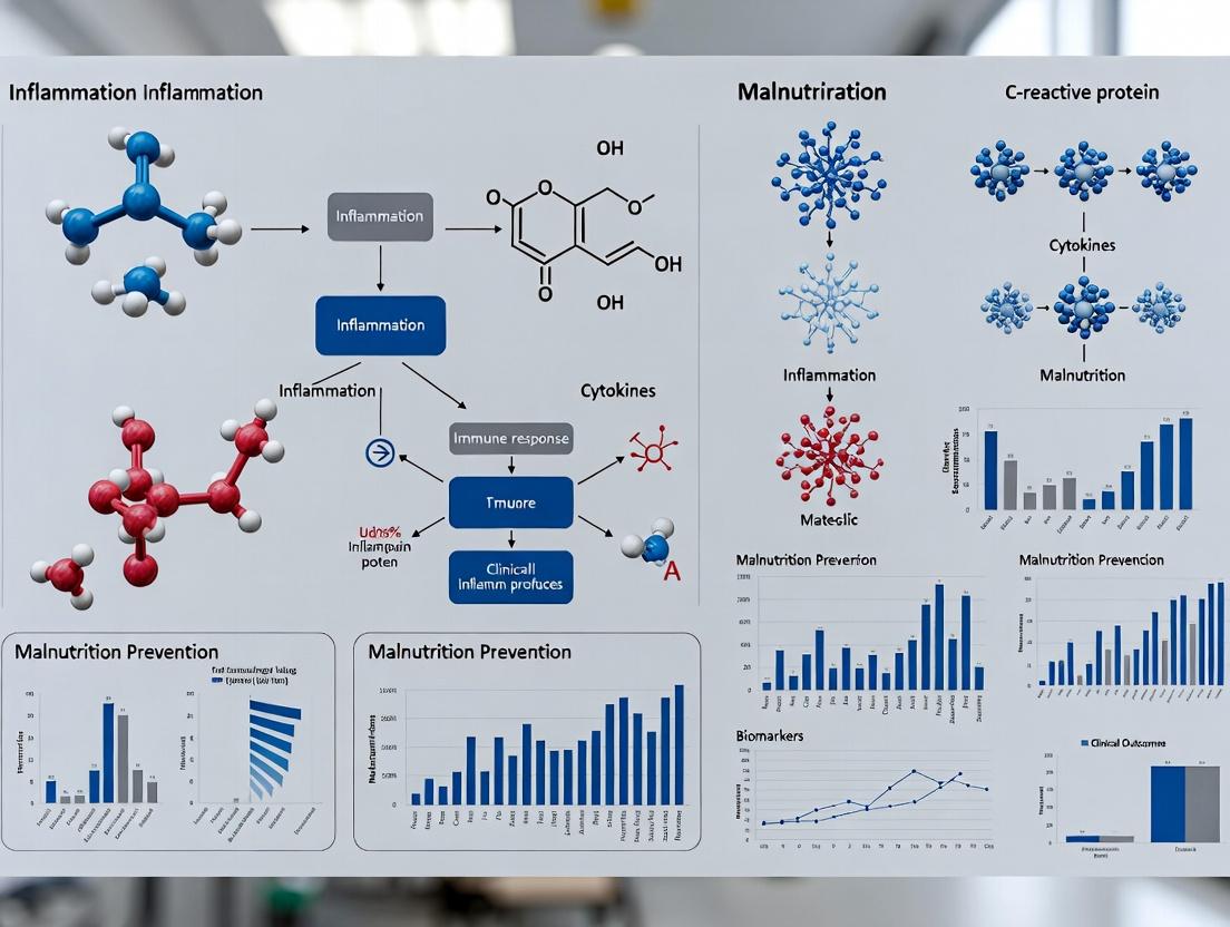
Inflammation, a key etiologic criterion in GLIM, drives malnutrition via complex signaling pathways that promote hypermetabolism, anorexia, and muscle proteolysis.
Title: Inflammatory Pathways Linking Disease to GLIM Criteria
The Scientist's Toolkit: Key Reagent Solutions for GLIM-Aligned Research
Table 4: Essential Research Reagents and Materials
| Item / Reagent | Function in GLIM Research | Example / Specification |
|---|---|---|
| Validated Nutritional Risk Screening Tool | First-step identification of 'at-risk' population per GLIM. | MUST, NRS-2002, or MNA-SF forms and scoring guides. |
| Bioelectrical Impedance Analysis (BIA) Device | Objective assessment of muscle mass (phenotypic criterion). | Multi-frequency, tetrapolar device with validated population-specific equations (e.g., Seca mBCA 515). |
| Inflammatory Biomarker Assay Kits | Quantification of inflammation (etiologic criterion support). | High-sensitivity ELISA kits for CRP, IL-6, TNF-α. |
| Dual-Energy X-ray Absorptiometry (DXA) Scanner | Reference or research-grade body composition analysis. | Enables precise measurement of lean soft tissue mass. |
| Computed Tomography (CT) Image Analysis Software | Gold-standard for muscle mass quantification at L3 vertebra. | Software like Slice-O-Matic for analyzing existing CT scans. |
| Standardized Anthropometric Kit | Portable, low-cost muscle mass assessment. | Includes non-stretch tape for calf circumference, skinfold calipers. |
| Indirect Calorimeter | Measurement of resting energy expenditure. | Supports assessment of hypermetabolism in etiologic evaluation. |
| Dietary Analysis Software | Accurate quantification of energy/protein intake. | Software like Nutritics or 24-hour recall analysis tools. |
GLIM Application Workflow in Clinical Research
The practical application of GLIM in a research cohort follows a defined logical pathway.
Title: GLIM Diagnostic Workflow in Research Studies
The mission of the GLIM initiative is to create a universal platform for malnutrition research. By providing consensus criteria, it enables the generation of comparable data across populations and settings, which is indispensable for meta-analyses, understanding the global burden of disease, and designing targeted drug and nutritional interventions. For the drug development sector, GLIM offers a validated framework for patient enrollment in clinical trials, ensuring homogeneity in study populations and defining clear, clinically relevant endpoints for therapeutic efficacy. The ongoing validation and refinement of GLIM criteria represent a critical, collaborative scientific endeavor to combat malnutrition through evidence-based standardization.
Within the framework of the Global Leadership Initiative on Malnutrition (GLIM) consensus criteria, the implementation of a rigorous two-step diagnostic model is paramount for robust research and clinical trials. This whitepaper details the core principles, technical methodologies, and experimental protocols underpinning the screening and assessment paradigm, tailored for researchers and drug development professionals.
The GLIM Two-Step Model: Conceptual Framework
The GLIM approach operationalizes malnutrition diagnosis through a sequential filter: initial screening for risk, followed by a confirmatory phenotypic and etiologic assessment. This model enhances specificity, ensures efficient resource allocation in trials, and creates phenotypically homogeneous cohorts for intervention studies.
Diagram Title: GLIM Two-Step Diagnostic Workflow for Cohort Identification
Quantitative Rationale: Sensitivity, Specificity, and Efficiency
A two-step model balances sensitivity (Sn) and specificity (Sp) to optimize positive predictive value (PPV) in target populations. Data from validation studies supports this approach.
Table 1: Performance Characteristics of a Two-Step vs. Single-Step Model in a Hypothetical Cohort of 1000 Hospitalized Patients
| Model | Sensitivity | Specificity | PPV | Patients for Full Assessment | Correctly Identified Cases |
|---|---|---|---|---|---|
| Single-Step (Assessment Only) | 95% | 90% | 66% | 1000 | 95 |
| Two-Step (Screening→Assessment) | 92% | 98% | 88% | ~300 | 92 |
Assumptions: Prevalence = 20%. Screening tool Sn=96%, Sp=85%. Assessment (GLIM) Sn=96%, Sp=95%. PPV=Positive Predictive Value.
Experimental Protocols for Validation Research
Protocol A: Validation of the Screening Step
- Objective: To validate the sensitivity and specificity of a chosen screening tool (e.g., MUST) against the full GLIM criteria as the reference standard.
- Design: Prospective, observational cohort study.
- Population: Consecutive adult patients (n>500) admitted to a defined clinical setting.
- Methodology:
- Within 24 hours of admission, a trained researcher administers the screening tool.
- Independently, a blinded assessor conducts the full GLIM assessment:
- Phenotypic Criteria: Measures weight loss (historical/records), low BMI (measured), and reduced muscle mass (using protocolized anthropometry, BIA, or DXA).
- Etiologic Criteria: Assesses reduced food intake/assimilation (≤50% of needs >1 week) and inflammation/disease burden (CRP, disease diagnosis).
- Results are recorded on a standardized case report form (CRF).
- Analysis: Calculate Sn, Sp, PPV, NPV, and area under the ROC curve (AUC) for the screening tool.
Protocol B: Assessing the Impact of Muscle Mass Measurement Techniques
- Objective: To compare the prevalence of the GLIM phenotypic criterion "reduced muscle mass" using different diagnostic modalities.
- Design: Cross-sectional, methodological comparison study.
- Population: Sub-cohort (n=200) from Protocol A.
- Methodology:
- Participants undergo muscle mass assessment via three methods on the same day:
- Anthropometry: Mid-upper arm circumference (MUAC) and calf circumference (CC) per ISAK guidelines.
- Bioelectrical Impedance Analysis (BIA): Using a validated device, standardized conditions (fasting, supine).
- Reference Method: Dual-energy X-ray Absorptiometry (DXA) full body scan.
- GLIM-compliant cut-offs are applied for each method (e.g., ASMMI from BIA, appendicular lean mass index from DXA).
- Participants undergo muscle mass assessment via three methods on the same day:
- Analysis: Determine agreement (Cohen's kappa) and correlation (Pearson's r) between methods. Calculate prevalence difference.
The Scientist's Toolkit: Research Reagent Solutions
Table 2: Essential Materials for GLIM-Based Malnutrition Research
| Item / Reagent | Function / Purpose in Research |
|---|---|
| Validated Screening Tool (e.g., MUST, NRS-2002) | Standardized, reproducible instrument for initial risk stratification in Step 1. |
| Calibrated Digital Scales & Stadiometer | Accurate measurement of weight and height for BMI calculation (phenotypic criterion). |
| Seca 201/214 Measuring Tape | For standardized measurement of mid-upper arm (MUAC) and calf circumference (CC). |
| Bioelectrical Impedance Analyzer (e.g., Seca mBCA 515) | Provides quantitative, objective data on fat-free mass and phase angle for muscle mass assessment. |
| Dual-Energy X-ray Absorptiometry (DXA) Scanner | Gold-standard reference method for body composition (muscle mass) validation studies. |
| High-Sensitivity C-Reactive Protein (hsCRP) Assay Kit | Quantifies systemic inflammation, a key etiologic criterion in GLIM. |
| Standardized 24-Hour Dietary Recall Protocol | Validated methodology for quantifying reduced food intake (<50% of requirements). |
| Electronic Case Report Form (eCRF) with GLIM Algorithm | Ensures consistent, auditable data capture for both screening and assessment steps. |
Signaling Pathways in Malnutrition Pathophysiology
The etiologic criteria of GLIM (inflammation/reduced intake) converge on molecular pathways driving muscle catabolism, a key phenotypic endpoint.
Diagram Title: Molecular Pathways Linking GLIM Etiologic to Phenotypic Criteria
The two-step (screening + assessment) model is the cornerstone of scientifically rigorous malnutrition research within the GLIM framework. It ensures diagnostic accuracy, enhances cohort homogeneity for clinical trials, and allows for precise investigation into the pathophysiological mechanisms linking disease and inflammation to the functional outcome of muscle loss. Adherence to detailed experimental protocols and utilization of standardized tools, as outlined, are critical for generating reproducible, high-quality data to inform therapeutic development.
Within the evolving landscape of clinical nutrition, the Global Leadership Initiative on Malnutrition (GLIM) consensus provides a standardized, multi-step framework for diagnosing malnutrition in adults. A core innovation of GLIM is its bifurcated diagnostic approach, requiring the identification of at least one phenotypic and one etiologic criterion. This whitepaper deconstructs this diagnostic framework, detailing the underlying phenotypic and etiologic components, their operational definitions, and their critical interplay within a research context, particularly for validating outcomes and developing targeted therapeutics.
Phenotypic Criteria: The Observable Manifestations
Phenotypic criteria are direct measures of the physical and compositional consequences of malnutrition. They are the observable, measurable outcomes of negative nutrient balance.
Core Phenotypic Components
The three phenotypic criteria, with their quantitative cut-offs, are summarized below.
Table 1: GLIM Phenotypic Criteria and Diagnostic Cut-offs
| Criterion | Parameter | Severity Threshold (Moderate) | Severity Threshold (Severe) | Measurement Method |
|---|---|---|---|---|
| Non-Volitional Weight Loss | % Weight loss over time | >5% within past 6 months, or >10% beyond 6 months | >10% within past 6 months, or >20% beyond 6 months | Serial weight measurement (calibrated scale). |
| Low Body Mass Index (BMI) | BMI (kg/m²) | <20 if <70 years; <22 if ≥70 years | <18.5 if <70 years; <20 if ≥70 years | Single measurement (stadiometer, scale). |
| Reduced Muscle Mass | Appendicular Skeletal Muscle Mass Index (ASMI) | ASMI <7.0 kg/m² (men), <5.7 kg/m² (women) via DXA | Further reduction from baseline or population norms. | Dual-energy X-ray Absorptiometry (DXA), Bioelectrical Impedance Analysis (BIA), CT/MRI. |
Experimental Protocol: Quantifying Muscle Mass via Bioelectrical Impedance Analysis (BIA)
A common protocol for assessing the phenotypic criterion of reduced muscle mass in research settings.
Title: BIA Protocol for Appendicular Skeletal Muscle Mass Assessment
Objective: To determine appendicular lean mass (ALM) and calculate the Appendicular Skeletal Muscle Mass Index (ASMI) for GLIM phenotypic classification.
Materials:
- Tetrapolar, multi-frequency BIA device (e.g., Seca mBCA 515, InBody 770).
- Standardized examination table.
- Skin alcohol wipes.
- Measurement tape and stadiometer.
Procedure:
- Pre-Test Conditions: The subject fasts for ≥4 hours, avoids moderate/strenuous exercise for ≥12 hours, and voids bladder completely within 30 minutes prior.
- Subject Positioning: The subject lies supine on a non-conductive surface, limbs abducted ~45° from torso, not touching the body. Arms and legs are separated from each other.
- Electrode Placement: After cleaning skin, place two current-injecting electrodes on the dorsal surfaces of the hand and foot at the metacarpophalangeal and metatarsophalangeal joints, respectively. Place two voltage-sensing electrodes at the pisiform prominence of the wrist and between the medial and lateral malleoli of the ankle.
- Measurement: Enter subject data (age, sex, height, weight) into the device. Initiate the measurement sequence. Ensure the subject remains motionless.
- Data Extraction: Record the device-reported values for lean mass of each limb. Calculate ALM = Sum of lean mass (kg) of all four limbs.
- Calculation: Calculate ASMI = ALM (kg) / height (m²). Compare to GLIM cut-offs (Table 1).
Title: Phenotypic Assessment Workflow
Etiologic Criteria: The Underlying Drivers
Etiologic criteria identify the root causes driving the phenotypic alterations. They are essential for understanding pathogenesis and guiding intervention.
Core Etiologic Components
The two etiologic criteria, with their associated metrics, are defined below.
Table 2: GLIM Etiologic Criteria and Supporting Metrics
| Criterion | Primary Definition | Supporting Metrics / Assessment Tools | Research Application |
|---|---|---|---|
| Reduced Food Intake or Assimilation | <50% of estimated energy requirement for >1 week, or any reduction for >2 weeks; OR chronic GI conditions impairing absorption. | Food records, 24-hr recalls, Indirect Calorimetry (REE). Gut Function Tests (fecal elastase, D-xylose). | Quantifies energy deficit. Links intake to phenotype. |
| Inflammation / Disease Burden | Acute disease/injury (e.g., sepsis, trauma) OR chronic disease-related (e.g., cancer, organ failure). | Acute: CRP, ESR. Chronic: CRP, IL-6, TNF-α, Clinical disease activity scores (e.g., APACHE II, SOFA). | Stratifies patients by inflammatory driver. Measures catabolic stimulus. |
Experimental Protocol: Assessing Systemic Inflammation via CRP and Cytokines
A protocol to objectively measure the inflammation etiologic criterion.
Title: Protocol for Quantifying Systemic Inflammation Biomarkers
Objective: To measure plasma/serum levels of C-reactive protein (CRP) and pro-inflammatory cytokines (IL-6, TNF-α) to support the GLIM inflammation criterion.
Materials:
- EDTA or serum separation tubes.
- Centrifuge.
- -80°C freezer.
- High-sensitivity CRP (hs-CRP) ELISA kit.
- Human IL-6 & TNF-α multiplex assay (e.g., Luminex, MSD) or ELISA kits.
- Microplate reader/luminescence analyzer.
Procedure:
- Sample Collection: Draw venous blood into appropriate tubes. Invert gently. For plasma, centrifuge EDTA tubes at 1000-2000 x g for 10 min at 4°C within 30 min. For serum, allow blood to clot at RT for 30 min, then centrifuge.
- Aliquot & Storage: Immediately aliquot supernatant (plasma/serum) into cryovials. Flash-freeze in liquid nitrogen and store at -80°C. Avoid freeze-thaw cycles.
- Biomarker Analysis:
- hs-CRP: Perform per ELISA kit instructions. Typically involves incubating samples in antibody-coated wells, washing, adding enzyme-conjugated detection antibody, washing, adding substrate, and measuring absorbance.
- Cytokines (IL-6, TNF-α): Using a multiplex assay, add samples to wells containing antibody-coupled magnetic beads. Follow with biotinylated detection antibodies and streptavidin-phycoerythrin. Read on a Luminex analyzer.
- Data Interpretation: Compare hs-CRP values to clinical thresholds (e.g., >3 mg/L indicates chronic inflammation, >10 mg/L indicates acute inflammation). Compare cytokine levels to assay-specific reference ranges or control groups.
Title: Etiologic Pathways to Phenotype
The Scientist's Toolkit: Research Reagent Solutions
Table 3: Essential Reagents and Materials for GLIM-Focused Research
| Item | Function | Example Product/Assay |
|---|---|---|
| High-Sensitivity CRP ELISA Kit | Quantifies low-grade chronic inflammation precisely. | R&D Systems Quantikine ELISA HsCRP, Abcam ab99995. |
| Multiplex Cytokine Panel (Human) | Simultaneously measures IL-6, TNF-α, IL-1β from a single sample. | Thermo Fisher Scientific ProcartaPlex, Bio-Rad Bio-Plex Pro. |
| Dual-Energy X-ray Absorptiometry (DXA) Scanner | Gold-standard for body composition (fat, lean, bone mass). | Hologic Horizon, GE Lunar iDXA. |
| Medical-Grade Multi-Frequency BIA Analyzer | Validated tool for estimating skeletal muscle mass (ASMI). | Seca mBCA, InBody 970. |
| Indirect Calorimetry System | Measures Resting Energy Expenditure (REE) to calculate energy deficit. | COSMED Quark RMR, MGC CareFusion. |
| Validated 24-Hour Dietary Recall Software | Standardizes assessment of reduced food intake. | USDA Automated Multiple-Pass Method, NDS-R. |
| Stable Isotope Tracers (e.g., D₃-Creatine) | Directly measures whole-body muscle protein synthesis and breakdown rates. | Cambridge Isotope Laboratories D₃-Creatine (methyl-d3). |
Integrating the Framework: The Diagnostic Algorithm
The GLIM framework's diagnostic power emerges from the mandatory combination of at least one phenotypic AND one etiologic criterion.
Title: GLIM Diagnostic Algorithm
The GLIM diagnostic framework, by decoupling phenotypic manifestations from their etiologic origins, provides a robust, mechanism-informed model for malnutrition research. This granularity enables precise patient stratification, elucidation of distinct pathophysiological pathways, and the development of targeted nutritional and pharmacologic interventions. For the research and drug development community, rigorous application of the standardized experimental protocols for each criterion is paramount to generating high-quality, comparable data that validates the framework and uncovers novel therapeutic targets.
The Strategic Impact of GLIM on Standardizing Clinical and Research Outcomes
The Global Leadership Initiative on Malnutrition (GLIM) consensus criteria, established in 2018, were developed to provide a unified, global approach for the diagnosis of malnutrition in adults. Within the broader thesis of malnutrition research, GLIM represents a pivotal effort to harmonize phenotypic and etiologic criteria, thereby enabling standardized data collection, comparable research outcomes, and enhanced clinical decision-making. This whitepaper examines the strategic impact of this standardization on clinical trials, epidemiological research, and drug development.
GLIM Criteria: Core Components and Diagnostic Workflow
The GLIM approach operates via a two-step model: first, screening for nutritional risk using a validated tool (e.g., MUST, NRS-2002), followed by a diagnostic assessment based on at least one phenotypic and one etiologic criterion.
Table 1: GLIM Diagnostic Criteria for Malnutrition
| Criterion Type | Specific Criteria | Threshold for Diagnosis |
|---|---|---|
| Phenotypic (Require 1+) | Non-volitional weight loss | >5% within past 6 months, or >10% beyond 6 months |
| Low body mass index (BMI) | <20 kg/m² if <70 years; <22 kg/m² if ≥70 years | |
| Reduced muscle mass | Reduced by validated body composition measurement techniques | |
| Etiologic (Require 1+) | Reduced food intake or assimilation | ≤50% of energy requirement >1 week, or any reduction for >2 weeks, or GI conditions impairing assimilation |
| Inflammation or disease burden | Acute disease/injury or chronic disease-related inflammation |
Diagram Title: GLIM Diagnostic Algorithm
Quantitative Impact on Research Standardization
Adoption of GLIM facilitates meta-analyses and cross-study comparisons by providing a common diagnostic endpoint. Recent validation studies demonstrate its performance characteristics.
Table 2: Performance Characteristics of GLIM in Selected Recent Studies (2022-2024)
| Study Population | Sample Size (n) | Reference Standard | GLIM Sensitivity | GLIM Specificity | Agreement (Kappa) |
|---|---|---|---|---|---|
| Hospitalized Oncology Patients | 412 | SGA | 78% | 85% | 0.72 |
| Elderly in Community | 567 | ESPEN 2015 | 82% | 89% | 0.68 |
| ICU Patients | 298 | Clinical Assessment + CT* | 65% | 92% | 0.61 |
| Surgery (GI) Cohort | 334 | ICD-10 | 71% | 88% | 0.70 |
*CT: Computed Tomography for muscle mass.
Experimental Protocol for Validating GLIM in a Clinical Cohort
Title: Prospective Validation of GLIM Criteria Against a Comprehensive Nutritional Assessment in Hospitalized Adults.
Objective: To determine the criterion validity and prognostic value of GLIM-defined malnutrition for 90-day post-discharge morbidity.
Methodology:
Participant Recruitment:
- Consecutive sampling of adults (≥18 years) admitted to general medical and surgical wards.
- Exclusion: Pregnancy, length of stay <48 hours, critical illness requiring immediate ICU admission.
- Target sample: n=500 (calculated for 80% power, α=0.05).
Baseline Assessment (Within 48h of Admission):
- Step 1 - Screening: Trained researchers administer the Nutritional Risk Screening 2002 (NRS-2002).
- Step 2 - GLIM Phenotypic Criteria:
- Weight Loss: Documented from medical history or patient recall. Verified with pre-admission records if available.
- Low BMI: Height and weight measured using calibrated stadiometer and scale. BMI calculated (kg/m²).
- Reduced Muscle Mass: Assessed via mid-upper arm circumference (MUAC) and calf circumference (CC) as per GLIM-supported proxies. A subset (n=100) undergoes bioelectrical impedance analysis (BIA) for cross-validation.
- Step 3 - GLIM Etiologic Criteria:
- Reduced Intake: Estimated from 24-hour dietary recall and nurse/patient interview (<50% of estimated needs for >1 week).
- Inflammation: Serum C-reactive protein (CRP) >5 mg/L and/or clinical diagnosis of acute/chronic inflammatory condition.
Reference Standard Assessment (Blinded):
- A certified dietitian, blinded to GLIM results, performs a Subjective Global Assessment (SGA), classifying patients as well-nourished (A), moderately malnourished (B), or severely malnourished (C). SGA rating B/C is considered the reference for malnutrition.
Outcome Measurement (Prognostic Validity):
- Primary Outcome: Composite of unplanned readmission, major complication (Clavien-Dindo ≥II), or mortality within 90 days post-discharge.
- Data collected via electronic health record review and structured telephone interview.
Statistical Analysis:
- Sensitivity, specificity, PPV, NPV calculated against SGA.
- Inter-rater reliability (kappa) for GLIM components.
- Cox proportional hazards models to assess GLIM diagnosis as a predictor of the 90-day composite outcome, adjusted for age, sex, and comorbidity index.
Research Reagent Solutions Toolkit
Table 3: Essential Materials for GLIM-Focused Clinical Research
| Item | Function/Description | Example Product/Model |
|---|---|---|
| Validated Screening Tool | Standardized form for initial nutritional risk identification. | NRS-2002 or MUST paper/electronic form |
| Calibrated Digital Scale | Accurate measurement of body weight (to 0.1 kg). | Seca 767 or equivalent medical grade scale |
| Stadiometer | Accurate measurement of height (to 0.1 cm). | Harpenden stadiometer or wall-mounted model |
| Non-Stretch Tape Measure | Measurement of mid-upper arm circumference (MUAC) and calf circumference (CC). | LuFlex or Gulick anthropometric tape |
| Bioelectrical Impedance Analyzer (BIA) | Validated device for estimating fat-free mass and skeletal muscle mass. | Seca mBCA 515 or InBody 770 |
| CRP Assay Kit | Quantitative measurement of serum C-reactive protein to assess inflammatory etiologic criterion. | Roche Cobas CRP Latex assay or Siemens Atellica CH CRP |
| Dietary Assessment Software | Aids in quantifying energy/protein intake from 24-hour recalls. | NDS-R, Nutritics, or ASA24 |
| Electronic Data Capture (EDC) System | Platform for standardized, secure data collection (REDCap, Medidata Rave). | Custom REDCap project with GLIM module |
GLIM in Drug Development and Clinical Trials
GLIM provides a standardized endpoint for patient stratification and outcome measurement in trials targeting muscle wasting, cachexia, or nutritional intervention.
Diagram Title: GLIM in Clinical Trial Design
Strategic Impact: By using GLIM, sponsors can define a consistent, severity-graded malnutrition endpoint, improving the interpretability of trial results for regulatory bodies (FDA, EMA) and enabling robust pooled analyses across studies.
The GLIM criteria have introduced a critical and necessary framework for standardizing the diagnosis of malnutrition. For researchers and drug development professionals, the strategic adoption of GLIM minimizes heterogeneity in case definition, strengthens the validity of prognostic studies, and provides a clear, consensus-based endpoint for clinical trials. This standardization is fundamental for advancing the science of clinical nutrition, developing effective therapies, and ultimately improving patient outcomes on a global scale.
Implementing GLIM in Practice: A Step-by-Step Methodology for Research Settings
Within the framework of the Global Leadership Initiative on Malnutrition (GLIM) consensus criteria for diagnosing malnutrition in adults, Step 1 involves mandatory risk screening. This initial step is crucial for identifying individuals at nutritional risk who should proceed to the subsequent diagnostic assessment (Steps 2 and 3). This technical guide provides an in-depth analysis of three validated screening tools—Malnutrition Universal Screening Tool (MUST), Mini Nutritional Assessment-Short Form (MNA-SF), and Nutritional Risk Screening 2002 (NRS-2002)—detailing their operational parameters, selection criteria, and application within clinical and research settings, particularly for drug development and interventional studies.
Validated Screening Tools: Quantitative Comparison
Table 1: Core Characteristics of MUST, MNA-SF, and NRS-2002
| Feature | MUST | MNA-SF | NRS-2002 |
|---|---|---|---|
| Primary Population | Adults, all settings, BMI >20 kg/m² | Adults ≥65 years, community/ hospital | Hospitalized adults |
| Components Scored | BMI, weight loss, acute disease effect | Food intake, weight loss, mobility, psychological stress/acute disease, neuropsychological problems, BMI | Impaired nutritional status (weight loss, BMI, food intake) + Severity of disease (e.g., major surgery, APACHE >10) + Age (≥70 years adds 1 point) |
| Scoring Range | 0 to 6+ | 0 to 14 | 0 to 7+ |
| Risk Categories | Low (0), Medium (1), High (≥2) | Normal nutrition (12-14), At risk (8-11), Malnourished (0-7) | Not at risk (<3), At risk (≥3) |
| Time to Administer | ~3-5 minutes | ~5-10 minutes | ~5-10 minutes |
| Validation Context | Community, hospital, care homes | Geriatric hospital, community, long-term care | Hospital inpatients |
| GLIM Step 1 Use | Suitable for general adult populations | Recommended for older adults (≥65 years) | Recommended for acute care/hospitalized patients |
Table 2: Diagnostic Performance Metrics in Selected Studies
| Tool | Sensitivity (%) | Specificity (%) | Positive Predictive Value (%) | Negative Predictive Value (%) | Reference Standard |
|---|---|---|---|---|---|
| MUST | 66 - 92 | 76 - 93 | 42 - 89 | 91 - 99 | GLIM/Subjective Global Assessment (SGA) |
| MNA-SF | 83 - 97 | 70 - 89 | 62 - 85 | 87 - 99 | Full MNA/GLIM |
| NRS-2002 | 75 - 90 | 60 - 93 | 32 - 88 | 85 - 98 | Clinical assessment/GLIM |
Note: Performance varies based on population and setting.
Selection Framework and Experimental Protocols
Rationale for Tool Selection
Selection must align with the target population, setting, and the overarching research objectives of the GLIM-based study.
Protocol 1: Selecting the Appropriate Screening Tool for a GLIM Study
- Define Study Cohort: Determine age, clinical setting (community, hospital, ICU, long-term care), and primary diagnosis.
- Map to Tool Validity: Match cohort to the tool with the strongest validation evidence in that specific demographic and setting (see Table 1).
- Assess Feasibility: Consider required training, time constraints, and availability of anthropometric data.
- Align with Endpoints: For drug trials, ensure the tool's risk categories correlate with clinical outcomes of interest (e.g., infection rates, length of stay, muscle function).
- Standardize Implementation: Train all research personnel on standardized administration and scoring to minimize inter-rater variability.
Protocol 2: Validating a Screening Tool Against GLIM Criteria in a Research Cohort
- Recruitment: Enroll a representative sample of the target population.
- Blinded Screening: A researcher administers the chosen screening tool (MUST, MNA-SF, or NRS-2002) following its standard operating procedure.
- Reference Standard Assessment: A separate, blinded assessor conducts the full GLIM diagnostic assessment (Step 2: Phenotypic Criteria – weight loss, low BMI, reduced muscle mass; Step 3: Etiologic Criteria – reduced food intake/assimilation, inflammation/disease burden).
- Data Analysis: Calculate sensitivity, specificity, positive/negative predictive values, and area under the ROC curve for each screening tool's risk categories against confirmed GLIM malnutrition.
- Statistical Determination: Establish the optimal screening threshold (e.g., MUST ≥2 vs. ≥1) for the specific research cohort using ROC analysis.
Visualizing the GLIM Workflow and Screening Tool Logic
Diagram 1: GLIM Diagnostic Pathway with Risk Screening Step
Diagram 2: Screening Tool Logic Comparison
The Scientist's Toolkit: Research Reagent Solutions
Table 3: Essential Materials for Malnutrition Screening & GLIM-Based Research
| Item | Function/Application in Research |
|---|---|
| Calibrated Digital Scales | Accurate measurement of body weight for BMI calculation and weight loss history (core for MUST, MNA-SF, NRS-2002, and GLIM phenotypic criterion). |
| Stadiometer / Height Measure | Accurate measurement of height for BMI calculation. For non-ambulatory subjects, use knee-height calipers or segmental measures. |
| Non-Stretchable Tape Measure | Measurement of mid-upper arm circumference (MUAC) or calf circumference (alternative in MNA-SF). Calibrated for tension. |
| Bioelectrical Impedance Analysis (BIA) | Device for estimating body composition (fat-free mass, skeletal muscle mass) to assess the reduced muscle mass criterion for GLIM Step 2. |
| Standardized Screening Forms | Validated, translated questionnaires for MUST, MNA-SF, and NRS-2002 to ensure consistency in data collection across research sites. |
| Electronic Data Capture (EDC) System | Secure platform for direct entry of screening and assessment data, with built-in logic checks for scoring algorithms and GLIM criteria application. |
| Reference Standards Kit | Materials for validation studies: e.g., Dual-Energy X-ray Absorptiometry (DXA) for muscle mass, Indirect Calorimetry for resting energy expenditure, validated dietary intake software. |
| Clinical Pathology Assays | Kits for measuring inflammatory biomarkers (e.g., CRP, IL-6) to support assessment of the inflammation etiologic criterion in GLIM Step 3. |
| Training & Certification Modules | Standardized video and written protocols to train research personnel on tool administration, anthropometry, and GLIM assessment to ensure inter-rater reliability. |
The Global Leadership Initiative on Malnutrition (GLIM) framework provides a consensus-based, two-step model for diagnosing malnutrition in adults. Step 1 involves malnutrition risk screening, while Step 2 is the phenotypic and etiologic assessment for diagnosis. This document details Step 2a, the phenotypic component, which requires the presence of at least one of three core phenotypic criteria: weight loss, low body mass index (BMI), and reduced muscle mass. Accurate and standardized assessment of these criteria is critical for research reproducibility, patient stratification in clinical trials, and the development of targeted nutritional or pharmacologic interventions.
Phenotypic Criterion 1: Weight Loss
Definition & Thresholds: Unintentional weight loss is a primary indicator of catabolic stress and negative energy balance. GLIM proposes the following thresholds for significant weight loss:
- ≥5% within the past 6 months, or
- ≥10% beyond 6 months.
Measurement Protocol:
- Tool: Calibrated digital scale (e.g., SECA 813).
- Procedure:
- Measure weight in light clothing, without shoes, at the same time of day (preferably morning, post-void).
- Record to the nearest 0.1 kg.
- Obtain a documented historical weight from medical records or a verified patient report. In a research setting, use a measured weight from a prior study visit.
- Calculate percentage weight loss:
[(Usual Weight - Current Weight) / Usual Weight] * 100.
- Considerations: Adjust for edema/ascites (clinical assessment). The reliability of patient-reported historical weight is a known limitation.
Phenotypic Criterion 2: Low Body Mass Index (BMI)
Definition & Thresholds: Low BMI reflects chronic energy deficiency. GLIM recommends age-specific cut-offs:
- <18.5 kg/m² for adults <70 years
- <20 kg/m² for adults ≥70 years
Measurement Protocol:
- Tools: Calibrated digital scale and stadiometer (e.g., SECA 217).
- Procedure for Height:
- Measured standing height: Participant stands without shoes, heels together, back straight, head in the Frankfort horizontal plane. Height is measured at maximum inspiration.
- For non-ambulatory or kyphotic patients, use knee-height calipers (e.g., Ross Laboratories) and validated equations (e.g., Chumlea equations) to estimate stature.
- Calculation: BMI =
Weight (kg) / [Height (m)]². - Considerations: BMI may be misleading in individuals with high adiposity but low muscle mass (sarcopenic obesity). It should be interpreted in conjunction with Criterion 3.
Phenotypic Criterion 3: Reduced Muscle Mass
Definition & Thresholds: This is the most specific phenotypic criterion for malnutrition, indicating protein depletion. GLIM endorses the use of validated body composition techniques with population-specific cut-offs.
Measurement Protocols & Techniques:
| Technique | Principle | GLIM-Suggested Cut-offs (Examples) | Research-Grade Protocol Summary |
|---|---|---|---|
| Bioelectrical Impedance Analysis (BIA) | Measures resistance and reactance to a low-level electrical current to estimate fat-free mass. | Appendicular Skeletal Muscle Mass (ASMM) divided by height²: <7.0 kg/m² (men), <5.5 kg/m² (women) (Caucasian). | Device: Seca mBCA 515 or equivalent. Protocol: Supine position for 10 mins, electrodes on hand and foot per manufacturer. No exercise/alcohol 24h prior, euvolemic state. |
| Dual-Energy X-ray Absorptiometry (DXA) | Uses low-dose X-ray at two energies to differentiate bone mineral, lean soft tissue, and fat mass. | ASMM Index: <7.26 kg/m² (men), <5.5 kg/m² (women) (AWGS 2019). | Device: Hologic or Lunar DXA scanner. Protocol: Supine, centered, in standardized clothing. System calibration daily with phantom. Analysis using enCore software. |
| Computed Tomography (CT) | Cross-sectional imaging at the L3 vertebra; muscle area is identified by Hounsfield Unit thresholds (-29 to +150). | L3 Skeletal Muscle Index (SMI): <52.4 cm²/m² (men), <38.5 cm²/m² (women) (Martin et al., 2013). | Analysis: SliceOmatic or NIH ImageJ software. Single axial slice at L3 landmark. Automate area calculation with H.U. thresholds. Normalize to height². |
| Mid-Upper Arm Circumference (MUAC) | Anthropometric surrogate for muscle mass. | <23.5 cm (men), <22.0 cm (women) (ESPEN 2015). | Tool: Non-stretchable tape. Protocol: Measure midpoint between acromion and olecranon on non-dominant arm, arm hanging relaxed. Record mean of three measurements. |
The Scientist's Toolkit: Key Research Reagent Solutions
| Item / Assay | Function in Phenotypic Assessment Research |
|---|---|
| Calibrated Digital Metabolic Scale | High-precision measurement of body weight and, in indirect calorimetry models, resting energy expenditure to link phenotype with metabolic alteration. |
| Whole-Body DXA Phantom | Quality control device for daily calibration of DXA scanners, ensuring longitudinal consistency in body composition measurements across multi-center trials. |
| BIA Validation Phantom | Test object with known electrical properties to calibrate and validate BIA devices, critical for standardization in large cohort studies. |
| CT Biomarker Software (e.g., SliceOmatic) | Enables semi-automated segmentation and quantification of skeletal muscle area from clinical CT scans, allowing retrospective and prospective analysis. |
| Standardized Anthropometric Kit | Includes sliding caliper for knee-height, non-stretchable tape for MUAC, and skinfold calipers, essential for field studies or low-resource settings. |
| Stable Isotope Tracers (e.g., D₂O) | Gold-standard for measuring total body water and, by extension, body composition (fat-free mass) in metabolic research studies. |
Visualizing Assessment Pathways & Method Selection
Diagram 1: Phenotypic Criteria Decision Pathway (78 chars)
Diagram 2: Muscle Mass Assessment Modality Selection (85 chars)
Rigorous and consistent application of the Step 2a phenotypic criteria is foundational for robust malnutrition research under the GLIM framework. The choice of assessment tools—from gold-standard imaging to pragmatic anthropometry—must align with study objectives, population, and resources. Standardized protocols, as outlined, ensure data comparability across trials, which is essential for advancing the science of malnutrition, validating biomarkers, and developing effective therapeutics.
Within the Global Leadership Initiative on Malnutrition (GLIM) framework, Step 2 involves the identification of at least one etiologic criterion, which must be combined with a phenotypic criterion for the diagnosis of malnutrition. This document provides an in-depth technical guide on the rigorous assessment of the three primary etiologic criteria: Reduced Food Intake or Assimilation, Chronic or Acute Inflammation, and Disease Burden. Accurate evaluation of these components is critical for researchers and clinical scientists conducting precise, reproducible malnutrition phenotyping in adult populations, particularly within clinical trials and pathophysiological studies.
Quantification of Reduced Food Intake or Assimilation
Reduced intake is a primary driver of malnutrition. Assessment must move beyond qualitative recall to objective, quantifiable measures.
Key Measurement Methodologies
- Direct Calorimetry & Doubly Labeled Water: Gold standards for measuring energy expenditure, against which intake data is often compared.
- Precise Dietary Logging: Utilized in research settings with digital tools or weighted food records over a minimum of 3-7 days.
- Digital Photography of Meals: Computer-aided analysis of pre- and post-meal images to estimate consumed volume and nutrient composition.
- Nutrient Database Integration: Intake data is coded and analyzed using standardized databases (e.g., USDA FoodData Central, national nutrient tables).
Thresholds & Interpretation
The GLIM consensus suggests a >50% reduction in energy intake for >1 week, or any reduction for >2 weeks, as a significant criterion. Research protocols often use more granular stratification:
Table 1: Stratification of Reduced Food Intake for Research Protocols
| Intake Level (% of Estimated Requirement) | Duration | GLIM Criterion Met | Research Severity Grade |
|---|---|---|---|
| >75% | >1 week | No | Mild Risk |
| 50-75% | >1 week | Yes | Moderate Reduction |
| <50% | >1 week | Yes | Severe Reduction |
| Any reduction (e.g., <90%) | >2 weeks | Yes | Chronic Suboptimal Intake |
Experimental Protocol: Protocol for High-Fidelity Food Intake Measurement in a Clinical Research Unit
- Participant Admission: Admit to controlled metabolic research unit for 5-7 days.
- Baseline Energy Requirement: Calculate resting energy expenditure (REE) via indirect calorimetry on Day 1. Multiply by a standardized physical activity factor (e.g., 1.2-1.3 for bedrest).
- Food Provision: Provide all meals. Each item is precisely weighed (to 0.1g) using calibrated scales prior to service. Duplicate meals are created and stored for compositional analysis.
- Intake Measurement: Weigh all returned items and plate waste post-meal. Calculate consumed weight per item.
- Nutrient Analysis: Use recipe decomposition and chemical analysis of duplicate meals to determine exact nutrient (energy, protein, micronutrient) intake.
- Data Synthesis: Daily and total study period intake is expressed as absolute values and as a percentage of calculated energy and protein requirements.
Assessment of Inflammation
Inflammation drives the catabolic response and alters nutrient utilization. Its assessment requires a multi-modal approach.
Inflammatory Biomarkers: Panels and Interpretation
No single biomarker is perfectly sensitive or specific. A panel approach is recommended.
Table 2: Core and Extended Inflammatory Biomarker Panels for Malnutrition Research
| Biomarker | Half-Life | Primary Source | Indicates | Typical GLIM Cut-point | Research-Grade Cut-point |
|---|---|---|---|---|---|
| C-Reactive Protein (CRP) | 19 hrs | Hepatocyte (IL-6 driven) | Acute phase response, systemic inflammation | >5 mg/L (acute/chronic disease) | >3 mg/L (high-sensitivity assay) |
| Albumin | 19 days | Hepatocyte (negative acute phase) | Chronic inflammation, synthetic function | <3.5 g/dL | Rate of change >0.5 g/dL/week |
| Interleukin-6 (IL-6) | 1-4 hrs | Macrophages, T-cells, adipocytes | Pro-inflammatory cytokine, upstream of CRP | Elevated | >3-5 pg/mL (plasma, via ELISA) |
| Tumor Necrosis Factor-alpha (TNF-α) | 10-20 mins | Macrophages, lymphocytes | Pro-inflammatory cytokine, cachexia driver | Elevated | >8 pg/mL (serum, via high-sensitivity assay) |
| Neutrophil-to-Lymphocyte Ratio (NLR) | N/A | Derived from CBC | Systemic inflammatory stress | >3-5 | >3 (validated for prognosis) |
Signaling Pathways in Inflammation-Driven Malnutrition
The interplay between inflammatory cytokines, cellular signaling, and tissue catabolism.
Title: Inflammatory Signaling in Malnutrition Pathogenesis
Experimental Protocol: Protocol for Serial Inflammatory Biomarker Analysis in a Longitudinal Cachexia Study
- Baseline Sampling: Collect fasting venous blood at study enrollment (Day 0). Process within 60 minutes.
- Sample Processing: For serum: allow clotting, centrifuge at 1500-2000xg for 10min. For plasma: collect in EDTA tubes, centrifuge immediately. Aliquot and store at -80°C.
- Assay Selection:
- CRP: High-sensitivity immunoturbidimetric assay on clinical chemistry analyzer.
- Cytokines (IL-6, TNF-α): Multiplex electrochemiluminescence (e.g., Meso Scale Discovery) or high-sensitivity ELISA. Run in duplicate.
- Longitudinal Collection: Repeat sampling at predefined intervals (e.g., weekly during active treatment, monthly during follow-up).
- Data Analysis: Analyze trajectories (absolute values, rate of change). Correlate with concurrent measures of body composition (e.g., DEXA, CT) and food intake.
Evaluation of Disease Burden
Disease burden reflects the severity and catabolic impact of the underlying condition.
Disease Classification Frameworks
Table 3: Disease Burden Classification for GLIM Etiologic Criterion
| Disease Category | Examples | GLIM Relevance & Rationale |
|---|---|---|
| Acute Disease/Injury | Major infection, burns, trauma, major surgery | High inflammatory drive and hypermetabolism. Burden is often time-limited but intense. |
| Chronic Disease | Chronic heart failure, COPD, chronic kidney disease | Persistent low-grade inflammation, anorexia, and increased energy expenditure. |
| Organ Failure | End-stage liver disease, dialysis-dependent CKD | Severe metabolic disruption, synthetic failure, and frequent dietary restrictions. |
| Oncologic Disease | Active cancer, particularly pancreatic, gastric, lung | Direct cachectic effects of tumor-derived factors (e.g., PIF), compounded by treatment effects. |
| Conditions Affecting Intake | Dysphagia, GI obstruction, severe depression | Primary barrier to meeting nutritional requirements, leading to direct starvation pathology. |
Composite Scoring Systems
Research often employs validated scores to quantify burden:
- Charlson Comorbidity Index (CCI): Weighted index of 19 conditions. Predicts 10-year mortality. A score ≥5 indicates high burden.
- Cumulative Illness Rating Scale (CIRS): Grades severity (0-4) of impairment in 14 organ systems. The Severity Index (average score) and Comorbidity Index (number of affected systems) are useful.
- Disease-Specific Staging: e.g., NYHA Class for CHF, GOLD stage for COPD, TNM stage and performance status (ECOG/WHO) for cancer.
Workflow for Integrated Etiologic Assessment
A logical pathway for determining if a GLIM etiologic criterion is fulfilled.
Title: Decision Logic for GLIM Etiologic Criteria
The Scientist's Toolkit: Research Reagent Solutions
Table 4: Essential Research Materials for Investigating GLIM Etiologic Criteria
| Item / Solution | Supplier Examples | Function in Research |
|---|---|---|
| High-Sensitivity CRP Assay Kit | Roche Diagnostics, Siemens Healthineers | Quantifies low-grade inflammation with precision for clinical research. |
| Multiplex Cytokine Panel (Human) | Meso Scale Discovery, R&D Systems, Luminex | Simultaneously measures IL-6, TNF-α, IL-1β, and other cytokines from a single small-volume sample. |
| ELISA for Myostatin/GDF-11 | Abcam, Thermo Fisher Scientific | Assesses specific pathways of muscle catabolism and wasting. |
| Indirect Calorimeter | MGC Diagnostics, COSMED, Maastricht | Gold-standard measurement of resting and total energy expenditure. |
| Dietary Analysis Software | Nutrition Data System for Research (NDSR), Nutritics | Standardizes nutrient intake analysis from food records against comprehensive databases. |
| Stable Isotopes (¹³C-Leucine, D₂O) | Cambridge Isotope Laboratories | Tracer for in vivo studies of protein metabolism and whole-body composition. |
| RNA/DNA Stabilization Tubes (PAXgene) | PreAnalytiX, Qiagen | Preserves whole-blood transcriptome for gene expression analysis related to inflammation/cachexia. |
| Body Composition Phantom (for CT) | QRM, CIRS | Calibrates CT/MRI scanners for precise, longitudinal quantification of muscle and adipose tissue. |
Thesis Context: This technical guide is framed within a broader research thesis examining the validation, applicability, and prognostic implications of the Global Leadership Initiative on Malnutrition (GLIM) consensus criteria for diagnosing malnutrition in adults, with a specific focus on the critical step of severity grading.
The GLIM framework provides a two-step approach for malnutrition diagnosis: first, screening for malnutrition risk, and second, assessment for diagnosis and severity grading. Case ascertainment is completed only after severity is assigned. Severity grading—distinguishing moderate (Stage 1) from severe (Stage 2) malnutrition—is pivotal for prognostic stratification, guiding intervention intensity, and serving as a key endpoint in clinical trials, particularly in drug development for cachexia and muscle-wasting disorders.
Quantitative Criteria for Severity Grading
Severity is determined by the most severe phenotypic criterion meeting the threshold.
Table 1: GLIM Phenotypic Criteria for Severity Grading
| Phenotypic Criterion | Moderate (Stage 1) Malnutrition | Severe (Stage 2) Malnutrition |
|---|---|---|
| Non-Volitional Weight Loss | 5-10% within the past 6 months, OR 10-20% beyond 6 months | >10% within the past 6 months, OR >20% beyond 6 months |
| Low Body Mass Index (BMI) | <20 kg/m² if <70 years; <22 kg/m² if ≥70 years | <18.5 kg/m² if <70 years; <20 kg/m² if ≥70 years |
| Reduced Muscle Mass | Mild to moderate deficit, according to local standards* | Severe deficit, according to local standards* |
*Quantitative cut-offs for muscle mass reduction are population and method-specific.
Table 2: Supporting Quantitative Measures for Muscle Mass (Examples)
| Assessment Method | Moderate Defcut-off Examples (Males / Females) | Severe Deficit Cut-off Examples (Males / Females) | Key Citations |
|---|---|---|---|
| CT-based SMI (L3) | Varies by population; e.g., <55 / <39 cm²/m² | e.g., <45 / <34 cm²/m² (Cancer-specific) | Martin et al., 2013; Prado et al., 2008 |
| DEXA (ASM/ht²) | <7.0 / <5.5 kg/m² | Further reduction below moderate threshold | Baumgartner et al., 1998 |
| BIA (Phase Angle) | <5.0 / <4.6 degrees | <4.5 / <4.2 degrees (Disease-specific norms) | Norman et al., 2010 |
| Anthropometry (AMC) | <5th to <10th percentile | <5th percentile | WHO Technical Report, 1995 |
Experimental Protocols for Key Assessment Modalities
Protocol: Mid-Upper Arm Circumference (MUAC) and Anthropometry
Purpose: To provide a rapid, bedside assessment of muscle mass proxy. Materials: Non-stretchable measuring tape, skinfold calipers. Procedure:
- Locate the mid-point of the patient's left upper arm (between acromion and olecranon).
- Ensure arm is relaxed and hanging. Wrap tape around arm at midpoint perpendicular to the long axis.
- Record measurement to the nearest 0.1 cm. Perform in triplicate, calculate mean.
- For AMC: Measure triceps skinfold (TSF) at the same midpoint. Apply calipers vertically. Record mean of three readings in mm.
- Calculate AMC: AMC (cm) = MUAC (cm) - [π * TSF (cm)]. Severity Grading: Compare AMC to reference percentiles (e.g., NHANES III). <5th percentile supports severe deficit.
Protocol: Bioelectrical Impedance Analysis (BIA) for Phase Angle
Purpose: To assess cellular health and integrity as a proxy for body cell mass. Materials: Tetrapolar, multi-frequency BIA device, alcohol wipes, examination table. Procedure:
- Calibrate device according to manufacturer instructions.
- Patient supine for ≥5 minutes, limbs abducted from body. Electrodes placed on hand, wrist, foot, and ankle of the same side.
- Input patient data (height, weight, age, sex). Ensure no metal contacts the electrodes.
- Run impedance measurement. Record Resistance (R), Reactance (Xc) at 50 kHz.
- Calculate Phase Angle (PhA): PhA (degrees) = arctan(Xc/R) * (180/π). Severity Grading: Compare PhA to validated, age- and disease-specific references. Lower values indicate worse status.
Protocol: CT-Derived Skeletal Muscle Index (SMI) at L3
Purpose: Gold-standard for quantifying cross-sectional muscle area. Materials: Existing abdominal/pelvic CT scan (±5 cm from L3), imaging software (e.g., Slice-O-Matic, Osirix), Hounsfield Unit (HU) threshold range (-29 to +150). Procedure:
- Identify the L3 lumbar vertebra on the CT scout or sagittal view.
- Select the single axial slice at the L3 level or average three consecutive slices centered on L3.
- Using semi-automated software, set HU thresholds for skeletal muscle (typically -29 to +150).
- Manually correct for inclusion of visceral muscle, vessels, and tumors.
- Software calculates total cross-sectional area (cm²) of skeletal muscle on the slice.
- Calculate SMI: SMI (cm²/m²) = Total Muscle Area (cm²) / Height (m)². Severity Grading: Apply validated, disease-specific cut-offs (see Table 2).
Visualization of Case Ascertainment Workflow
Title: GLIM Case Ascertainment and Severity Grading Workflow
Key Signaling Pathways in Disease-Related Malnutrition
Title: Inflammatory Pathways Driving Severe Muscle Loss
The Scientist's Toolkit: Research Reagent Solutions
Table 3: Essential Research Materials for GLIM-Related Investigations
| Item / Reagent | Function in Research Context | Example Application |
|---|---|---|
| Human Myoblast Cell Lines (e.g., LHCN-M2) | In vitro model of human skeletal muscle. | Studying cytokine-induced proteolysis and testing anabolic compounds. |
| Recombinant Human TNF-α / IL-6 | Induce inflammatory signaling mimicking disease-related malnutrition. | Creating cell culture models of muscle wasting to investigate pathways. |
| Anti-MuRF1 / Anti-Atrogin-1 Antibodies | Detect and quantify key E3 ubiquitin ligases in muscle catabolism. | Western blot, immunohistochemistry on muscle biopsies from patients. |
| CT Imaging Software with Body Composition Module (e.g., Slice-O-Matic) | Precise quantification of muscle cross-sectional area from medical images. | Retrospective/prospective measurement of L3 SMI for GLIM criteria application. |
| Multi-Frequency Bioimpedance Analyzer (e.g., Seca mBCA) | Objectively measures body composition compartments (FFM, ASM) and phase angle. | Validating GLIM muscle mass criteria against reference methods in cohorts. |
| Dual-Energy X-ray Absorptiometry (DEXA) Scanner | Gold-standard for lean soft tissue mass (LSTM) and bone mineral density. | Providing reference data for validating simpler muscle mass assessment tools. |
| Validated Food Frequency Questionnaire (FFQ) | Quantifies habitual nutrient and energy intake. | Assessing the GLIM etiologic criterion of "reduced food intake" in studies. |
| ELISA Kits for CRP, Prealbumin (Transthyretin) | Measure systemic inflammation and short-term visceral protein status. | Correlating inflammation with severity of phenotypic criteria in patients. |
Within the evolving framework of malnutrition research, the Global Leadership Initiative on Malnutrition (GLIM) consensus criteria provide a standardized, evidence-based approach for diagnosing malnutrition in adults. The broader thesis asserts that while GLIM offers a robust phenotypic-etiological diagnostic model, its operationalization within dynamic research environments—specifically longitudinal cohort studies and randomized controlled trials (RCTs)—requires significant protocol adaptation and integration. This technical guide addresses the methodological challenges and solutions for embedding GLIM within such rigorous scientific protocols, ensuring consistent, comparable, and valid endpoint ascertainment across time and treatment arms.
Core GLIM Criteria: A Baseline for Protocol Design
The GLIM approach operates on a two-step model: first, screening for nutritional risk, and second, assessment for diagnosis based on phenotypic and etiological criteria.
Table 1: Core GLIM Diagnostic Criteria for Malnutrition
| Criterion Type | Specific Criterion | Cut-off Threshold |
|---|---|---|
| Phenotypic (Requires 1) | Non-volitional weight loss | >5% within past 6 months, or >10% beyond 6 months |
| Low body mass index (BMI) | <20 kg/m² if <70 years; <22 kg/m² if ≥70 years | |
| Reduced muscle mass | Reduced by validated body composition techniques | |
| Etiological (Requires 1) | Reduced food intake/assimilation | ≤50% of ER >1 week, or any reduction >2 weeks, or GI dysfunction |
| Inflammation/ disease burden | Acute disease/injury, or chronic disease-related |
A diagnosis requires at least one phenotypic AND one etiological criterion.
Adapting GLIM for Longitudinal Cohort Studies
Longitudinal studies introduce variables of time, repeated measures, and fluctuating health states. Protocol integration must ensure stability and sensitivity of the GLIM construct.
Key Methodological Challenges & Solutions
Challenge 1: Temporal Variability of Phenotypic Criteria.
- Protocol: Implement fixed, pre-specified assessment time points (e.g., baseline, 6, 12, 24 months). Weight must be measured consistently (calibrated digital scale, standardized clothing). For muscle mass, specify the primary technique (e.g., DXA, BIA) and mandate identical model/software use at all sites and time points.
- Experiment Protocol (Muscle Mass via BIA):
- Participant Preparation: Fast for ≥4 hours, void bladder, abstain from vigorous exercise for 12 hours, no alcohol for 24 hours.
- Equipment Calibration: Validate BIA device (e.g., Seca mBCA 515) daily against manufacturer's phantom.
- Positioning: Supine position, arms abducted 30°, legs separated. Clean skin, attach electrodes to right hand/wrist and foot/ankle per Tanita or NHANES positioning.
- Measurement: Record resistance (R) and reactance (Xc) at 50 kHz. Perform duplicate measurements; a third if difference >1%.
- Analysis: Input R, Xc, height, weight, age, sex into validated population-specific equation (e.g., Sergi et al. 2015 for older adults) to derive fat-free mass.
Challenge 2: Defining Incident GLIM Cases.
- Protocol: Establish clear rules for "new onset" diagnosis. Example rule: A participant not meeting GLIM at baseline/T1 must meet full criteria (1 pheno + 1 etio) at a subsequent visit. Document the exact visit of criterion fulfillment.
Challenge 3: Handling Fluctuating Inflammatory Status.
- Protocol: Pre-define biomarkers and thresholds for the "inflammation" etiological criterion (e.g., CRP >5 mg/L or IL-6 >5 pg/mL). Collect and bank serum/plasma at each visit for batch analysis to minimize assay drift.
Longitudinal GLIM Assessment Workflow
Diagram 1: GLIM Flow in Longitudinal Studies
Integrating GLIM into Clinical Trial Protocols
In RCTs, GLIM can serve as a stratification factor, a safety endpoint, or a secondary efficacy outcome. Integration demands precision and blinding.
Protocol Specifications for Drug Development
Role as Stratification/Enrollment Criterion:
- Protocol: Define the GLIM assessment window (e.g., 14 days pre-randomization). Standardize tools across all study sites via centralized training and certification. Document full criteria leading to diagnosis.
Role as an Efficacy Endpoint:
- Protocol: Pre-specify "resolution of GLIM-defined malnutrition" as a binary secondary endpoint. Resolution is defined as the absence of all previously met phenotypic criteria at two consecutive visits, sustained until trial end, while the etiological criterion may be treatment-dependent.
Table 2: GLIM as Endpoint in an Oncology Trial Protocol
| Trial Phase | GLIM Integration Point | Assessment Method | Primary Purpose |
|---|---|---|---|
| Screening | Inclusion/Stratification | MUST → Full GLIM (CT-based muscle mass) | Define high-risk population |
| Baseline | Biomarker Correlation | GLIM status + CRP, albumin, banked serum | Subgroup analysis baseline |
| On-Treatment (Cycles 2,4,6) | Safety/Tolerability | Weight, PG-SGA short form | Monitor nutritional impact |
| Treatment End | Secondary Efficacy Endpoint | Full GLIM assessment | Rate of GLIM resolution |
| Follow-up | Prognostic Outcome | GLIM status, PFS, OS | Correlation with survival |
Experimental Protocol: CT-Based Muscle Mass for GLIM in Trials
For high-precision trials, lean body mass via CT at L3 is the gold standard.
- Image Acquisition: Perform CT scan per protocol (120 kVp, slice thickness ≤5 mm). Ensure L3 vertebra is centered.
- Analysis Software: Utilize validated software (e.g., Slice-O-Matic, TomoVision).
- Tissue Segmentation: At the L3 landmark, a single trained analyst (blinded to time point/arm) defines Hounsfield Unit thresholds: Skeletal Muscle (-29 to +150), Subcutaneous/Visceral Adipose (-190 to -30). Inter/intra-observer CV must be <5%.
- Calculation: Software computes cross-sectional area (cm²) for each tissue. Muscle area is normalized for height² to derive the L3 Skeletal Muscle Index (SMI, cm²/m²).
- GLIM Application: Apply pre-defined, consensus SMI cut-offs (e.g., Males: <55 cm²/m²; Females: <39 cm²/m²) to fulfill the "reduced muscle mass" criterion.
The Scientist's Toolkit: Research Reagent Solutions
Table 3: Essential Materials for GLIM-Integrated Research Protocols
| Item/Category | Example Product/Specifics | Function in GLIM Protocol |
|---|---|---|
| Body Composition Analyzer | Seca mBCA 515; InBody 770 | Provides medically validated, segmental BIA for estimating skeletal muscle mass, fulfilling the GLIM phenotypic criterion. |
| Dual-Energy X-ray Absorptiometry (DXA) | Hologic Horizon A; GE Lunar iDXA | Gold-standard for lean soft tissue mass measurement in research settings. Requires standardized scanning and analysis protocol. |
| CT Image Analysis Software | Slice-O-Matic (TomoVision) | Enables precise quantification of skeletal muscle area from routine CT scans for GLIM diagnosis in oncology trials. |
| Biomarker Assay Kits | R&D Systems Human IL-6 Quantikine ELISA; Roche cobas c-reactive protein (CRP) assay | Quantifies inflammatory markers (e.g., CRP, IL-6) to objectively apply the GLIM etiological "inflammation" criterion. |
| Indirect Calorimeter | COSMED Quark RMR; Vyaire MedGraphics Ultima | Measures resting metabolic rate (RMR) to calculate energy requirements and objectively assess hypometabolism or hypermetabolism. |
| Food Intake Monitoring Software | ASA24 (Automated Self-Administered 24-hr Recall); Glooko | Captures detailed dietary intake data to quantify reduced food intake/assimilation (<50% of needs) for the etiological criterion. |
| Handheld Dynamometer | Jamar Hydraulic; MicroFET2 | Measures handgrip strength as a supportive, functional correlate of reduced muscle mass (not a core GLIM criterion but recommended). |
Data Synthesis and Reporting Standards
Table 4: Minimum Data Set for GLIM Reporting in Clinical Trials
| Data Domain | Specific Variables Required | Timing |
|---|---|---|
| Phenotypic | Measured weight (kg), height (m), BMI (kg/m²). % weight loss from recalled usual weight. Muscle mass (kg or index) by specified method. | Screening, Baseline, Primary Endpoint |
| Etiological | Estimated energy/protein intake (% of requirements). Documented GI dysfunction. CRP (mg/L) + other pre-defined inflammatory markers. | Screening, Baseline, Primary Endpoint |
| Diagnostic | Final GLIM status (Yes/No). Which specific phenotypic + etiological criteria were met. | Each Assessment Time Point |
| Outcomes | Trial primary outcome (e.g., PFS). Adverse events (especially related to intake). Health-related quality of life score. | Trial End |
Diagram 2: GLIM Data Analysis Pathways in RCTs
The integration of GLIM criteria into longitudinal and clinical trial protocols necessitates a meticulous, pre-specified methodological approach. By standardizing the assessment of phenotypic and etiological criteria, defining clear endpoints, and employing robust, blinded measurement techniques, researchers can reliably incorporate malnutrition diagnosis as a key variable in understanding disease trajectory and therapeutic efficacy. This protocol integration elevates GLIM from a diagnostic tool to a rigorous research construct, capable of generating high-quality evidence within the broader thesis of malnutrition's impact on clinical outcomes.
Navigating GLIM Challenges: Solutions for Measurement, Cut-offs, and Complex Populations
The Global Leadership Initiative on Malnutrition (GLIM) consensus establishes a two-step model for malnutrition diagnosis: first, a screening risk identification, followed by phenotypic and etiologic criteria assessment. The phenotypic criterion of reduced muscle mass is a key determinant of severity and prognostic outcomes. However, its reliable and consistent measurement across multiple clinical trial sites presents significant operational hurdles, directly impacting the validity of nutrition intervention studies framed within GLIM.
Core Challenges in Multicenter Muscle Mass Assessment
The primary obstacles stem from technological variability, protocol standardization, and biological confounding factors.
| Challenge Category | Specific Issues | Impact on Data Consistency |
|---|---|---|
| Technological Heterogeneity | Use of different manufacturer devices (e.g., DXA from Hologic vs. GE Lunar); Varying MRI/CT scanner specifications and software versions. | Introduces systematic bias; limits pooled analysis. |
| Protocol Standardization | Lack of uniform patient positioning, time of day, pre-test conditions (fasting, hydration), and analysis region of interest (ROI). | Increases intra- and inter-site variability, obscuring true treatment effects. |
| Operator Dependency | Differences in technician training and skill for ultrasound probe placement or BIA electrode positioning. | Major source of measurement error, especially for ultrasound. |
| Biological Confounding | Acute changes in hydration status significantly affect BIA and DXA readings; Edema in ill patients. | Muscle mass estimates may reflect fluid shifts rather than true lean tissue. |
| Cost & Logistics | High cost and low portability of DXA, CT, MRI; Regulatory hurdles for moving equipment; Subject burden for travel. | Limits patient enrollment and frequency of measurements, favoring less accurate but portable methods. |
Detailed Methodologies for Key Modalities
Dual-Energy X-ray Absorptiometry (DXA) Protocol
Pre-test Requirements: Subjects must fast for ≥4 hours, avoid strenuous exercise for 24 hours, and be euvolemic. Clothing must be metal-free. Positioning: Supine position on scanning table with limbs straightened and secured using manufacturer-supplied straps to ensure consistent placement. Arms pronated and slightly separated from torso. Feet secured in a neutral position with a strap. Scanning & Analysis: Whole-body scan performed according to manufacturer guidelines. Analysis uses standardized regions: arms, legs, and trunk. Appendicular Lean Mass (ALM) is calculated as the sum of lean mass in arms and legs. Cross-calibration phantoms must be circulated and scanned at all sites.
Bioelectrical Impedance Analysis (BIA) Protocol
Pre-test Conditions: Standardized hydration: no food/drink for 4 hours prior, no alcohol for 24 hours, no vigorous exercise for 12 hours. Void bladder immediately before test. Positioning: Supine position on a non-conductive surface, limbs abducted from the body. Electrodes placed on the dorsal surfaces of the hand and foot (distal to the metacarpophalgeal and metatarsophalgeal joints) and between the radial and ulnar styloid processes and the medial and lateral malleoli. Measurement: Use a fixed frequency (50 kHz) or multi-frequency device. Record resistance (R) and reactance (Xc). Apply a validated, population-specific equation to estimate fat-free mass. The same device model must be used at all trial sites.
Bedside Ultrasound (Rectus Femoris) Protocol
Patient Position: Supine with legs fully extended and relaxed. A bolster may be placed under knees for comfort if it does not cause flexion. Probe Placement: Longitudinal and transverse views of the dominant-side rectus femoris measured at the midpoint between the anterior superior iliac spine (ASIS) and the superior patellar border. Image Acquisition: Use a linear array probe (≥12 MHz). Ensure minimal compression. Capture transverse image with clear delineation of the fascial borders. Measure Cross-Sectional Area (CSA) and/or Muscle Thickness using built-in calipers. Store raw DICOM images for centralized analysis.
Quantitative Data Comparison of Modalities
| Modality | Typical Precision (CV%) | Relative Cost | Portability | Influence of Hydration | GLIM-Recommended for Trials? |
|---|---|---|---|---|---|
| Computed Tomography (CT) | 0.5 - 2% | Very High | Low | Low | Yes (Gold Standard for cross-sectional area) |
| Magnetic Resonance Imaging (MRI) | 1 - 3% | Very High | Low | Low | Yes |
| Dual-energy X-ray Absorptiometry (DXA) | 1 - 3% | High | Medium | High | Yes, with strict protocol |
| Bioelectrical Impedance Analysis (BIA) | 3 - 5% | Low | High | Very High | Conditional (requires validated equation) |
| Bedside Ultrasound | 5 - 10% (operator-dependent) | Medium | High | Low | Emerging, requires standardization |
The Scientist's Toolkit: Key Research Reagent Solutions
| Item / Solution | Primary Function in Muscle Mass Measurement |
|---|---|
| Cross-Calibration Phantoms (DXA/CT) | Anthropomorphic phantoms circulated to all sites to quantify and correct for inter-device measurement bias. |
| Standardized Electrolyte Solutions | Used for pre-BIA hydration status normalization in highly controlled sub-studies. |
| Centralized Image Analysis Software | Software (e.g., Slice-O-Matic, ImageJ with customized macros) for blinded, uniform analysis of CT/MRI/US DICOM images from all sites. |
| Certified Reference Materials for Body Composition | Phantoms with known tissue-equivalent materials to validate device accuracy during trial setup. |
| Structured Technician Training & Certification Modules | Online and in-person training with competency assessment for operators, especially for ultrasound and BIA. |
Multicenter Muscle Mass Assessment Workflow
Modality Trade-off Analysis
Standardization Framework for Multicenter Trials
To mitigate these hurdles, a rigorous standardization framework is non-negotiable:
- Central Protocol Development: A detailed, step-by-step manual of operations (MOOP) covering all procedures.
- Cross-Calibration: Pre-trial and periodic during-trial calibration using circulating phantoms for imaging modalities.
- Centralized Analysis: All raw images (CT, MRI, US) and data files should be transferred to a core lab for blinded, uniform analysis.
- Harmonization Equations: Use published equations (e.g., for DXA) to convert measures from different devices to a common standard.
- Quality Assurance Audits: Regular monitoring of site adherence to protocol and technician performance.
Integrating a reliable, operationally feasible measure of muscle mass into multicenter trials adhering to GLIM criteria is a complex but surmountable challenge. Success hinges on acknowledging the limitations of each modality, selecting the optimal tool for the trial context, and implementing an uncompromising standardization and quality control protocol across all sites. The resultant high-quality data are essential for validating muscle mass as a robust endpoint in clinical nutrition research.
Within the framework of the Global Leadership Initiative on Malnutrition (GLIM) consensus criteria for diagnosing malnutrition in adults, the application of phenotypic criteria—specifically, low body mass index (BMI) and non-volitional weight loss—presents significant challenges across diverse ethnic populations and age groups. This whitepaper provides a technical analysis of the ongoing debates and proposed adaptations for these cut-offs, grounded in current physiological and epidemiological research. The imperative for precision in malnutrition diagnosis directly impacts clinical trial enrollment, endpoint validation, and therapeutic development in chronic and acute disease states.
The GLIM Framework and the Challenge of Universal Cut-offs
The GLIM criteria operate on a two-step model: first, screening for nutritional risk, and second, applying at least one phenotypic and one etiologic criterion for diagnosis. The phenotypic criteria central to this discussion are:
- BMI Cut-off: <18.5 kg/m² for individuals under 70 years, and <20 kg/m² for those 70 years or older (Cederholm et al., 2019).
- Weight Loss Cut-off: >5% within the past 6 months, or >10% beyond 6 months.
Emerging evidence contests the universal applicability of these values, citing variations in body composition, metabolic health, and mortality risk associations across ethnicities and ages.
Quantitative Data: Ethnicity-Specific BMI and Mortality Risk
Epidemiological studies reveal that the relationship between BMI and all-cause mortality varies significantly by ethnic group. The universally applied WHO BMI categories may not accurately reflect health risks in non-European populations.
Table 1: Proposed Ethnic-Specific BMI Cut-offs for Increased Health Risk (Adapted from WHO Expert Consultations and Contemporary Cohort Studies)
| Ethnic Group / Population | Proposed BMI Cut-off for Increased Risk (kg/m²) | Rationale and Evidence Summary |
|---|---|---|
| European, Sub-Saharan African, Middle Eastern | 25.0 (Overweight) 30.0 (Obesity) | Baseline WHO standards derived largely from European data. |
| Asian (including South, East, and SE Asian) | 23.0 (Overweight) 27.5 (Obesity) | Higher percentage of body fat and visceral adiposity at lower BMI; increased cardiometabolic risk observed at lower thresholds. |
| Polynesian & Melanesian | 26.0 (Overweight) 32.0 (Obesity) | Tendency toward higher lean body mass; mortality risk associated with higher BMI ranges than Europeans. |
| Hispanic/Latino | ~24.0 (Overweight) ~29.0 (Obesity) | Intermediate risk profile, with evidence suggesting lower optimal BMI than Europeans but higher than Asians. |
| South Asian | 23.0 (Overweight) 27.5 (Obesity) | Particularly high risk of insulin resistance and cardiovascular disease at low BMI; some proposals suggest even lower public health action thresholds (e.g., 22 kg/m²). |
Age-Related Considerations in Body Composition and Weight Loss
Aging is associated with sarcopenia (loss of muscle mass and function), which can be masked by stable weight or even elevated BMI due to increased fat mass (sarcopenic obesity). This decouples BMI from nutritional status.
Table 2: Age-Related Adaptations for Phenotypic GLIM Criteria
| Parameter | Younger & Middle-Aged Adults (<65 years) | Older Adults (≥65 years) | Rationale |
|---|---|---|---|
| BMI Cut-off | Standard GLIM (<18.5) may be applicable but requires ethnicity adjustment. | GLIM cut-off of <20 is a start, but <22 may better identify risk. Muscle mass is more critical than BMI alone. | Age-related loss of muscle mass and function (sarcopenia) increases morbidity/mortality risk at higher BMIs. |
| Weight Loss Significance | >5% non-volitional loss is a robust indicator of metabolic stress. | >5% loss is significant, but smaller, sustained loss (e.g., 2% per month) is highly predictive of poor outcomes due to reduced physiologic reserve. | Rapid weight loss in older adults often reflects catabolism of lean mass. |
| Key Companion Metric | BMI, Waist Circumference. | Calf Circumference (<31 cm), Handgrip Strength, Appendicular Skeletal Muscle Index (ASMI). | Direct measures of muscle mass and function are essential to diagnose malnutrition/sarcopenia. |
Experimental Protocols for Validating Adapted Cut-offs
Protocol 1: Cohort Study for Validating Ethnicity-Specific BMI Cut-offs in Malnutrition Diagnosis
- Objective: To determine the BMI threshold associated with increased 6-month post-hospitalization morbidity in different ethnic groups within a GLIM-defined malnourished cohort.
- Design: Multi-center, prospective observational cohort study.
- Participants: Adults (n>2000) screened as at-risk via NRS-2002 or MUST, diagnosed with malnutrition via GLIM (using etiologic criteria + weight loss).
- Grouping: Stratified by self-reported ethnicity (Asian, Black, White, Hispanic, etc.).
- Measurements:
- Primary Exposure: BMI at diagnosis.
- Primary Outcome: Composite of unplanned readmission, major infection, or mortality within 6 months.
- Confounders: Age, disease severity (e.g., ICD codes), GLIM etiologic criterion.
- Analysis: Use Receiver Operating Characteristic (ROC) analysis within each ethnic group to identify the BMI value that best predicts the primary outcome. Compare against standard GLIM cut-off.
Protocol 2: Longitudinal Bioimpedance Analysis (BIA) Study on Weight Loss Composition
- Objective: To quantify the proportion of lean mass vs. fat mass loss in older adults experiencing >5% weight loss over 6 months.
- Design: Longitudinal, case-control.
- Participants:
- Cases: Older adults (≥70 yrs) with >5% non-volitional weight loss.
- Controls: Weight-stable older adults (≤2% change).
- Measurements (at 0 and 6 months):
- Gold Standard: Dual-energy X-ray Absorptiometry (DXA) for body composition.
- Practical Method: Bioimpedance Spectroscopy (BIS) for extracellular/intracellular water and phase angle.
- Functional: Handgrip strength, gait speed.
- Analysis: Compare change in appendicular lean mass index (ALMI) between groups. Determine if a % weight loss threshold combined with a phase angle or ALMI decline improves outcome prediction over weight loss alone.
Visualizing the Decision Pathway for Contextualized Phenotypic Criteria
GLIM Phenotype Assessment with Age/Ethnicity Context
The Scientist's Toolkit: Key Research Reagent Solutions
Table 3: Essential Materials for Body Composition and Metabolic Research in Malnutrition
| Item / Reagent Solution | Function in Research Context | Example / Supplier |
|---|---|---|
| Bioimpedance Analyzer (BIA/BIS) | Measures body water compartments (ECW, ICW) to estimate fat-free mass and phase angle, a marker of cellular health. | Seca mBCA 515; ImpediMed SFB7. |
| Dual-Energy X-ray Absorptiometry (DXA) | Gold standard for regional and whole-body composition analysis (lean, fat, bone mass). Essential for validating field methods. | Hologic Horizon A; GE Lunar iDXA. |
| Indirect Calorimetry System | Measures resting energy expenditure (REE) and respiratory quotient (RQ). Critical for determining metabolic adaptation in malnutrition. | COSMED Quark RMR; MGCcare Ultima CardiO2. |
| Standardized Body Composition Phantoms | Calibration devices for ensuring accuracy and cross-device comparability of BIA and DXA measurements. | Europhant DXA phantom; BIA calibration resistors. |
| Electronic Hand Dynamometer | Objective measure of muscle strength (handgrip strength), a key functional criterion for sarcopenia and malnutrition severity. | Jamar Hydraulic; CAMRY EH101. |
| ELISA Kits for Catabolic/Anabolic Hormones | Quantify serum levels of cortisol, ghrelin, leptin, IGF-1 to understand endocrine drivers of weight loss and anabolic resistance. | R&D Systems; Mercodia; Abcam. |
| Stable Isotope Tracers (e.g., D₂O, ¹³C-Leucine) | Used in metabolic studies to dynamically measure protein synthesis and breakdown rates, and total body water for composition. | Cambridge Isotope Laboratories. |
| Body Composition Tracking Software | Specialized software for analyzing longitudinal DXA/BIA data and calculating derived indices (ASMI, Fat Mass Index). | Encore (GE); Apex (Hologic); specific BIA manufacturer software. |
The Global Leadership Initiative on Malnutrition (GLIM) consensus criteria provide a standardized framework for diagnosing malnutrition in adults. A critical thesis in current research posits that while GLIM offers a robust operational structure, its application in complex, overlapping clinical phenotypes—specifically obesity, critical illness, chronic inflammation, and frailty—requires nuanced interpretation and advanced methodological approaches. This whitepaper serves as a technical guide for researchers navigating these complexities, emphasizing experimental validation of GLIM components within intricate pathophysiology.
Phenotype-Specific Challenges & Current Data
The following table synthesizes key challenges and supportive quantitative data from recent studies for applying GLIM in complex cases.
Table 1: GLIM Application in Complex Phenotypes: Challenges and Supporting Data
| Phenotype | Core Challenge to GLIM | Key Supporting Data (Prevalence/Impact) | Primary Research Gap |
|---|---|---|---|
| Obesity | Unmasking sarcopenic obesity; reduced muscle mass amidst high BMI. | 5-10% of obese adults have sarcopenic obesity; up to 40% of hospitalized obese patients meet GLIM criteria. | Validated, accessible body composition cut-offs for obesity. |
| Critical Illness | Dynamic, intense inflammation confounding etiology; fluid shifts distorting anthropometry. | >40% of ICU patients diagnosed with malnutrition via GLIM; CRP >150 mg/L predicts 3x higher malnutrition risk. | Disentangling acute inflammatory driven loss from chronic malnutrition. |
| Chronic Inflammation (e.g., CKD, RA, Cancer) | Persistent, low-grade inflammation as a primary etiologic factor. | In RA, GLIM prevalence is 31%; inflammation (CRP>10mg/dL) is the leading etiologic criterion in 89% of cases. | Quantifying the dose-response between inflammation severity and phenotypic criteria. |
| Frailty | Overlap between malnutrition and frailty phenotypes (e.g., low strength, exhaustion). | 68% overlap between GLIM malnutrition and frailty (Clinical Frailty Scale ≥4) in geriatric inpatients. | Causal pathways linking GLIM's etiologic and phenotypic criteria to frailty components. |
Experimental Protocols for Validating GLIM in Complex Cases
Protocol 1: Body Composition Analysis in Sarcopenic Obesity
- Objective: To validate the GLIM phenotypic criterion of "reduced muscle mass" in obese populations (BMI ≥30 kg/m²).
- Methodology (DXA Protocol):
- Participant Preparation: Overnight fast, voided bladder, wearing light clothing without metal.
- Equipment Calibration: Daily quality assurance scan using manufacturer's phantom.
- Scanning: Participant positioned supine on DXA table (Lunar iDXA or Hologic Horizon). Arms pronated, slight separation from torso. Feet secured in neutral position.
- Analysis: Use enCore software (v18.0+) to determine:
- Appendicular Lean Mass (ALM): Sum of lean mass in arms and legs.
- ALM Index (ALMI): ALM/height² (kg/m²).
- GLIM Application: Apply sex-specific cut-offs (e.g., ALMI <7.0 kg/m² for men, <5.5 kg/m² for women) irrespective of BMI to diagnose reduced muscle mass.
Protocol 2: Serial Biomarker & Functional Assessment in Critical Illness
- Objective: To differentiate inflammatory etiology and monitor phenotypic change in ICU.
- Methodology (Longitudinal Cohort Design):
- Baseline (ICU Admission D1):
- Etiologic: Plasma CRP (immunoturbidimetric assay), IL-6 (ELISA).
- Phenotypic: Ultrasound of rectus femoris muscle thickness (RFMT) and cross-sectional area (CSA). Hand grip strength (Jamar dynamometer) if alert.
- Follow-up (ICU Day 7 & Hospital Discharge):
- Repeat biomarkers and muscle ultrasound.
- Perform Medical Research Council (MRC) sum-score for muscle strength upon awakening.
- GLIM Application: Diagnose malnutrition if D1 shows inflammation (CRP>10mg/dL) AND reduced RFMT vs. population norm. Confirm diagnosis if Day 7 shows >10% loss in RFMT CSA.
- Baseline (ICU Admission D1):
Signaling Pathways in Inflammation-Driven Muscle Wasting
Diagram 1: Inflammation-Driven Muscle Wasting Pathways
GLIM Diagnostic Logic in Complex Cases
Diagram 2: GLIM Diagnostic Flow with Complexity Check
The Scientist's Toolkit: Research Reagent Solutions
Table 2: Essential Reagents & Kits for GLIM-Related Mechanistic Research
| Item | Function/Application | Example Product (Research-Use Only) |
|---|---|---|
| Human CRP Immunoassay Kit | Quantifies inflammatory etiologic criterion (CRP >10 mg/L) in serum/plasma. | Roche Cobas CRP Gen.3 (Turbidimetric) or R&D Systems ELISA. |
| Human IL-6 / TNF-α ELISA Kit | Measures specific pro-inflammatory cytokines driving muscle catabolism. | DuoSet ELISA (R&D Systems) or Simplex Multiplex Assay (Bio-Rad). |
| Myostatin (GDF-8) ELISA Kit | Assesses levels of this negative regulator of muscle mass. | Abcam Human GDF-8/Myostatin ELISA. |
| Ubiquitin Ligase Antibody (MuRF-1/MAFbx) | Western blot detection of atrogenes in cell/tissue lysates. | Cell Signaling Technology: Anti-TRIM63 (MuRF-1) Antibody. |
| Phospho-/Total mTOR Pathway Antibody Sampler Kit | Investigates mTOR signaling inhibition in muscle protein synthesis. | Cell Signaling Technology #9862. |
| C2C12 Mouse Myoblast Cell Line | In vitro model for studying inflammation-induced muscle atrophy. | ATCC CRL-1772. |
| Differentiation & Atrophy Induction Media | To differentiate myoblasts to myotubes and induce atrophy (e.g., with TNF-α). | ThermoFisher Gibco Horse Serum, Dexamethasone, Cytokine supplements. |
In the context of research applying the Global Leadership Initiative on Malnutrition (GLIM) consensus criteria for diagnosing malnutrition in adults, data quality is paramount. The GLIM framework involves a two-step process: initial screening followed by phenotypic and etiologic criterion assessment. This inherently subjective classification, reliant on clinician or researcher judgment, introduces significant risk of inter-rater variability (IRV). High IRV threatens the validity, reliability, and generalizability of multisite trials and epidemiological studies, directly impacting drug development pipelines and clinical guidelines. This guide details technical strategies to minimize IRV, ensuring robust, reproducible data in malnutrition research.
Quantifying the Problem: Inter-Rater Reliability Metrics
Inter-rater reliability (IRR) is the converse measure of variability. Standard statistical metrics are used to quantify agreement.
Table 1: Key Metrics for Assessing Inter-Rater Reliability
| Metric | Best For | Interpretation | Common Thresholds |
|---|---|---|---|
| Percent Agreement | Quick, initial assessment. | Simple proportion of times raters agree. | >80% often cited, but inflated by chance. |
| Cohen's Kappa (κ) | 2 raters, categorical data (e.g., GLIM Yes/No). | Agreement corrected for chance. | <0: Poor; 0-0.2: Slight; 0.21-0.4: Fair; 0.41-0.6: Moderate; 0.61-0.8: Substantial; 0.81-1: Almost Perfect. |
| Fleiss' Kappa (K) | >2 raters, categorical data. | Generalized Cohen's Kappa for multiple raters. | Same thresholds as Cohen's Kappa. |
| Intraclass Correlation Coefficient (ICC) | 2+ raters, continuous data (e.g., muscle mass measurements). | Measures consistency or absolute agreement. | <0.5: Poor; 0.5-0.75: Moderate; 0.75-0.9: Good; >0.9: Excellent. |
Recent studies applying GLIM in clinical cohorts report Cohen's Kappa values ranging from 0.45 to 0.78 for overall malnutrition diagnosis, highlighting the variability challenge. Phenotypic criteria like muscle mass assessment (via ultrasound or CT) often show higher IRV (ICC: 0.6-0.8) compared to more objective weight loss.
Core Strategies for Minimizing Variability
Pre-Data Collection: Protocol & Training Standardization
Experimental Protocol: Standardized Rater Training and Certification
- Objective: To ensure all raters (clinicians, researchers) have a unified understanding and application of the GLIM criteria.
- Materials: GLIM consensus papers, standardized case vignettes, digital training modules, reference image libraries (e.g., for muscle wasting), certification quiz.
- Methodology:
- Didactic Training: Develop a mandatory module covering: a) GLIM algorithm flowchart, b) operational definitions for each criterion (e.g., ">5% weight loss within past 6 months" must be documented, not recalled), c) handling of borderline cases.
- Case-Based Learning: Raters independently assess 20-30 standardized patient vignettes with comprehensive data (medical history, weight charts, lab values, images).
- Calibration Session: Facilitated discussion of vignette answers, focusing on discrepancies to build consensus.
- Certification Test: Raters must assess a new set of 10 vignettes, achieving a predefined IRR threshold (e.g., κ > 0.8 against a gold-standard panel) to be certified for the study.
- Documentation: Maintain a training manual with all decisions on ambiguous criterion interpretations.
During Data Collection: Structured Tools & Automation
Experimental Protocol: Integration of Automated Data Extraction
- Objective: To remove subjective judgment from data acquisition for GLIM criteria where possible.
- Materials: Electronic health record (EHR) systems with application programming interfaces (APIs), automated data scraping scripts (Python, R), structured data fields.
- Methodology:
- Identify Automatable Criteria: Weight loss percentage (from serial weight data in EHR), BMI (from height/weight), inflammation biomarkers (CRP from lab data).
- Develop Algorithms: Write explicit code to calculate values. E.g.,
Weight Loss % = [(Usual Weight - Current Weight) / Usual Weight] * 100. Flag values meeting GLIM threshold (>5% or >10%). - Implement Validation Rules: Scripts check for data errors (e.g., negative height). Ambiguous cases (e.g., "usual weight" not recorded) are flagged for manual review.
- Present Structured Data: Create a "GLIM Dashboard" for each patient, presenting pre-calculated metrics and source data, limiting rater input to validation and assessment of non-automatable criteria (e.g., physical exam findings).
Post-Collection: Ongoing Monitoring & Reconciliation
Experimental Protocol: Scheduled Inter-Rater Reliability Checks
- Objective: To monitor for "rater drift" and maintain consistency throughout a longitudinal study.
- Materials: A shared, blinded database; statistical software (R, SPSS); a pre-defined subset of study subjects (e.g., 10% randomly selected monthly).
- Methodology:
- Blinded Re-assessment: Monthly, each certified rater is assigned the same 5-10 randomly selected subject cases (with all source data) for independent re-assessment.
- Statistical Analysis: Calculate IRR metrics (κ, ICC) for the month's batch and trend over time.
- Root Cause Analysis: If IRR falls below a pre-specified threshold (e.g., κ < 0.7), convene the raters to review discrepant cases without revealing identities, retraining on problematic criteria.
- Data Reconciliation: For the main study, consider dual independent rating for a subset of all subjects, with a third senior rater adjudicating disagreements.
Visualizing Workflows and Relationships
Diagram Title: End-to-End Workflow for Minimizing Inter-Rater Variability in GLIM Studies
Diagram Title: GLIM Diagnostic Pathway Highlighting Subjective Assessment Nodes
The Scientist's Toolkit: Research Reagent Solutions
Table 2: Essential Tools for Standardized GLIM Criteria Assessment
| Item / Solution | Function in Minimizing IRV | Example / Specification |
|---|---|---|
| Standardized Training Vignettes | Calibrates rater judgment using realistic, pre-scored patient cases. | A library of 50+ de-identified cases with expert-consensus GLIM diagnosis and severity. |
| Electronic Case Report Form (eCRF) with Branching Logic | Ensures consistent data capture by forcing adherence to GLIM algorithm; prevents skipping steps. | REDCap or commercial EDC system with built-in GLIM workflow, auto-calculations for weight loss/BMI. |
| Reference Image Library for Muscle Wasting | Provides visual standard for subjective assessments (e.g., temporal region, clavicle prominence). | Curated, high-resolution photos of graded muscle loss (mild/moderate/severe) at key anatomical sites. |
| Body Composition Analyzer (BIA) | Provides a more objective, quantitative measure of muscle mass than visual assessment alone. | Seca mBCA 515 or similar, with validated equations for patient population. Standardized measurement protocol (hydration, time of day). |
| Digital Calipers for Grip Strength | Objective functional measure correlated with muscle mass. Reduces device variability. | Jamar Hydraulic Hand Dynamometer. Protocol: best of 3 attempts, standardized patient position. |
| Central Adjudication Committee Charter | Formal process for resolving rating discrepancies, ensuring final consistency. | Document defining committee composition, blinding procedures, and decision rules for borderline cases. |
| Statistical Software Scripts for IRR | Automates monthly or quarterly reliability checks, providing consistent metrics. | R scripts using irr or psych packages to batch-calculate Fleiss' Kappa and ICC from rater output files. |
The Global Leadership Initiative on Malnutrition (GLIM) consensus criteria established a standardized, two-step model for diagnosing malnutrition in adults, comprising phenotypic (weight loss, low BMI, reduced muscle mass) and etiologic (reduced food intake/assimilation, inflammation/disease burden) criteria. While crucial for harmonizing diagnosis, a significant research gap remains: the translation of a GLIM diagnosis into predictive insights for functional clinical outcomes and the elucidation of underlying biological mechanisms. This whitepaper posits that the next critical phase in this research thesis must move beyond diagnosis to establish causal and correlative links between specific GLIM phenotypic-etiologic criterion combinations and downstream functional impairments (e.g., muscle function, immune competence, wound healing) via defined molecular pathways. This linkage is essential for developing targeted nutritional and pharmacological interventions.
Current evidence links GLIM-defined malnutrition to adverse outcomes. Data is synthesized from recent meta-analyses and cohort studies.
Table 1: Associations between GLIM-Defined Malnutrition and Clinical/Functional Outcomes
| Outcome Metric | Population | Reported Effect (vs. Non-Malnourished) | Key Supporting Studies (Year) |
|---|---|---|---|
| Overall Mortality | Mixed hospital patients | Hazard Ratio (HR): 2.05 (95% CI: 1.61–2.60) | Zhang et al., 2021; Correia et al., 2021 |
| Surgical Oncology patients | Odds Ratio (OR): 4.72 (95% CI: 2.25–9.92) | Song et al., 2022 | |
| Postoperative Complications | Gastrointestinal surgery | Risk Ratio (RR): 1.84 (95% CI: 1.60–2.11) | Kondrup et al., 2022 |
| Length of Hospital Stay | General inpatients | Mean Increase: 3.2 days (p<0.001) | de van der Schueren et al., 2023 |
| Muscle Function (Handgrip Strength) | Older adults | Mean Difference: -5.1 kg (95% CI: -7.2 to -3.0) | Xu et al., 2022 |
| Quality of Life (EQ-5D Index) | Chronic disease patients | Mean Difference: -0.15 (p<0.01) | Sanchez-Rodriguez et al., 2023 |
Table 2: Proposed Mechanistic Pathways Linked to GLIM Etiologic Criteria
| GLIM Etiologic Criterion | Primary Mechanistic Pathway | Key Mediators | Functional Impact |
|---|---|---|---|
| Reduced Food Intake / Assimilation | Anabolic Resistance & Autophagy | ↓mTORC1, ↑FOXO, ↑Ubiquitin-Proteasome | Muscle wasting, hypoalbuminemia |
| Chronic Inflammation / Disease Burden | Inflammatory Cytokine Signaling | IL-6, TNF-α, CRP, NF-κB | Metabolic dysfunction, cachexia |
Experimental Protocols for Linking GLIM to Mechanisms & Function
Protocol A: Longitudinal Cohort Study Linking GLIM Phenotype to Muscle Function Decline
- Objective: To establish a causal relationship between specific GLIM phenotypic criteria and the rate of decline in physical function.
- Population: N=500 adults (≥60 years) from geriatric outpatient clinics.
- Methodology:
- Baseline Assessment: Apply full GLIM criteria. Categorize as: (1) GLIM-negative, (2) GLIM with only weight loss/BMI phenotype, (3) GLIM with only low muscle mass (by DXA), (4) GLIM with combined phenotypes.
- Functional Outcomes: Measure at baseline and quarterly for 2 years.
- Primary: Handgrip strength (Jamar dynamometer, 3 trials).
- Secondary: Short Physical Performance Battery (SPPB), 6-minute walk test.
- Biomarker Analysis: Monthly serum via multiplex ELISA for: IL-6, TNF-α, Myostatin, IGF-1.
- Statistical Analysis: Use linear mixed-effects models to compare slopes of functional decline between GLIM phenotype groups, adjusting for age, sex, and etiology.
Protocol B:In VitroModel of GLIM Etiologic Drivers on Myotube Metabolism
- Objective: To dissect the contribution of reduced nutrients vs. inflammatory stimuli to skeletal muscle catabolism.
- Cell Model: Differentiated C2C12 mouse myotubes or human primary myotubes.
- Intervention Groups (n=6/group, 24h exposure):
- Control: Normal growth medium.
- Low Amino Acids (AA): Mimics reduced intake. Medium with 50% reduction in essential AAs.
- Inflammatory Challenge: Control medium + recombinant mouse/human IL-6 (10ng/mL) & TNF-α (5ng/mL).
- Combined: Low AA + Cytokines.
- Outcome Measures:
- Protein Synthesis: Surface Sensing of Translation (SUnSET) assay using puromycin incorporation (Western blot).
- Protein Degradation: Fluorescently-labeled casein proteasome activity assay; qPCR for Atrogin-1 and MuRF-1.
- Signaling Pathways: Western blot for phospho/total Akt, mTOR (pS2448), p70S6K, and FoxO3a.
- Analysis: ANOVA with Tukey's post-hoc test.
Visualization of Mechanistic Pathways and Workflows
Title: GLIM Etiologic Criteria Drive Catabolic Pathways
Title: Cohort Study Workflow for GLIM & Function
The Scientist's Toolkit: Key Research Reagent Solutions
Table 3: Essential Reagents for Investigating GLIM-Linked Pathways
| Reagent / Material | Supplier Examples | Function in GLIM-Related Research |
|---|---|---|
| Recombinant Human IL-6 & TNF-α Proteins | R&D Systems, PeproTech | To mimic the inflammatory etiologic criterion in cell culture models (e.g., myotubes, hepatocytes). |
| Phospho-/Total Antibody Kits (Akt/mTOR/FoxO) | Cell Signaling Technology | To interrogate anabolic and catabolic signaling pathway activation in tissue lysates or cell models. |
| Multiplex ELISA Panels (Human Cytokine/Metabolic) | Meso Scale Discovery (MSD), Bio-Rad | To profile inflammatory and metabolic biomarkers from patient serum/plasma longitudinally. |
| Puromycin (for SUnSET Assay) | Sigma-Aldrich, Thermo Fisher | To measure global protein synthesis rates in cells or muscle tissue ex vivo. |
| Proteasome Activity Assay Kit | Cayman Chemical, Abcam | To measure chymotrypsin-like, trypsin-like, and caspase-like activity of the 20S proteasome. |
| DXA (Dual-energy X-ray Absorptiometry) | GE Lunar, Hologic | Gold-standard for quantifying lean body mass (muscle mass) to assess GLIM phenotypic criterion. |
| Electronic Hand Dynamometer | Jamar, Biodex | Objective, reliable measurement of handgrip strength as a primary functional outcome. |
GLIM Validation and Comparative Analysis: Evidence, Critiques, and Future Directions
1. Introduction Within the Global Leadership Initiative on Malnutrition (GLIM) consensus framework, the validation of the criteria against robust clinical and functional outcomes is paramount. This whitepaper provides an in-depth technical review of the current evidence base from validation studies, detailing methodologies, results, and essential research tools. The synthesis is critical for researchers and drug development professionals aiming to standardize malnutrition diagnostics in clinical trials and therapeutic development.
2. Key Validation Studies: Methodologies and Outcomes
Table 1: Summary of Key GLIM Validation Studies Against Clinical Outcomes
| Study (First Author, Year) | Population & Sample Size | GLIM Assessment Methodology (Phase 1 + 2) | Primary Clinical/Functional Outcomes Validated Against | Key Quantitative Findings (Adjusted Risk) |
|---|---|---|---|---|
| Cederholm, 2019 | Community-Dwelling Older Adults (n=4,844) | Screening: MNA-SF; Phenotype: Weight loss, Low BMI; Etiology: Reduced intake/inflammation | 3-Year Mortality, Physical Function (Gait speed) | Mortality: OR 1.92 (1.52–2.43); Low gait speed: OR 2.40 (1.96–2.95) |
| Zhang, 2021 | Hospitalized Patients (n=2,491) | Screening: NRS-2002; Phenotype: FFMI (BIA), Weight loss; Etiology: Disease burden | Hospital Length of Stay, 90-Day Readmission | Length of Stay: +3.2 days (p<0.01); Readmission: HR 1.68 (1.21–2.33) |
| Vázquez-Lorente, 2022 | Oncology Patients (n=312) | Screening: PG-SGA; Phenotype: Fat-free mass (DEXA); Etiology: Disease/inflammation | Chemotherapy Toxicity (CTCAE), Postoperative Complications | Grade 3-4 Toxicity: RR 2.15 (1.40–3.31); Complications: RR 2.89 (1.62–5.16) |
| de van der Schueren, 2023 | Mixed Clinical Settings (n=6,321) | Various screeners; Phenotype: GLIM consensus; Etiology: Clinical assessment | 6-Month Mortality, Quality of Life (EQ-5D) | Mortality: HR 2.01 (1.70–2.38); EQ-5D Index: -0.12 points (p<0.001) |
3. Detailed Experimental Protocols
3.1. Protocol for a Prospective Cohort Validation Study (Exemplar)
- Objective: To validate GLIM criteria against 180-day all-cause mortality and functional decline (measured by Hand Grip Strength, HGS) in hospitalized adults.
- Phase 1 – Screening: Within 48h of admission, trained researchers administer the MNA-SF or NRS-2002. A positive screen triggers Phase 2.
- Phase 2 – Phenotypic Criteria Assessment:
- Non-volitional weight loss: Documented from medical history or previous records. A loss of >5% within 6 months is confirmed.
- Low BMI: Measured height and weight are used to calculate BMI (<20 kg/m² if <70y, <22 kg/m² if ≥70y).
- Reduced muscle mass: Assessed via Bioelectrical Impedance Analysis (BIA) using a standardized device (e.g., Seca mBCA). Measurements are taken in the fasting state, supine position. Appendicular Skeletal Muscle Mass (ASMM) is calculated and compared to reference standards.
- Phase 2 – Etiologic Criteria Assessment:
- Reduced food intake/assimilation: A quantified intake <50% of estimated requirement for >1 week, via 24-hour dietary recall.
- Disease burden/inflammation: Clinical diagnosis of chronic disease (e.g., cancer, CHF) or acute disease/injury with a measured CRP >5 mg/L.
- GLIM Diagnosis: At least one phenotypic AND one etiologic criterion must be present.
- Outcome Assessment:
- Mortality: Tracked via hospital records and national death registry at 180 days post-enrollment.
- Functional Status: HGS is measured at admission and at 180-day follow-up using a calibrated hydraulic dynamometer (Jamar). Decline is defined as a reduction >10%.
- Statistical Analysis: Cox proportional hazards models for mortality, logistic regression for functional decline, adjusting for age, sex, and primary diagnosis.
3.2. Protocol for Body Composition Analysis via DEXA
- Principle: Dual-Energy X-ray Absorptiometry (DEXA) differentiates tissue types based on X-ray attenuation.
- Equipment Calibration: Daily quality assurance calibration using manufacturer-supplied phantoms.
- Subject Preparation: Fasted state (≥4h), voided bladder, light clothing without metal.
- Positioning: Supine position on scanning table, arms at sides, palms down, feet secured with toes pointing upward.
- Scanning: A fan-beam scanner (e.g., Hologic Horizon, GE Lunar iDXA) performs a whole-body scan from head to toe. Scan time is ~6-10 minutes.
- Analysis: Software (e.g., APEX) segments the body into regions (arms, legs, trunk). Fat-Free Mass (FFM) and Appendicular Lean Mass (ALM) are derived. Low muscle mass is defined as ALM/height² below published cut-offs (e.g., from the Foundation for the National Institutes of Health Sarcopenia Project).
4. Diagram: GLIM Validation Study Workflow
Diagram Title: GLIM Validation Study Design Flowchart
5. The Scientist's Toolkit: Key Research Reagent Solutions
Table 2: Essential Materials and Tools for GLIM Validation Research
| Item / Reagent | Function / Rationale in GLIM Research |
|---|---|
| Validated Screening Tool (MNA-SF, NRS-2002) | Standardized, reproducible tool for Phase 1 malnutrition risk identification. Essential for consistent cohort stratification. |
| Calibrated Digital Scale & Stadiometer | For accurate measurement of body weight and height, critical for BMI calculation and weight loss documentation. |
| Bioelectrical Impedance Analysis (BIA) Device | Portable, non-invasive method for estimating body composition (fat-free mass, phase angle). Key for assessing the low muscle mass phenotypic criterion. |
| Dual-Energy X-ray Absorptiometry (DEXA) Scanner | Gold-standard for body composition analysis (lean mass, fat mass, bone density). Used as reference method in validation studies. |
| Hand Grip Dynamometer | Objective, quantitative measure of functional strength and a key functional outcome correlated with nutritional status and prognosis. |
| Standardized 24-Hour Dietary Recall Protocol | Systematic method to quantify energy and protein intake, providing objective data for the reduced food intake/assimilation etiologic criterion. |
| High-Sensitivity C-Reactive Protein (hsCRP) Assay | Quantitative measurement of systemic inflammation, supporting the inflammation etiologic criterion within GLIM. |
| Quality of Life Questionnaire (e.g., EQ-5D, SF-36) | Patient-reported outcome measure (PROM) to assess the impact of malnutrition on well-being, a key functional endpoint. |
| Electronic Data Capture (EDC) System | Secure, compliant platform for managing longitudinal study data, linking GLIM criteria to clinical outcomes over time. |
Within the evolving framework of malnutrition research, the Global Leadership Initiative on Malnutrition (GLIM) criteria represent a consensus effort to standardize diagnosis. This whitepaper, situated within a broader thesis on GLIM validation and application, provides a technical, evidence-based comparison of GLIM against established diagnostic tools including ESPEN 2015 criteria, Subjective Global Assessment (SGA), and others. The analysis is targeted at researchers and clinical scientists engaged in mechanistic studies, diagnostic validation, and therapeutic development.
Comparative Diagnostic Criteria: Core Definitions and Thresholds
The following table delineates the core phenotypic and etiologic criteria, along with severity thresholds, for each diagnostic framework.
Table 1: Diagnostic Criteria and Thresholds Comparison
| Criterion | GLIM (Consensus, 2019) | ESPEN (2015) | Subjective Global Assessment (SGA) | MNA (Mini Nutritional Assessment) |
|---|---|---|---|---|
| Core Approach | 2-step: Risk screening then diagnostic assessment. | Direct diagnostic criteria. | Clinical, subjective assessment. | Screening & assessment tool for elderly. |
| Phenotypic Criteria | 1. Non-volitional weight loss (% over time).2. Low BMI (kg/m²).3. Reduced muscle mass. | 1. BMI <18.5 kg/m² OR2. Weight loss >10% indefinite time or >5% over 3 mo + low BMI or FFMI. | Weight loss, dietary intake, GI symptoms, functional capacity, physical exam (loss of subcutaneous fat, muscle wasting, edema). | Appetite, weight loss, mobility, psychological stress, neuropsychological problems, BMI. |
| Etiologic Criteria | 1. Reduced food intake/assimilation.2. Inflammation/disease burden. | Not explicitly separated; disease burden considered. | Integrated subjectively into overall assessment (disease, GI symptoms). | Integrated into questions. |
| Diagnostic Threshold | Requires ≥1 phenotypic AND ≥1 etiologic criterion. | Meets one of the listed criteria. | Categorized as A (well nourished), B (moderately/suspected malnourished), or C (severely malnourished). | Score ≤7 indicates malnutrition; 8-11 risk of malnutrition; 12-14 normal. |
| Severity Grading | Stage 1 (moderate) and Stage 2 (severe) based on phenotypic cut-offs. | Implicit in criteria (e.g., BMI <18.5 severe vs. <20 with weight loss). | Implicit in categorization (B=moderate, C=severe). | Score defines severity. |
| Target Population | Adults in clinical settings. | Adults in clinical settings. | Broad clinical inpatient/outpatient. | Elderly (65+). |
| Reference | Cederholm et al., Clin Nutr. 2019. | Cederholm et al., Clin Nutr. 2015. | Detsky et al., JPEN. 1987. | Guigoz et al., Facts Res Gerontol. 1994. |
Validation Studies: Quantitative Data Synthesis
Recent validation studies have compared the prevalence, agreement, and prognostic value of these criteria across diverse patient cohorts.
Table 2: Validation Study Data Summary (Selected Studies)
| Study (First Author, Year) | Population (n) | Prevalence GLIM (%) | Prevalence ESPEN (%) | Prevalence SGA (%) | Statistical Agreement (κ vs. SGA) | Prognostic Value (HR for Mortality, GLIM) |
|---|---|---|---|---|---|---|
| de van der Schueren, 2021 | Oncology (211) | 29.9 | 32.2 | 30.3 | κ=0.84 | HR: 2.34 (p<0.05) |
| Xu, 2021 | GI Surgery (305) | 25.2 | 19.3 | 24.6 | κ=0.71 | HR: 1.92 (p<0.05) |
| Yilmaz, 2022 | Cirrhosis (150) | 38.7 | 31.3 | 40.0 | κ=0.76 | HR: 2.15 (p<0.01) |
| Liang, 2023 | Elderly Inpatients (452) | 23.5 | 21.0 | 22.1 | κ=0.78 | HR: 1.87 (p<0.05) |
| Blanco, 2024 | Critical Care (187) | 41.2 | 38.5 | N/A | vs. ESPEN: κ=0.81 | HR: 2.41 (p<0.01) |
Experimental Protocols for Method Comparison Studies
A standard protocol for head-to-head validation is essential for robust research.
Protocol 1: Concurrent Validity Assessment Against SGA as Reference
- Subject Recruitment: Consecutively recruit a target patient cohort (e.g., n>200) from a defined clinical setting (e.g., oncology, gastroenterology).
- Blinded Assessment:
- Investigator A: Conducts SGA (Forms A/B/C) following standardized methodology, including patient interview and physical examination for muscle wasting, fat loss, and edema.
- Investigator B: Applies GLIM criteria.
- Step 1: Screen all subjects using a validated tool (e.g., MUST, NRS-2002).
- Step 2: For at-risk subjects, apply GLIM phenotypic criteria (weight loss history, measured BMI, muscle mass via BIA or CT at L3).
- Step 3: Apply GLIM etiologic criteria (≤50% intake >1 week or any reduction >2 weeks; inflammation via CRP >5 mg/L or disease diagnosis).
- Data Analysis: Calculate prevalence for each method. Using SGA Class B/C as the reference standard, compute sensitivity, specificity, positive/negative predictive values, and Cohen's kappa (κ) for agreement for GLIM.
Protocol 2: Prognostic Validation for Clinical Outcomes
- Cohort Definition & Baseline Diagnosis: Establish a prospective cohort with baseline nutritional status diagnosed by GLIM, ESPEN 2015, and SGA.
- Outcome Measures: Define primary endpoints (e.g., 6-month all-cause mortality, postoperative complications graded by Clavien-Dindo, hospital length of stay, readmission rate).
- Follow-up: Conduct scheduled follow-up at 30 days, 90 days, and 1 year via medical record review and/or direct contact.
- Statistical Modeling: Perform Cox proportional hazards regression for time-to-event data (mortality), adjusting for relevant confounders (age, disease severity, comorbidities). Compare hazard ratios (HR) and their 95% confidence intervals between diagnostic criteria. Use logistic regression for binary complication outcomes.
Visualizing Diagnostic Pathways and Workflows
Diagram 1: GLIM 2-Step Diagnostic Algorithm
Diagram 2: Criteria Component Relationships
The Scientist's Toolkit: Key Research Reagent Solutions
Table 3: Essential Materials and Reagents for Malnutrition Criteria Research
| Item / Solution | Function in Research Context | Example / Specification |
|---|---|---|
| Bioelectrical Impedance Analysis (BIA) Device | Objective, bedside assessment of body composition (Fat-Free Mass, Skeletal Muscle Mass) for GLIM phenotypic criterion. | Medical-grade, multi-frequency BIA (e.g., Seca mBCA 515, InBody S10). Requires standardized protocol (hydration, fasting). |
| Computed Tomography (CT) Image Analysis Software | Gold-standard for quantifying muscle mass at L3 vertebra. Critical for retrospective or oncological studies validating GLIM. | SliceOmatic (TomoVision), Horos (open-source). Enables analysis of cross-sectional area (cm²) of skeletal muscle. |
| High-Sensitivity C-Reactive Protein (hsCRP) Assay | Quantifies systemic inflammation, a key GLIM etiologic criterion. | ELISA or immunoturbidimetric assays. Cut-off >5 mg/L commonly used. |
| Indirect Calorimetry System | Measures resting energy expenditure (REE) to objectively assess metabolic adaptation and correlate with etiologic criteria. | Portable canopy or mouthpiece systems (e.g., Vyntus CPX, COSMED Quark RMR). |
| Validated Screening Tool Kits | For initial risk stratification in GLIM Step 1. Must be validated in target population. | Standardized forms for NRS-2002, MUST, or MNA-SF with official guidelines. |
| Dual-Energy X-ray Absorptiometry (DXA) Scanner | Reference method for body composition (lean soft tissue mass). Used in validation studies for simpler tools like BIA. | Hologic Horizon A, GE Lunar iDXA. Provides regional and total body analysis. |
| Standardized Nutritional Intake Assessment Protocol | To objectively quantify reduced food intake/assimilation (GLIM etiologic criterion). | 3-day weighed food records, 24-hour multiple-pass recalls using software (e.g., NDS-R). |
| Quality of Life & Functional Assessment Questionnaires | To correlate diagnostic status with patient-centered outcomes in prognostic studies. | EORTC QLQ-C30, EQ-5D, Handgrip Strength Dynamometry. |
The GLIM framework provides a structured, etiology-informed diagnostic approach that shows strong convergent validity with existing tools like SGA and ESPEN 2015, while offering standardized severity grading. Discrepancies in prevalence stem from its mandatory two-step process and dual (phenotypic+etiologic) requirement. For researchers, the choice of comparator and objective measurement techniques for muscle mass and inflammation (The Scientist's Toolkit) directly impacts validation study outcomes. Ongoing research must focus on refining operational definitions, especially for etiologic criteria, and establishing universally accessible measurement protocols to ensure GLIM's reliability across diverse clinical and research settings.
The Global Leadership Initiative on Malnutrition (GLIM) consensus criteria provide a standardized, multi-step framework for diagnosing malnutrition in adults. This whitepaper, framed within a broader thesis on GLIM's validation, provides an in-depth technical guide to the predictive validity of the GLIM criteria—specifically, its association with morbidity, mortality, and healthcare costs. Establishing robust predictive validity is paramount for researchers, clinicians, and health economists to justify the implementation of GLIM in clinical practice and research trials, including those in drug development where nutritional status is a critical confounding or effect-modifying variable.
Core Concepts: GLIM Framework and Predictive Validity
The GLIM approach involves a two-step process: 1) Screening using a validated tool (e.g., MUST, MNA-SF), followed by 2) Diagnostic Assessment for those at risk. Diagnosis requires at least one phenotypic criterion (non-volitional weight loss, low BMI, reduced muscle mass) and one etiologic criterion (reduced food intake/assimilation, inflammation/disease burden).
Predictive validity refers to the extent to which a GLIM diagnosis predicts future health outcomes. High predictive validity indicates that the criteria successfully identify individuals at genuine risk of adverse events, a cornerstone for prognostic research and healthcare planning.
Quantitative Synthesis of Predictive Validity Evidence
The following tables summarize key findings from recent meta-analyses and large-scale cohort studies on GLIM's predictive validity.
Table 1: GLIM Association with Mortality (Pooled Data from Meta-Analyses)
| Outcome Measure | Patient Population | Hazard Ratio / Odds Ratio (95% CI) | Number of Studies | Reference Year |
|---|---|---|---|---|
| Overall Mortality | Mixed Hospitalized | 2.37 (1.86, 3.02) | 12 | 2023 |
| 1-Year Mortality | Oncology | 2.05 (1.63, 2.57) | 8 | 2024 |
| Long-Term Mortality | Community-Dwelling Elderly | 1.72 (1.41, 2.10) | 5 | 2023 |
| Post-Operative Mortality | Surgical (GI, Hepatobiliary) | 3.12 (2.11, 4.61) | 7 | 2024 |
Table 2: GLIM Association with Morbidity & Healthcare Utilization
| Outcome Category | Specific Outcome | Risk Ratio / Mean Difference (95% CI) | Notes |
|---|---|---|---|
| Complications | Postoperative Complications | 1.92 (1.64, 2.25) | Major & minor |
| Chemotherapy Toxicity (≥Grade 3) | 1.81 (1.45, 2.26) | Dose-limiting | |
| Healthcare Utilization | Length of Hospital Stay | +3.2 days (+2.1, +4.3) | vs. Non-Malnourished |
| Hospital Readmission (30-day) | 1.65 (1.38, 1.97) | All-cause | |
| Functional Decline | Loss of ADL Independence | 2.11 (1.70, 2.62) | 6-month follow-up |
Table 3: GLIM and Direct Healthcare Costs
| Study Setting | Cost Increase Associated with GLIM Malnutrition | Timeframe | Cost Driver Analysis |
|---|---|---|---|
| Tertiary Hospital (EU) | +€5,900 per admission | Per patient | Longer LOS, more procedures, ICU use |
| US Medicare Cohort | +$12,300 per patient-year | Annual | Primarily hospitalization & post-acute care |
| Oncology Care (Asia) | +41% total care costs | Treatment cycle | Management of complications, supportive care |
Detailed Experimental Protocols for Validating Predictive Validity
The following protocols outline the core methodologies employed in high-quality GLIM predictive validity studies.
Protocol 1: Prospective Cohort Study for Mortality & Morbidity
Aim: To determine the association between GLIM-defined malnutrition at baseline and time-to-event outcomes.
- Subject Recruitment: Consecutive sampling of a defined population (e.g., all patients admitted to a gastroenterology service within 6 months).
- Baseline Assessment:
- Step 1 - Screening: Administer a validated tool (e.g., MUST) within 48 hours of admission/enrollment.
- Step 2 - Phenotypic Assessment: Measure weight, height (for BMI), and document history of weight loss (% from usual). Appendicular skeletal muscle mass via BIA or DXA.
- Step 3 - Etiologic Assessment: Document reduced food intake (<50% of needs for >1 week) via intake records. Assess inflammation via CRP >5 mg/L or clinical diagnosis of chronic disease.
- GLIM Diagnosis: Apply consensus thresholds (e.g., >5% weight loss in 6 months, BMI <20 if <70yrs, etc.). Record severity stage.
- Blinding: Outcome assessors are blinded to GLIM status.
- Follow-up: Active follow-up at 30 days, 6 months, and 1 year via electronic health records and/or direct contact.
- Outcome Ascertainment:
- Primary: All-cause mortality (verified by death registry).
- Secondary: Incidence of major complications (Clavien-Dindo ≥II), unplanned readmission, functional decline (change in Barthel Index).
- Statistical Analysis: Kaplan-Meier survival curves and multivariable Cox proportional hazards regression, adjusting for age, sex, disease severity (e.g., Charlson Comorbidity Index), and cancer stage.
Protocol 2: Health Economic Analysis (Cost-of-Illness Study)
Aim: To quantify the attributable increase in healthcare costs associated with GLIM malnutrition.
- Study Design: Retrospective matched cohort using hospital administrative/claims data.
- Case Identification: Identify patients with a GLIM diagnosis from a prior prospective study or via retrospective application of GLIM using available data (dietitian notes, weights, lab values).
- Matching: For each GLIM+ patient, match 1-2 GLIM- controls by age (±5 years), primary diagnosis (ICD-10 code), and admission date (±3 months) using propensity score matching.
- Cost Data Extraction: Extract direct medical costs from billing systems for the index admission and a fixed follow-up period (e.g., 6 months). Categorize into: room & board, pharmacy, laboratory, imaging, procedures, rehabilitation.
- Analysis: Compare mean total costs between cohorts using generalized linear models (gamma distribution with log link). Calculate the incremental cost attributable to malnutrition (GLIM+ cost minus GLIM- cost). Perform bootstrapping to generate 95% confidence intervals.
Visualizing the GLIM Validation Workflow and Pathophysiological Links
Title: GLIM Diagnostic and Validation Workflow
Title: Pathophysiological Links from GLIM to Outcomes
The Scientist's Toolkit: Key Research Reagent Solutions
Table 4: Essential Materials for GLIM Predictive Validity Research
| Item / Reagent Solution | Function in Research | Technical Notes |
|---|---|---|
| Validated Screening Tools (MUST, MNA-SF, NRS-2002) | Standardized initial risk identification. Essential for first step of GLIM process. | MUST is preferred in acute care for simplicity; MNA-SF validated in geriatrics. |
| Bioelectrical Impedance Analysis (BIA) Device (e.g., Seca mBCA, InBody) | Objective, rapid assessment of fat-free mass and skeletal muscle mass for GLIM phenotypic criterion. | Use medically-approved, multi-frequency devices. Follow standardized protocols (hydration, posture). |
| Dual-Energy X-ray Absorptiometry (DXA) | Gold-standard for body composition (muscle mass quantification). | Higher cost and limited mobility; used for validation sub-studies. |
| High-Sensitivity C-Reactive Protein (hs-CRP) Assay | Quantifies systemic inflammation, supporting the GLIM etiologic criterion. | Standard ELISA or immunoturbidimetric kits. Threshold >5 mg/L often used. |
| Electronic Medical Record (EMR) Data Abstraction Platform (e.g., REDCap, Epic SlicerDicer) | For efficient, HIPAA/GDPR-compliant collection of clinical variables, outcomes, and cost data. | Essential for large cohort studies and health economic analyses. |
Statistical Software with Survival Analysis (e.g., R survival package, SAS PROC PHREG, Stata stcox) |
To perform time-to-event analyses (Cox regression) for mortality/morbidity outcomes. | Key for calculating adjusted hazard ratios and survival curves. |
| Health Economic Modeling Software (e.g., TreeAge Pro, Microsoft Excel with VBA) | To build models for cost analysis and calculate incremental cost-effectiveness ratios. | Used in advanced health economic validation studies. |
The Global Leadership Initiative on Malnutrition (GLIM) consensus criteria, introduced in 2012019, established a standardized framework for diagnosing malnutrition in adults. This whitepaper examines the ongoing scholarly debate concerning specific methodological criteria and elements perceived as missing, which may impact the validity, reliability, and applicability of GLIM in diverse clinical and research settings, particularly in drug development trials where nutritional status is a critical outcome or covariate.
Core Critiques: Specific Criteria and Their Limitations
The implementation of GLIM's two-step model—screening followed by phenotypic and etiologic criteria assessment—has revealed several points of contention.
Table 1: Key Scholarly Critiques of GLIM Criteria
| Critiqued Criterion | Primary Limitation Cited | Impact on Research | Proposed Alternatives/Enhancements |
|---|---|---|---|
| Phenotypic Criterion: Low BMI | Fixed, population-agnostic cut-offs (e.g., <18.5 kg/m² for <70 years) may not account for ethnic, age, or disease-specific variations in body composition and mortality risk. | Potential for misclassification in diverse cohorts; reduces sensitivity in obesity-prone populations with sarcopenic obesity. | Use of ethnic-specific BMI cut-offs; integration of body composition (e.g., fat-free mass index) via BIA or DEXA. |
| Phenotypic Criterion: Unintentional Weight Loss | Reliance on recalled weight, often inaccurate. The timeframe (>5% within past 6 months) may be too long for acute conditions (e.g., sepsis, major surgery). | Recall bias compromises data quality; may miss acute, clinically significant malnutrition in ICU or oncology trials. | Mandate documented weight records; consider shorter, disease-specific timeframes (e.g., 2-4 weeks in ICU). |
| Etiologic Criterion: Reduced Food Intake/Assimilation | Qualitative ("compared to normal") and vague quantification (e.g., "≤50% of ER for >1 week"). Lacks precision for nutritional intake assessment. | High inter-rater variability; difficult to standardize across multi-center drug trials, affecting endpoint consistency. | Standardized tools like 24-hour recalls or food diaries with explicit percentage of energy/protein targets. |
| Etiologic Criterion: Inflammation | Dichotomization (present/absent) oversimplifies a continuum. Guidance on measuring CRP or interpreting disease burden is non-specific. | Fails to grade severity of inflammation, which is crucial for prognostic stratification and understanding catabolic drive. | Graded scales (e.g., mild/moderate/severe based on CRP levels and clinical context). |
| Diagnostic Workflow | Mandatory initial screening tool not specified, leading to use of various tools with different sensitivities. | Inconsistent patient identification from the outset, threatening internal validity of nutrition-focused clinical trials. | Consensus on a specific, validated screening tool (e.g., MUST, NRS-2002) for research contexts. |
Experimental Protocols for Key Validating Studies
Understanding the evidence base for these critiques requires examination of core validation study methodologies.
Protocol 1: Validation of GLIM Criteria vs. Subjective Global Assessment (SGA) in a Hospitalized Cohort
- Objective: To assess the concordance, sensitivity, and specificity of GLIM criteria against the SGA as a reference standard.
- Population: Consecutive adult patients (n=500) admitted to internal medicine wards within 48 hours of admission.
- Methodology:
- Screening: All patients screened using the Nutritional Risk Screening 2002 (NRS-2002). Patients at risk (score ≥3) proceed.
- Reference Assessment: A trained dietitian performs SGA, classifying patients as well-nourished (A), moderately malnourished (B), or severely malnourished (C). This is blinded to GLIM assessment.
- GLIM Assessment:
- Phenotypic: Measured height and weight (for BMI); documented weight history from medical records.
- Etiologic: Food intake assessed via interview with patient/nurse; inflammation status based on medical diagnosis and CRP >10 mg/L.
- Diagnosis: GLIM malnutrition diagnosis (moderate or severe) assigned per consensus algorithm.
- Statistical Analysis: Calculate Cohen's kappa for agreement, sensitivity, specificity, and predictive values using SGA as reference.
Protocol 2: Evaluating the Missing Element of Body Composition in GLIM (Observational Cohort Study)
- Objective: To determine the prevalence of low muscle mass (sarcopenia) in patients meeting GLIM criteria and in those not meeting GLIM criteria (esp. with normal/high BMI).
- Population: Oncology patients (n=300) starting chemotherapy.
- Methodology:
- Baseline Assessment:
- Perform GLIM assessment as per Protocol 1.
- Measure body composition via Dual-energy X-ray Absorptiometry (DEXA) or Bioelectrical Impedance Analysis (BIA) to obtain appendicular skeletal muscle mass index (ASMI).
- Apply validated sarcopenia cut-offs (e.g., EWGSOP2).
- Outcome Correlation: Assess 6-month outcomes (chemotherapy toxicity, unplanned hospitalizations, survival).
- Analysis: Compare the prognostic value of GLIM alone versus GLIM combined with low muscle mass for the specified outcomes using multivariate regression models.
- Baseline Assessment:
Visualizing the Debate and Workflows
Title: Scholarly Critique Pathway of GLIM Criteria
Title: Experimental Protocol for GLIM Validation Studies
The Scientist's Toolkit: Research Reagent Solutions
Table 2: Essential Research Materials for GLIM-Related Studies
| Item / Reagent Solution | Function in GLIM Research | Technical Notes |
|---|---|---|
| Validated Nutritional Screening Tool (e.g., NRS-2002, MUST) | Mandatory first step to identify "at-risk" patients for full GLIM assessment in research protocols. | Ensures standardized entry point; MUST is preferred for community/outpatient studies. |
| Calibrated Digital Scale & Stadiometer | Accurate measurement of weight and height for BMI calculation and weight loss history. | Must follow ISO standards; regular calibration logs are required for trial audit. |
| Bioelectrical Impedance Analysis (BIA) Device | Provides estimates of fat-free mass, body cell mass, and phase angle to assess the "missing element" of body composition. | Use multifrequency, validated devices; standardize measurement conditions (hydration, posture, time). |
| Dual-energy X-ray Absorptiometry (DEXA) | Gold-standard for measuring appendicular skeletal muscle mass to diagnose sarcopenia alongside GLIM. | High cost and low portability limit use to dedicated research centers. |
| High-Sensitivity C-Reactive Protein (hsCRP) Assay | Quantifies the inflammatory etiologic criterion, allowing for graded rather than binary assessment. | Enables analysis of inflammation severity correlation with phenotypic criteria. |
| Standardized 24-Hour Dietary Recall Software | Objectively quantifies reduced food intake/assimilation, replacing subjective estimation. | Improves precision of the etiologic criterion; multiple recalls increase accuracy. |
| Electronic Health Record (EHR) Data Abstraction Tool | Systematic collection of documented weight history, diagnosis codes (inflammation), and clinical outcomes. | Reduces recall bias; essential for retrospective and large-scale pragmatic trials. |
| Statistical Analysis Software (e.g., R, SAS, STATA) | For calculating diagnostic test metrics, survival analysis, and predictive modeling of GLIM vs. outcomes. | Advanced packages needed for kappa statistics, ROC analysis, and multivariate regression. |
1. Introduction: The GLIM Framework in Modern Research
The Global Leadership Initiative on Malnutrition (GLIM) consensus criteria provide a standardized, multi-step model for diagnosing and grading malnutrition in adults. Its core involves a phenotypic component (non-volitional weight loss, low body mass index, reduced muscle mass) and an etiologic component (reduced food intake/assimilation, inflammation/disease burden). As a research framework, GLIM's strength lies in its operationalizability, making it a critical tool for integrating malnutrition assessment into advanced research paradigms: digital phenotyping, multi-omics, and clinical trials.
2. GLIM in Digital Health: From Criteria to Digital Biomarkers
Digital health technologies enable the continuous, objective capture of GLIM phenotypic criteria, transforming them into dynamic digital biomarkers.
Table 1: Digital Modalities for GLIM Phenotype Capture
| GLIM Phenotypic Criterion | Digital Health Technology | Measured Parameter | Accuracy/Precision Range (Recent Studies) |
|---|---|---|---|
| Non-volitional weight loss | Bluetooth/Wi-Fi smart scales | Daily body weight | ± 0.1% to 0.5% of measured weight |
| Low BMI | Smartphone photogrammetry | Estimated height/BMI | BMI error margin: ± 0.8 - 1.5 kg/m² |
| Reduced muscle mass | Bioelectrical impedance (BIA) via wearable electrodes | Phase angle, Fat-free mass | FFM correlation (r) with DXA: 0.85-0.92 |
| Reduced muscle mass | Wearable accelerometry/EMG | Activity counts, muscle quality signals | Distinguishes sarcopenia with >80% specificity |
Experimental Protocol: Validation of Smartphone-Derived Body Composition
- Objective: Validate a smartphone-based 3D body model against dual-energy X-ray absorptiometry (DXA).
- Population: Recruit N=200 adults across BMI categories (normal, overweight, obese).
- Procedure: Participants undergo a DXA scan (gold standard). Within 2 hours, two standardized full-body photos (frontal, lateral) are taken using a calibrated smartphone app.
- 3D Modeling: Photos are processed using a convolutional neural network (CNN) to generate a 3D mesh, from which limb circumferences and volumes are estimated.
- Statistical Analysis: Use Bland-Altman plots to assess agreement between DXA-derived appendicular lean mass and photo-derived limb volume estimates. Calculate Pearson's r and root mean square error (RMSE).
Digital Health Pipeline for GLIM Biomarkers
3. GLIM in Omics Research: Unraveling the Molecular Substrate
GLIM-defined malnutrition cohorts provide a rigorous phenotypic anchor for multi-omics investigations, moving beyond association to mechanism.
Experimental Protocol: Integrative Omics in GLIM-Defined Patients
- Cohort Stratification: Recruit patients with GI cancer. Apply GLIM criteria to create two groups: GLIM-positive severe malnutrition (n=50) and GLIM-negative well-nourished (n=50).
- Sample Collection: Collect fasting plasma and peripheral blood mononuclear cells (PBMCs) at diagnosis.
- Multi-Omics Profiling:
- Proteomics/ Metabolomics: Perform LC-MS/MS on plasma.
- Transcriptomics: Conduct RNA sequencing on PBMCs.
- Epigenomics: Perform methylated DNA immunoprecipitation sequencing (MeDIP-seq) on PBMCs.
- Data Integration: Use multi-omics factor analysis (MOFA) to identify latent factors driving variation between groups. Pathway enrichment analysis (KEGG, Reactome) is applied to omics features loading strongly on GLIM-associated factors.
Omics Workflow for GLIM-Defined Cohorts
Table 2: Key Research Reagent Solutions for GLIM-Omics Protocols
| Item | Function in Protocol | Example Product/Target |
|---|---|---|
| Stable Isotope-Labeled Amino Acids | Enables precise measurement of protein synthesis/breakdown (kinetic phenotyping). | L-[ring-¹³C₆]Phenylalanine |
| Olink Target 96 or 384 Panel | High-sensitivity, multiplex immunoassay for inflammatory/ metabolic plasma proteins. | Inflammation, Metabolism, Oncology Panels |
| TruSeq Stranded Total RNA Kit | Library preparation for transcriptome-wide gene expression profiling via RNA-seq. | Illumina (Cat# 20020596) |
| Cell Separation Tubes (CPT) | Efficient isolation of PBMCs from whole blood for functional and molecular assays. | BD Vacutainer CPT Mononuclear Cell Preparation Tube |
| Methylated DNA IP (MeDIP) Kit | Immunoprecipitation of methylated DNA for epigenomic profiling. | Diagenode MagMeDIP Kit |
4. GLIM as an Endpoint in Drug Development
GLIM offers a validated, patient-centric composite endpoint for clinical trials, particularly in oncology, geriatrics, and gastroenterology.
Table 3: GLIM as a Trial Endpoint vs. Traditional Measures
| Endpoint Characteristic | GLIM Composite Endpoint | Traditional Measures (e.g., Weight Loss) |
|---|---|---|
| Diagnostic Specificity | High (requires ≥1 phenotypic AND ≥1 etiologic criterion) | Low (can reflect fluid shifts, fat loss alone) |
| Clinical Relevance | Directly linked to functional impairment and outcomes | Indirect, often a surrogate |
| Grading Capability | Yes (Stage 1/2 based on severity) | Limited |
| Regulatory Acceptability | Emerging; endorsed by professional societies. | Established but recognized as suboptimal. |
Experimental Protocol: GLIM as a Secondary Endpoint in a Phase III Oncology Trial
- Trial Design: Randomized, double-blind study of a novel anti-cachexia drug vs. placebo in advanced non-small cell lung cancer.
- GLIM Assessment Points: Baseline, Week 4, Week 8, and Week 12.
- Assessment Procedure: a. Step 1 - Screening: Use Patient-Generated Subjective Global Assessment (PG-SGA) as a screening tool. b. Step 2 - Phenotypic Criteria: Measure weight loss (historical), BMI (measured), and muscle mass (via trial-site BIA). c. Step 3 - Etiologic Criteria: Document reduced food intake (<50% of needs for >1 week) and inflammation (C-reactive protein >5 mg/L). d. Step 4 - Diagnosis & Grading: Apply GLIM algorithm. Severe malnutrition (Stage 2) is defined by any one of: >10% weight loss, BMI <18.5 (<70 years) or <20 (>70 years), or low muscle mass plus a severe etiologic criterion.
- Statistical Analysis: Compare the proportion of patients who revert from GLIM-positive to GLIM-negative between treatment and placebo arms at Week 12 using a Chi-square test. Time-to-GLIM-resolution is analyzed using Kaplan-Meier curves and Cox regression.
GLIM as a Composite Trial Endpoint
Conclusion
The GLIM consensus criteria represent a pivotal, though evolving, milestone in standardizing the diagnosis of malnutrition for adult research populations. This synthesis underscores that while GLIM provides a much-needed unified framework—enhancing reproducibility and comparability across studies—practical challenges in measurement and population-specific adaptations remain active areas of investigation. For researchers and drug developers, proficient use of GLIM facilitates improved patient phenotyping, more robust trial enrollment criteria, and clearer links between nutritional status and clinical endpoints. Future directions must focus on refining operational tools for muscle mass assessment, validating criteria in diverse global and clinical contexts, and exploring GLIM's utility in predictive modeling and as a biomarker-responsive outcome in therapeutic interventions, ultimately bridging nutritional diagnosis with precision medicine approaches.