GLIM Criteria for Malnutrition: A Comprehensive Guide to Phenotypic and Etiologic Diagnosis for Researchers and Clinicians
This article provides a detailed, evidence-based examination of the Global Leadership Initiative on Malnutrition (GLIM) criteria for the diagnosis of malnutrition.
GLIM Criteria for Malnutrition: A Comprehensive Guide to Phenotypic and Etiologic Diagnosis for Researchers and Clinicians
Abstract
This article provides a detailed, evidence-based examination of the Global Leadership Initiative on Malnutrition (GLIM) criteria for the diagnosis of malnutrition. Aimed at researchers, scientists, and drug development professionals, it explores the foundational principles, stepwise methodological application, and clinical validation of the GLIM framework. The content covers the two core components—phenotypic (weight loss, low BMI, reduced muscle mass) and etiologic (reduced food intake/assimilation, inflammation/disease burden) criteria—and addresses practical challenges in implementation, optimization strategies, and comparative analysis against traditional screening tools. The synthesis offers critical insights for standardizing malnutrition diagnosis in clinical trials, epidemiological research, and the development of targeted nutritional interventions.
Understanding GLIM: The Foundational Framework for Standardizing Malnutrition Diagnosis
The Global Leadership Initiative on Malnutrition (GLIM) emerged as a pivotal consensus framework to standardize the diagnosis of malnutrition across clinical settings worldwide. This initiative was born from the critical need to unify disparate diagnostic criteria, enabling consistent research, clinical practice, and therapeutic development. Framed within the broader thesis of enhancing reliability in malnutrition phenotyping and etiologic categorization, GLIM provides a two-step model: first, screening for malnutrition risk, followed by a formal diagnosis using at least one phenotypic and one etiologic criterion. This whitepaper details the core technical principles, validation protocols, and research applications of the GLIM criteria for a scientific audience.
The GLIM framework is built upon specific, measurable components. The following tables summarize the quantitative thresholds for phenotypic and etiologic criteria.
Table 1: GLIM Phenotypic Criteria and Diagnostic Thresholds
| Phenotypic Criterion | Threshold for Diagnosis |
|---|---|
| Non-volitional Weight Loss | >5% within past 6 months, or >10% beyond 6 months |
| Low Body Mass Index (BMI) | <20 kg/m² if <70 years; <22 kg/m² if ≥70 years |
| Reduced Muscle Mass | Reduced by validated body composition techniques |
Table 2: GLIM Etiologic Criteria and Diagnostic Thresholds
| Etiologic Criterion | Operational Definition |
|---|---|
| Reduced Food Intake or Assimilation | ≤50% of estimated energy requirement for >1 week, or any reduction for >2 weeks, or gastrointestinal dysfunction. |
| Inflammation or Disease Burden | Acute disease/injury, chronic disease, or organ failure associated with chronic or acute inflammation. |
Experimental Protocols for Validating GLIM Criteria
The validation of GLIM requires rigorous methodological approaches. Below are detailed protocols for key research experiments cited in the literature.
Protocol for Assessing the Criterion of Reduced Muscle Mass
Objective: To quantify reduced muscle mass as a phenotypic criterion using bioelectrical impedance analysis (BIA). Materials: Medical-grade BIA device, standardized measurement protocol, population-specific reference values. Procedure:
- Calibrate the BIA device according to manufacturer specifications.
- Ensure the subject is in a supine position for ≥5 minutes, with limbs abducted from the body.
- Place electrodes on the dorsal surfaces of the hand and foot on the dominant side of the body.
- Record resistance and reactance at a 50 kHz frequency.
- Calculate appendicular skeletal muscle mass (ASM) using validated population-specific equations (e.g., Janssen et al. or Sergi et al.).
- Calculate ASM/height² (kg/m²). Compare to reference cut-offs (e.g., <7.0 kg/m² for men, <5.7 kg/m² for women using BIA). Validation: Results should be correlated with outcomes such as grip strength, physical performance, or post-operative complications.
Protocol for Validating the Inflammation Criterion via CRP Measurement
Objective: To objectively define the presence of inflammation using high-sensitivity C-reactive protein (hs-CRP). Materials: Serum collection tubes, centrifuge, hs-CRP immunoassay kit (e.g., ELISA or particle-enhanced immunoturbidimetric assay). Procedure:
- Collect venous blood sample in a serum-separator tube.
- Allow blood to clot for 30 minutes at room temperature.
- Centrifuge at 1000-2000 x g for 10 minutes to separate serum.
- Aliquot serum and store at -80°C if not analyzed immediately.
- Perform hs-CRP assay strictly following kit instructions. Include standard curve samples in duplicate.
- Interpret results: Inflammation is confirmed for hs-CRP >3 mg/L, or >10 mg/L in acute disease. Statistical Analysis: Use receiver operating characteristic (ROC) curves to determine the optimal hs-CRP threshold for predicting adverse clinical outcomes in the study population.
Visualizing the GLIM Diagnostic Pathway and Biological Mechanisms
Graphviz Diagram 1: GLIM Diagnostic Algorithm Workflow
Title: GLIM Diagnostic Decision Pathway
Graphviz Diagram 2: Inflammation-Driven Muscle Catabolism Pathway
Title: Core Inflammatory Pathway in Malnutrition
The Scientist's Toolkit: Research Reagent Solutions
Table 3: Essential Research Materials for GLIM-Related Investigations
| Item / Reagent | Function & Application |
|---|---|
| High-Sensitivity CRP (hs-CRP) ELISA Kit | Quantifies low-grade chronic inflammation to objectively apply the GLIM inflammation criterion. |
| Bioelectrical Impedance Analysis (BIA) Device | Validated tool for estimating body composition, specifically appendicular skeletal muscle mass. |
| Dual-Energy X-ray Absorptiometry (DXA) Scanner | Gold-standard method for validating muscle mass measurements against BIA or other techniques. |
| Standardized Nutritional Risk Screening Tool (e.g., NRS-2002) | Essential for the first step of the GLIM process to identify at-risk individuals. |
| Validated Food Intake/Assimilation Questionnaire | Assesses reduced food intake/assimilation (<50% energy requirement) etiologic criterion. |
| Myosin Heavy Chain (MyHC) Antibodies (Type I & II) | For histochemical analysis of muscle fiber type and cross-sectional area in mechanistic studies. |
| Ubiquitin Ligase (Atrogin-1/MuRF1) PCR Assay | Molecular quantification of key markers of muscle protein breakdown in catabolic states. |
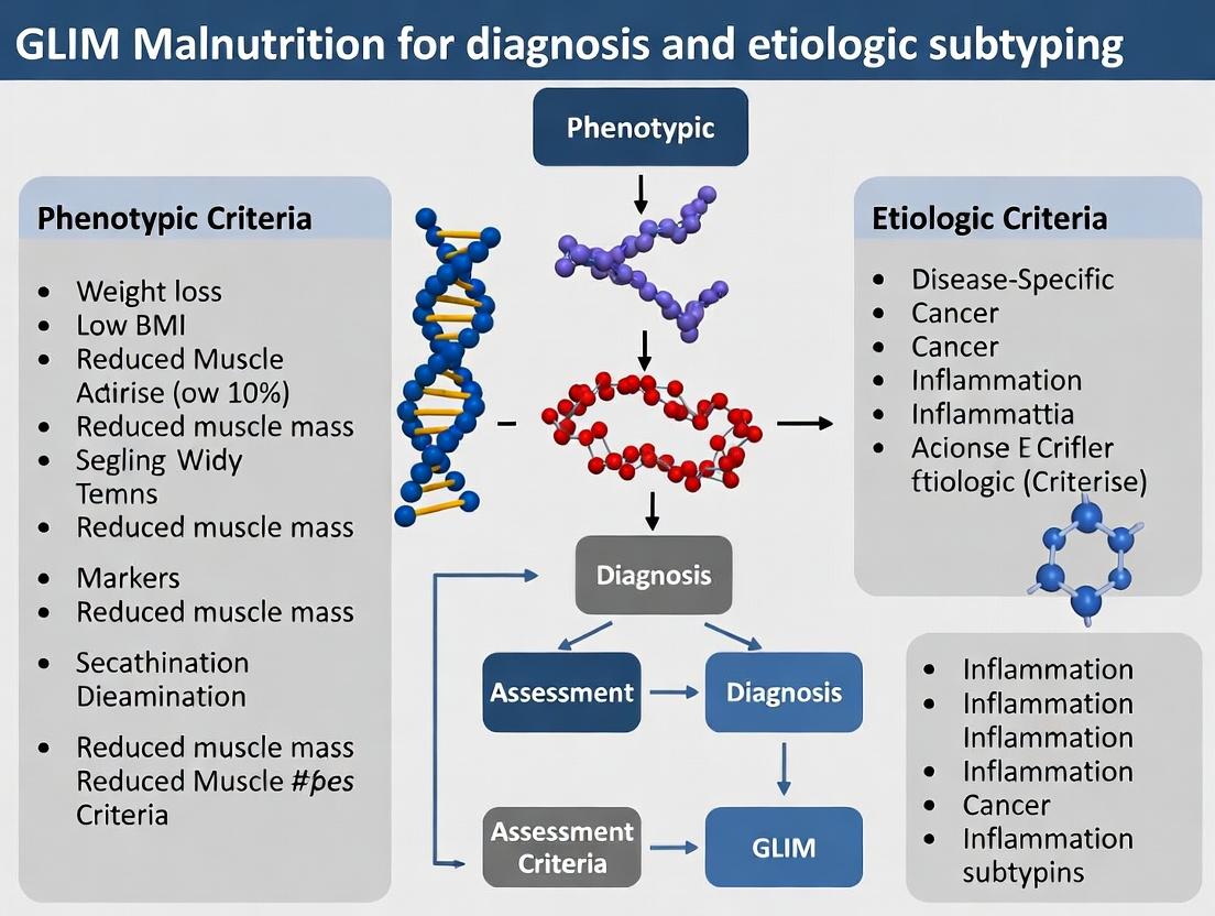
Within the Global Leadership Initiative on Malnutrition (GLIM) framework, diagnosing malnutrition requires the concurrence of at least one phenotypic and one etiologic criterion. This technical guide delineates the core components, underlying biological pathways, and standardized research protocols for operationalizing these criteria in clinical and translational research, pivotal for patient stratification and therapeutic development.
The GLIM Framework: A Diagnostic Algorithm
The GLIM approach employs a two-step model: first, screening for malnutrition risk, followed by a diagnostic assessment applying phenotypic and etiologic criteria. Diagnosis is confirmed by the presence of ≥1 phenotypic AND ≥1 etiologic criterion.
| Criterion Type | Specific Criteria | Operational Cut-points (Adults) | Primary Measurement Method |
|---|---|---|---|
| Phenotypic | 1. Non-volitional weight loss | >5% within past 6 months, or >10% beyond 6 months | Serial weight measurement; patient recall. |
| 2. Low body mass index (BMI) | <18.5 kg/m² for <70y; <20 kg/m² for ≥70y | Weight and height measurement. | |
| 3. Reduced muscle mass | Below gender/age-specific percentiles | DXA, BIA, CT/MRI at L3, Anthropometry. | |
| Etiologic | 1. Reduced food intake/assimilation | ≤50% of ER >1 week, or any reduction >2 weeks, or GI dysfunction | Food records, intake surveys, malabsorption tests. |
| 2. Inflammation/disease burden | Acute disease/injury, chronic disease, or advanced age-related inflammation | CRP >5 mg/L, IL-6, Clinical diagnosis of chronic/infectious disease. |
Abbreviations: DXA: Dual-energy X-ray Absorptiometry; BIA: Bioelectrical Impedance Analysis; CT: Computed Tomography; MRI: Magnetic Resonance Imaging; CRP: C-Reactive Protein; IL-6: Interleukin-6; ER: Energy Requirements.
Biological Pathways Linking Etiology to Phenotype
Inflammation is a primary etiologic driver, activating catabolic pathways that lead to phenotypic changes.
Experimental Protocols for Criterion Assessment
Protocol 3.1: Quantification of Muscle Mass via CT at L3
Objective: To objectively measure reduced muscle mass (phenotypic criterion) by analyzing cross-sectional skeletal muscle area at the third lumbar vertebra (L3).
Materials:
- CT scanner.
- DICOM image analysis software (e.g., Slice-O-Matic, Horos, 3D Slicer).
- Hounsfield Unit (HU) thresholds for tissue demarcation.
Methodology:
- Image Acquisition: Obtain a single axial CT slice at the L3 vertebral level. Ensure patient's arms are raised if possible.
- Import & Calibration: Import DICOM file into analysis software. Verify spatial calibration using scale in images.
- Tissue Segmentation:
- Set HU thresholds for skeletal muscle: -29 to +150 HU.
- Manually or semi-automatically trace the total abdominal muscle area, including psoas, erector spinae, quadratus lumborum, transversus abdominis, external and internal obliques, and rectus abdominis.
- Area Calculation: Software calculates total cross-sectional area (cm²) of pixels within the defined HU range and manual trace.
- Normalization: Calculate the L3 Skeletal Muscle Index (SMI) = Muscle Area (cm²) / Height (m²). Compare to validated, population-specific cut-offs (e.g., SMI < 55 cm²/m² for men and < 39 cm²/m² for women for sarcopenia).
Protocol 3.2: Assessment of Inflammatory Burden via CRP & Cytokines
Objective: To quantify the presence and magnitude of inflammation (etiologic criterion) via circulating biomarkers.
Materials:
- Patient serum or plasma samples.
- High-sensitivity CRP (hs-CRP) ELISA kit.
- Multiplex cytokine assay panel (e.g., for IL-6, TNF-α).
- Microplate reader, multiplex analyzer.
Methodology:
- Sample Collection: Collect venous blood into serum separator or EDTA tubes. Process within 2 hours (centrifuge at 1000-2000 x g for 10 min). Aliquot and store at -80°C.
- hs-CRP ELISA:
- Follow manufacturer's protocol. Briefly, add standards and samples to antibody-coated wells.
- Incubate, wash, add detection antibody conjugate.
- Incubate, wash, add substrate solution. Stop reaction.
- Measure absorbance at 450nm. Calculate concentration from standard curve.
- Multiplex Cytokine Assay:
- Prepare magnetic bead cocktail.
- Add standards, controls, and samples to plate wells.
- Incubate, wash, add detection antibodies.
- Incubate, wash, add streptavidin-PE. Wash and resuspend in reading buffer.
- Analyze on multiplex analyzer. Use software to calculate concentrations from standard curves.
- Interpretation: Apply GLIM-aligned thresholds (e.g., CRP >5 mg/L indicates inflammation). Report cytokine levels as supportive mechanistic data.
The Scientist's Toolkit: Research Reagent Solutions
Table 2: Essential Research Materials for GLIM Criteria Investigation
| Item | Function in Research | Example Product/Catalog |
|---|---|---|
| hs-CRP ELISA Kit | Quantifies low-grade chronic inflammation, directly supporting the etiologic inflammation criterion. | R&D Systems Quantikine ELISA (DCRP00) |
| Human Cytokine Multiplex Panel | Profiles multiple inflammatory mediators (IL-6, TNF-α, IFN-γ) to elucidate specific catabolic drivers. | Bio-Plex Pro Human Cytokine 8-plex (M50000007A) |
| Myostatin (GDF-8) ELISA Kit | Measures myostatin, a negative regulator of muscle growth, linking inflammation to anabolic suppression. | Abcam Human GDF-8/Myostatin ELISA (ab99933) |
| Ubiquitin Ligase Antibody (MuRF1/MAFbx) | Detects expression of atrogenes via Western Blot/IHC, confirming activation of proteasomal degradation. | Cell Signaling Technology Anti-TRIM63 (43055) |
| D3-Creatine Dilution Kit | Provides a gold-standard, non-invasive method for quantifying total body skeletal muscle mass. | Creative Diagnostics D3-Creatine (DLS-3CR-10) |
| Bioelectrical Impedance Analyzer (BIA) | Enables rapid, bedside assessment of fat-free mass and phase angle for muscle mass estimation. | Seca mBCA 515/525 |
| Body Composition Phantom (for CT) | Calibrates CT scanners for consistent Hounsfield Unit measurement across sites/longitudinal studies. | CIRS Model 062 (Tissue Simulation Phantom) |
Integrated Workflow for GLIM-Based Research
A systematic research approach integrates phenotypic and etiologic measurement.
The precision of the GLIM framework hinges on the rigorous, reproducible assessment of its core phenotypic and etiologic components. For researchers and drug developers, mastering the associated biomarkers, imaging protocols, and integrated workflows is essential for defining homogeneous patient cohorts, identifying therapeutic targets, and validating interventions aimed at reversing the specific catabolic pathways of malnutrition.
Within the framework of the Global Leadership Initiative on Malnutrition (GLIM) diagnostic criteria, phenotypic components serve as the cornerstone for identifying malnutrition. This technical guide provides a focused, in-depth analysis of the three core phenotypic criteria: involuntary weight loss, low body mass index (BMI), and reduced muscle mass. For researchers and drug development professionals, a precise understanding of these parameters—their measurement, underlying pathophysiology, and interrelationships—is critical for advancing diagnostic accuracy, etiological research, and therapeutic interventions.
Core Phenotypic Criteria: Definitions and Current Thresholds
The GLIM consensus establishes specific, graded thresholds for each phenotypic criterion to ensure standardized diagnosis across research and clinical settings.
Table 1: GLIM Phenotypic Criteria and Diagnostic Thresholds
| Phenotypic Criterion | Severity Grade 1 (Moderate) | Severity Grade 2 (Severe) | Primary Measurement Method |
|---|---|---|---|
| Weight Loss | 5-10% within past 6 months, or 10-20% beyond 6 months | >10% within past 6 months, or >20% beyond 6 months | Documented historical weight; Patient/caregiver recall. |
| Low BMI (kg/m²) | <20.0 if <70 years; <22.0 if ≥70 years | <18.5 if <70 years; <20.0 if ≥70 years | Direct measurement of height and weight. |
| Reduced Muscle Mass | Reduced by an amount equivalent to the thresholds for low BMI. | Further reductions aligned with severe BMI thresholds. | CT/MRI (L3 slice); DXA; BIA; Anthropometry (adjusted AMC). |
Source: Adapted from Cederholm et al., Clinical Nutrition, 2019 and subsequent validation studies.
Pathophysiological Mechanisms and Interrelationships
The three phenotypic criteria are not independent; they are interconnected manifestations of a net negative balance between energy/protein intake and requirements, often driven by disease burden.
3.1. Signaling Pathways in Cachexia and Muscle Wasting A complex interplay of pro-inflammatory cytokines, hormonal changes, and disrupted anabolic signaling drives catabolism, linking systemic inflammation (an etiologic criterion) directly to phenotypic changes.
Title: Inflammatory Drivers of GLIM Phenotypic Criteria
3.2. Logical Diagnostic Workflow The application of phenotypic criteria within GLIM follows a specific, sequential logic to ensure consistent diagnosis.
Title: GLIM Phenotypic Assessment Workflow
Experimental Protocols for Key Measurements
4.1. Protocol: Quantification of Muscle Mass via Computed Tomography (CT) at L3 This is considered the reference standard for body composition analysis in research.
Objective: To precisely quantify cross-sectional skeletal muscle area (SMA) from a single abdominal CT scan slice at the third lumbar vertebra (L3).
Materials & Procedure:
- Image Acquisition: Obtain a routinely collected abdominal/pelvic CT scan. Identify the single axial slice at the midpoint of the L3 vertebra.
- Tissue Segmentation: Import the DICOM image into specialized analysis software (e.g., Slice-O-Matic, ImageJ with appropriate plugins).
- Hounsfield Unit (HU) Thresholding: Define skeletal muscle using established attenuation ranges (-29 to +150 HU).
- Area Calculation: Manually or semi-automatically delineate the total muscle area within the threshold, excluding bone and visceral organs. The software calculates the total cross-sectional area (cm²).
- Normalization: Normalize SMA to height squared to calculate the L3 Skeletal Muscle Index (SMI; cm²/m²). Compare to validated, population-specific cut-offs (e.g., Martin et al., J Clin Oncol, 2013).
4.2. Protocol: Bioelectrical Impedance Analysis (BIA) for Phase Angle and Body Composition A portable, non-invasive method for estimating body compartments.
Objective: To estimate fat-free mass (FFM) and derive phase angle (PhA), a biomarker of cellular health and integrity.
Materials & Procedure:
- Standardization: Perform measurement after a 4-hour fast, empty bladder, no strenuous exercise in prior 12 hours. Subject lies supine with limbs abducted.
- Electrode Placement: Place adhesive electrodes on the dorsal surfaces of the right hand and wrist, and right foot and ankle, according to manufacturer specifications.
- Measurement: A fixed or multi-frequency BIA device passes a low-amplitude alternating current. Resistance (R) and reactance (Xc) are recorded at 50 kHz.
- Calculations:
- Phase Angle: PhA = (Xc / R) * (180°/π).
- FFM: Use manufacturer or validated population-specific regression equations (e.g., Sergi et al., Clin Nutr, 2017) incorporating R, Xc, height, weight, sex, and age.
- SMM: Skeletal muscle mass may be derived from FFM using proprietary or published formulae.
The Scientist's Toolkit: Research Reagent Solutions
Table 2: Essential Research Reagents for Investigating Muscle Wasting Phenotypes
| Reagent / Material | Function in Research | Example Application |
|---|---|---|
| Recombinant Human Cytokines (TNF-α, IL-6, IL-1β) | To induce inflammatory signaling in vitro and in vivo. | Treating C2C12 myotubes to study proteolytic gene expression. |
| Proteasome Activity Assay Kit (e.g., Suc-LLVY-AMC substrate) | Fluorometric quantitation of 20S proteasome chymotrypsin-like activity. | Measuring proteasomal degradation activity in muscle homogenates from cachectic animal models. |
| Phospho- & Total Antibody Panels (Akt, mTOR, p70S6K, FoxO) | Western blot analysis of anabolic and catabolic signaling pathways. | Assessing mTOR pathway inhibition and FoxO transcription factor activation in atrophying muscle. |
| Murine Cachexia Models (e.g., C26 colon adenocarcinoma, LLC) | In vivo models exhibiting systemic inflammation, weight loss, and muscle wasting. | Testing efficacy of anti-cachexia drug candidates on lean mass preservation. |
| Myoblast Cell Line (e.g., C2C12, L6) | In vitro model for studying myogenesis, hypertrophy, and atrophy. | Screening compounds for their ability to inhibit dexamethasone-induced myotube diameter loss. |
| ELISA Kits for Myostatin, GDF-15, Activin A | Quantification of circulating or tissue-level negative regulators of muscle mass. | Correlating serum biomarker levels with CT-derived muscle mass in clinical cohorts. |
Within the Global Leadership Initiative on Malnutrition (GLIM) framework, the etiologic criteria of "Reduced Food Intake or Assimilation" and "Disease Burden/Inflammatory Condition" are central to diagnosing and classifying malnutrition. This whitepaper provides a technical dissection of these criteria, detailing their biological mechanisms, measurement methodologies, and interplay, framed within contemporary research on precision malnutrition diagnosis.
The GLIM consensus provides a two-step model for malnutrition diagnosis: first, a phenotypic criterion (e.g., weight loss, low BMI, reduced muscle mass), and second, at least one etiologic criterion. The two primary etiologic criteria are:
- Reduced food intake or assimilation: Encompassing diminished oral intake, malabsorption, and maldigestion.
- Inflammation/disease burden: Arising from acute or chronic disease-related inflammation.
These criteria are not mutually exclusive; they frequently interact, creating synergistic catabolic states that accelerate muscle and functional loss.
Reduced Intake/Assimilation: Mechanisms and Assessment
Pathophysiological Pathways
Reduced intake or assimilation leads to a pure "starvation" adaptation, characterized by hypoinsulinemia, increased lipolysis, and the suppression of pro-inflammatory cytokines. The body shifts to ketone metabolism to preserve lean mass. However, when compromised assimilation (e.g., intestinal failure, pancreatic insufficiency) is present, nutrient deprivation occurs despite adequate intake.
Quantitative Assessment Protocols
Key metrics and their measurement standards are summarized below.
Table 1: Methods for Assessing Reduced Intake/Assimilation
| Metric | Measurement Protocol | Threshold for GLIM Criterion | Tool/Instrument |
|---|---|---|---|
| Food Intake | 3-day weighed food record or 24-hour multiple-pass recall. | ≤50% of estimated energy requirement for >1 week. | Dietetic analysis software (e.g., NDS-R). |
| Malabsorption | 72-hour fecal fat collection while on a 100g fat/day diet. | Fecal fat >7g/day indicates steatorrhea. | Laboratory gravimetric analysis. |
| GI Function | D-Xylose absorption test: 5h urinary excretion after 5g oral dose. | <1.2g excretion indicates malabsorption. | Spectrophotometric assay. |
| Muscle Protein Synthesis | Stable isotope tracer (L-[ring-¹³C₆]phenylalanine) with serial muscle biopsies. | Fractional synthesis rate (FSR) depressed vs. controls. | Mass spectrometry (GC-MS/LC-MS). |
Key Experimental Protocol: Dual-Isotope Method for Assimilation
Objective: To simultaneously quantify whole-body protein breakdown and net absorption of an amino acid. Protocol:
- Tracer Infusion: Primed, continuous intravenous infusion of L-[¹³C₆]phenylalanine.
- Oral Tracer: Simultaneous ingestion of L-[²H₅]phenylalanine with a test meal.
- Sampling: Frequent arterialized venous blood sampling over 8 hours.
- Analysis: Plasma is analyzed by tandem mass spectrometry to determine enrichments of both tracers.
- Calculation: The appearance rate of the oral tracer in plasma reflects the systemic availability (absorption and first-pass metabolism) of dietary phenylalanine.
Inflammation: The Disease Burden Criterion
Inflammatory Pathways in Malnutrition
Inflammation, particularly from chronic or acute disease, disrupts normal anabolic responses. The primary mediators include:
- Pro-inflammatory cytokines: IL-1β, IL-6, TNF-α.
- Signaling Pathways: NF-κB and JAK/STAT activation.
- Cellular Effects: Increased muscle proteolysis via the ubiquitin-proteasome and autophagy-lysosome systems, inhibited protein synthesis via mTORC1 suppression, and altered appetite regulation.
Biomarkers and Measurement
Table 2: Inflammatory Biomarkers for GLIM Criterion Assessment
| Biomarker | Assay Protocol | Suggested Cut-off | Interpretation |
|---|---|---|---|
| C-Reactive Protein (CRP) | High-sensitivity immunoturbidimetric assay. | >5 mg/L (chronic), >10 mg/L (acute). | Acute phase responder, short half-life. |
| Interleukin-6 (IL-6) | Multiplex electrochemiluminescence (MSD) or ELISA. | >4 pg/mL. | Proximal driver of CRP production. |
| Albumin | Bromocresol green dye-binding method. | <3.5 g/dL. | Negative acute phase protein; confounded by hydration. |
| Neopterin | Competitive ELISA. | >10 nmol/L. | Marker of cell-mediated immune activation (IFN-γ). |
Key Experimental Protocol: Ex Vivo Muscle Strip Analysis
Objective: To measure cytokine-induced proteolysis in human muscle tissue. Protocol:
- Biopsy: Obtain percutaneous needle biopsy of vastus lateralis.
- Preparation: Dissect muscle into ~10mg strips in oxygenated (95% O₂/5% CO₂) Krebs-Henseleit buffer.
- Incubation: Incubate strips for 2-4 hours in buffer alone (control) or buffer containing recombinant human TNF-α (20 ng/mL) + IFN-γ (100 U/mL).
- Proteolysis Measurement: Use tyrosine release into the medium as a proxy for total proteolysis, measured fluorometrically or via HPLC.
- Pathway Inhibition: Parallel experiments can include inhibitors of the proteasome (MG-132) or autophagy (3-methyladenine) to delineate pathways.
The Scientist's Toolkit: Research Reagent Solutions
Table 3: Essential Reagents for Etiologic Criteria Research
| Reagent / Material | Supplier Examples | Primary Function in Research |
|---|---|---|
| Stable Isotope Tracers (L-[¹³C₆]Phenylalanine) | Cambridge Isotope Labs; Sigma-Aldrich | Precise metabolic flux studies of protein/AA kinetics. |
| Recombinant Human Cytokines (TNF-α, IL-1β, IL-6) | R&D Systems; PeproTech | In vitro and ex vivo modeling of inflammatory muscle wasting. |
| Multiplex Immunoassay Panels (Human Cytokine/Chemokine) | Meso Scale Discovery (MSD); Luminex | Simultaneous quantification of multiple inflammatory biomarkers. |
| Pathway Inhibitors (MG-132, Rapamycin) | Cell Signaling Technology; Cayman Chemical | Mechanistic studies to block specific proteolytic or synthetic pathways. |
| Anti-Myosin Heavy Chain Antibodies (for fiber typing) | DSHB; Abcam | Histological assessment of muscle morphology and fiber-type specific changes. |
| D-Xylose Test Kit | Fischer Scientific; Trinity Biotech | Standardized clinical assessment of carbohydrate malabsorption. |
Integrated Model and Diagnostic Workflow
The confluence of reduced intake and inflammation creates a vicious cycle. Inflammation induces anorexia and malabsorption, while reduced nutrient intake can impair gut barrier function, potentially exacerbating inflammation.
Precision application of GLIM's etiologic criteria requires rigorous, standardized measurement. Future research must focus on:
- Validating and refining biomarker cut-offs for diverse populations and diseases.
- Developing integrated "omics" signatures that capture the interaction between intake and inflammation.
- Creating point-of-care tools for reliable intake and inflammation assessment in clinical settings to enhance GLIM's utility in both research and patient care.
This technical guide outlines a rigorous diagnostic algorithm for the identification of malnutrition using the Global Leadership Initiative on Malnutrition (GLIM) criteria. Framed within the broader thesis that standardized, multi-step phenotypic and etiologic assessment improves diagnostic precision and clinical trial outcomes, this whitepaper details a structured pathway from initial screening to definitive confirmation. The stepwise approach is designed to enhance reproducibility in research settings and reliability in drug development targeting nutritional interventions.
The GLIM criteria provide a consensus framework for malnutrition diagnosis, requiring the identification of at least one phenotypic criterion (e.g., weight loss, low BMI, reduced muscle mass) and one etiologic criterion (e.g., reduced food intake, inflammation/disease burden). For researchers, a validated diagnostic algorithm is critical for subject stratification, endpoint adjudication in clinical trials, and elucidating the pathophysiology of disease-related malnutrition.
The Diagnostic Algorithm: A Stepwise Workflow
The algorithm proceeds through four discrete, sequential phases: 1) Risk Screening, 2) Phenotypic Assessment, 3) Etiologic Assessment, and 4) Severity Grading & Confirmation.
Phase 1: Initial Risk Screening
Objective: To identify "at-risk" individuals within a study population using a validated, rapid screening tool. Protocol: Administer the screening tool (e.g., MUST, MST, NRS-2002) to all potential subjects. Scoring must be performed by trained personnel according to the tool's manual. Decision Node: Subjects classified as "medium" or "high" risk proceed to Phase 2. "Low-risk" subjects are excluded from a malnutrition diagnosis but may serve as controls.
Phase 2: Phenotypic Criterion Assessment
Objective: To objectively measure and confirm at least one of the three GLIM phenotypic criteria. Experimental Protocols:
- Unintentional Weight Loss: Document weight change over a defined period (typically 6-12 months) using calibrated scales. Calculate percentage loss from a documented or recalled usual weight.
- Formula:
[(Usual Weight - Current Weight) / Usual Weight] * 100.
- Formula:
- Low Body Mass Index (BMI): Measure height with a stadiometer and weight in light clothing. Calculate BMI as
weight (kg) / [height (m)]^2. - Reduced Muscle Mass: This is the most technical assessment. Preferred research methods include:
- Dual-energy X-ray Absorptiometry (DXA): Standardized whole-body scan to estimate appendicular skeletal muscle mass (ASMM).
- Bioelectrical Impedance Analysis (BIA): Use a medical-grade, phase-sensitive device with population-specific equations to estimate fat-free mass.
- Computed Tomography (CT) at L3: In cohorts with existing abdominal CT scans, analyze a single cross-sectional slice at the third lumbar vertebra. Measure skeletal muscle area (SMA) using predefined Hounsfield Unit thresholds (-29 to +150). Normalize to height (SMI = SMA/height(m)^2).
Decision Node: Confirmation of at least one phenotypic criterion (meeting the predefined GLIM cut-offs) is required to proceed to Phase 3.
Phase 3: Etiologic Criterion Assessment
Objective: To identify and document the underlying cause driving the phenotypic alterations. Protocols:
- Reduced Food Intake/Assimilation: Quantify via 3-day food diaries analyzed with nutritional software, or by direct measurement of intake (e.g., in institutional settings). Malabsorption may be confirmed via fecal fat tests or specific nutrient biomarkers.
- Disease Burden/Inflammation: Assess via:
- Clinical Diagnosis: Documenting active disease states (e.g., metastatic cancer, major infection).
- Biomarkers: Measure C-reactive protein (CRP), interleukin-6 (IL-6), or other acute-phase proteins using standardized, validated immunoassays (e.g., ELISA). Cut-offs (e.g., CRP >5 mg/L) should be defined a priori.
Decision Node: Confirmation of at least one etiologic criterion, in conjunction with a confirmed phenotypic criterion from Phase 2, yields a provisional GLIM diagnosis of malnutrition.
Phase 4: Severity Grading & Final Confirmation
Objective: To classify malnutrition severity, a critical endpoint for interventional trials. Protocol: Grade severity based on the phenotypic criterion with the most severe finding.
- Phenotypic Grading Guide: Stage 1 (Moderate) malnutrition is defined by a 5-10% weight loss within 6 months, or BMI <20 kg/m² if <70 years (or <22 kg/m² if ≥70 years), or mild reductions in muscle mass. Stage 2 (Severe) malnutrition is defined by >10% weight loss, BMI <18.5 kg/m² if <70 years (or <20 kg/m² if ≥70 years), or severe reductions in muscle mass. Final Output: A confirmed GLIM diagnosis with severity staging.
Data Presentation: GLIM Criteria Cut-offs & Biomarkers
Table 1: GLIM Phenotypic Criteria and Research Cut-off Points
| Phenotypic Criterion | Measurement Tool | Cut-off for Diagnosis (Moderate/Severe) | Research-Grade Validation Notes |
|---|---|---|---|
| Weight Loss | Serial weight measurement | 5-10% / >10% (over 6 mo) | Must use documented or reliably recalled usual weight. |
| Low BMI (kg/m²) | Stadiometer & calibrated scale | <20 (<70y) / <18.5 (<70y) | Age-specific cut-offs are critical. <22 / <20 if age ≥70 years. |
| Reduced Muscle Mass | DXA | ASMI: <7.0 kg/m² (M), <5.5 kg/m² (F) | Use device and ethnicity-specific reference standards. |
| CT at L3 (SMI) | <55 cm²/m² (M), <39 cm²/m² (F) | Validated in oncology; emerging cut-offs for other diseases. | |
| BIA (Phase-sensitive) | Population-specific equations | Must be validated against a reference method (e.g., DXA). |
Table 2: Etiologic Criterion Assessment & Biomarkers
| Etiologic Criterion | Primary Assessment Method | Quantitative Supportive Biomarkers | Typical Research Cut-off |
|---|---|---|---|
| Reduced Intake | 3-day food diary (<50% of ER) | Serum prealbumin (transthyretin) | <0.2 g/L (rapid turnover). |
| Inflammation | Clinical diagnosis + CRP | CRP, IL-6, TNF-α, Albumin | CRP >5 mg/L; Albumin <35 g/L. |
Visualizing the Diagnostic Pathway
Title: GLIM Diagnostic Algorithm Workflow
Title: Pathophysiology of GLIM Criteria
The Scientist's Toolkit: Research Reagent Solutions
Table 3: Essential Materials for GLIM-Related Research
| Item / Reagent | Function in GLIM Research | Example / Specification |
|---|---|---|
| Validated Screening Tool | Standardized, rapid identification of at-risk subjects. | MUST (Malnutrition Universal Screening Tool) kit or digital form. |
| Calibrated Digital Scale | Accurate weight measurement for BMI and weight loss calculation. | SECA 767 or equivalent, with regular calibration. |
| Stadiometer | Accurate height measurement. | SECA 213 or wall-mounted model. |
| Bioelectrical Impedance Analyzer | Estimation of fat-free and muscle mass. | SECA mBCA 525 or InBody 770 (phase-sensitive, multi-frequency). |
| DXA System | Gold-standard for body composition (bone, fat, lean mass). | Hologic Horizon A or GE Lunar iDXA. |
| ELISA Kits (CRP, IL-6, Prealbumin) | Quantification of inflammatory and nutritional status biomarkers. | R&D Systems DuoSet ELISA, Abcam kits. Validated for serum/plasma. |
| Nutritional Analysis Software | Analysis of 3-day food diaries for energy/protein intake. | Nutrition Data System for Research (NDSR), Nutritics. |
| CT Image Analysis Software | Quantification of skeletal muscle area at L3 vertebra. | Slice-O-Matic (Tomovision), 3D Slicer with specialized plugins. |
The Global Leadership Initiative on Malnutrition (GLIM) represents a consensus framework for the diagnosis of malnutrition in adults, designed for global clinical implementation. This whitepaper contextualizes GLIM within its historical evolution, detailing its technical foundations and relationships to predecessor tools: Subjective Global Assessment (SGA), Malnutrition Universal Screening Tool (MUST), and Nutritional Risk Screening 2002 (NRS-2002). The analysis is framed within ongoing research into the validation and refinement of GLIM's phenotypic and etiologic criteria.
Historical Evolution of Malnutrition Assessment Tools
The development of GLIM is a direct response to the need for a unified, evidence-based diagnostic approach, reconciling methodologies from several historically significant tools.
Table 1: Historical Timeline and Core Focus of Key Malnutrition Tools
| Tool (Year) | Primary Setting | Core Methodology | Population Focus |
|---|---|---|---|
| SGA (1982) | Clinical (Inpatient) | Clinical Assessment (History, Physical Exam) | Surgical/Medical Inpatients |
| MUST (2003) | Community & Hospital | BMI, Weight Loss, Acute Disease Score | Adults in all settings |
| NRS-2002 (2003) | Hospital | Impaired Nutrition Status + Disease Severity Score | Hospitalized Patients |
| GLIM (2018) | All Clinical Settings | Phenotypic + Etiologic Criteria (Consensus) | Adults in all clinical settings |
Technical Comparison of Diagnostic Criteria
GLIM integrates and formalizes components from earlier systems into a two-step model: screening, then diagnosis based on at least one phenotypic and one etiologic criterion.
Table 2: Quantitative Criteria Comparison Across Assessment Tools
| Diagnostic Component | SGA | MUST | NRS-2002 | GLIM |
|---|---|---|---|---|
| Weight Loss | Qualitative History | >5% in 3-6 mo (Score) | >5% in 3 mo (1-3 pts) | >5% within 6 mo or >10% beyond 6 mo |
| Low BMI | Not Explicit | <18.5 kg/m² (Score) | <20.5 if <70y (Score) | <20 if <70y, <22 if ≥70y (Asia: <18.5/<20) |
| Reduced Muscle Mass | Subjective Loss (Exam) | Not Included | Not Included | Reduced by validated methods |
| Reduced Food Intake | Qualitative History | Not Included | 0-100% of requirements (0-3 pts) | ≤50% of >1 wk, or any reduction >2 wk |
| Inflammation/Disease Burden | Underlying Disease State | Acute Disease Effect (Score) | Severity of Disease (0-3 pts) | Acute/Chronic Disease-related Inflammation |
Core GLIM Diagnostic Framework: Phenotypic and Etiologic Criteria
The GLIM diagnosis requires at least one phenotypic AND one etiologic criterion.
Table 3: GLIM Diagnostic Criteria and Validation Thresholds
| Criterion Type | Specific Criterion | Operational Definition & Common Measurement Protocols |
|---|---|---|
| Phenotypic | Non-volitional Weight Loss | >5% within past 6 months or >10% beyond 6 months. Protocol: Serial weight measurement calibrated scale. |
| Phenotypic | Low BMI | <20 kg/m² (<70 years) or <22 kg/m² (≥70 years). Protocol: Height stadiometer, weight calibrated scale. |
| Phenotypic | Reduced Muscle Mass | Low quantity via BIA, DXA, CT (third lumbar vertebra), MRI, or anthropometry. Protocol (CT example): L3 single slice analyzed for skeletal muscle area, sex-specific cut-offs. |
| Etiologic | Reduced Food Intake/Absorption | ≤50% of energy needs >1 week, or any reduction >2 weeks. Protocol: 24-hour recall, food diary analyzed with nutritional software. |
| Etiologic | Inflammation/Disease Burden | Acute disease/injury or chronic disease-related. Protocol: CRP >5 mg/L, IL-6 assays, clinical diagnosis of chronic disease. |
Experimental Protocols for Validating GLIM Criteria
Protocol 4.1: Validation of Muscle Mass Criterion via CT Imaging
- Patient Selection: Recruit cohort meeting GLIM screening risk.
- Image Acquisition: Obtain abdominal CT scan within 72 hours of assessment. Use standard clinical parameters (120 kVp, automated mA).
- Analysis (SliceOmatic Software v5.0): Identify L3 vertebra. Highlight skeletal muscle tissue using Hounsfield Unit thresholds (-29 to +150). Calculate cross-sectional area (cm²).
- Normalization: Normalize area to height squared to obtain SMI (cm²/m²).
- Diagnosis Apply: Apply GLIM sex-specific cut-offs (e.g., SMI <55 cm²/m² for men, <39 cm²/m² for women). Correlate with other phenotypic/etiologic criteria.
Protocol 4.2: Assessing Reduced Food Intake via 24-Hour Recall
- Structured Interview: Conduct multiple-pass 24-hour dietary recall by trained dietitian.
- Data Entry: Input food items into standardized nutritional database (e.g., USDA FoodData Central, local equivalents).
- Energy Calculation: Compute total energy intake (kcal).
- Requirement Estimation: Estimate resting energy expenditure via indirect calorimetry or predictive equations (e.g., Mifflin-St Jeor), apply stress/activity factors.
- Criterion Fulfillment: Calculate intake as percentage of estimated requirement. Document if ≤50% for >1 week.
Visualizing the GLIM Diagnostic Pathway
Diagram Title: GLIM Diagnostic Algorithm Flowchart
Diagram Title: Tool Evolution to GLIM Consensus
The Scientist's Toolkit: Research Reagent Solutions for GLIM Research
Table 4: Essential Research Materials for GLIM Criteria Validation Studies
| Item/Category | Example Product/Source | Function in GLIM Research |
|---|---|---|
| Body Composition Analyzer | Seca mBCA 515 Bioelectrical Impedance Analysis (BIA) | Quantifies fat-free muscle mass for phenotypic criterion. |
| CT Image Analysis Software | TomoVision SliceOmatic (v5.0) | Analyzes L3 CT slices for skeletal muscle area, critical for muscle mass criterion validation. |
| Indirect Calorimeter | COSMED Quark RMR | Gold-standard measurement of resting energy expenditure for accurate calculation of reduced food intake criterion. |
| High-Sensitivity CRP Assay | Roche Cobas c503 (hsCRP) | Quantifies inflammatory marker (≥5 mg/L) to objectively support the inflammation etiologic criterion. |
| Cytokine Multiplex Panel | Bio-Plex Pro Human Cytokine 8-plex (Bio-Rad) | Measures IL-6, TNF-α, etc., for deep phenotyping of inflammatory etiology. |
| Nutritional Analysis Software | Nutrition Data System for Research (NDSR) | Analyzes detailed dietary intake from recalls/diaries to quantify reduced intake. |
| Calibrated Medical Scales & Stadiometer | Seca 767/217 series | Accurate, repeated measures of weight and height for BMI and weight loss criteria. |
| Standardized Patient-Reported Outcome | PG-SGA (Patient-Generated SGA) | Validated tool for capturing weight history and symptom impact related to intake. |
Implementing GLIM: A Step-by-Step Methodological Guide for Clinical and Research Settings
The Global Leadership Initiative on Malnutrition (GLIM) framework provides a consensus for the diagnosis of malnutrition, requiring the identification of at least one phenotypic (e.g., weight loss, low BMI, reduced muscle mass) and one etiologic (e.g., reduced food intake, inflammation) criterion. The selection of appropriate patients for full GLIM assessment is critical for resource efficiency and research validity. Pre-screening with validated tools is this essential first step, ensuring that comprehensive but labor-intensive phenotypic (e.g., DEXA, BIA) and etiologic (e.g., CRP, IL-6) measurements are targeted appropriately. This guide details the application of three cornerstone pre-screening tools—MUST, MNA, and NRS-2002—within a GLIM-oriented research protocol.
Quantitative Comparison of Pre-Screening Tools
A systematic review of current literature (2021-2024) reveals the performance characteristics of each tool against GLIM diagnosis as the reference standard in various adult populations.
Table 1: Performance Metrics of MUST, MNA-SF, and NRS-2002 Against GLIM Criteria
| Tool (Full Name) | Target Population | Key Components | Scoring & Risk Categories | Average Sensitivity vs. GLIM | Average Specificity vs. GLIM | Time to Administer |
|---|---|---|---|---|---|---|
| MUST (Malnutrition Universal Screening Tool) | All adult settings | BMI, weight loss, acute disease effect | 0 (Low), 1 (Medium), ≥2 (High) | 75-85% | 70-80% | 3-5 min |
| MNA-SF (Mini Nutritional Assessment-Short Form) | Geriatric (≥65 years) | Food intake, weight loss, mobility, neuropsychological, BMI | 12-14 (Normal), 8-11 (At Risk), 0-7 (Malnourished) | 85-95% | 60-75% | 5-10 min |
| NRS-2002 (Nutritional Risk Screening 2002) | Hospital inpatients | Weight loss, food intake, BMI + Disease severity (stress metabolism) | Score = Impaired Status + Disease Severity. ≥3 (At Risk) | 80-90% | 70-85% | 5-8 min |
Table 2: Alignment with GLIM Criteria and Recommended Research Context
| Tool | Directly Captured GLIM Phenotypic Criteria | Directly Captured GLIM Etiologic Criteria | Recommended Research Application |
|---|---|---|---|
| MUST | Unintentional weight loss, Low BMI | --- (Implied by acute disease component) | Large-scale epidemiological studies, mixed adult populations. |
| MNA-SF | Weight loss, Low BMI | Reduced food intake/assimilation | Geriatric and community-dwelling elderly studies. |
| NRS-2002 | Weight loss, Low BMI | Reduced food intake, Inflammation/Disease burden | Acute care, clinical trials, studies involving inflammatory states. |
Detailed Experimental Protocols for Tool Application
Protocol 3.1: MUST Application in a Cohort Study
Objective: To pre-screen a broad adult research cohort for risk of malnutrition prior to applying full GLIM criteria.
- Equipment: Calibrated scale, stadiometer, medical records.
- Step 1 – BMI Score: Measure height and weight. Calculate BMI (kg/m²). Score: >20=0, 18.5-20=1, <18.5=2.
- Step 2 – Weight Loss Score: Document unplanned weight loss in past 3-6 months via recall/records. Score: <5%=0, 5-10%=1, >10%=2.
- Step 3 – Acute Disease Score: If patient is acutely ill and there has been/no nutritional intake for >5 days, score=2.
- Step 4 – Aggregate Risk: Sum scores. 0=Low risk (routine follow-up), 1=Medium risk (observe), ≥2=High risk (trigger full GLIM assessment).
- Data Recording: Record individual component scores and total MUST score. Participants with score ≥2 proceed to Protocol 4.1.
Protocol 3.2: MNA-SF Application in a Geriatric Study
Objective: To identify malnutrition risk in subjects aged ≥65 years.
- Setting: Quiet, private area. Use validated MNA-SF form.
- Administration: Conduct as a structured interview.
- A1: Food intake decline over 3 months?
- A2: Weight loss over 3 months?
- A3: Mobility (bed/chair bound vs. goes out)?
- A4: Psychological stress/acute disease in 3 months?
- A5: Neuropsychological problems (dementia/depression)?
- B: BMI (measure or self-report). If unavailable, substitute calf circumference.
- Scoring: Sum points (0-14). 12-14: Normal, 8-11: At Risk, 0-7: Malnourished.
- Action: Subjects scoring ≤11 proceed to full GLIM assessment and/or full MNA.
Protocol 3.3: NRS-2002 Application in a Hospital-Based Trial
Objective: To screen for nutritional risk in hospitalized patients within 24 hours of admission.
- Initial Screening: Ask: Is BMI <20.5? Has there been weight loss in last 3 months? Has food intake decreased in last week? Is the patient severely ill (e.g., in ICU)? If "No" to all, re-screen weekly. If "Yes" to any, proceed to final screening.
- Final Screening - Impaired Nutritional Status Score (0-3): Grade severity based on weight loss, food intake, and BMI (see official grid).
- Final Screening - Disease Severity Score (0-3): Grade based on increased nutritional requirements (e.g., hip fracture=1, major abdominal surgery=2, severe sepsis=3).
- Age Adjustment: If age ≥70 years, add 1 point to total.
- Total Score: Sum of status + severity + age score. ≥3: Patient is nutritionally at risk – trigger full GLIM assessment and initiate nutritional care plan.
Integrated Workflow for GLIM-Centric Research
Diagram 1: Decision workflow for tool selection and GLIM assessment
Molecular Pathways Linking Screening to GLIM Etiology
Diagram 2: Inflammation pathway linking NRS-2002 to GLIM etiology
The Scientist's Toolkit: Key Research Reagent Solutions
Table 3: Essential Materials for Integrated Pre-Screening and GLIM Research
| Item / Reagent | Supplier Examples | Function in Pre-Screening/GLIM Context |
|---|---|---|
| Calibrated Digital Scales & Stadiometers | SECA, Tanita | Accurate measurement of weight and height for BMI calculation (MUST, MNA, NRS). |
| Bioelectrical Impedance Analysis (BIA) Devices | InBody, Seca mBCA | Quantifies phase angle, fat-free mass, and skeletal muscle mass for GLIM phenotypic criterion. |
| ELISA Kits for Inflammatory Markers (CRP, IL-6, TNF-α) | R&D Systems, Abcam, ThermoFisher | Measures etiologic GLIM criterion (inflammation) to correlate with pre-screen risk scores. |
| Validated MNA-SF & NRS-2002 Form Booklets | Nestlé Nutrition Institute, ESPEN | Standardized tools for consistent data collection across research sites. |
| Dual-Energy X-ray Absorptiometry (DEXA) | Hologic, GE Lunar | Gold-standard for body composition (muscle mass) assessment in GLIM phenotypic validation. |
| Electronic Data Capture (EDC) System with Built-in Calculators | REDCap, Castor EDC | Streamlines data entry, automates MUST/NRS scoring, and ensures protocol adherence. |
| Standardized Nutritional Supplement | Abbott, Nutricia, Fresenius Kabi | Used in interventional arms of trials following positive pre-screen/GLIM diagnosis. |
The Global Leadership Initiative on Malnutrition (GLIM) framework provides a consensus for diagnosing malnutrition. Its second step, the phenotypic assessment, relies on three measurable criteria: non-volitional weight loss, low body mass index (BMI), and reduced muscle mass. This technical guide details the practical, precise measurement techniques essential for robust research into these phenotypic criteria, enabling their validation, the exploration of their pathophysiological interrelationships, and the development of targeted nutritional or pharmacologic interventions.
Measurement Techniques for Each Phenotypic Criterion
Non-Volitional Weight Loss
This criterion assesses a history of weight loss over time, requiring accurate and sequential measurements.
Practical Protocol:
- Instrumentation: Use a calibrated digital floor scale (e.g., SECA 813) with a precision of ±0.1 kg. For bed-bound subjects, use integrated bed scales or hoist scales.
- Standardization: Weigh the subject at the same time of day (morning, after voiding, in fasting state), wearing standardized light clothing or a gown.
- Frequency: Document weight at consistent intervals (e.g., upon admission, weekly).
- Calculation:
- Percent Weight Loss = [(Usual Weight - Current Weight) / Usual Weight] x 100.
- Use recalled usual weight (from the past 3-6 months) if historical clinical weights are unavailable, acknowledging potential recall bias.
- Documentation: Clearly note if weight loss is non-volitional, excluding intentional weight loss from diet/exercise.
Low Body Mass Index (BMI)
BMI provides a population-level index of weight-for-height.
Practical Protocol:
- Height Measurement:
- For ambulatory patients: Use a wall-mounted stadiometer (e.g., SECA 213). Subject stands barefoot, heels together, back straight, head in the Frankfort horizontal plane. Measure to the nearest 0.1 cm.
- For non-ambulatory patients: Use knee-height calipers (e.g., Ross Laboratories caliper). Measure the distance from the heel to the anterior surface of the thigh just proximal to the patella. Estimate height using validated equations (e.g., Chumlea equations).
- Calculation:
- BMI = Current Weight (kg) / [Height (m)]².
- GLIM Cut-offs: Apply the agreed ethnicity-specific cut-offs (e.g., <18.5 kg/m² for Caucasians <70 years; <20 kg/m² for those >70 years).
Reduced Muscle Mass
This is the most technically complex criterion, requiring direct or indirect measurement of body composition.
Primary Research-Grade Techniques:
- Computed Tomography (CT): Considered the gold standard for cross-sectional muscle area measurement in research.
- Protocol (L3 Slice Analysis): Analyze a single axial CT image at the third lumbar vertebra (L3). Using specialized software (e.g., Slice-O-Matic, Horos), define the skeletal muscle tissue based on Hounsfield Unit thresholds (-29 to +150). Calculate the cross-sectional area (cm²) of all muscles (psoas, erector spinae, quadratus lumborum, transversus abdominis, external and internal obliques, rectus abdominis). Normalize to height squared to calculate the L3 Skeletal Muscle Index (SMI, cm²/m²).
- Bioelectrical Impedance Analysis (BIA): A more accessible, bedside technique.
- Protocol: Use a medical-grade, multi-frequency BIA device (e.g., SECA mBCA 515). Ensure subject is supine for ≥5 minutes, limbs abducted from the body. Place electrodes on the hand, wrist, foot, and ankle on the same side of the body. Measure resistance and reactance. Use validated population-specific equations (e.g., ESPEN consensus equations) to derive fat-free mass (FFM) and appendicular skeletal muscle mass (ASMM). Express ASMM as a height-adjusted index (ASMI, kg/m²).
Supporting Field Techniques:
- Mid-Upper Arm Circumference (MUAC): Measured at the midpoint between the acromion and olecranon using a non-stretch tape. Simple but correlates with muscle mass.
- Calf Circumference (CC): Measured at the widest point of the calf with the patient seated, knee at 90°. A value <31 cm is a suggested GLIM supportive measure.
Table 1: Quantitative Comparison of Muscle Mass Assessment Techniques
| Technique | Parameter Measured | Primary Output | GLIM Cut-off Examples (Research) | Key Advantages | Key Limitations |
|---|---|---|---|---|---|
| CT (L3) | Tissue cross-sectional area | SMI (cm²/m²) | <55 cm²/m² (Men), <39 cm²/m² (Women) | Gold standard, high precision, discriminates tissue types | Radiation exposure, cost, limited portability |
| BIA | Whole-body impedance | ASMI (kg/m²) | <7.0 kg/m² (Men), <5.7 kg/m² (Women)* | Portable, rapid, low cost, good for serial measures | Affected by hydration status; requires validated equation |
| MUAC | Limb circumference | MUAC (cm) | <23.5 cm (Men), <22 cm (Women) | Extremely simple, low cost, good for screening | Non-specific, includes subcutaneous fat |
| CC | Limb circumference | CC (cm) | <31 cm | Simple, good predictor of mobility | Non-specific, includes subcutaneous fat |
*Example cut-offs from ESPEN 2019 consensus; population-specific.
Experimental Protocol: Validating a Novel BIA Equation against CT
Title: Protocol for Cross-Validation of BIA-derived Muscle Mass against CT Reference Standard.
Aim: To develop/validate a disease-specific BIA equation for estimating ASMI using CT-derived SMI as the criterion method.
Methods:
- Subject Cohort: Recruit N=200 patients from target population (e.g., oncology, geriatric). Record age, sex, ethnicity, clinical diagnosis.
- CT Acquisition & Analysis (Reference):
- Perform a clinically indicated abdominal CT scan.
- Export the single axial image at the L3 vertebra in DICOM format.
- Two trained analysts, blinded to BIA results, will segment muscle tissue using semi-automated software (Hounsfield Unit range: -29 to +150).
- Calculate SMI (cm²/m²). Use the mean of both analysts' results. Inter-rater reliability (ICC) must be >0.95.
- BIA Measurement (Index):
- Within 24 hours of the CT scan, perform a standardized BIA measurement using a selected device.
- Record resistance (R), reactance (Xc), phase angle, and device-generated estimates of FFM/ASM.
- Statistical Analysis:
- Perform linear regression with CT-SMI as the dependent variable and BIA parameters (R, Xc, height²/R, weight, age, sex) as independent variables.
- Derive a new prediction equation.
- Assess agreement between the new BIA-predicted ASMI and CT-SMI using Bland-Altman plots, calculating the mean bias and limits of agreement.
Title: Research Workflow for BIA Equation Validation (83 chars)
The Scientist's Toolkit: Research Reagent Solutions
| Item / Reagent | Function in Phenotypic Assessment Research |
|---|---|
| Calibrated Digital Floor Scale (e.g., SECA 813) | Provides high-precision (±0.1 kg) weight measurements for accurate calculation of weight loss and BMI. |
| Wall-Mounted Stadiometer (e.g., SECA 213) | Ensures accurate, reproducible height measurement to the nearest 0.1 cm for valid BMI calculation. |
| Medical-Grade Multi-Frequency BIA Device (e.g., SECA mBCA 515) | Measures bioelectrical impedance at multiple frequencies to estimate body composition compartments (FFM, ASM) using phase-sensitive analysis. |
| CT Scanner with DICOM Export | Acquires cross-sectional images for gold-standard analysis of skeletal muscle area at the L3 vertebral level. |
| Image Analysis Software (e.g., Slice-O-Matic, Horos) | Allows for semi-automated segmentation and quantification of muscle tissue area on CT images based on Hounsfield Unit thresholds. |
| Knee-Height Caliper (e.g., Ross Laboratories) | Enables estimation of height in non-ambulatory patients for BMI calculation via validated anthropometric equations. |
| Non-Stretch Insertion Tape | Used for standardized measurement of Mid-Upper Arm Circumference (MUAC) and Calf Circumference (CC) as supportive phenotypic measures. |
| Standardized Operating Procedure (SOP) Documents | Critical for ensuring measurement consistency across different researchers and timepoints, minimizing technical error. |
Title: GLIM Phenotypic Criteria & Measurement Pathways (66 chars)
Within the Global Leadership Initiative on Malnutrition (GLIM) framework, the etiologic criteria—reduced food intake/assimilation and inflammation/disease burden—are essential for confirming malnutrition diagnosis. This step moves beyond phenotypic assessment to identify underlying causes, crucial for targeted intervention. For researchers and drug developers, precise quantification of inflammatory states and disease burden is vital for patient stratification, biomarker discovery, and evaluating therapeutic efficacy.
Quantifying the Inflammatory State
Chronic inflammation, a key GLIM etiologic criterion, drives catabolism and muscle wasting. Accurate assessment requires a multi-modal approach.
Key Biomarkers and Their Clinical Significance
Biomarkers provide objective measures of systemic inflammation. The table below summarizes primary analytes, their sources, and implications for malnutrition research.
Table 1: Core Inflammatory Biomarkers for Etiologic Assessment
| Biomarker | Primary Source | Half-Life | Key Function in Pathophysiology | Typical Assay Method | Elevated Threshold (Malnutrition Context) |
|---|---|---|---|---|---|
| C-Reactive Protein (CRP) | Hepatocyte (IL-6 driven) | 19 hrs | Acute-phase reactant; activates complement. | Immunoturbidimetry, ELISA | >5 mg/L (chronic low-grade), >10 mg/L (acute) |
| Interleukin-6 (IL-6) | Macrophages, T cells, adipocytes | <1 hr | Pro-inflammatory cytokine; chief driver of hepatic APR. | ELISA, Electrochemiluminescence | >3-5 pg/mL (plasma) |
| Tumor Necrosis Factor-alpha (TNF-α) | Macrophages, NK cells | 10-20 min | Pro-inflammatory cytokine; induces cachexia. | ELISA, Multiplex Bead Array | >8.1 pg/mL (serum) |
| Serum Amyloid A (SAA) | Hepatocyte (IL-6/IL-1 driven) | ~50 min | Acute-phase reactant; alters HDL metabolism. | ELISA, Nephelometry | >10 mg/L |
| Albumin | Hepatocyte | 19-21 days | Negative acute-phase reactant; carrier protein. | Bromocresol Green dye-binding | <3.5 g/dL (considering half-life) |
| Neopterin | Macrophages (IFN-γ stimulated) | - | Marker of cell-mediated immune activation. | ELISA, HPLC | >10 nmol/L |
Experimental Protocol: Multiplex Cytokine Profiling from Human Plasma/Sera
Objective: To simultaneously quantify a panel of pro- and anti-inflammatory cytokines from a single small-volume sample.
Materials & Workflow:
- Sample Collection: Collect venous blood into serum separator or EDTA tubes. Process within 30-60 minutes (centrifuge at 1000-2000 x g for 10 min at 4°C). Aliquot and store at -80°C. Avoid freeze-thaw cycles.
- Assay Principle: Magnetic bead-based multiplex immunoassay (e.g., Luminex xMAP technology).
- Procedure:
- Thaw samples on ice. Dilute samples 1:2 or 1:4 in provided assay buffer.
- Prepare standards in serial dilution and quality controls.
- Add 50 µL of standards, controls, and samples to designated wells of a 96-well filter plate pre-washed with wash buffer.
- Add 50 µL of antibody-conjugated magnetic bead mix. Seal plate and incubate for 2 hours on a plate shaker at room temperature, protected from light.
- Wash plate 3x using a magnetic plate washer with 100 µL wash buffer per well.
- Add 50 µL of biotinylated detection antibody cocktail. Incubate for 1 hour with shaking.
- Wash 3x.
- Add 50 µL of streptavidin-phycoerythrin (SA-PE). Incubate for 30 minutes with shaking, protected from light.
- Wash 3x.
- Resuspend beads in 100-150 µL of drive fluid. Shake for 5 minutes.
- Read plate on a multiplex array analyzer (e.g., Luminex instrument). Analyze data using a 5-parameter logistic curve fit from standard concentrations.
Documenting Disease Burden
Disease burden refers to the cumulative impact of a disease on functional status, metabolic demand, and catabolic drive. It is intrinsically linked to inflammation.
Scoring Systems and Functional Measures
Table 2: Disease Burden Assessment Tools in Malnutrition Research
| Tool/Measure | Domain Assessed | Administration | Scoring & Interpretation | Relevance to GLIM Etiology |
|---|---|---|---|---|
| Charlson Comorbidity Index (CCI) | Comorbidity burden | Chart review | Weighted sum of 19 conditions. Higher score indicates greater mortality risk. | Directly captures "disease burden." Score ≥1 often used. |
| Karnofsky Performance Status (KPS) | Functional performance | Clinician-rated | 0% (dead) to 100% (normal). <70% indicates unable to work/care for self. | Proxy for burden's functional impact. |
| NYHA Functional Classification | Cardiac-specific limitation | Clinician-rated | Class I (no limitation) to IV (symptoms at rest). | Disease-specific burden quantification. |
| Clinical Frailty Scale (CFS) | Frailty/Vulnerability | Clinician-rated | 1 (very fit) to 9 (terminally ill). ≥5 indicates vulnerable/mild frailty. | Overlaps with phenotypic criterion of reduced muscle mass. |
| Hand Grip Strength (HGS) | Functional capacity | Dynamometer | Sex/BMI-specific cutoffs. Low HGS indicates sarcopenia and functional impairment. | Links etiology (inflammation/burden) to phenotype. |
Experimental Protocol: In Vivo Assessment of Cancer Cachexia Burden in a Murine Model
Objective: To document disease burden and associated inflammation in a preclinical model of cancer-associated malnutrition/cachexia.
Materials & Workflow:
- Model Induction: Implant murine colon-26 (C26) adenocarcinoma cells (1 x 10^6 cells) subcutaneously into the flank of syngeneic BALB/c mice. Use age/weight-matched sham-injected controls.
- Longitudinal Monitoring: Track daily food intake and body weight. Measure tumor volume 2-3 times weekly via calipers (Volume = (length x width^2)/2).
- Terminal Assessment (Day 14-21):
- Functional Burden: Assess grip strength using a rodent grip strength meter (average of 5 trials).
- Body Composition: Euthanize mouse. Dissect and weigh specific muscles (tibialis anterior, gastrocnemius, quadriceps), epididymal/perigonadal fat pads, and spleen (as an immune/inflammation organ).
- Blood Collection: Perform cardiac puncture. Isolate serum for CRP, IL-6, TNF-α analysis via ELISA.
- Tissue Analysis: Snap-freeze muscles in liquid N2. Later, homogenize for mRNA/protein extraction to assess ubiquitin-proteasome (Atrogin-1, MuRF1) and autophagy (LC3-II, p62) pathway markers via qRT-PCR/Western blot.
- Key Outcome Metrics: Compare between tumor-bearing and control groups: body weight change, muscle/fat mass, grip strength, and circulating inflammatory markers.
Signaling Pathways Linking Inflammation to Muscle Wasting
The interplay between systemic inflammation (TNF-α, IL-6) and muscle protein turnover is central to disease-related malnutrition.
Inflammatory Pathways to Muscle Wasting
The Scientist's Toolkit: Essential Research Reagents & Materials
Table 3: Key Research Reagent Solutions for Etiologic Assessment Studies
| Item | Function/Application in Etiologic Assessment | Example Product/Catalog | Critical Notes |
|---|---|---|---|
| Human/Mouse Cytokine Multiplex Panel | Simultaneous quantification of inflammatory mediators (IL-6, TNF-α, IFN-γ, IL-1β) from low-volume samples. | Bio-Plex Pro Human Cytokine 27-plex Assay; Milliplex Mouse Cytokine/Chemokine Panel. | Choose panels aligned with study hypothesis (Th1/Th2/acute phase). |
| High-Sensitivity CRP (hsCRP) ELISA Kit | Precise measurement of low-grade chronic inflammation. | Abcam Human hsCRP ELISA Kit; R&D Systems Mouse/Rat CRP Quantikine ELISA. | Distinguish from standard CRP kits; higher sensitivity range (0.1-10 mg/L). |
| Phospho-Specific Antibodies (Western Blot) | Detection of activated signaling pathways (p-STAT3, p-p65 NF-κB, p-p38 MAPK) in muscle or immune cell lysates. | Cell Signaling Technology Phospho-STAT3 (Tyr705) (D3A7) XP Rabbit mAb #9145. | Always run alongside total protein antibody for ratio quantification. |
| RNA Isolation Kit (Fibrous Tissue) | High-yield RNA extraction from skeletal muscle for qRT-PCR analysis of atrogenes. | Qiagen RNeasy Fibrous Tissue Mini Kit; Norgen's Animal Tissue RNA Purification Kit. | Includes specialized lysis for tough muscle tissue; DNase treatment essential. |
| Luminex Magnetic Bead Assay Platform | Infrastructure for performing multiplex biomarker assays. | Luminex MAGPIX or xMAP INTELLIFLEX system; Bio-Rad Bio-Plex 200. | Requires compatible magnetic bead-based kits and analysis software. |
| Murine Cancer Cachexia Model Cells | Preclinical study of disease burden and inflammation. | Colon-26 (C26) adenocarcinoma cells (for BALB/c mice); Lewis Lung Carcinoma (LLC) cells (for C57BL/6). | Source from reputable cell banks (ATCC, JCRB). Characterize cytokine secretion profile. |
| Handheld Digital Dynamometer | Objective measurement of functional strength as a proxy for disease burden impact. | Bioseb Grip Strength Test for rodents; Jamar Hydraulic Hand Dynamometer for humans. | Follow standardized positioning protocols. Report average of multiple trials. |
Integrated Assessment Workflow
A systematic approach to etiologic assessment combines biomarker data with clinical indices.
GLIM Etiologic Assessment Workflow
Robust etiologic assessment is fundamental to validating GLIM-based malnutrition diagnosis in research settings. By implementing standardized protocols for biomarker quantification and disease burden documentation, scientists can ensure precise patient phenotyping. This rigor is indispensable for uncovering mechanistic pathways linking inflammation to cachexia and for developing targeted nutritional or pharmacologic interventions. The integration of quantitative laboratory data with validated clinical indices forms the evidence base required for advancing the science of disease-related malnutrition.
1. Introduction: Context within GLIM Research
The Global Leadership Initiative on Malnutrition (GLIM) framework provides a consensus-based, stepwise approach for diagnosing malnutrition, integrating phenotypic (weight loss, low BMI, reduced muscle mass) and etiologic (reduced food intake, inflammation/disease burden) criteria. Within the broader thesis of validating and refining GLIM criteria, oncology clinical trial cohorts represent a critical proving ground. These cohorts offer highly characterized patients, longitudinal data, and validated clinical outcomes, allowing for rigorous assessment of GLIM's predictive validity, operational feasibility, and interplay with cancer-specific pathophysiology. This whitepaper details the technical implementation of GLIM within such a cohort.
2. Methodological Protocol for GLIM Application
The following protocol is designed for retrospective or prospective application within an oncology trial database.
Phase 1: Screening
- Tool: Must use a validated screening tool (e.g., Patient-Generated Subjective Global Assessment Short Form [PG-SGA SF]).
- Threshold: A score of ≥4 indicates "at risk" and proceeds to GLIM assessment.
Phase 2: GLIM Diagnosis
- Rule: At least one phenotypic and one etiologic criterion must be present for diagnosis.
- Assessment Window: Criteria are assessed within a ±2-week window of the index date (e.g., treatment initiation).
Phase 3: Severity Grading
- Post-Diagnosis: Use phenotypic criteria to grade severity (Stage 1: Moderate, Stage 2: Severe).
3. Data Synthesis & Quantitative Findings
Table 1: Prevalence of GLIM Criteria in a Hypothetical Phase III NSCLC Trial Cohort (N=500)
| GLIM Criterion | Operational Definition in Trial | Prevalence, n (%) |
|---|---|---|
| Phenotypic | ||
| Weight Loss >5% (6 mo) | Calculated from baseline/ historical weight in CRF | 165 (33.0) |
| Low BMI (<20/22 kg/m²) | Calculated from baseline measurements | 75 (15.0) |
| Low Muscle Mass (CT) | SMI <41 cm²/m² (M), <34 cm²/m² (F) | 210 (42.0) |
| Etiologic | ||
| Reduced Intake | PG-SGA Item 4 score ≥2 or clinician note | 140 (28.0) |
| Disease Burden | Stage IV disease or CRP >5 mg/L | 450 (90.0) |
| GLIM Diagnosis | ≥1 Phenotypic + ≥1 Etiologic Criterion | 200 (40.0) |
Table 2: Association Between Baseline GLIM Malnutrition and 12-Month Clinical Outcomes
| Outcome Measure | GLIM (+) (n=200) | GLIM (-) (n=300) | p-value |
|---|---|---|---|
| Grade 3+ Treatment Toxicity, % | 48.5 | 28.7 | <0.001 |
| Treatment Interruption, % | 62.0 | 40.3 | <0.001 |
| Median Progression-Free Survival, months | 8.2 | 11.5 | 0.003 |
| Median Overall Survival, months | 14.1 | 19.8 | 0.001 |
4. Visualizing Pathways and Workflows
GLIM Assessment Workflow in an Oncology Trial
Cancer Inflammation Drives GLIM Criteria
5. The Scientist's Toolkit: Key Research Reagent Solutions
| Item / Solution | Function in GLIM Oncology Research |
|---|---|
| PG-SGA (SF) Forms | Validated tool for mandatory initial screening and quantifying reduced intake/symptoms. |
| CT Image Analysis Software | Enables objective measurement of skeletal muscle mass from routine oncology scans. |
| Body Composition Phantoms | Calibration tools for ensuring consistency and accuracy across CT scanners and analysis software. |
| CRP & Albumin Assays | Quantifies inflammatory etiologic criterion; high-sensitivity CRP preferred. |
| Bioelectrical Impedance Analysis | Alternative for muscle mass assessment where CT is not feasible; requires cancer-specific equations. |
| Electronic Patient-Reported Outcome Platform | Facilitates real-time capture of weight and nutrition impact symptoms directly from patients. |
| Standardized Operating Procedures (SOPs) | Critical for ensuring consistent application of GLIM criteria across multi-center trial sites. |
Within the field of malnutrition research, particularly for the Global Leadership Initiative on Malnutrition (GLIM) framework, robust data collection is the cornerstone of validating phenotypic and etiologic criteria for diagnosis. This guide details the technical protocols and documentation standards essential for building datasets that enable precise etiological classification and phenotypic severity grading, which are critical for clinical trials and therapeutic development.
1. Core Data Domains for GLIM Criteria Data collection must be structured to capture both phenotypic and etiologic components as defined by GLIM. The following table summarizes the quantitative and qualitative measures required.
Table 1: Core Data Domains for GLIM Malnutrition Diagnosis
| GLIM Criterion | Data Variable | Measurement Protocol | Required Precision | Common Tools/Sources |
|---|---|---|---|---|
| Phenotypic: Weight Loss | Percentage body weight loss | (Current Usual Weight - Current Weight) / Usual Weight x 100% | To nearest 0.1 kg; historical recall validated where possible. | Digital scale, patient history, serial medical records. |
| Phenotypic: Low BMI | Body Mass Index (BMI) | Weight (kg) / [Height (m)]² | Height: to nearest 0.1 cm. Weight: to nearest 0.1 kg. | Stadiometer, calibrated scale. |
| Phenotypic: Reduced Muscle Mass | Appendicular Skeletal Muscle Mass Index (ASMI) | Dual-energy X-ray Absorptiometry (DXA) scan of limbs. Calculated as ASM (kg) / height (m)². | DXA machine with standardized positioning protocol. | DXA scanner, bioelectrical impedance analysis (BIA) with population-specific equations. |
| Etiologic: Reduced Food Intake | Average daily energy intake | 3-day weighed food record or 24-hour recall repeated 3x. | Analyzed using standardized food composition tables (e.g., USDA, local databases). | Dietetic assessment software, food scales. |
| Etiologic: Inflammation/Disease Burden | C-Reactive Protein (CRP) | Venous blood sample analyzed via immunoturbidimetric assay. | Serum/plasma; assay detection limit <0.3 mg/dL. | Clinical chemistry analyzer, ELISA kits for low-range detection. |
| Supporting: Handgrip Strength | Isometric grip strength | Jamar dynamometer, three trials per hand, highest value recorded. | Calibrated dynamometer; protocol per ESPEN/EWGSOP. | Handheld dynamometer. |
2. Experimental Protocol: Validating Muscle Mass Measurement via BIA against DXA Objective: To validate bioelectrical impedance analysis (BIA) equations for estimating appendicular skeletal muscle mass (ASMM) in a specific patient population (e.g., elderly with chronic disease) against the reference method DXA. Materials: DXA scanner (e.g., Hologic, GE Lunar), multi-frequency BIA device (e.g., Seca mBCA 515), calibration phantoms, data collection forms, anthropometric tape, scale, stadiometer. Population: n=200 target population participants. Procedure: 1. Ethics & Consent: Obtain IRB approval and informed consent. 2. Preparation: Participants fast for 4 hours, avoid strenuous exercise for 12 hours, and void bladder 30 minutes prior. 3. Anthropometry: Measure height and weight in light clothing. 4. BIA Measurement: Position participant supine, arms 30° from body, legs not touching. Place electrodes on right hand and foot per manufacturer's guide. Record impedance values at 50 kHz. 5. DXA Measurement: Perform whole-body DXA scan with participant in standardized position following manufacturer's calibration protocol. Analyze to derive ASMM. 6. Data Analysis: Use linear regression and Bland-Altman analysis to compare BIA-predicted ASMM (using device equations) against DXA-derived ASMM. Develop population-specific correction equations if bias is detected.
Title: GLIM Muscle Mass Validation Workflow
3. Inflammatory Pathway Documentation in Disease-Related Malnutrition Chronic disease drives malnutrition via inflammatory pathways. Documenting specific mediators is key for etiologic attribution.
Title: Inflammatory Pathways in Disease-Related Malnutrition
4. The Scientist's Toolkit: Research Reagent Solutions Table 2: Essential Reagents and Materials for GLIM-Related Research
| Item | Function | Example/Specification |
|---|---|---|
| ELISA Kit for Cytokines (IL-6, TNF-α) | Quantifies low concentrations of inflammatory markers in serum/plasma to document etiologic inflammation. | High-sensitivity kits (detection limit <0.5 pg/mL). |
| Certified DNA/RNA Shield Tubes | Stabilizes whole blood or buccal swab samples for genomic or transcriptomic analysis of metabolic pathways. | Enables ambient temperature storage, preserving sample integrity. |
| Stable Isotope Tracers (e.g., ¹³C-Leucine) | Allows precise measurement of whole-body or muscle protein synthesis rates in kinetic studies. | ≥ 98 atom% ¹³C; administered via controlled infusion. |
| SDS-PAGE & Western Blot Reagents | Detects and semi-quantifies proteins related to muscle atrophy (e.g., MuRF-1, atrogin-1). | Precast gels, validated antibodies, chemiluminescent substrate. |
| Liquid Chromatography-Mass Spectrometry (LC-MS) System | Gold standard for metabolomic profiling and quantifying specific nutrient or hormone levels. | High-resolution MS with reverse-phase chromatography. |
| Validated Food Frequency Questionnaire (FFQ) | Assesses habitual dietary intake over time to document reduced intake/assimilation. | Population-specific, nutrient database-linked. |
| Electronic Handheld Dynamometer | Objectively measures muscle strength as a functional correlate of muscle mass. | Jamar-type, calibrated annually with known weights. |
This whitepaper provides an in-depth technical guide for differentiating moderate (Stage 1) from severe (Stage 2) malnutrition using the Global Leadership Initiative on Malnutrition (GLIM) criteria. Framed within ongoing research on the refinement of phenotypic and etiologic diagnostic criteria, this document serves researchers, scientists, and drug development professionals engaged in metabolic and nutritional studies. Accurate severity stratification is critical for prognostication, intervention triaging, and endpoint selection in clinical trials.
GLIM Criteria Recap: Phenotypic and Etiologic Criteria
The GLIM framework operates via a two-step approach: first, screening for malnutrition risk, then confirmation and severity grading by assessing at least one phenotypic and one etiologic criterion.
Table 1: Core GLIM Criteria for Diagnosis
| Criterion Type | Specific Criteria |
|---|---|
| Phenotypic (Require ≥1) | 1. Non-volitional weight loss (%)2. Low body mass index (BMI; kg/m²)3. Reduced muscle mass (measured by validated methods) |
| Etiologic (Require ≥1) | 1. Reduced food intake or assimilation (≤50% of ER >1 week, or any reduction >2 weeks, or GI dysfunction)2. Inflammation or disease burden (acute disease/injury, chronic disease, or organ failure) |
Quantitative Thresholds for Severity Grading
Severity is graded based on the magnitude of the phenotypic criterion. The most severe phenotypic finding determines the overall stage.
Table 2: GLIM Severity Grading for Adults
| Phenotypic Criterion | Moderate (Stage 1) Malnutrition | Severe (Stage 2) Malnutrition |
|---|---|---|
| Weight Loss (Past 6 months) | 5-10% | >10% |
| Low BMI (kg/m²) | <20 (if <70 years)<22 (if ≥70 years) | <18.5 (if <70 years)<20 (if ≥70 years) |
| Reduced Muscle Mass | Mild to moderate deficit* | Severe deficit* |
Note: Precise cut-offs for muscle mass reduction are population and method-specific. Commonly referenced standards include Skeletal Muscle Index (SMI) via BIA (<10.75 kg/m² men, <6.75 kg/m² women for severe) or appendicular skeletal muscle mass index (ASMI) via DEXA.
Methodological Protocols for Phenotypic Criterion Assessment
Protocol for Weight Loss Assessment
- Objective: Quantify non-volitional weight loss percentage over a defined retrospective period (typically 6 months, or 3 months if acute).
- Materials: Calibrated scale, documented historical weight (medical record), structured patient interview.
- Procedure:
- Record current weight (kg) in light clothing, post-void, using a calibrated scale.
- Obtain a previously documented usual weight (from 6 months prior). If unavailable, use a patient's self-reported usual weight, noting the source.
- Calculate percentage weight loss:
[(Usual Weight - Current Weight) / Usual Weight] x 100. - Classify severity per Table 2.
Protocol for Body Composition Analysis (Muscle Mass)
- Objective: Quantify appendicular skeletal muscle mass (ASMM) via Dual-Energy X-ray Absorptiometry (DEXA) as a reference standard.
- Materials: DEXA scanner (e.g., Hologic, GE Lunar), calibration phantoms, analysis software.
- Procedure:
- Calibration: Perform daily quality assurance scans using manufacturer-specific calibration phantoms.
- Subject Preparation: Instruct subject to fast for 4 hours, void, wear metal-free clothing, and remove jewelry.
- Scanning: Position subject supine on the scanning table. Perform a whole-body scan according to manufacturer protocols.
- Analysis: Use machine software to define regions of interest. ASMM is calculated as the sum of lean soft tissue mass in the arms and legs.
- Indexing: Calculate ASMI:
ASMI = ASMM (kg) / height (m²). Compare to reference populations (e.g., FNIH, EWGSOP2 cut-offs) for severity grading.
Visualizing the GLIM Assessment Workflow
GLIM Diagnosis and Severity Grading Pathway
Key Inflammatory Pathways Linking Etiology to Phenotype
Chronic disease and inflammation, a key etiologic criterion, drive muscle catabolism via defined signaling pathways.
Inflammatory Pathways Driving Muscle Wasting
The Scientist's Toolkit: Key Research Reagent Solutions
Table 3: Essential Reagents for Investigating GLIM-Related Mechanisms
| Reagent/Material | Function in Research | Example Application |
|---|---|---|
| Recombinant Human TNF-α / IL-6 | To induce inflammatory signaling in vitro in myotube cultures. | Modeling inflammation-driven muscle atrophy in C2C12 or primary human myotubes. |
| Proteasome Inhibitor (MG-132) | To block the ubiquitin-proteasome system, allowing measurement of ubiquitinated protein accumulation. | Determining the contribution of UPS to muscle protein degradation in experimental models. |
| Phospho-specific Antibodies (p-Akt, p-S6, p-STAT3) | To assess activity status of key anabolic and catabolic signaling pathways via Western blot. | Quantifying mTORC1 inhibition (p-S6↓) or JAK/STAT activation (p-STAT3↑) in muscle biopsies. |
| DEXA Calibration Phantom | To ensure accuracy and precision of body composition measurements over time. | Daily quality assurance for longitudinal studies tracking ASMM changes. |
| Stable Isotope Tracers (e.g., D₂O, [¹³C]Leucine) | To measure in vivo rates of muscle protein synthesis and breakdown. | Directly quantifying the impact of reduced food intake (etiologic criterion) on muscle protein kinetics in humans. |
Challenges and Refinements: Troubleshooting GLIM Implementation in Complex Patient Populations
Within the framework of the Global Leadership Initiative on Malnutrition (GLIM) diagnostic criteria, accurate phenotypic assessment is paramount. This technical guide details the profound confounding effects of edema, acute fluid shifts, and body composition dynamics on core phenotypic measures—fat-free mass (FFM), muscle mass, and body weight—critical for the reduced muscle mass and non-volitional weight loss criteria. We provide methodologies to mitigate these pitfalls, ensuring rigor in etiologic and phenotypic research for malnutrition.
The Clinical and Research Challenge
Edema and rapid fluid shifts, common in acute illness, chronic disease, and therapeutic interventions, introduce significant error in body composition measurement. This compromises the precision of GLIM's phenotypic criteria, potentially leading to misclassification. For researchers, this noise obscures true treatment effects in nutritional and pharmacological intervention trials.
Impact on Key Phenotypic Measures
Table 1: Impact of Fluid Dynamics on Common Assessment Modalities
| Measurement Modality | Primary Target | Susceptibility to Edema/Fluid | Estimated Error Range | Primary Mechanism of Interference |
|---|---|---|---|---|
| Bioelectrical Impedance Analysis (BIA) | FFM, Total Body Water | Very High | FFM: ±5–15% in edema | Extracellular water expansion alters impedance vectors, invalidating standard equations. |
| Dual-Energy X-ray Absorptiometry (DXA) | Lean Soft Tissue, Fat Mass | Moderate-High | LSTM: ±2–5% in severe edema | Increased hydration of lean tissue elevates attenuation, overestimating lean mass. |
| Anthropometry (Mid-Upper Arm Circumference) | Muscle/Adipose Stores | High | Circumference: +1–4 cm | Subcutaneous fluid accumulation directly increases measurement. |
| Body Weight | Mass Change | Very High | Acute: ±1–5 kg/day | Rapid intravascular/interstitial fluid shifts unrelated to tissue mass. |
| CT/MRI (Skeletal Muscle Index) | Skeletal Muscle Area | Low (for analysis) | Minimal when properly analyzed | Direct visualization excludes fluid; but segmentation must distinguish edema. |
Experimental Protocols for Controlled Measurement
Protocol 1: Standardized Pre-Measurement Protocol for Fluid Stabilization
Objective: Minimize acute fluid shift noise in longitudinal studies.
- Posture & Time: Supine rest for 10 minutes pre-measurement.
- Consistency: Measurements at same time of day (±1 hour), post-void.
- Hydration: Standardized fluid intake (5 mL/kg) 2 hours prior.
- Diet: Avoid large meals or high sodium 12 hours prior.
- Documentation: Record clinical edema score (e.g., 0-3+ pitting) at each timepoint.
Protocol 2: BIA with Cole-Cplot and Bioimpedance Spectroscopy (BIS) Analysis
Objective: Distinguish intra- (ICW) and extracellular (ECW) water to adjust FFM estimates.
- Equipment: Multi-frequency or bioimpedance spectroscopy device.
- Procedure: Measure resistance (R) and reactance (Xc) at ≥50 frequencies (e.g., 5kHz-1MHz).
- Data Fitting: Fit measured data to Cole-Cole model to derive R at zero (R0) and infinite (R∞) frequency.
- Calculation: ECW = Kecw * (Height²/R0)^(2/3); ICW = Kicw * (Height²/R∞)^(2/3) – ECW. Use population-specific constants.
- Adjusted FFM: Apply hydration fraction (e.g., 0.73) to estimated dry lean mass.
Protocol 3: CT-Derived Muscle Analysis with Edema Assessment
Objective: Accurately segment skeletal muscle area (SMA) while quantifying intermuscular fluid.
- Image Acquisition: Single-slice axial CT at L3 vertebra.
- Segmentation: Threshold -29 to +150 Hounsfield Units (HU) for skeletal muscle.
- Edema Identification: Pixels within muscle mask with HU range -5 to +30 indicate edema-infiltrated muscle.
- Calculation:
Normal Muscle Area = Total Muscle Area - Edema Area (pixels in -5 to +30 HU range). - Normalization: Skeletal Muscle Index (SMI) = Normal Muscle Area (cm²) / Height (m²).
Visualizing Pathways and Workflows
Title: Fluid Shift Impact on GLIM Phenotypic Criteria
Title: Protocol for BIS-Adjusted Body Composition
The Scientist's Toolkit: Research Reagent Solutions
Table 2: Essential Research Materials for Mitigating Fluid-Related Pitfalls
| Item / Reagent Solution | Primary Function | Application in This Context |
|---|---|---|
| Bioimpedance Spectroscopy (BIS) Device (e.g., ImpediMed SFB7, Seca mBCA) | Measures impedance across multiple frequencies to model ICW/ECW. | Gold-standard for in vivo fluid compartment analysis; critical for adjusting BIA-derived FFM. |
| Validated BIA/BIS Prediction Equations | Converts raw impedance data to body composition estimates. | Must be selected for population (critically ill, elderly, specific disease states) and account for hydration. |
| Standardized Hydration Beverage | Provides consistent pre-measurement fluid load. | Controls for variation in hydration status at time of measurement in longitudinal protocols. |
| CT Scan with Standardized Protocol | Provides cross-sectional imaging for tissue demarcation. | Enables precise segmentation of skeletal muscle and identification of low-attenuation edema areas. |
| Image Analysis Software (e.g., Slice-O-Matic, Analyze, Horos) | Segments and analyzes tissue areas from CT/MRI DICOM images. | Quantifies muscle area and mean attenuation; separates normal muscle from edema-infiltrated regions. |
| Clinical Edema Assessment Scale | Semi-quantifies severity of pitting edema. | Provides a quick, complementary clinical correlate to instrument-based fluid measures. |
| Criterion Method References (e.g., Deuterium Oxide, Bromide Dilution) | Directly measures total body water and ECW. | Used to validate and calibrate BIS/BIA devices and equations in specific study populations. |
| High-Precision Digital Scale | Measures body weight to ±0.1 kg. | Essential for detecting true weight change against background of daily fluid fluctuation. |
The Global Leadership Initiative on Malnutrition (GLIM) operationalizes a two-step model for diagnosing malnutrition, requiring the identification of at least one phenotypic and one etiologic criterion. The phenotypic criteria (e.g., weight loss, low BMI, reduced muscle mass) are often the measurable outcome. The etiologic criteria, specifically "inflammation/disease burden," present a significant diagnostic challenge. Accurate application of this criterion requires a precise understanding of the inflammatory context. Chronic inflammation, characterized by a persistent, low-grade, non-resolving immune response, is a primary driver of the metabolic and anabolic resistance seen in disease-related malnutrition. In contrast, acute inflammation is a self-limiting, protective response. Misattribution of acute inflammation as chronic can lead to incorrect GLIM categorization and inappropriate nutritional intervention strategies. This guide details the experimental and analytical methodologies essential for distinguishing these states within clinical and research contexts pertinent to GLIM implementation and drug development targeting malnutrition.
Quantitative Biomarker Profiles: Acute vs. Chronic Inflammation
The distinction relies on a multi-analyte approach, as no single biomarker is pathognomonic. The following table summarizes key differentiating mediators, their sources, and dynamics.
Table 1: Core Biomarker Profiles in Acute vs. Chronic Inflammation
| Biomarker (Source) | Acute Inflammation Profile | Chronic Inflammation Profile | Primary Function & Interpretation |
|---|---|---|---|
| C-Reactive Protein (CRP) (Hepatocyte) | Rapid, sharp increase (hours); peaks 24-48h; rapid decline with resolution. | Sustained, moderate elevation (≥5 mg/L for weeks). | Acute-phase reactant; High sensitivity but low specificity. Serial measurement tracks kinetics. |
| Erythrocyte Sedimentation Rate (ESR) | Rises slower than CRP; can remain elevated during recovery. | Persistently elevated, often correlated with disease activity. | Indirect measure of acute-phase proteins (fibrinogen); non-specific but widely available. |
| Procalcitonin (PCT) (Multiple cell types) | Markedly elevated in systemic bacterial infection; low in viral or non-infectious inflammation. | Typically low or mildly elevated (e.g., in autoimmune disease). | Strongly associated with bacterial sepsis; useful for distinguishing etiology of acute phase. |
| IL-6 (Immune cells, endothelium, muscle) | Early, sharp peak; drives CRP production. | Persistent, low-level production; may be elevated in adipose tissue. | Pro-inflammatory cytokine; master regulator of acute phase; key target for therapeutic blockade. |
| TNF-α (Macrophages, T-cells) | Transient increase. | Chronically elevated; contributes to cachexia and insulin resistance. | Pro-inflammatory cytokine; central mediator in inflammatory diseases and malnutrition. |
| IL-1β (Monocytes, macrophages) | Sharp increase in response to pathogens/injury. | Can be chronically elevated (e.g., in autoinflammatory diseases). | Pyrogenic cytokine; induces IL-6; involved in innate immunity. |
| IL-10 (Regulatory T cells, macrophages) | Rises later to dampen acute response. | Often elevated but insufficient to resolve inflammation (failed resolution). | Anti-inflammatory cytokine; high IL-10/IL-6 ratio may indicate attempted resolution. |
| Serum Amyloid A (SAA) (Hepatocyte) | Very rapid, dramatic increase (can exceed CRP). | Sustained elevation, may promote pro-inflammatory HDL. | Acute-phase reactant; may be a more sensitive marker than CRP for low-grade inflammation. |
| Neopterin (Macrophages upon IFN-γ stimulation) | Moderate increase. | Consistently elevated, indicating sustained Th1/ macrophage activation. | Marker of cell-mediated immunity; correlates with disease activity in chronic disorders. |
| Albumin (Hepatocyte) | Decreases (negative acute-phase reactant). | Chronic low levels (hypoalbuminemia) indicative of prolonged inflammatory burden. | Nutritional and inflammatory marker; synthesis suppressed by IL-6, IL-1, TNF-α. |
Experimental Protocols for Etiologic Distinction
Protocol: Multiplex Cytokine Profiling from Human Plasma/Serum
Objective: To quantitatively profile a panel of pro- and anti-inflammatory cytokines to establish inflammatory signature. Materials: EDTA or serum separator tubes, centrifuge, -80°C freezer, multiplex assay kit (e.g., Luminex xMAP, Meso Scale Discovery ELISA), plate reader. Procedure:
- Sample Collection & Prep: Collect venous blood. For plasma, use EDTA tubes, centrifuge at 1000-2000 x g for 10 min at 4°C within 30 min. For serum, use serum tubes, allow to clot 30 min at RT, then centrifuge. Aliquot and store at -80°C. Avoid freeze-thaw cycles.
- Assay Setup: Thaw samples on ice. Follow manufacturer's protocol for the chosen multiplex panel (typically including IL-6, TNF-α, IL-1β, IL-10, IL-8). Dilute samples if necessary.
- Plate Incubation: Add standards, controls, and samples to pre-coated or bead-coupled plates. Incubate (typically 2h). Wash.
- Detection: Add biotinylated detection antibody cocktail. Incubate (1h). Wash. Add streptavidin-phycoerythrin (Luminex) or electrochemiluminescence reagent (MSD). Incubate.
- Reading & Analysis: Read plate on appropriate analyzer. Generate standard curves using 5-parameter logistic regression. Calculate cytokine concentrations in pg/mL.
Protocol: Gene Expression Analysis of Inflammatory Mediators in Peripheral Blood Mononuclear Cells (PBMCs)
Objective: To assess the transcriptional activation state of immune cells, revealing sustained signaling in chronic inflammation. Materials: Cell preparation tubes (CPTs) with sodium citrate, RNA stabilization reagent (e.g., Tempus, PAXgene), RNA extraction kit, cDNA synthesis kit, qPCR system, TaqMan assays. Procedure:
- PBMC Isolation: Draw blood into CPTs. Centrifuge at room temperature at 1500-1800 x g for 20-30 min. Recover the PBMC layer. Wash with PBS. Alternatively, lyse whole blood for RNA.
- RNA Stabilization & Extraction: Immediately add cells to RNA stabilization reagent. Store at -80°C. Extract total RNA using silica-membrane columns. Assess purity (A260/A280 ~2.0) and integrity (RIN >7).
- cDNA Synthesis: Use 100 ng – 1 µg total RNA for reverse transcription with random hexamers and reverse transcriptase.
- Quantitative PCR (qPCR): Perform TaqMan qPCR for target genes (e.g., IL6, TNF, NFKB1, SOCS3, ARG1) and housekeeping genes (e.g., GAPDH, HPRT1, B2M). Use 20-40 cycles.
- Data Analysis: Calculate ΔΔCq values. Express results as fold-change relative to a control group (e.g., healthy donors) or an internal reference gene.
Protocol: Functional Immune Cell Phenotyping by Flow Cytometry
Objective: To characterize immune cell subsets and activation states associated with chronic inflammation (e.g., monocyte priming, exhausted T cells). Materials: Fresh whole blood (heparin or EDTA), fluorescently conjugated antibodies (CD14, CD16, HLA-DR, CD86, CD3, CD4, CD8, CD28, PD-1), red cell lysis buffer, flow cytometer. Procedure:
- Staining: Aliquot 100 µL whole blood per tube. Add surface antibody cocktails. Incubate 20-30 min in the dark at 4°C.
- Lysis & Fixation: Add 2 mL of 1x red cell lysis buffer. Incubate 10-15 min at RT in dark. Centrifuge at 500 x g for 5 min. Aspirate supernatant.
- Wash & Resuspend: Wash cells with PBS + 2% FBS. Centrifuge. Aspirate. Resuspend in 300-400 µL of fixation buffer (1% paraformaldehyde) or sheath fluid.
- Acquisition: Acquire data on a flow cytometer, collecting ≥100,000 events in the lymphocyte/monocyte gate. Use single-stained controls for compensation.
- Analysis: Identify populations: Classical (CD14++CD16-), intermediate (CD14++CD16+), non-classical (CD14+CD16++) monocytes; assess HLA-DR/CD86 expression. Identify T cell subsets and PD-1 expression.
Visualizing Inflammatory Pathways and Workflows
Diagram 1: Signaling Pathways in Acute vs. Chronic Inflammation
Diagram 2: Etiologic Attribution Decision Workflow
The Scientist's Toolkit: Essential Research Reagents
Table 2: Key Research Reagent Solutions for Inflammatory Profiling
| Reagent / Material | Function & Application in Etiologic Distinction |
|---|---|
| Luminex xMAP Multiplex Kits | Simultaneous quantification of 30+ cytokines/chemokines from low-volume samples. Essential for generating comprehensive inflammatory signatures. |
| Meso Scale Discovery (MSD) U-PLEX Assays | Electrochemiluminescence-based multiplex immunoassays. High sensitivity and broad dynamic range for detecting low-abundance inflammatory markers. |
| Tempus Blood RNA Tubes | Stabilizes RNA expression profile at point of collection. Critical for accurate PBMC gene expression analysis reflecting in vivo state. |
| TaqMan Gene Expression Assays | Fluorogenic probe-based qPCR assays for precise quantification of mRNA for inflammatory mediators (e.g., IL6, TNF, NFKB1). |
| Flow Cytometry Antibody Panels (e.g., CD14/CD16/HLA-DR, CD3/CD4/CD8/PD-1) | Enable immunophenotyping of monocyte subsets and T-cell exhaustion markers linked to chronic inflammatory states. |
| Recombinant Cytokines & Neutralizing Antibodies | Used as standards in assays and for functional validation experiments (e.g., blocking IL-6/TNF to assess metabolic endpoints in cell models). |
| Phorbol Myristate Acetate (PMA) / Ionomycin | Cell stimulation controls for flow cytometry and functional assays to determine maximal immune cell activation capacity. |
| LPS (Lipopolysaccharide) | Toll-like receptor 4 agonist. Used in vitro to model acute inflammatory responses in immune cells. |
| Protease & Phosphatase Inhibitor Cocktails | Added to protein lysates during sample preparation for western blot or phosphoprotein assays to preserve signaling pathway integrity. |
| CRP & SAA High-Sensitivity ELISA Kits | Quantify low-level baseline inflammation crucial for identifying subclinical chronic inflammation. |
The Global Leadership Initiative on Malnutrition (GLIM) framework establishes a consensus for diagnosing malnutrition based on phenotypic (weight loss, low BMI, reduced muscle mass) and etiologic (reduced food intake/assimilation, inflammation/disease burden) criteria. A core thesis in contemporary clinical nutrition research posits that while GLIM provides a robust generalizable structure, its application must be phenotypically and mechanistically adapted for complex, high-risk populations where malnutrition pathophysiology is masked or divergent. This technical guide explores the requisite adaptations for three such groups: individuals with obesity, critically ill patients, and the elderly, aligning with the broader research imperative to validate GLIM criteria across diverse phenotypes and etiologies.
Population-Specific Adaptations: Criteria and Protocols
Obesity (The Paradox of Sarcopenic Obesity)
In obesity, excess adipose tissue masks the phenotypic criterion of low body weight (BMI). The etiologic driver is often chronic, low-grade inflammation.
- Adapted Phenotypic Criterion: Focus shifts entirely to reduced muscle mass, necessitating direct assessment.
- Recommended Protocol: Bioelectrical Impedance Analysis (BIA) with phase-sensitive devices using population-specific equations, or Computed Tomography (CT) at the L3 vertebral level from clinically obtained scans.
Table 1: Muscle Mass Assessment Cut-offs in Obesity
| Assessment Method | Parameter | Adjusted Cut-off for Class II/III Obesity (BMI ≥35) | Standard GLIM Cut-off (Reference) |
|---|---|---|---|
| BIA (Phase-sensitive) | Appendicular Skeletal Muscle Mass Index (ASMI) | < 9.2 kg/m² (men), < 7.3 kg/m² (women)* | < 7.0 kg/m² (men), < 5.7 kg/m² (women) |
| CT at L3 | Skeletal Muscle Index (SMI) | < 52.4 cm²/m² (men), < 38.5 cm²/m² (women) | < 50 cm²/m² (men), < 39 cm²/m² (women) |
Derived from ESPEN-specific recommendations. *Based on recent cohort analyses in severe obesity.
Experimental Protocol (CT-Based SMI Analysis):
- Image Selection: Identify the axial CT slice at the third lumbar vertebra (L3).
- Muscle Segmentation: Use specialized software (e.g., Slice-O-Matic, Horos) with Hounsfield Unit (HU) thresholds of -29 to +150 to delineate skeletal muscle (psoas, paraspinal, abdominal wall).
- Area Calculation: Software computes the total cross-sectional area (cm²) of the segmented muscle.
- Indexation: Normalize the area to height squared (m²) to calculate SMI (cm²/m²).
- Diagnosis: Compare SMI to validated, obesity-adjusted cut-offs (see Table 1).
Critical Illness (Hypercatabolic-Inflammatory State)
In ICU patients, universal systemic inflammation (GLIM etiologic criterion) is present, making phenotypic criteria primary. Fluid resuscitation confounds weight-based measures.
- Adapted Phenotypic Criteria: Weight loss is unreliable. Low BMI may be used if pre-admission weight is known. Reduced muscle mass is the most reliable but requires specific timing.
- Recommended Protocol: Muscle ultrasound of the quadriceps or CT (if available for clinical reasons) at ICU admission and serially to track loss.
Table 2: Muscle Change in Critical Illness: Quantitative Measures
| Method | Parameter | Significant Loss Indicative of Malnutrition | Measurement Frequency |
|---|---|---|---|
| Quadriceps Muscle Ultrasound | Rectus Femoris Cross-Sectional Area (RFCSA) or Thickness | >10-15% decrease over 7-10 days | Baseline (Day 1-3), then weekly |
| CT at L3 | Skeletal Muscle Index (SMI) | >5% decrease per week of ICU stay | When clinically obtained scans are available |
| Bioimpedance Spectroscopy (BIS) | Phase Angle (PhA) | PhA < 3.5° - 4.0° (population-dependent) | Daily/Weekly, accounting for fluid status |
Experimental Protocol (Quadriceps Ultrasound):
- Positioning: Patient supine, knee extended and relaxed.
- Landmarking: Identify the midpoint between the anterior superior iliac spine and the superior patellar border.
- Imaging: Use a linear probe (8-12 MHz) placed perpendicular to the long axis of the thigh. Ensure no compression.
- Measurement: Capture image. For thickness, measure distance from superficial to deep fascia. For RFCSA, trace the rectus femoris perimeter.
- Standardization: Repeat identical positioning for serial measurements. Use three-image average.
The Elderly (The Anorexia of Aging)
Etiology is multifactorial: anorexia (reduced intake), chronic inflammation ("inflammaging"), and anabolic resistance. Phenotypic criteria are applicable but require age-conscious interpretation.
- Adapted Phenotypic Criterion: Weight loss remains key but a lower threshold (>2% in 1 month) may increase sensitivity. Low BMI cut-off should be adjusted upward for age >70 years.
- Adapted Etiologic Criterion: Reduced food intake must be quantified meticulously.
Table 3: Adapted GLIM Criteria for the Elderly (≥70 years)
| GLIM Criterion | Standard Recommendation | Proposed Elder-Specific Adaptation |
|---|---|---|
| Weight Loss | >5% in past 6 months or >10% beyond 6 months | >2% in 1 month is clinically significant. |
| Low BMI | <20 kg/m² if <70 years; <22 kg/m² if >70 years | Consider <23 kg/m² as risk-enhancing. |
| Reduced Muscle Mass | Validated ethnicity-specific cut-offs | Use EWGSOP2 cut-offs (e.g., gait speed + low muscle quantity). |
| Reduced Intake | ≤50% of ER for >1 week | <75% of ER for ≥1 month or any involuntary loss >5% in 3 months. |
Experimental Protocol (Quantified Food Intake Assessment in Elderly):
- Method: 3-day weighed food record (preferred) or detailed dietary recall using digital photography aids.
- Tools: Provided digital food scales, detailed food diaries, and visual portion size guides.
- Analysis: Nutrient analysis software (e.g., NDS-R, Nutritics) to calculate total energy and protein intake.
- Benchmarking: Compare intake to estimated energy requirement (ER) using the Mifflin-St Jeor equation, multiplied by a physical activity level (PAL) factor of 1.3-1.5 for sedentary to moderately active elderly.
- Criterion Fulfillment: Document intake as a consistent percentage of ER over the assessment period (see Table 3).
Mechanistic Pathways and Diagnostic Workflows
Pathway: Inflammatory Drivers of Sarcopenic Obesity
Workflow: GLIM Diagnostic Algorithm for All Populations
Workflow: Elderly GLIM Diagnosis with Key Confounders
The Scientist's Toolkit: Research Reagent & Material Solutions
Table 4: Essential Research Materials for GLIM Validation Studies
| Item/Category | Function in GLIM Research | Example/Note |
|---|---|---|
| Phase-Sensitive BIA Device | Assesses body composition (ASMI) via resistance/reactance. Critical for obesity/elderly studies. | Seca mBCA 515, InBody 770. Must use validated, population-specific equations. |
| CT Image Analysis Software | Quantifies skeletal muscle area from clinical CT scans at L3. Gold standard for obesity/ICU. | Slice-O-Matic (TomoVision), Horos (open-source), 3D Slicer. Uses specific HU thresholds. |
| High-Frequency Ultrasound System | Measures muscle thickness/CSA at bedside for serial ICU monitoring. | Philips Lumify, SonoSite X-Porte with linear array probe (8-12 MHz). |
| Calibrated Digital Food Scales | Enables precise weighed food records for intake quantification (elderly, critical illness). | Must have ±1g precision. Integrated with dietary apps for data logging. |
| CRP & IL-6 ELISA Kits | Quantifies inflammatory burden (etiologic criterion) in obesity, elderly, critical illness. | High-sensitivity assays required for chronic low-grade inflammation ("inflammaging"). |
| Dual-Energy X-ray Absorptiometry (DXA) | Reference method for lean soft tissue mass. Used to validate BIA equations in specific populations. | Hologic Horizon, GE Lunar iDXA. Requires standardized positioning and analysis. |
| Standardized Nutritional Supplement | Used in controlled trials to test the "reduced intake" criterion's reversibility. | Ensure uniform protein/energy density (e.g., 2.0 kcal/mL, 20% protein). |
| Electronic Medical Record (EMR) Data Extraction Tools | For retrospective validation of GLIM criteria using documented weight, diagnosis, lab values. | i2b2, Epic Healthy Planet, custom SQL queries. |
Within the framework of research validating the Global Leadership Initiative on Malnutrition (GLIM) criteria, achieving high inter-rater reliability (IRR) across multiple clinical trial sites is a fundamental prerequisite for robust, generalizable findings. This technical guide provides a comprehensive strategy for quantifying and enhancing IRR, focusing on the application to GLIM's phenotypic (non-volitional weight loss, low body mass index, reduced muscle mass) and etiologic (reduced food intake/assimilation, inflammation/disease burden) criteria. Inconsistent application of these criteria directly threatens the validity of malnutrition prevalence studies and intervention trials.
The adoption of the GLIM framework represents a major advancement in standardizing malnutrition diagnosis. However, its operationalization relies on clinician and researcher judgment, particularly for phenotypic components like muscle mass assessment (e.g., physical exam, ultrasound) and etiologic criteria like inflammation. Multi-center trials, essential for adequate patient recruitment, inherently introduce variability. High IRR ensures that a diagnosis of "moderate malnutrition" via GLIM criteria is equivalent in Boston, Berlin, and Beijing, enabling pooled data analysis and credible cross-study comparisons.
Core Metrics for Quantifying Inter-Rater Reliability
Selecting the appropriate statistical metric is critical and depends on the data type of the GLIM criterion being assessed.
Table 1: IRR Metrics for GLIM Criteria Data Types
| GLIM Criterion Example | Data Type | Recommended IRR Metric | Interpretation Threshold |
|---|---|---|---|
| Presence/Absence of Reduced Muscle Mass (via palpation) | Nominal (Binary) | Cohen's Kappa (κ) | κ > 0.8: Excellent; 0.6-0.8: Substantial |
| Severity of Inflammation (e.g., None, Low, High) | Ordinal | Weighted Kappa (κ_w) | κ_w > 0.7 indicates acceptable agreement |
| Percentage of Weight Loss | Continuous (Interval/Ratio) | Intraclass Correlation Coefficient (ICC) | ICC > 0.75: Good; >0.9: Excellent |
| Continuous Muscle Thickness (cm via ultrasound) | Continuous (Interval/Ratio) | Intraclass Correlation Coefficient (ICC) | ICC > 0.75: Good; >0.9: Excellent |
Strategic Protocol for Establishing and Maintaining High IRR
A proactive, multi-phase approach is required throughout the trial lifecycle.
Phase 1: Pre-Trial Calibration & Charter Development
- Objective: Achieve baseline consensus before patient enrollment.
- Protocol:
- Develop a Rater Manual of Operations (RMOOP): Create a detailed, illustrated document defining every GLIM criterion. Include decision trees for borderline cases (e.g., how to classify "mild" vs. "moderate" inflammation in patients with chronic kidney disease).
- Centralized Training: Conduct mandatory, interactive virtual or in-person training sessions led by the core expert committee. Use standardized patient videos and case vignettes.
- Initial Reliability Testing: All site raters independently assess the same set of 20-30 benchmark patient cases (real or simulated). Calculate IRR metrics (κ, ICC) for each GLIM criterion.
- Feedback & Recertification: Raters falling below thresholds undergo targeted retraining and must recertify on a new case set.
Phase 2: In-Trial Monitoring & Drift Correction
- Objective: Prevent systematic divergence in criteria application over time.
- Protocol:
- Scheduled Re-Calibration: Implement quarterly or bi-annual reliability checks using new, core-committee-validated case sets.
- Ongoing Data Quality Checks: The data coordinating center runs scripts to flag outliers (e.g., a site with a 50% higher rate of "severe muscle loss" than others). This triggers a targeted review.
- Adjudication Committee: For complex or borderline cases identified by sites, a pre-defined central committee (blinded to site and initial assessment) makes the final GLIM diagnosis.
Phase 3: Post-Hoc Analysis & Reporting
- Objective: Quantify and report the achieved IRR as a measure of study quality.
- Protocol: Calculate final IRR statistics on a randomly selected subset of patient assessments that were independently rated by multiple, blinded raters at the same site or across sites. Report these metrics alongside the primary trial results.
Visualization of IRR Strategy Workflow
Diagram Title: Three-Phase IRR Workflow for GLIM Trials
The Scientist's Toolkit: Essential Reagents & Materials for GLIM Reliability Studies
Table 2: Key Research Reagent Solutions for IRR in GLIM Studies
| Item / Reagent | Primary Function in IRR Context | Example / Specification |
|---|---|---|
| Standardized Patient Case Library | Serves as the "gold standard" benchmark for training and testing rater agreement. Must be validated by expert consensus. | Digital repository of 50+ cases with fully documented GLIM criteria, including de-identified medical history, lab values (CRP, albumin), and key images. |
| Rater Manual of Operations (RMOOP) | The definitive reference document to minimize criterion ambiguity. | A living PDF with explicit definitions, flowcharts, and high-quality reference images for muscle wasting, fat loss, and edema. |
| Digital Data Capture Platform with IRR Module | Enforces standardized data entry and facilitates blinded re-assessment for IRR calculation. | REDCap or commercial EDC system configured with dual/multiple independent data entry fields for key GLIM variables. |
| Ultrasound System with Standardized Protocol | For objective, quantitative assessment of the muscle mass phenotype (e.g., rectus femoris thickness). | Portable B-mode ultrasound with a linear array probe (≥7.5 MHz). Must be used with a standardized Scanning Protocol SOP (patient position, probe placement, measurement landmark). |
| Calibration Phantom | Ensures measurement consistency across different ultrasound machines and sites for muscle quantification. | Tissue-mimicking phantom with known acoustic properties to verify machine calibration and linear measurement accuracy. |
| Central Adjudication Committee Charter | Formalizes the process for resolving diagnostic discrepancies, a key IRR safeguard. | Document defining committee membership, blinding procedures, quorum, and decision rules (e.g., majority vote) for borderline GLIM assessments. |
| Statistical Software IRR Packages | To calculate Kappa, ICC, and confidence intervals for ongoing monitoring. | R (irr package), SPSS (Reliability Analysis), or Stata (kappa and icc commands) with pre-written scripts for consistent analysis. |
Advanced Statistical Protocol: Calculating ICC for Muscle Mass Measurements
This protocol is critical for the GLIM phenotypic criterion of "reduced muscle mass" when assessed via quantitative techniques like ultrasound or bioelectrical impedance analysis (BIA).
- Objective: To assess the consistency of continuous muscle measurement data (e.g., thigh muscle layer thickness in cm) across different raters or devices.
- Experimental Design: A two-way random-effects, single measurement, absolute agreement model is typically used. This assumes a random sample of patients and a random sample of raters from larger populations, and it values exact agreement on the same numerical value.
- Methodology:
- Sample: Randomly select a subset of n=15-20 patients from the trial population.
- Raters: A group of k=3-4 raters (or devices), representing those from different trial sites, are assigned.
- Blinding & Independence: Each rater measures each patient's predefined anatomical site (e.g., dominant side rectus femoris) independently and blinded to the others' results and patient clinical details. The order of patient presentation is randomized per rater.
- Data Structure: Record data in a matrix where rows are patients and columns are raters.
- Analysis: Use statistical software (e.g., R
psychpackageICC()function) to calculate the ICC(2,1) according to Shrout & Fleiss.- R Code Example:
- Interpretation: Report the ICC point estimate and its 95% confidence interval. An ICC(2,1) < 0.75 indicates unacceptable reliability for pooling data across those raters/sites, triggering review of the measurement protocol and retraining.
In multi-center research employing the GLIM criteria, inter-rater reliability is not a peripheral quality check but a central pillar of scientific validity. A systematic, resource-informed strategy encompassing pre-training standardization, continuous monitoring, and rigorous post-hoc analysis is non-negotiable. By implementing the protocols and utilizing the toolkit outlined in this guide, researchers can ensure that diagnoses of malnutrition are consistent, credible, and capable of supporting global nutritional health initiatives.
Within the framework of the Global Leadership Initiative on Malnutrition (GLIM) criteria, accurate assessment of the phenotypic criterion of reduced muscle mass is paramount for robust diagnosis and etiologic stratification. This whitepaper provides a technical analysis of three core imaging and bioimpedance technologies—Bioelectrical Impedance Analysis (BIA), Dual-Energy X-ray Absorptiometry (DEXA), and Computed Tomography (CT)—detailing their methodologies, comparative diagnostic performance, and integrative application in clinical research and drug development for cachexia and sarcopenia.
The GLIM consensus identifies reduced muscle mass as a key phenotypic criterion for diagnosing malnutrition. Precise quantification is critical for staging severity, predicting outcomes, and evaluating therapeutic interventions in clinical trials. The evolution from simple anthropometry to advanced technological assessment reflects the need for accuracy, reproducibility, and prognostic relevance.
Core Technology Deep Dive: Methodologies & Protocols
Bioelectrical Impedance Analysis (BIA)
Principle: Measures the opposition of body tissues to a small, alternating electric current. Fluid-filled tissues conduct well (low impedance), while fat and bone impede current. Predictive equations derive fat-free mass.
Detailed Experimental Protocol (Single-Frequency, Tetrapolar):
- Subject Preparation: 4-hour fast, 12-hour abstention from strenuous exercise and alcohol, void bladder 30 minutes prior. No metal objects.
- Positioning: Supine position, limbs abducted from body. Electrodes placed on the dorsal surfaces of the right hand and foot at specific anatomical landmarks (wrist, ankle).
- Measurement: A 50 kHz, 800 µA current is applied. Impedance (Z), Resistance (R), and Reactance (Xc) are recorded.
- Calculation: Use population-specific or device-specific regression equations (e.g., Sergi equation for elderly) to estimate skeletal muscle mass (SMM) or appendicular lean mass (ALM).
Dual-Energy X-ray Absorptiometry (DEXA)
Principle: Uses two low-dose X-ray beams with distinct energy levels to differentiate tissue types based on differential attenuation, providing a three-compartment model (fat mass, lean soft tissue mass, bone mineral content).
Detailed Experimental Protocol (Whole-Body Scan):
- Calibration: Perform daily quality assurance with manufacturer-supplied spine and block phantoms.
- Subject Positioning: Centered supine on scanning table, arms at sides with palms down, feet secured with Velcro straps to maintain internal rotation. A positioning foam pad may be used under the knees.
- Scanning: The C-arm traverses from head to toe at a constant speed (~1 cm/s for pencil-beam systems). Scan mode (e.g., standard, thin, or thick) is selected based on subject thickness.
- Analysis: Regions of interest (ROIs) are auto-defined (arms, legs, trunk) and manually verified. ALM is calculated as the sum of lean soft tissue in arms and legs. Skeletal Muscle Index (SMI) = ALM/height².
Computed Tomography (CT)
Principle: Cross-sectional X-ray imaging providing precise anatomical visualization. Tissue is identified by Hounsfield Units (HU). Muscle cross-sectional area (CSA) at a specific vertebral level (L3) is a validated surrogate for whole-body muscle mass.
Detailed Experimental Protocol (L3 Skeletal Muscle Analysis):
- Image Acquisition: Standard abdominal/pelvic CT scan (120 kVp, slice thickness ≤5 mm).
- Slice Selection: Identify the single axial slice at the caudal end of the L3 vertebra where both transverse processes are fully visible.
- Segmentation: Using specialized software (e.g., Slice-O-Matic, 3D Slicer), define tissue HU thresholds: Skeletal Muscle: -29 to +150 HU; Visceral Adipose Tissue (VAT): -150 to -50 HU; Subcutaneous Adipose Tissue (SAT): -190 to -30 HU.
- Quantification: Automatically calculate the total cross-sectional area (cm²) of all muscle tissues within the threshold. Skeletal Muscle Index (SMI) = Muscle CSA (cm²) / height² (m²).
Comparative Quantitative Analysis
Table 1: Technical Specifications & Performance Metrics
| Feature | BIA | DEXA | CT (L3 Analysis) |
|---|---|---|---|
| Primary Output | Estimated TBW, FFM, ALM (kg) | Lean Soft Tissue Mass, ALM (kg), BMC (g) | Skeletal Muscle Cross-Sectional Area (cm²) |
| Precision (CV%) | 1-3% (same device/condition) | 1-2% for lean mass | < 0.5% for area measurement |
| Accuracy Limitation | Affected by hydration status; equation-dependent | Overestimates lean mass in edema; software-variant | Gold standard for compartmental mass; requires specific slice |
| Radiation Exposure | None | Very Low (1-10 µSv) | Moderate to High (2-10 mSv) |
| Scan Time | < 5 minutes | 5-20 minutes | < 5 minutes (analysis time added) |
| Cost | Low | Moderate | High (equipment & analysis) |
| Key Prognostic Cut-points | ALM/height²: M<7.26 kg/m²; F<5.45 kg/m² | ALM/height²: M<7.0 kg/m²; F<5.5 kg/m² | SMI: M<43 cm²/m² (BMI<25), <53 cm²/m² (BMI≥25); F<41 cm²/m² |
Table 2: Suitability for GLIM & Research Contexts
| Application | BIA | DEXA | CT |
|---|---|---|---|
| High-Volume Screening | Excellent | Good | Poor |
| Longitudinal Monitoring | Good (with strict protocol) | Excellent | Limited (due to radiation) |
| Cancer Cachexia Trials | Limited (hydration confounders) | Good | Excellent (gold standard, prognostic) |
| Etiologic Criterion Mapping | Low specificity | Moderate | High (discriminates sarcopenia, cachexia) |
| Drug Development Endpoint | Exploratory/Secondary | Primary (common) | Primary/Reference Standard |
Integrative Pathway for GLIM Diagnosis & Research
A strategic, tiered approach optimizes resource use and diagnostic certainty within GLIM workflows.
Diagram 1: Integrative Muscle Assessment Pathway for GLIM
The Scientist's Toolkit: Essential Research Reagents & Materials
Table 3: Key Reagents & Solutions for Muscle Mass Assessment Research
| Item | Function & Specification | Example Vendor/Product |
|---|---|---|
| BIA Calibration Phantom | Validates device accuracy using resistors/capacitors simulating known impedance. | Impedimed SFB7 Calibration Box |
| DEXA Anthropomorphic Phantom | Quality assurance for bone density, lean, and fat mass measurements across scanners. | Gammex RMI 462 Whole-Body Phantom |
| CT Calibration Phantom | Converts Hounsfield Units to true physical density for quantitative imaging. | Mindways CT Calibration Phantom |
| Segmentation Software | Semi-automated analysis of muscle/adipose tissue from CT/MRI scans. | Tomovision Slice-O-Matic, 3D Slicer |
| Standardized Positioning Aids | Ensures consistent limb positioning for DEXA/BIA, reducing measurement variance. | Hologic Positioning Foam, foot braces |
| Biobanking Aliquot Kits | Links muscle imaging data with biologic correlates (e.g., inflammatory cytokines). | Cryogenic vials, serum separators |
Optimizing the GLIM phenotypic criterion for reduced muscle mass requires a nuanced understanding of BIA, DEXA, and CT. BIA offers scalable screening, DEXA provides a robust low-radiation quantitative standard, and CT delivers etiologic and prognostic granularity for complex cases and high-stakes trials. An integrated, context-driven application of these technologies strengthens malnutrition diagnosis, etiologic classification, and the evaluation of novel therapeutic agents.
The Global Leadership Initiative on Malnutrition (GLIM) framework establishes a two-step model for diagnosing malnutrition, requiring at least one phenotypic (e.g., weight loss, low BMI, reduced muscle mass) and one etiologic criterion (e.g., reduced food intake, inflammation). Inflammation is a core etiologic driver, directly contributing to metabolic dysregulation, muscle catabolism, and anorexia. While C-Reactive Protein (CRP) is the most clinically accessible inflammatory biomarker, it provides a limited, one-dimensional view of a complex biological state. This technical guide argues for the operationalization of a broader inflammatory signature, integrating clinical data with a panel of pathophysiologically relevant biomarkers to refine the inflammation criterion within GLIM, thereby enhancing diagnostic precision, prognostic accuracy, and targeted intervention strategies in research and drug development.
Limitations of CRP as a Sole Inflammatory Metric
CRP, an acute-phase protein synthesized by hepatocytes primarily in response to IL-6, serves as a sensitive but non-specific indicator of systemic inflammation. Its limitations include:
- Non-Specificity: Elevated levels occur in a wide range of conditions (infection, trauma, chronic disease, obesity).
- Kinetic Lag: Its half-life (~19 hours) means it may not reflect real-time inflammatory changes.
- Single Pathway: It primarily reflects IL-6-mediated hepatic synthesis, ignoring other key inflammatory axes (e.g., TNF-α, IL-1β direct tissue effects).
- Modulation by Nutritional Status: Hepatic synthesis can be impaired in severe malnutrition, potentially yielding falsely low values.
An Expanded Biomarker Panel for Inflammation Operationalization
To move beyond CRP, a multi-dimensional assessment is required. The following table summarizes key candidate biomarkers, their physiological roles, and their relevance to malnutrition-associated inflammation.
Table 1: Expanded Biomarker Panel for Inflammation Operationalization
| Biomarker Category | Specific Biomarker | Primary Source/Cause | Physiological Role | Relevance to GLIM & Malnutrition |
|---|---|---|---|---|
| Acute Phase Proteins | C-Reactive Protein (CRP) | Hepatocyte (IL-6 driven) | Opsonin, complement activation | Standard, but limited, etiologic marker. |
| Fibrinogen | Hepatocyte | Coagulation, wound healing | Links inflammation to hypercoagulable state in chronic disease. | |
| Serum Amyloid A (SAA) | Hepatocyte (IL-1/IL-6 driven) | HDL modification, chemotaxis | More sensitive than CRP in some chronic states. | |
| Pro-inflammatory Cytokines | Interleukin-6 (IL-6) | Immune cells, fibroblasts, adipocytes | Pleiotropic; induces APR, B-cell growth | Key driver of muscle proteolysis and hepatic APR. |
| Tumor Necrosis Factor-alpha (TNF-α) | Macrophages, T-cells, adipocytes | Apoptosis, cachexia, insulin resistance | Directly promotes muscle wasting via NF-κB. | |
| Interleukin-1 Beta (IL-1β) | Macrophages, monocytes | Pyrogen, T-cell activation, bone resorption | Potent anorexigenic cytokine; drives sickness behavior. | |
| Anti-inflammatory & Regulatory Mediators | Interleukin-10 (IL-10) | Tregs, macrophages | Suppresses cytokine production | Imbalance with pro-inflammatory cytokines is critical. |
| Soluble TNF Receptors (sTNFR1/2) | Cleavage of membrane receptors | Modulate TNF-α bioavailability | May be more stable markers of chronic TNF activity. | |
| Oxidative Stress Markers | Advanced Oxidation Protein Products (AOPP) | Protein chlorination by myeloperoxidase | Oxidative damage quantitation | Links inflammation to oxidative protein damage. |
| Glutathione (GSH/GSSG Ratio) | Intracellular antioxidant | Redox balance indicator | Depletion indicates oxidative stress and compromised defense. | |
| Functional/Cellular Markers | Neutrophil-to-Lymphocyte Ratio (NLR) | Complete Blood Count (CBC) | Integrated stress/inflammatory index | Readily available, prognostic in many conditions. |
| Platelet-to-Lymphocyte Ratio (PLR) | Complete Blood Count (CBC) | Indicator of inflammation/thrombosis | Associated with poor outcomes in chronic diseases. |
Methodologies for Biomarker Assessment
Experimental Protocol: Multiplex Cytokine Analysis (Luminex/xMAP Technology)
Objective: To simultaneously quantify a panel of pro- and anti-inflammatory cytokines (e.g., IL-6, TNF-α, IL-1β, IL-10) in human serum/plasma.
Detailed Protocol:
- Sample Collection & Preparation: Collect venous blood into serum separator or EDTA plasma tubes. Process within 2 hours (centrifuge at 1000-2000 x g for 10 min at 4°C). Aliquot and store at -80°C. Avoid freeze-thaw cycles.
- Reagent Preparation: Thaw samples, assay buffer, and standards on ice. Prepare serial dilutions of the provided cytokine standard cocktail in the specified matrix.
- Bead Incubation: Add 50 µL of standards, controls, and pre-diluted samples to a 96-well filter plate. Add 50 µL of the mixed antibody-conjugated magnetic bead suspension. Seal and incubate on a plate shaker (500-600 rpm) in the dark at room temperature for 2 hours.
- Wash: Aspirate, then wash wells 3x with 100 µL of wash buffer using a magnetic plate washer.
- Detection Antibody Incubation: Add 50 µL of biotinylated detection antibody cocktail. Seal, incubate with shaking for 1 hour.
- Wash: Repeat wash step as in #4.
- Streptavidin-PE Incubation: Add 50 µL of Streptavidin-Phycoerythrin (SA-PE). Seal, incubate with shaking for 30 minutes.
- Final Wash & Resuspension: Wash 3x, then add 100-150 µL of sheath fluid or reading buffer. Resuspend beads by shaking for 5 minutes.
- Data Acquisition: Analyze on a Luminex MAGPIX or FLEXMAP 3D analyzer. Acquire a minimum of 50 events per bead region.
- Analysis: Use instrument software to generate standard curves (5-parameter logistic fit) and calculate sample concentrations from median fluorescence intensity (MFI).
Diagram Title: Multiplex Cytokine Assay Workflow
Clinical Data Integration: The NLR & PLR
Objective: To calculate NLR and PLR from routine complete blood count (CBC) data. Methodology:
- Obtain a standard automated CBC with differential.
- Extract the absolute neutrophil count (ANC) and absolute lymphocyte count (ALC).
- Calculate NLR: NLR = ANC / ALC.
- Extract the absolute platelet count (PLT) and ALC.
- Calculate PLR: PLR = PLT / ALC. Interpretation: NLR >3 and PLR >150 are commonly used cut-offs indicating systemic inflammation, though condition-specific thresholds should be applied.
Inflammatory Signaling Pathways in Muscle Wasting
Chronic inflammation promotes muscle atrophy primarily via the NF-κB and JAK/STAT pathways, downstream of cytokines like TNF-α and IL-6.
Diagram Title: Pro-inflammatory Pathways in Muscle Wasting
The Scientist's Toolkit: Key Research Reagent Solutions
Table 2: Essential Reagents for Inflammation & Malnutrition Research
| Item/Category | Example Product/Source | Function in Research |
|---|---|---|
| Multiplex Cytokine Kits | Bio-Plex Pro Human Cytokine Assays (Bio-Rad), LEGENDplex (BioLegend) | Simultaneous quantification of multiple cytokines from small sample volumes. |
| High-Sensitivity CRP ELISA | Human CRP Quantikine ELISA Kit (R&D Systems) | Accurate measurement of low-grade inflammation (sub-µg/mL range). |
| Oxidative Stress Assays | AOPP Assay Kit (Cell Biolabs), GSH/GSSG-Glo Assay (Promega) | Quantification of protein oxidation and cellular redox state. |
| Myokine/Cachexia Factors | Human GDF-15, Myostatin ELISA Kits | Assessment of specific factors linking inflammation to muscle metabolism. |
| Stable Isotope Tracers | ¹³C-Labeled Leucine, D₃-Creatine (Cambridge Isotopes) | For dynamic metabolic studies of protein turnover and muscle synthesis. |
| Cell-Based Reporter Assays | NF-κB/AP-1 Reporter HEK293 Cells (InvivoGen) | Screening for compounds that modulate inflammatory signaling pathways. |
| Bioimpedance Analysis (BIA) | InBody 770, SECA mBCA 525 | Clinical-grade assessment of body composition (muscle mass, phase angle). |
| Muscle Histology Antibodies | Anti-Dystrophin, Anti-Myosin Heavy Chain (Developmental Studies Hybridoma Bank) | Validation of muscle fiber size and morphology in tissue samples. |
Operationalizing inflammation requires a paradigm shift from single-marker reliance (CRP) to a data-integration approach. By combining a carefully selected biomarker panel—encompassing acute-phase proteins, cytokines, oxidative stress markers, and cellular indices—with clinical assessment, researchers can construct a more nuanced and mechanistically informative Quantitative Inflammation Index (QII). This QII can serve as a robust, gradable etiologic criterion within the GLIM framework, directly linking inflammatory biology to phenotypic outcomes like muscle mass loss. For drug development, this integrated approach enables better patient stratification, more precise target engagement biomarkers, and clearer efficacy readouts for interventions aimed at breaking the cycle of inflammation and malnutrition.
Validating GLIM: Comparative Evidence, Predictive Value, and Future Research Directions
Within the ongoing research thesis on the Global Leadership Initiative on Malnutrition (GLIM) diagnostic framework, validating its criteria against hard clinical endpoints is paramount. This whitepaper synthesizes current evidence from validation studies assessing GLIM's performance in predicting mortality and complications across diverse patient populations. The core thesis posits that the consistent application of GLIM's phenotypic (non-volitional weight loss, low BMI, reduced muscle mass) and etiologic (reduced food intake/assimilation, inflammation/disease burden) criteria will reliably identify nutritional risk associated with adverse outcomes, thereby providing a robust tool for clinical research and patient stratification in therapeutic trials.
The following tables summarize quantitative data from recent studies investigating GLIM-defined malnutrition and its association with clinical outcomes.
Table 1: GLIM Criteria Validation Against Mortality
| Study Population (Author, Year) | Sample Size | GLIM Prevalence (%) | Outcome Measured | Adjusted Hazard/Odds Ratio (95% CI) | P-value |
|---|---|---|---|---|---|
| Hospitalized Patients (Zhang et al., 2023) | 2451 | 28.5% | 6-month Mortality | HR: 2.41 (1.87-3.11) | <0.001 |
| Gastrointestinal Cancer (Cederholm et al., 2022) | 819 | 31.0% | 1-year Mortality | HR: 2.10 (1.50-2.94) | <0.001 |
| Community-Dwelling Elderly (Mo et al., 2024) | 1123 | 12.8% | 3-year Mortality | HR: 1.82 (1.25-2.65) | 0.002 |
| ICU Patients (De Groot et al., 2023) | 567 | 48.0% | In-Hospital Mortality | OR: 3.05 (1.92-4.85) | <0.001 |
Table 2: GLIM Criteria Validation Against Postoperative Complications
| Surgical Cohort (Author, Year) | Sample Size | GLIM Prevalence (%) | Complication Type | Adjusted Risk Ratio/Odds Ratio (95% CI) | P-value |
|---|---|---|---|---|---|
| Colorectal Cancer Surgery (Li et al., 2023) | 721 | 26.2% | Major Complications (Clavien-Dindo ≥ III) | OR: 2.58 (1.64-4.06) | <0.001 |
| Hepatobiliary Surgery (Sánchez-Torralvo et al., 2022) | 454 | 34.6% | Infectious Complications | RR: 2.12 (1.44-3.12) | <0.001 |
| Orthopedic Surgery (Kuzu et al., 2023) | 389 | 19.0% | 30-day Readmission | OR: 2.95 (1.60-5.45) | <0.001 |
Experimental Protocols for Key Cited Studies
The predictive validity of GLIM is typically assessed through prospective or retrospective cohort studies. The following details a generalized methodological protocol representative of the cited research.
Protocol: Prospective Cohort Study Linking GLIM to Clinical Outcomes
- Patient Recruitment & Setting: Consecutively recruit patients from a defined clinical setting (e.g., hospital admission, pre-operative clinic). Apply inclusion/exclusion criteria (e.g., adult patients, specific diagnosis, expected stay >48 hours).
- Baseline Assessment (Within 48h of Admission/Enrollment):
- Phenotypic Criteria:
- Weight Loss: Document historical weight loss (%) over the past 3-6 months via patient recall or records.
- Low BMI: Measure height and current weight to calculate BMI (kg/m²).
- Reduced Muscle Mass: Assess via computed tomography (CT) at the L3 level (analyzing skeletal muscle index), bioelectrical impedance analysis (BIA), or validated anthropometric measures (e.g., calf circumference).
- Etiologic Criteria:
- Reduced Food Intake/Assimilation: Document intake ≤50% of estimated requirement for >1 week via interview/dietary records, or presence of gastrointestinal disorders impairing absorption.
- Inflammation/Disease Burden: Record acute disease/injury (e.g., infection, trauma) or chronic disease states (e.g., cancer, organ failure) associated with chronic inflammation (elevated CRP often used as supportive evidence).
- Phenotypic Criteria:
- GLIM Diagnosis: Apply the GLIM algorithm. Diagnosis requires at least one phenotypic AND one etiologic criterion.
- Outcome Ascertainment:
- Mortality: Track via hospital records, national death registries, or patient/family follow-up at pre-specified time points (e.g., 30-day, 6-month, 1-year).
- Complications: Monitor prospectively for post-operative or hospital-acquired complications, graded by standardized systems (e.g., Clavien-Dindo, CDC criteria for infections). Readmission data is collected from hospital system records.
- Statistical Analysis:
- Use Cox proportional hazards regression for time-to-event data (mortality) to calculate Hazard Ratios (HR).
- Use logistic or Poisson regression for binary/complication count data to calculate Odds Ratios (OR) or Risk Ratios (RR).
- Adjust for Confounders: Models are adjusted for key variables such as age, sex, disease severity (e.g., ASA score, TNM stage), and comorbidities (e.g., Charlson Comorbidity Index).
Visualizations
GLIM Diagnostic Algorithm Workflow
GLIM's Link to Adverse Clinical Outcomes
The Scientist's Toolkit: Research Reagent Solutions
Table 3: Essential Materials for GLIM Validation Research
| Item | Function in GLIM Research | Example/Notes |
|---|---|---|
| Validated Screening Tool | Initial risk identification for GLIM assessment. | MUSTT, MST, NRS-2002. Required to identify "at-risk" status per GLIM protocol. |
| CT Imaging Software | Gold-standard for quantifying muscle mass (phenotypic criterion). | Slice-O-Matic, Aquarius iNtuition for analyzing L3 CT slices to compute Skeletal Muscle Index (SMI). |
| Bioelectrical Impedance Analyzer (BIA) | Portable, non-invasive method for estimating fat-free mass. | Seca mBCA, InBody series. Must use population-specific, validated equations. |
| Calibrated Medical Scales & Stadiometer | Accurate measurement of weight and height for BMI calculation. | Digital, calibrated scales; wall-mounted stadiometer. |
| Anthropometric Tape | Measuring calf/mid-arm circumference as a surrogate for muscle mass. | Non-stretchable, flexible tape. Must follow standardized measurement protocols. |
| High-Sensitivity C-Reactive Protein (hsCRP) Assay | Quantitative biomarker to support the "inflammation" etiologic criterion. | ELISA or immunoturbidimetric kits. Provides objective data on systemic inflammation. |
| Electronic Data Capture (EDC) System | Secure, organized collection of patient data, criteria, and outcomes. | REDCap, Castor EDC. Essential for managing cohort study data and ensuring audit trails. |
| Standardized Complication Criteria | Consistent classification of clinical outcome events. | Clavien-Dindo Classification, CDC/NHSN definitions for infections. Critical for endpoint adjudication. |
This technical guide provides a comparative analysis of three principal frameworks for diagnosing malnutrition: the Global Leadership Initiative on Malnutrition (GLIM) criteria, the European Society for Clinical Nutrition and Metabolism (ESPEN) 2015 consensus, and the Subjective Global Assessment (SGA). This analysis is framed within a broader thesis investigating the operational validation, diagnostic accuracy, and clinical applicability of the GLIM criteria's phenotypic and etiologic components. For researchers and drug development professionals, understanding the methodological nuances and comparative performance of these tools is critical for patient stratification, endpoint selection, and outcome interpretation in clinical trials.
Diagnostic Criteria: Structural Comparison
Table 1: Core Diagnostic Framework Comparison
| Feature | GLIM | ESPEN 2015 | Subjective Global Assessment (SGA) |
|---|---|---|---|
| Classification | Two-step: Risk筛查 + Diagnostic | Single-step: Diagnostic categories | Single-step: Clinical assessment |
| Phenotypic Criteria | 1. Non-volitional weight loss2. Low BMI3. Reduced muscle mass | 1. BMI <18.5 kg/m²2. Unintentional weight loss + low BMI3. Low FFMI with weight loss | Integrated clinical judgment (weight loss, dietary intake, GI symptoms, functional capacity, physical exam) |
| Etiologic Criteria | 1. Reduced food intake/assimilation2. Inflammation/disease burden | Not explicitly separated; underlying disease is a key determinant | Incorporated into history (disease state, metabolic demand) and physical exam |
| Diagnostic Thresholds | Defined cut-offs (e.g., >5% weight loss in 6 mo, BMI <20 if <70y, <22 if ≥70y) | Defined cut-offs for BMI and weight loss | Categorical (SGA A=well nourished, B=moderately malnourished, C=severely malnourished) |
| Muscle Mass Assessment | Mandatory for severity grading; any validated method (e.g., CT, BIA, DXA) | Low FFMI by BIA as one criterion | Subjective loss on physical exam (wasting of temples, clavicles, shoulders, etc.) |
Quantitative Comparative Performance Data
Table 2: Selected Validation Study Outcomes (Meta-Analysis Data)
| Comparison | Prevalence Concordance | Sensitivity | Specificity | Kappa Statistic (Agreement) | Key Study (Year) |
|---|---|---|---|---|---|
| GLIM vs. ESPEN 2015 | GLIM typically higher | 0.85 - 0.92 | 0.88 - 0.94 | 0.70 - 0.82 | Zhang et al. (2021) |
| GLIM vs. SGA | Moderate to High | 0.78 - 0.89 | 0.82 - 0.90 | 0.60 - 0.75 | de van der Schueren et al. (2020) |
| ESPEN 2015 vs. SGA | Variable by population | 0.70 - 0.85 | 0.90 - 0.95 | 0.55 - 0.70 | Cederholm et al. (2017) |
| GLIM (with CT muscle mass) vs. SGA | High | 0.91 | 0.93 | 0.81 | Yin et al. (2022) |
Experimental Protocols for Key Validation Studies
Protocol 1: Diagnostic Accuracy Study Comparing GLIM, ESPEN, and SGA
- Objective: To determine the concordance, sensitivity, and specificity of GLIM criteria against ESPEN 2015 and SGA as reference standards.
- Population: Consecutive cohort of hospitalized patients (n≥300).
- Screening: All patients screened for nutritional risk using MUST (for GLIM step 1) or NRS-2002.
- Assessment:
- SGA: Performed by trained clinicians blinded to other assessments. Classified as A, B, or C.
- ESPEN 2015: Applied based on documented weight loss, BMI, and fat-free mass index (FFMI via Bioelectrical Impedance Analysis - BIA).
- GLIM: For those at risk (MUST ≥1), apply phenotypic criteria (weight loss, low BMI, muscle mass via BIA) and etiologic criteria (reduced intake, inflammation).
- Muscle Mass Measurement: BIA performed using a standardized device (e.g., Seca mBCA) after 10-min supine rest. FFMI calculated.
- Statistical Analysis: Calculate prevalence, sensitivity, specificity, positive/negative predictive values, and Cohen's kappa for inter-method agreement.
Protocol 2: CT-Based Muscle Mass Validation for GLIM Severity Grading
- Objective: To validate the use of computed tomography (CT) at the L3 level as the objective phenotypic criterion for low muscle mass in GLIM.
- Population: Oncology patients with available abdominal CT scans within ±14 days of nutritional assessment.
- Image Analysis:
- Slice Selection: Identify the single axial CT slice at the third lumbar vertebra (L3).
- Muscle Segmentation: Using specialized software (e.g., Slice-O-Matic, Tomovision), delineate the cross-sectional areas (CSA) of skeletal muscles (psoas, erector spinae, quadratus lumborum, transversus abdominis, external and internal obliques, rectus abdominis).
- Calculation: Sum all muscle CSAs. Calculate skeletal muscle index (SMI) = Total Muscle CSA (cm²) / Height (m)².
- GLIM Application: Apply pre-defined sex-specific SMI cut-offs (e.g., Males: <55 cm²/m²; Females: <39 cm²/m²) as the phenotypic criterion for low muscle mass.
- Correlation: Correlate CT-derived SMI with BIA-derived FFMI and functional outcomes (grip strength, 6-minute walk test).
Visualization of Diagnostic Pathways
Title: GLIM Diagnostic Algorithm Workflow
Title: Data Integration Logic Across Diagnostic Tools
The Scientist's Toolkit: Key Research Reagent Solutions
Table 3: Essential Materials for Malnutrition Criteria Validation Research
| Item / Reagent | Function / Application | Example Product / Method |
|---|---|---|
| Bioelectrical Impedance Analyzer (BIA) | Measures body composition (fat-free mass, body cell mass) for FFMI calculation in ESPEN and GLIM criteria. | Seca mBCA 515; multifrequency, medically graded. |
| CT Image Analysis Software | Quantifies skeletal muscle area at L3 vertebra for objective assessment of the GLIM low muscle mass criterion. | Tomovision Slice-O-Matic (v.5.0); allows specific tissue demarcation. |
| Hand Dynamometer | Assesses handgrip strength as a functional correlate and severity marker for malnutrition. | Jamar Hydraulic Hand Dynamometer; gold standard for grip strength. |
| Indirect Calorimetry System | Objectively measures resting energy expenditure (REE) to assess metabolic alteration, an etiologic factor. | Cosmed Quark CPET; canopy hood method for precise REE. |
| Validated Screening Tool Kit | Standardized forms for initial nutritional risk screening (Step 1 in GLIM). | Malnutrition Universal Screening Tool (MUST) kit. |
| ELISA / Immunoassay Kits | Quantifies inflammatory biomarkers (CRP, IL-6) to objectively define the "inflammation" etiologic criterion in GLIM. | R&D Systems Human HS CRP Quantikine ELISA. |
| Dual-Energy X-ray Absorptiometry (DXA) | Reference method for body composition analysis, validating BIA and CT muscle mass measurements. | Hologic Horizon A DXA System. |
Within the framework of research on the Global Leadership Initiative on Malnutrition (GLIM) diagnostic criteria, the rigorous assessment of diagnostic tools is paramount. The GLIM approach employs a two-step model: first, malnutrition risk screening, followed by a diagnostic assessment incorporating phenotypic (e.g., weight loss, low BMI, reduced muscle mass) and etiologic (reduced food intake, inflammation/disease burden) criteria. Evaluating the sensitivity (ability to correctly identify malnourished individuals) and specificity (ability to correctly identify non-malnourished individuals) of these components and related assessment tools through meta-analysis is critical for evidence-based implementation and refinement. This review critically examines recent meta-analyses focusing on sensitivity and specificity in the context of GLIM-related phenotypic and etiologic assessment.
Core Concepts & Recent Meta-Analytic Findings
The performance of diagnostic criteria is summarized by sensitivity (Se) and specificity (Sp), often analyzed collectively using hierarchical models (e.g., bivariate model, HSROC) in contemporary meta-analyses. These models account for the inherent trade-off between Se and Sp across studies and the correlation between them.
Table 1: Summary of Key Recent Meta-Analyses Relevant to GLIM Phenotypic/Etiologic Assessment
| Meta-Analysis Focus (Author, Year) | Pooled Sensitivity (95% CI) | Pooled Specificity (95% CI) | Number of Studies (Participants) | Reference Standard | Key Relevance to GLIM |
|---|---|---|---|---|---|
| CT Scans for Low Muscle Mass (Chen et al., 2023) | 0.89 (0.85–0.92) | 0.92 (0.88–0.95) | 15 (n=4,502) | Histology/Expert Consensus | Validates a key phenotypic criterion (reduced muscle mass) assessment. |
| PG-SGA vs. GLIM Criteria (Zhang et al., 2024) | 0.78 (0.71–0.84) | 0.82 (0.76–0.87) | 8 (n=2,317) | Full clinical assessment | Benchmarks a common tool against the GLIM framework. |
| CRP for Inflammation (Köhler et al., 2023) | 0.72 (0.65–0.78) | 0.81 (0.74–0.86) | 22 (n=10,455) | Clinical diagnosis of inflammatory state | Informs the etiologic criterion of inflammation/disease burden. |
| MUST for Malnutrition Risk (Tang et al., 2023) | 0.81 (0.77–0.85) | 0.83 (0.79–0.86) | 18 (n=6,889) | GLIM or ESPEN criteria | Evaluates the first-step screening preceding GLIM diagnosis. |
Note: CI = Confidence Interval; PG-SGA = Patient-Generated Subjective Global Assessment; CRP = C-Reactive Protein; MUST = Malnutrition Universal Screening Tool.
Critical Appraisal of Methodologies
A critical review reveals common methodological strengths and weaknesses in recent meta-analyses.
Experimental Protocol for a Typical Diagnostic Test Accuracy (DTA) Meta-Analysis:
- Protocol Registration: The analysis is registered a priori in PROSPERO.
- Search Strategy: Systematic searches of PubMed, Embase, Web of Science, and Cochrane Library are performed. Search strings combine terms for the index test (e.g., "mid-arm muscle circumference"), the target condition (e.g., "sarcopenia," "muscle wasting"), and diagnostic accuracy.
- Study Selection: Two reviewers independently screen titles/abstracts and full texts against inclusion criteria (e.g., original DTA studies, adult populations, use of a defined reference standard).
- Data Extraction: For each study, extract: 2x2 contingency table data (True Positive, False Positive, False Negative, True Negative), study demographics, index test and reference standard details, and risk of bias domains.
- Quality Assessment: The Quality Assessment of Diagnostic Accuracy Studies 2 (QUADAS-2) tool is applied to evaluate risk of bias in patient selection, index test, reference standard, and flow/timing.
- Statistical Synthesis:
- Data is pooled using a bivariate random-effects model or a Hierarchical Summary Receiver Operating Characteristic (HSROC) model.
- The model jointly estimates the logit-transformed sensitivity and specificity, accounting for their correlation and between-study heterogeneity.
- A summary ROC curve is generated.
- Heterogeneity is investigated via subgroup analysis (e.g., by patient setting, reference standard cut-off) or meta-regression.
- Reporting: Results are reported per PRISMA-DTA guidelines.
Diagram 1: Diagnostic Test Accuracy Meta-Analysis Workflow
Key issues identified include:
- Reference Standard Heterogeneity: Variability in the "gold standard" for malnutrition (e.g., full clinical assessment vs. older tools like SGA) compromises comparability.
- Spectrum Bias: Studies focusing only on severe cases inflate sensitivity; those with healthy controls inflate specificity.
- Partial Verification Bias: Not all patients receiving the index test undergo the same reference standard.
- Threshold Effect: Varying cut-offs for continuous measures (e.g., CRP level, BMI %) create a negative correlation between Se and Sp across studies, visible in an S-shaped ROC plot.
Diagram 2: GLIM Diagnostic Pathway and Meta-Analysis Targets
The Scientist's Toolkit: Research Reagent Solutions
Table 2: Essential Materials for Research in GLIM & Diagnostic Accuracy
| Item / Reagent | Function / Application in Research |
|---|---|
| DEXA (Dual-Energy X-ray Absorptiometry) | Reference standard for body composition analysis; quantifies lean muscle mass for the GLIM phenotypic criterion. |
| Bioelectrical Impedance Analysis (BIA) Devices | Portable tool for estimating muscle mass and phase angle (a marker of cellular health) in clinical and research settings. |
| ELISA Kits for CRP, IL-6, TNF-α | Quantify inflammatory biomarkers to objectively define the GLIM etiologic criterion of inflammation. |
| Standardized Nutritional Intake Software | Accurately measure and analyze reduced food intake/assimilation (etiologic criterion) in research cohorts. |
Statistical Software (R with mada/metafor, STATA metandi) |
Perform complex bivariate meta-analyses and generate hierarchical summary ROC curves for DTA synthesis. |
| QUADAS-2 Checklist | Critical appraisal tool to assess risk of bias and applicability in diagnostic accuracy studies included in meta-analyses. |
The Global Leadership Initiative on Malnutrition (GLIM) criteria provide a consensus framework for the diagnosis and grading of malnutrition. This whitepaper posits that the standardized, phenotype- and etiology-based GLIM diagnosis is not merely a clinical tool but a critical, robust endpoint for clinical trials. Within the broader thesis of GLIM research, its utility extends beyond identification to enabling precise measurement of intervention efficacy, thereby accelerating advancements in both clinical nutrition and pharmaceutical development, particularly for conditions where malnutrition is a key comorbidity or outcome.
GLIM Criteria: A Structured Endpoint Framework
The GLIM approach involves a two-step process: 1) Screening and 2) Diagnostic Assessment. For endpoint use, the diagnostic assessment is paramount, requiring at least one phenotypic criterion AND one etiologic criterion.
Table 1: GLIM Diagnostic Criteria for Malnutrition Endpoint Definition
| Criterion Type | Specific Criterion | Threshold for Diagnosis |
|---|---|---|
| Phenotypic (Required: ≥1) | Non-volitional weight loss | >5% within past 6 months, or >10% beyond 6 months |
| Low body mass index (BMI) | <20 kg/m² if <70 years; <22 kg/m² if ≥70 years | |
| Reduced muscle mass | Reduced by validated body composition techniques | |
| Etiologic (Required: ≥1) | Reduced food intake/assimilation | ≤50% of ER >1 week, or any reduction for >2 weeks, or GI dysfunction |
| Inflammation/disease burden | Acute disease/injury, or chronic disease-related |
Table 2: GLIM Severity Grading for Endpoint Stratification
| Severity Grade | Phenotypic Criterion | Threshold |
|---|---|---|
| Stage 1 (Moderate) | Weight loss | 5-10% within 6 months |
| Low BMI | <20 if <70y; <22 if ≥70y | |
| Reduced muscle mass | Mild to moderate deficit | |
| Stage 2 (Severe) | Weight loss | >10% within 6 months |
| Low BMI | <18.5 if <70y; <20 if ≥70y | |
| Reduced muscle mass | Severe deficit |
Experimental Protocols for Utilizing GLIM as an Endpoint
Protocol 1: GLIM Assessment in a Phase III Nutrition Intervention Trial
Objective: To determine the efficacy of a high-protein oral nutritional supplement (ONS) in reducing the prevalence of moderate/severe GLIM-diagnosed malnutrition over 12 weeks.
- Screening: Recruit at-risk patients (e.g., >65 years, hospitalized). Use MUST or MNA-SF tool.
- Baseline Assessment (Day 0): For screen-positive subjects:
- Phenotype: Measure weight (historical from records), height, BMI. Assess muscle mass via bioelectrical impedance analysis (BIA) using population-specific cut-offs.
- Etiology: Document dietary intake via 24-hour recall (≤50% of estimated requirement). Document presence of chronic inflammation (C-reactive protein >5 mg/L).
- Diagnosis: Apply GLIM criteria. Record severity stage.
- Randomization: Stratify by GLIM severity (Stage 1 vs. 2). Randomize to Intervention (Standard diet + ONS) or Control (Standard diet).
- Follow-up Assessments (Weeks 4, 12): Repeat full phenotypic measures. Re-assess dietary intake.
- Endpoint Analysis: Primary endpoint: Change in GLIM severity stage or reversal to "no malnutrition" status. Secondary: Change in individual criteria (e.g., weight, muscle mass).
Protocol 2: GLIM as a Secondary Endpoint in a Drug Trial for Cachexia
Objective: To evaluate the impact of a novel myostatin inhibitor on nutritional status in cancer cachexia using GLIM.
- Population: Patients with advanced non-small cell lung cancer, >5% weight loss in 6 months.
- Baseline: Full GLIM assessment (phenotype: weight loss %, BMI, muscle mass via CT scan at L3; etiology: reduced intake, inflammation via CRP).
- Intervention: Administer drug or placebo per trial protocol.
- Monitoring: Assess phenotypic criteria monthly. Monitor dietary intake and inflammation biomarkers.
- Endpoint Integration: Co-primary endpoints: overall survival and lean body mass change. Key Secondary Endpoint: Proportion of patients with improvement in GLIM stage (e.g., from Severe to Moderate) at 16 weeks.
Signaling Pathways in Malnutrition and Intervention Targets
Title: GLIM Pathways & Intervention Targets
The Scientist's Toolkit: Key Research Reagent Solutions
Table 3: Essential Reagents & Tools for GLIM-Endpoint Research
| Item | Function/Application | Example/Notes |
|---|---|---|
| Validated Screening Tools | Initial risk identification in study population. | MUST (Malnutrition Universal Screening Tool), MNA-SF (Mini Nutritional Assessment-Short Form). |
| Bioelectrical Impedance Analysis (BIA) | Assess fat-free mass and muscle mass for phenotypic criterion. | Devices with population-specific equations (e.g., seca mBCA, InBody). Must be validated against reference methods. |
| CT/MRI Analysis Software | Gold-standard for quantifying muscle mass at specific anatomical landmarks. | SliceOmatic, NIH ImageJ with specific plugins for analyzing L3 CT scans. |
| High-Sensitivity CRP Assay | Quantify inflammatory burden as part of etiologic criterion. | ELISA or immunoturbidimetric kits (e.g., R&D Systems, Roche Diagnostics). |
| Indirect Calorimeter | Measure resting energy expenditure to assess hypermetabolism. | Used to contextualize reduced food intake/assimilation criterion. |
| Standardized 24-Hour Recall Software | Objectively assess dietary intake for etiologic criterion. | Automated self-administered 24-hour dietary assessment (ASA24) system. |
| Phase Angle from BIA | Research parameter for cellular health and prognosis. | Emerging biomarker correlated with GLIM severity and outcomes. |
GLIM Endpoint Integration Workflow
Title: GLIM as an Endpoint in Trial Workflow
Integrating GLIM as a primary or key secondary endpoint provides a standardized, physiologically grounded, and clinically meaningful measure for trials. It moves beyond single-parameter outcomes (e.g., weight alone) to capture the multifaceted syndrome of malnutrition. This enhances the validity of nutritional intervention studies and provides a crucial patient-centric outcome for drug development in cachexia, sarcopenia, and other disease-related malnutrition states, directly supporting the core thesis that GLIM criteria are fundamental for advancing research and therapeutic innovation.
1. Introduction within the GLIM Thesis Context
The Global Leadership Initiative on Malnutrition (GLIM) framework provides a consensus for diagnosing malnutrition across populations. Its core involves applying at least one phenotypic (e.g., weight loss, low BMI, reduced muscle mass) and one etiologic criterion (reduced food intake, inflammation/disease burden). While validation is advancing in general adult populations, critical gaps remain in pediatric and intensive care unit (ICU) cohorts. This whitepaper, framed within a broader thesis on GLIM criterion validation, identifies specific research gaps in these populations and proposes standardized experimental protocols to address them, thereby enhancing diagnostic rigor for drug development and clinical trial stratification.
2. Current Evidence & Quantitative Data Gaps
Table 1: Key Research Gaps in Pediatric GLIM Validation
| Gap Area | Specific Challenge | Current Data Deficiency | Impact on GLIM Criteria |
|---|---|---|---|
| Phenotypic: Growth Standards | Transition from growth velocity/Z-scores to adult-equivalent metrics (e.g., BMI). | Lack of standardized Z-score cut-offs for GLIM’s “low BMI” in children >5 years. | Phenotypic criterion application is inconsistent. |
| Phenotypic: Muscle Mass | Age-appropriate, accessible body composition methods (e.g., vs. DXA, BIA). | Limited reference data for psoas muscle area (CT) or phase angle (BIA) in illness. | Reduced muscle mass is rarely assessed or validated. |
| Etiologic: Inflammation | Differentiating chronic disease inflammation from acute illness or normal growth. | No pediatric-specific thresholds for inflammatory markers (e.g., CRP) as a GLIM etiologic criterion. | Over-diagnosis in acute illness, under-diagnosis in chronic disease. |
| Outcome Linkage | Linking GLIM-defined malnutrition to pediatric-specific outcomes (linear growth, neurodevelopment). | Few prospective studies correlating GLIM diagnosis with infection risk or recovery time. | Weak predictive validity for clinical endpoints. |
Table 2: Key Research Gaps in ICU GLIM Validation
| Gap Area | Specific Challenge | Current Data Deficiency | Impact on GLIM Criteria |
|---|---|---|---|
| Phenotypic: Fluid Resuscitation | Weight change and edema confound weight loss and low BMI measures. | No validated method to adjust for fluid balance in weight-based criteria. | Phenotypic criteria may be invalid in early ICU stay. |
| Phenotypic: Muscle Mass | Rapid muscle loss necessitates frequent assessment; tools limited. | Ultrasound muscle thickness change rates not calibrated to GLIM “severe” loss. | Dynamic loss is measured but not diagnostically categorized. |
| Etiologic: Inflammation | Universal high inflammation from critical illness invalidates this as a discriminator. | No data on whether inflammation severity/duration improves GLIM specificity. | Etiologic criterion is nearly always present, reducing utility. |
| Pre-ICU Malnutrition | Distinguishing chronic pre-admission malnutrition from acute catabolism. | Insufficient protocols to obtain pre-admission weight history reliably. | GLIM may diagnose incident, not pre-existing, malnutrition. |
3. Proposed Experimental Protocols for Validation Studies
Protocol 1: Pediatric Cut-Point Derivation for BMI-Z Scores
- Objective: Establish GLIM-compliant BMI-for-age Z-score cut-offs for moderate and severe malnutrition in hospitalized children aged 5-18.
- Design: Multicenter, prospective observational cohort.
- Population: n=2000 hospitalized children, stratified by age and diagnosis (chronic vs. acute).
- Methodology:
- Anthropometry: Measure weight, height/length at admission. Calculate BMI and convert to Z-scores using WHO growth standards.
- Reference Method: Perform dual-energy X-ray absorptiometry (DXA) within 48h to measure fat-free mass index (FFMI).
- Outcome: Record composite outcome of hospital-acquired infections, length of stay >90th percentile, or readmission within 30 days.
- Analysis: Use Receiver Operating Characteristic (ROC) analysis to determine the BMI-Z score that best predicts both low FFMI (phenotypic validity) and adverse outcome (predictive validity). Cross-validate in separate cohort.
Protocol 2: ICU Muscle Ultrasound Longitudinal Calibration
- Objective: Define the rate of rectus femoris muscle thickness loss correlating with GLIM “severe” malnutrition and poor functional outcomes.
- Design: Longitudinal observational study within ICU.
- Population: n=500 mechanically ventilated adult medical/surgical ICU patients.
- Methodology:
- Baseline: Within 24h of admission, perform bilateral rectus femoris muscle ultrasound (RF-US) to measure cross-sectional area (CSA) and thickness. Apply GLIM criteria using pre-admission weight history.
- Longitudinal Tracking: Repeat RF-US every 72-96 hours until ICU discharge or day 28.
- Outcome Measures: Primary: 60-day mortality. Secondary: ventilator-free days, Medical Research Council (MRC) sum score at hospital discharge.
- Analysis: Use joint-model statistical analysis to link the rate of muscle loss (% change per day) to mortality risk, identifying critical thresholds. Validate against nitrogen balance and 3-methylhistidine excretion in a sub-study.
4. Visualizing the Research Pathway & Pathophysiology
Title: Research Pathway to Address GLIM Validation Gaps
Title: ICU Catabolism Pathophysiology & GLIM Interface
5. The Scientist's Toolkit: Key Research Reagent Solutions
Table 3: Essential Materials for Proposed Validation Studies
| Item / Reagent | Function in Protocol | Specific Application / Rationale |
|---|---|---|
| Dual-Energy X-ray Absorptiometry (DXA) | Gold-standard reference for body composition. | Quantifies fat-free mass (FFM) and fat mass in pediatric Protocol 1 for criterion validation. |
| WHO Anthro/AnthroPlus Software | Standardized Z-score calculation. | Converts pediatric anthropometric data to Z-scores based on WHO growth standards. |
| High-Frequency Linear Ultrasound Probe (e.g., 12-15 MHz) | Bedside muscle morphology imaging. | Measures rectus femoris cross-sectional area and thickness in ICU Protocol 2. |
| Ultrasound Gel Standoff Pad | Acoustic coupling for superficial structures. | Ensures accurate measurement of muscle tissue directly beneath skin in ultrasound. |
| 3-Methylhistidine ELISA Kit | Biomarker of myofibrillar protein breakdown. | Used in ICU sub-study to validate muscle loss rates measured by ultrasound against proteolytic activity. |
| C-Reactive Protein (CRP) & Prealbumin Assays | Quantify inflammatory and visceral protein status. | Assesses the severity and persistence of the inflammatory etiologic criterion in both populations. |
| Validated Pre-Admission Weight History Questionnaire | Clinical data collection tool. | Critical for accurate application of GLIM weight loss criterion, especially in ICU setting. |
| Nitrogen Balance Calculation Tools | Net protein catabolism assessment. | Requires 24-hour urea nitrogen collection kits and precise dietary protein intake logs for ICU sub-studies. |
The Global Leadership Initiative on Malnutrition (GLIM) has established a consensus framework for diagnosing malnutrition, utilizing phenotypic (weight loss, low BMI, reduced muscle mass) and etiologic (reduced food intake, inflammation/disease burden) criteria. While a pivotal step, its operationalization faces challenges in precision, early detection, and etiological granularity. This whitepaper posits that the integration of multi-omics profiling and digital health technologies (DHTs) with the GLIM criteria is essential for evolving malnutrition diagnosis from a syndromic classification to a mechanistically defined, predictive condition. This integration aligns with a broader thesis that malnutrition research must transition towards systems biology and continuous phenotyping to drive personalized interventions and robust drug development.
Current GLIM Framework: Strengths and Diagnostic Gaps
The GLIM approach requires at least one phenotypic and one etiologic criterion for diagnosis. While providing standardization, key limitations exist:
Table 1: GLIM Criteria and Associated Measurement Challenges
| Criterion Type | Specific Criterion | Common Assessment Method | Key Limitations |
|---|---|---|---|
| Phenotypic | Non-volitional weight loss | Patient recall, periodic weighing | Imprecise recall, infrequent measurement. |
| Phenotypic | Low BMI | Height/weight measurement | Insensitive to body composition changes. |
| Phenotypic | Reduced muscle mass | CT, DXA, BIA, anthropometry | CT/DXA are not bedside; BIA confounded by hydration. |
| Etiologic | Reduced food intake | Dietary recall, intake charts | Subjective, prone to error. |
| Etiologic | Inflammation/Disease | CRP, disease diagnosis | Single-timepoint CRP may not reflect chronicity; disease burden is crude. |
Integrating Multi-Omics for Mechanistic Subtyping
Omics technologies can delineate malnutrition endotypes, moving beyond phenotype to underlying molecular drivers.
Proteomics and Metabolomics for Phenotype Refinement
Muscle mass loss (sarcopenia) in malnutrition is driven by an imbalance between protein synthesis and breakdown. Proteomic and metabolomic profiles can identify specific pathway disruptions.
Experimental Protocol: Serum Proteomic Profiling for Catabolic Signaling
- Objective: To quantify proteins associated with muscle protein turnover and inflammation in GLIM-diagnosed patients.
- Methodology:
- Cohort: 100 patients (50 GLIM+, 50 GLIM- controls), matched for age and primary disease.
- Sample Collection: Fasting serum samples at diagnosis (T0) and after 8 weeks (T1).
- Sample Processing: Deplete high-abundance proteins (albumin, IgG). Digest with trypsin.
- Mass Spectrometry (MS): Use data-independent acquisition (DIA) on a high-resolution tandem MS platform (e.g., timsTOF Pro).
- Data Analysis: Align spectra against a human proteome library. Quantify fold-changes. Pathway analysis (Ingenuity, Metascape) for enriched catabolic (ubiquitin-proteasome, autophagy) and inflammatory (acute phase, cytokine) pathways.
- Key Outputs: Identification of protein signatures (e.g., elevated GDF15, activin A, specific ubiquitin ligases) correlating with phenotypic severity and predicting trajectory.
Diagram 1: Omics-Informed GLIM Phenotype Refinement
Genomics and Transcriptomics for Etiologic Elucidation
Genetic susceptibility and tissue-specific gene expression can explain variable responses to identical etiologic stressors (e.g., inflammation).
Experimental Protocol: Skeletal Muscle Transcriptomics in Cachexia
- Objective: To define gene expression signatures in muscle biopsies differentiating inflammatory vs. starvation-driven sarcopenia within GLIM.
- Methodology:
- Patients: GLIM-confirmed malnourished patients with cancer (inflammatory) or anorexia nervosa (starvation). Healthy controls.
- Biopsy: Percutaneous needle biopsy of vastus lateralis under local anesthetic.
- RNA Extraction: Use TRIzol/phenol-chloroform method, assess integrity (RIN > 7).
- Sequencing: Poly-A selection, prepare stranded RNA-seq libraries. Sequence on Illumina NovaSeq (30M paired-end reads/sample).
- Bioinformatics: Alignment (STAR), quantification (featureCounts). Differential expression (DESeq2). Gene set enrichment analysis (GSEA) for pathways like NF-κB, FOXO, mTOR.
- Key Outputs: Molecular definition of etiologic subtypes, identifying potential drug targets (e.g., specific inflammatory mediators).
Incorporating Digital Health Technologies for Continuous Phenotyping
DHTs enable frequent, objective, and remote measurement of GLIM criteria, transforming episodic diagnosis into continuous monitoring.
Table 2: Digital Health Tools for GLIM Criterion Assessment
| GLIM Criterion | Digital Health Technology | Data Collected | Advantage over Traditional |
|---|---|---|---|
| Weight Loss | Smart scales with cellular IoT | Frequent, longitudinal weight | Objective, tracks trends in real-time. |
| Reduced Intake | Smartphone apps with image-based dietary analysis | Estimated calorie/nutrient intake | Passive, more accurate than recall. |
| Muscle Mass/Function | Wearable accelerometers & EMG sensors | Gait speed, step count, muscle activation | Continuous functional assessment, correlates with mass. |
| Inflammation (Proxy) | Consumer-grade PPG sensors (smartwatches) | Resting heart rate, heart rate variability | Continuous physiologic proxy for systemic inflammation. |
Diagram 2: Digital Health-Enabled GLIM Assessment Workflow
A Unified Framework: From Data to Diagnostic Criteria
The future diagnostic model is a closed-loop system integrating omics, DHTs, and GLIM.
Diagram 3: Integrated Diagnostic Framework for Precision Malnutrition
The Scientist's Toolkit: Essential Research Reagents & Materials
Table 3: Key Reagent Solutions for Integrated Malnutrition Research
| Item | Function/Application | Example/Note |
|---|---|---|
| Olink Explore Proximity Extension Assay Panels | High-throughput, high-sensitivity multiplex proteomics from low-volume serum/plasma. | Ideal for validating inflammatory and metabolic protein signatures in longitudinal cohorts. |
| Seahorse XF Analyzer Reagents | Measure cellular metabolic function (glycolysis, mitochondrial respiration) in primary myocytes or adipocytes. | Links serum omics findings to functional tissue-level metabolism. |
| TruSeq Stranded mRNA Library Prep Kit | Prepares high-quality RNA-seq libraries from degraded or low-input RNA (e.g., from archived biopsies). | Essential for transcriptomic profiling of clinical samples. |
| Promega MMP GLO Assay | Quantifies muscle protein synthesis rates in vitro by measuring puromycin incorporation. | Functional validation of proteomic hits related to anabolic resistance. |
| Cellular IoT-Enabled Smart Scales (e.g., Seca) | Provides reliable, automated weight data transmission for remote patient monitoring studies. | Critical for objective, frequent phenotypic data capture in DHT studies. |
| ActiGraph GT9X Link Accelerometer | Research-grade wearable for objective measurement of physical activity, energy expenditure, and gait. | Quantifies the functional "reduced muscle mass" criterion digitally. |
| RStudio with Bioconductor Packages (DESeq2, limma) | Open-source environment for statistical analysis of differential gene expression and proteomic data. | Core computational toolkit for omics data analysis. |
| REDCap (Research Electronic Data Capture) | Secure web platform for building and managing integrated clinical, DHT, and omics metadata databases. | Ensures reproducible data management in complex, multi-modal studies. |
Conclusion
The GLIM criteria represent a significant leap forward in standardizing the diagnosis of malnutrition, offering a pragmatic, two-step framework grounded in phenotypic and etiologic evidence. For the research and pharmaceutical community, its adoption promises enhanced consistency in patient stratification for clinical trials, more reliable epidemiological data, and clearer endpoints for evaluating nutritional and pharmacologic interventions. However, successful implementation requires careful attention to methodological details, especially in complex clinical scenarios and special populations. Future directions must focus on broader validation across diverse settings, refinement of tools for muscle mass assessment, and exploration of GLIM's integration with novel biomarkers and digital phenotyping. Ultimately, GLIM provides a robust, much-needed foundation for advancing malnutrition science and developing targeted therapies to improve patient outcomes globally.