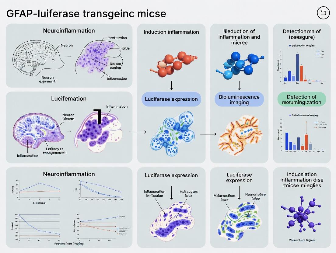
In Vivo Imaging of Neuroinflammation: A Comprehensive Guide to GFAP-Luciferase Transgenic Mouse Models
This article provides a detailed resource for researchers utilizing GFAP-luciferase transgenic mice to study neuroinflammation in vivo.
In Vivo Imaging of Neuroinflammation: A Comprehensive Guide to GFAP-Luciferase Transgenic Mouse Models
Abstract
This article provides a detailed resource for researchers utilizing GFAP-luciferase transgenic mice to study neuroinflammation in vivo. It begins with foundational knowledge of the GFAP promoter and bioluminescence imaging principles. It then covers methodological protocols for inducing models of CNS injury and disease, followed by a critical troubleshooting guide for optimizing signal-to-noise ratios and data interpretation. Finally, the article validates the model by comparing it with traditional histological methods and alternative in vivo imaging technologies, offering a holistic perspective on its applications in preclinical drug development for conditions like Alzheimer's disease, multiple sclerosis, and traumatic brain injury.
Understanding the GFAP-Luciferase Model: Principles, Components, and Reporter System Mechanics
This whitepaper details the core mechanism by which glial fibrillary acidic protein (GFAP) gene expression is quantitatively linked to bioluminescent light emission in vivo, forming the foundation for non-invasive neuroinflammation research using GFAP-luciferase transgenic mouse models. Within the broader thesis that GFAP-luciferase reporters provide a sensitive, dynamic, and translational platform for monitoring astrocyte activation, this guide elucidates the molecular and biophysical principles enabling this critical link. The technology allows researchers and drug development professionals to longitudinally track neuroinflammatory progression and therapeutic efficacy in real time.
Molecular Mechanism: From Transcriptional Activation to Photon Emission
The linkage is established through a transgenic construct where the regulatory elements of the Gfap gene drive the expression of a luciferase reporter enzyme, typically firefly luciferase (Fluc). Under neuroinflammatory conditions, activated astrocytes undergo significant molecular remodeling, leading to the upregulation of GFAP. This increase in GFAP transcription is directly mirrored by increased transcription of the downstream luciferase gene.
Once translated, the luciferase enzyme catalyzes a reaction that produces visible light. The substrate, D-luciferin, is injected systemically, crosses the blood-brain barrier, and enters cells. In the presence of oxygen, ATP, and Mg²⁺, luciferase oxidizes D-luciferin to oxyluciferin in an electronically excited state. As oxyluciferin relaxes to its ground state, a photon of light (~560-610 nm) is emitted. The number of photons detected per unit time is proportional to the amount of luciferase enzyme present, which itself is proportional to Gfap promoter activity.
Key Quantitative Relationships and Data
The correlation between bioluminescence signal and biological variables is foundational. The following tables summarize core quantitative relationships established in recent literature.
Table 1: Correlation between Bioluminescence Signal and Molecular/Cellular Metrics
| Measured Biological Variable | Correlation Coefficient (r) with BLI Signal | Experimental Model & Reference | Key Insight |
|---|---|---|---|
| GFAP mRNA levels (qPCR) | 0.85 - 0.92 | GFAP-Fluc mouse, LPS model | BLI reflects transcriptional activation. |
| GFAP Protein (Western blot) | 0.78 - 0.88 | GFAP-Fluc mouse, TBI model | Signal correlates with protein upregulation. |
| Astrocyte Cell Count (IHC) | 0.80 - 0.90 | GFAP-Fluc mouse, ALS model | Linear relationship in focal regions. |
| Inflammatory Cytokine IL-1β (ELISA) | 0.75 - 0.82 | GFAP-Fluc mouse, Systemic Inflammation | Links astrogliosis to innate immune response. |
Table 2: Typical Baseline and Activated Bioluminescence Signal Parameters
| Parameter | Naive / Baseline State | Acute Neuroinflammation (e.g., LPS) | Chronic Neurodegeneration (e.g., APP/PS1) |
|---|---|---|---|
| Peak Photon Flux (p/s/cm²/sr) | 5.0 x 10³ - 1.0 x 10⁴ | 1.0 x 10⁵ - 5.0 x 10⁵ | 5.0 x 10⁴ - 2.0 x 10⁵ |
| Signal-to-Background Ratio | ~2:1 | 20:1 - 100:1 | 10:1 - 50:1 |
| Time to Peak Post-Induction | N/A | 24 - 48 hours | Weeks to months (progressive) |
| Signal Localization | Diffuse, low brain signal | Focal (e.g., hippocampus) or whole-brain | Plaque-associated or region-specific |
Experimental Protocols for Key Assays
Protocol: In Vivo Bioluminescence Imaging (BLI) of GFAP-Luc Mice
Objective: To acquire quantitative, longitudinal bioluminescent data reflecting GFAP expression.
- Substrate Administration: Inject D-luciferin potassium salt (150 mg/kg, i.p.) in sterile PBS.
- Incubation: Place mouse in a clean, dark cage for 10 minutes to allow for systemic distribution and blood-brain barrier penetration.
- Anesthesia: Induce anesthesia (e.g., 3% isoflurane) and maintain at 1-2% during imaging.
- Imaging Setup: Place mouse in the imaging chamber of a cooled CCD camera system (e.g., IVIS Spectrum). Ensure nose is in anesthetic nose cone.
- Acquisition Parameters: Use medium binning, f/stop = 1, and an exposure time auto-adjusted to avoid saturation (typically 1 sec - 5 min). Acquire a grayscale reference photograph.
- Image Analysis: Define consistent regions of interest (ROIs) over the brain region. Quantify signal as total flux (photons/second) or average radiance (p/s/cm²/sr). Subtract background from a similar ROI outside the animal.
Protocol: Ex Vivo Validation via qPCR
Objective: To biochemically validate in vivo BLI data by measuring Gfap and luciferase mRNA levels.
- Tissue Collection: Following final BLI session, perfuse mouse transcardially with cold PBS. Dissect and flash-freeze brain regions of interest.
- RNA Extraction: Homogenize tissue in TRIzol. Extract total RNA following chloroform separation and isopropanol precipitation. Quantify using a Nanodrop.
- cDNA Synthesis: Use 1 µg of total RNA with a reverse transcription kit (e.g., High-Capacity cDNA Reverse Transcription Kit) including random hexamers.
- qPCR Reaction: Prepare reactions with SYBR Green master mix. Use the following primers:
- Gfap: Forward 5'-CGGAGACGCATCACCTCTG-3', Reverse 5'-AGGGAGTGGAGGAGTCATTCG-3'
- Fluc: Forward 5'-TTCGAAAGTCGATGCCCC-3', Reverse 5'-ACCGGGCGATCTTGTCATAG-3'
- Housekeeper (e.g., Gapdh): Use validated primers.
- Analysis: Calculate ΔΔCt values relative to a control group and housekeeper. Plot against corresponding BLI flux values for correlation analysis.
Visualizations
Title: Molecular Pathway from Neuroinflammation to Bioluminescence
Title: In Vivo BLI Imaging and Validation Workflow
The Scientist's Toolkit: Essential Research Reagents and Materials
Table 3: Key Reagent Solutions for GFAP-Bioluminescence Research
| Item | Function/Benefit | Example/Catalog Consideration |
|---|---|---|
| GFAP-Luc Transgenic Mouse | Expresses firefly luciferase under GFAP promoter; foundational model. | Available from repositories (e.g., JAX Stock #025575). |
| D-Luciferin, Potassium Salt | Cell-permeable substrate for firefly luciferase. Essential for BLI. | High-purity, sterile-filtered for in vivo use (e.g., GoldBio LUCK-1G). |
| In Vivo Imaging System (IVIS) | Cooled CCD camera for sensitive, quantitative bioluminescence detection. | PerkinElmer IVIS Spectrum or comparable system. |
| Isoflurane Anesthesia System | For humane animal restraint and stable imaging conditions. | Precision vaporizer with induction chamber and nose cones. |
| qPCR Primers for Gfap & Fluc | For ex vivo mRNA validation of transgenic expression and astrocyte response. | Validated, intron-spanning primer sets from sources like IDT. |
| Anti-GFAP Antibody (IHC Validated) | For histological validation of astrocyte activation and transgene correlation. | Clone GA5 (Millipore) or D1F4Q (CST). |
| Neuroinflammation Inducers | Positive controls to activate the GFAP pathway (e.g., LPS, TNF-α). | Ultrapure LPS from E. coli (InvivoGen). |
| Living Image or FIJI Software | For image acquisition, ROI analysis, and quantification of photon flux. | Standard software packages for data analysis. |
This whitepaper provides a technical dissection of the core components of a transgene, framed within the critical context of constructing and utilizing GFAP-luciferase transgenic mouse models for neuroinflammation research. The precise interplay between promoter specificity, reporter sensitivity, and host genetic background dictates the reliability, applicability, and translational value of these in vivo biosensor systems.
Core Components of the Transgene
The Promoter: Glial Fibrillary Acidic Protein (GFAP)
The GFAP promoter drives astrocyte-specific expression. In neuroinflammation, astrocyte reactivity (astrogliosis) is a hallmark, characterized by upregulated GFAP expression. Modern constructs use minimal or truncated GFAP promoters (often human or murine, ~2.0-2.5 kb upstream sequence) to direct expression while reducing transgene silencing. Key regulatory elements within this region (e.g., AP-1, NF-κB, STAT3 binding sites) confer inducibility upon inflammatory challenge.
The Reporter: Luciferase
Firefly luciferase (Photinus pyralis; luc) is the standard reporter. Its reaction with D-luciferin, ATP, and O₂ yields oxyluciferin and bioluminescent photons (λmax ~560 nm). Quantification via in vivo imaging systems (IVIS) provides a non-invasive, longitudinal readout of promoter activity.
Table 1: Quantitative Characteristics of Common Luciferase Reporters
| Reporter Enzyme | Source | Peak Emission (nm) | Cofactor/Substrate | Relative Signal Half-life | Relative Sensitivity |
|---|---|---|---|---|---|
| Firefly Luciferase | Photinus pyralis | 560 | D-luciferin, ATP, O₂ | ~30 min (medium) | High |
| Gaussia Luciferase | Gaussia princeps | 480 | Coelenterazine | ~5 min (fast) | Very High (secreted) |
| NanoLuc | Engineered | 460 | Furimazine | >2 hours (slow) | Extremely High |
The Genetic Background
The strain onto which the transgene is bred (e.g., C57BL/6J, FVB/N) is not a passive container. It profoundly affects neuroinflammatory responses, transgene expression patterns, and baseline bioluminescence. Background-dependent differences in immune cell recruitment, cytokine profiles, and blood-brain barrier integrity can confound results if not standardized.
Table 2: Impact of Common Mouse Genetic Backgrounds on Neuroinflammation Research
| Background Strain | Key Neuroinflammatory Phenotype Characteristics | Transgene Expression Considerations |
|---|---|---|
| C57BL/6J | Th1-biased response; common "standard" for disease models (e.g., EAE). | Lower baseline transgene silencing; preferred for most studies. |
| FVB/N | Pronounced visual system deficits; high fecundity for transgenesis. | Susceptible to retinal degeneration; can have variable transgene copy number. |
| BALB/c | Th2-biased response; less susceptible to some neurodegenerative insults. | May exhibit weaker GFAP-driven responses in some paradigms. |
Experimental Protocols for Validation & Application
Protocol: Longitudinal In Vivo Bioluminescence Imaging in GFAP-Luc Mice
Purpose: To quantify neuroinflammatory dynamics in real-time. Materials: GFAP-luc transgenic mice, LPS or disease-inducing agent, D-luciferin potassium salt (150 mg/kg in sterile PBS), Anesthesia system (isoflurane), In Vivo Imaging System (IVIS), Living Image or equivalent software. Procedure:
- Baseline Imaging: Anesthetize mouse. Inject D-luciferin intraperitoneally (i.p.). Place mouse in IVIS chamber. Acquire image 10-15 minutes post-injection (peak signal).
- Induction: Administer neuroinflammatory agent (e.g., LPS, 5 mg/kg i.p.).
- Time-course Imaging: Repeat imaging at defined intervals (e.g., 6, 24, 48, 72h post-induction) using identical anesthesia, luciferin dose, and imaging parameters (exposure time, f/stop, binning).
- Analysis: Define consistent regions of interest (ROIs) over the brain. Quantify total flux (photons/sec). Normalize to baseline or sham-treated controls.
Protocol: Ex Vivo Validation via Immunohistochemistry
Purpose: To correlate bioluminescence signal with cellular GFAP expression. Materials: Perfusion pump, 4% paraformaldehyde (PFA), Cryostat, Primary antibodies: anti-GFAP (chicken, 1:1000), anti-Iba1 (microglia, rabbit, 1:500), Fluorescent secondary antibodies, Mounting medium with DAPI. Procedure:
- Perfusion & Fixation: At imaging endpoint, deeply anesthetize mouse. Transcardially perfuse with cold PBS followed by 4% PFA. Dissect brain and post-fix for 24h.
- Sectioning: Cryoprotect in 30% sucrose, embed in OCT, section coronally (30 µm thickness) using a cryostat.
- Immunostaining: Perform free-floating immunofluorescence. Block in 5% normal serum. Incubate in primary antibody cocktail for 48h at 4°C. Wash, incubate in fluorescent secondaries for 2h at RT.
- Imaging & Analysis: Image using a confocal microscope. Quantify GFAP+ area or intensity in relevant brain regions (e.g., hippocampus, cortex) and correlate with in vivo bioluminescence from the same animal.
Signaling Pathways in GFAP Induction During Neuroinflammation
Diagram Title: Inflammatory Signaling to GFAP-Luc Reporter Activation
Experimental Workflow for a Neuroinflammation Study
Diagram Title: GFAP-Luc Neuroinflammation Study Workflow
The Scientist's Toolkit: Research Reagent Solutions
Table 3: Essential Reagents and Materials for GFAP-Luc Neuroinflammation Studies
| Item | Function & Specification | Key Consideration |
|---|---|---|
| GFAP-luc Transgenic Mouse Line | In vivo biosensor for astrocyte activation. Available from repositories (e.g., JAX). | Confirm promoter fragment (species, length) and backcrossed genetic background (e.g., C57BL/6J). |
| D-Luciferin, Potassium Salt | Substrate for firefly luciferase. Required for in vivo imaging. | Use sterile, endotoxin-free formulation. Prepare fresh in PBS or aliquot and store at -20°C protected from light. |
| Lipopolysaccharide (LPS) | Tool to induce sterile neuroinflammation. | Serotype (e.g., E. coli O111:B4) and purity (ultra-pure) determine TLR4-specificity and response magnitude. |
| In Vivo Imaging System (IVIS) | Camera system for quantifying bioluminescence. | Calibrate regularly. Use low background, light-tight chamber. |
| Isoflurane Anesthesia System | For animal restraint during imaging. | Provides stable, rapid anesthesia induction/recovery, minimizing stress confounds. |
| Anti-GFAP Antibody | Validation of astrocyte activation via IHC/IF. | Select species reactivity (e.g., anti-mouse). Monoclonal antibodies offer higher specificity. |
| Cryostat | For sectioning fixed brain tissue. | Maintain blade and chamber at -20°C for optimal 30 µm sectioning of CNS tissue. |
| Confocal Microscope | High-resolution imaging of immunofluorescent validation. | Enables co-localization studies (e.g., GFAP with other cell markers). |
Within the broader thesis investigating GFAP-luciferase transgenic mice for in vivo neuroinflammation research, the specificity of Glial Fibrillary Acidic Protein (GFAP) as a marker for reactive astrocytes is a fundamental cornerstone. This whitepaper provides a technical guide on GFAP's expression dynamics, its role in astrocyte reactivity, and methodological considerations for its quantification, particularly within transgenic reporter models.
GFAP Expression & Astrocyte Reactivity
GFAP, a Class-III intermediate filament, is the canonical marker for astrocytes. In the healthy central nervous system (CNS), GFAP is constitutively expressed but at relatively low levels, with significant regional heterogeneity. Neuroinflammatory states trigger astrocyte reactivity (astrogliosis), characterized by hypertrophic morphology and a pronounced upregulation of GFAP expression and filament formation.
The GFAP-luciferase transgenic mouse model utilizes the GFAP promoter to drive the expression of firefly luciferase. This allows for non-invasive, longitudinal bioluminescence imaging (BLI) of astrocyte activation, correlating luciferase signal intensity with the degree of neuroinflammation. However, GFAP upregulation is not binary and varies with inflammatory stimulus, CNS region, and disease stage.
Table 1: GFAP Expression Dynamics in Neuroinflammatory Models
| Neuroinflammatory Model | GFAP Upregulation Onset | Peak GFAP Expression | Key Signaling Pathways Involved | Notes on Specificity |
|---|---|---|---|---|
| Systemic LPS Injection | 6-12 hours | 24-48 hours | NF-κB, JAK/STAT3, MAPK | Rapid, widespread activation; can involve other glia. |
| Focal Mechanical Injury | 1-2 days | 5-7 days | TGF-β, BMP, STAT3 | Localized to lesion penumbra; correlates with scar formation. |
| EAE (MS Model) | Pre-clinical phase | Clinical peak | JAK/STAT3, NF-κB, IL-6 signaling | Heterogeneous; prominent in spinal cord lesions. |
| APP/PS1 (AD Model) | Chronic, age-dependent | Late-stage pathology | JAK/STAT3, Complement C3a | Co-localizes with amyloid plaques; nuanced reactivity states. |
Key Signaling Pathways in GFAP Upregulation
GFAP transcription is regulated by a complex interplay of signaling cascades initiated by inflammatory mediators.
Diagram Title: Signaling Pathways Leading to GFAP Upregulation in Reactive Astrocytes
Experimental Protocols for Validation
Protocol 1: Ex Vivo Validation of GFAP-luciferase Signal
Objective: Correlate in vivo bioluminescence with post-mortem GFAP protein levels. Materials: GFAP-luciferase transgenic mouse, IVIS Spectrum Imaging System, luciferin, tissue homogenizer, GFAP ELISA kit. Method:
- Perform in vivo BLI: Inject mouse intraperitoneally with D-luciferin (150 mg/kg), anesthetize with isoflurane, and acquire image 10-15 minutes post-injection.
- Euthanize mouse and perfuse with ice-cold PBS. Dissect brain regions of interest (e.g., cortex, hippocampus).
- Homogenize tissue in RIPA buffer with protease inhibitors.
- Quantification:
- Luciferase Activity: Use a portion of homogenate in a luminometer assay with luciferin substrate.
- GFAP Protein: Perform GFAP ELISA on homogenate per manufacturer's protocol.
- Perform linear regression analysis between BLI signal (photons/sec) and GFAP concentration (pg/mg tissue).
Protocol 2: Immunofluorescence Co-localization Analysis
Objective: Determine specificity of GFAP upregulation to reactive astrocytes in a lesion model. Materials: Perfused brain tissue, cryostat, primary antibodies (anti-GFAP, anti-Iba1, anti-NeuN), fluorescent secondary antibodies, confocal microscope. Method:
- Generate focal cortical injury using controlled stereotaxic impact.
- At peak GFAP expression (e.g., 5 days post-injury), perfuse-fix mouse with 4% PFA. Section brain (30µm) on a cryostat.
- Perform immunofluorescence: Block sections, incubate with chicken anti-GFAP (1:1000), rabbit anti-Iba1 (1:800), and mouse anti-NeuN (1:500) overnight at 4°C.
- Incubate with species-specific Alexa Fluor-conjugated secondary antibodies (488, 568, 647) for 2 hours.
- Image using confocal microscopy. Quantify GFAP+ cell morphology (process thickness, territory area) and intensity using Fiji/ImageJ software. Confirm co-localization is specific to astrocytes (GFAP+/Iba1-/NeuN-).
The Scientist's Toolkit: Research Reagent Solutions
Table 2: Essential Reagents for GFAP-Based Neuroinflammation Research
| Reagent/Material | Function & Application | Example Product/Catalog # |
|---|---|---|
| GFAP-luciferase Transgenic Mouse | In vivo model for longitudinal imaging of astrocyte reactivity. | The Jackson Laboratory, Stock #025854 (FVB-Tg(Gfap-luc)Xen) |
| D-Luciferin, Potassium Salt | Substrate for firefly luciferase; injected for BLI. | PerkinElmer, #122799 |
| Chicken Anti-GFAP Primary Antibody | High-specificity antibody for immunohistochemistry and Western blot. | Abcam, ab4674 |
| Anti-Iba1 Antibody (Rabbit) | Microglial marker to distinguish astrocytes from activated microglia. | Fujifilm Wako, 019-19741 |
| GFAP ELISA Kit | Quantitative measurement of GFAP protein levels from tissue homogenates. | Thermo Fisher Scientific, EHGFAP |
| Cell Lysis Buffer (RIPA) | For tissue homogenization and protein extraction for luciferase/ELISA assays. | MilliporeSigma, R0278 |
| Recombinant IL-1β / TNF-α | Pro-inflammatory cytokines used to induce astrocyte reactivity in vitro or in vivo. | PeproTech, 200-01B / 315-01A |
| STAT3 Inhibitor (S3I-201) | Small molecule inhibitor to probe the JAK/STAT pathway's role in GFAP upregulation. | MilliporeSigma, SML0330 |
Workflow for Integrated Analysis
A comprehensive research approach in the GFAP-luciferase model integrates in vivo, ex vivo, and in vitro data.
Diagram Title: Integrated Experimental Workflow Using GFAP-Luciferase Mice
Limitations and Complementary Markers
While GFAP is specific for astrocytes, its upregulation does not capture the full heterogeneity of reactive states. A1/A2 or neurotoxic/neuroprotective astrocyte paradigms require complementary markers. Quantitative data from recent studies (2023-2024) highlight this:
Table 3: Complementary Markers for Astrocyte Reactivity
| Marker | Expression in Resting Astrocytes | Change in Neuroinflammation | Association with GFAP+ Cells | Functional Implication |
|---|---|---|---|---|
| S100β | High | Often upregulated | Co-expressed in most GFAP+ cells | Calcium signaling, trophic support. |
| Vimentin | Low | Sharply upregulated | Co-localizes with GFAP filaments | Dynamic cytoskeletal remodeling. |
| C3 (A1) | Negligible | Strongly induced (A1) | Subset of hypertrophic GFAP+ cells | Complement activation, neurotoxicity. |
| PTX3 (A2) | Very Low | Induced (A2) | Subset of GFAP+ cells | Tissue repair, anti-inflammatory. |
| ALDH1L1 | Very High | Often downregulated | Lost in severely reactive astrocytes | Metabolic shift in reactivity. |
GFAP remains a highly specific and indispensable marker for identifying and quantifying reactive astrocytes in neuroinflammatory states. Within the thesis framework of GFAP-luciferase transgenic models, rigorous ex vivo validation and the integration of complementary markers are critical to accurately interpret the in vivo bioluminescence signal and deconvolve the complex functional phenotypes of astrocyte reactivity in disease progression and therapeutic intervention.
This whitepaper details the technical advantages of bioluminescence imaging (BLI), focusing on its application within neuroinflammation research using GFAP-luciferase transgenic mouse models. BLI provides unparalleled sensitivity for in vivo longitudinal tracking of astrocyte activation, a core component of neuroinflammatory responses. The non-invasive nature of BLI allows for quantification of dynamic biological processes within the same animal over time, significantly enhancing statistical power and reducing inter-subject variability.
Core Principles: Sensitivity and Quantification
Bioluminescence results from the enzymatic oxidation of a substrate (e.g., D-luciferin) by a luciferase (e.g., firefly luciferase). This reaction emits photons detectable by sensitive charge-coupled device (CCD) cameras.
- Sensitivity: BLI benefits from an exceptionally low background due to the absence of endogenous mammalian luciferase, enabling detection of small cell populations (as few as 100-1000 cells in vivo).
- Quantification: Photon flux (measured in photons/second/cm²/steradian, p/s/cm²/sr) is proportional to the number of luciferase-expressing cells, allowing for robust longitudinal quantification of signal changes.
GFAP-luciferase Transgenic Mice in Neuroinflammation
In GFAP-luciferase mice, the firefly luciferase gene is under the control of the glial fibrillary acidic protein (GFAP) promoter. As GFAP is upregulated in reactive astrocytes during neuroinflammation, BLI signal intensity provides a quantitative measure of astrogliosis in real time. This model is pivotal for studying conditions like multiple sclerosis, Alzheimer's disease, traumatic brain injury, and stroke.
Table 1: BLI Performance Characteristics
| Parameter | Typical Value/Range | Notes |
|---|---|---|
| Detection Threshold (Cells in vivo) | 100 - 1,000 cells | Dependent on luciferase expression level and tissue depth. |
| Signal-to-Noise Ratio (SNR) | > 100:1 in vivo | Can exceed 1000:1 for superficial or in vitro assays. |
| Linear Dynamic Range | 3-4 orders of magnitude | Linear correlation between cell number and photon flux. |
| Temporal Resolution | Minutes to Hours | Limited by substrate kinetics; peak signal ~10-20 min post-injection. |
| Spatial Resolution (FWHM) | 2-5 mm in vivo | Diffuse light scattering in tissue limits precise anatomical localization. |
Table 2: GFAP-BLI Response in Common Neuroinflammatory Models
| Disease Model | Typical BLI Signal Increase (Fold over Baseline) | Peak Signal Time Post-Induction | Key Reference Compound/Intervention |
|---|---|---|---|
| Experimental Autoimmune Encephalomyelitis (EAE) | 8 - 15x | 14-21 days | Dexamethasone (reduces signal by ~60%) |
| Lipopolysaccharide (LPS) Intracranial Injection | 10 - 25x | 24 - 48 hours | Minocycline (inhibits signal by ~40-50%) |
| Traumatic Brain Injury (TBI) | 5 - 10x | 3 - 7 days | NA |
| Neurodegenerative (APP/PS1) Model | 2 - 4x | Chronic, age-dependent (e.g., 12 months) | NA |
Key Experimental Protocols
Protocol: LongitudinalIn VivoBLI of Neuroinflammation in GFAP-Luc Mice
Objective: To non-invasively monitor the progression and intervention of neuroinflammation over time.
Materials: See "The Scientist's Toolkit" below.
Procedure:
- Animal Preparation: Anesthetize GFAP-luciferase transgenic mouse using an isoflurane/oxygen mixture (2-3% induction, 1-2% maintenance).
- Substrate Administration: Inject D-luciferin potassium salt (150 mg/kg body weight in sterile PBS) intraperitoneally. Uniform injection volume and site are critical.
- Image Acquisition:
- Place the anesthetized mouse in the imaging chamber of the BLI system.
- Maintain body temperature at 37°C using a heating pad.
- Begin image acquisition 10 minutes post-injection (allowing for systemic distribution and blood-brain barrier penetration).
- Acquire a grayscale reference photograph under low light.
- Acquire bioluminescence images. Typical parameters: exposure time = 1-5 minutes, binning = medium (8x8), field of view = 15-20 cm, f/stop = 1.
- Data Quantification:
- Using system software, define a consistent region of interest (ROI) over the brain region for all mice and time points.
- Quantify total flux (p/s) or average radiance (p/s/cm²/sr) within the ROI.
- Normalize data to baseline (pre-induction) values or to a contralateral control ROI if applicable.
- Longitudinal Schedule: Image mice at predetermined intervals (e.g., daily, weekly) post-disease induction/treatment. Always image at the same time of day to minimize circadian variability.
Protocol:Ex VivoOrgan Imaging for Signal Verification
Objective: To confirm the anatomical source of the in vivo BLI signal and assess biodistribution.
Procedure:
- Following the final in vivo imaging session, euthanize the mouse as per approved protocol.
- Rapidly dissect out the brain and other organs of interest (e.g., spleen, liver).
- Place organs in a Petri dish and immerse in a D-luciferin solution (150 µg/mL in PBS).
- Image organs immediately using the BLI system with a short exposure time (10-60 seconds).
- Quantify signal from specific brain regions (e.g., cortex, hippocampus, cerebellum) for precise correlation with histology.
Signaling Pathways and Experimental Workflows
Title: GFAP-BLI Signaling Pathway in Neuroinflammation
Title: Longitudinal BLI Experiment Workflow
The Scientist's Toolkit: Research Reagent Solutions
Table 3: Essential Materials for GFAP-BLI Neuroinflammation Studies
| Item | Function/Description | Example Vendor/Product |
|---|---|---|
| GFAP-luciferase Transgenic Mice | Express firefly luciferase under GFAP promoter for astrocyte-specific imaging. | The Jackson Laboratory (Stock #: 024698 - FVB-Tg(Gfap-luc)Xen). |
| D-Luciferin, Potassium Salt | Bioluminescent substrate for firefly luciferase. Critical for consistent dosing. | PerkinElmer (122799), GoldBio (LUCK-1G). |
| In Vivo Imaging System (IVIS) | High-sensitivity CCD camera system for low-light photon detection. | PerkinElmer IVIS Spectrum, Bruker In-Vivo Xtreme. |
| Isoflurane Anesthesia System | Provides safe, consistent, and reversible anesthesia during imaging. | Parkland Scientific VetFlo, Summit Medical Systems. |
| Living Image or Equivalent Software | For image acquisition, ROI analysis, and quantification of photon flux. | PerkinElmer Living Image. |
| Sterile PBS | Vehicle for dissolving D-luciferin to a consistent concentration (e.g., 15 mg/mL). | Thermo Fisher (10010023). |
| Heating Pad/Stage | Maintains mouse body temperature at 37°C during anesthesia to ensure consistent physiology and substrate metabolism. | RightTemp (Kent Scientific). |
| Matrigel or LPS | For establishing disease models (e.g., Matrigel for EAE induction, LPS for acute inflammation). | Corning (356234), Sigma (L2880). |
| Microsyringe (e.g., Hamilton) | For accurate intracranial injections to induce focal neuroinflammation. | Hamilton Company (7000 Series). |
Within the broader thesis on utilizing GFAP-luciferase transgenic mice for non-invasive, longitudinal neuroinflammation research, this technical guide details the core experimental applications of this model system. The GFAP-luciferase mouse, where firefly luciferase expression is driven by the Glial Fibrillary Acidic Protein promoter, enables in vivo bioluminescence imaging (BLI) to quantify astrocyte activation dynamically. This guide provides a technical framework for applying this model to two primary research domains: chronic neurodegenerative diseases and acute central nervous system (CNS) injury.
Core Technical Mechanism and Validation
The transgenic model relies on the GFAP promoter's responsiveness to neuroinflammatory stimuli. Upon astrocyte activation, luciferase is transcribed and translated. Following systemic injection of its substrate, D-luciferin, a quantifiable photon signal is emitted, proportional to the degree of activation.
Key Validation Protocol:
- Objective: Correlate bioluminescence signal with canonical molecular markers of neuroinflammation.
- Procedure:
- Acquire baseline BLI in GFAP-luc mice.
- Administer disease model or injury (see Sections 3 & 4).
- Perform longitudinal BLI at defined time points (e.g., days 1, 3, 7, 14 post-induction).
- Following final imaging, perfuse and harvest brain/spinal cord.
- Process tissue for:
- Immunohistochemistry (IHC): Co-staining for GFAP and luciferase to confirm cellular specificity.
- Western Blot/RT-qPCR: Quantify endogenous GFAP, IBA1 (microglia), TNF-α, IL-1β levels from dissected regions of interest.
- Expected Outcome: A strong positive correlation between in vivo photon flux and post-mortem protein/mRNA levels of inflammatory markers validates the model's specificity and sensitivity.
Application in Neurodegenerative Disease Models
Chronic neuroinflammation is a hallmark of neurodegenerative diseases. GFAP-luc mice enable the tracking of astrogliosis throughout disease progression and in response to therapeutic intervention.
Alzheimer's Disease (AD) Model
- Common Model: Intracerebroventricular (ICV) or hippocampal injection of oligomeric Aβ42, or cross-breeding with APP/PS1 transgenic mice.
- Experimental Workflow & BLI Profile:
- Acute Aβ injection: Peak BLI signal at 3-7 days post-injection, gradual resolution over 14-21 days.
- Genetic AD models: A slow, steady increase in BLI signal over months, correlating with plaque deposition.
- Key Data Output:
Table 1: BLI Signal Progression in Aβ42-Induced AD Model
| Time Point (Post-Injection) | Mean Photon Flux (p/s/cm²/sr) ± SEM | Corresponding Histopathology (GFAP+ Area %) |
|---|---|---|
| Baseline (Day 0) | 5.2 x 10³ ± 0.8 x 10³ | 2.1 ± 0.5 |
| Day 3 | 1.8 x 10⁵ ± 2.1 x 10⁴ | 15.4 ± 3.2 |
| Day 7 | 2.9 x 10⁵ ± 3.3 x 10⁴ | 28.7 ± 4.1 |
| Day 14 | 1.1 x 10⁵ ± 1.5 x 10⁴ | 18.3 ± 3.8 |
Diagram 1: Aβ-induced neuroinflammatory signaling leading to BLI.
Parkinson's Disease (PD) Model
- Common Model: Intrastriatal injection of 6-hydroxydopamine (6-OHDA) or systemic MPTP administration.
- Experimental Insight: BLI reveals a biphasic astrocyte response—an acute peak (Day 3-5) followed by a sustained chronic activation phase, aligning with dopaminergic neuron loss.
Application in CNS Injury Models
Acute injuries provide a paradigm for studying the dynamics of the neuroinflammatory cascade.
Controlled Cortical Impact (CCI) – Traumatic Brain Injury
- Protocol: Under anesthesia, a craniotomy is performed, and a pneumatic or electromagnetic impactor delivers a precise impact to the dura.
- BLI Kinetic Profile: Signal rises sharply within 24 hours, peaks at Day 3-5 (10-50 fold increase), and slowly resolves over 2-4 weeks, mirroring the transition from acute astrogliosis to glial scar formation.
Spinal Cord Injury (SCI) – Compression/Contusion
- Protocol: Laminectomy followed by compression with forceps or contusion using an Infinite Horizon or NYU impactor.
- BLI Kinetic Profile: Similar acute peak to CCI, with signal intensity and spatial spread correlating with injury severity (e.g., 100 kdyn vs. 200 kdyn impact force).
Table 2: Comparative BLI Kinetics in Acute CNS Injury Models
| Model (Severity) | Peak Signal Time | Peak Flux Range (p/s/cm²/sr) | Time to Return to Baseline |
|---|---|---|---|
| CCI (Moderate) | Day 3-4 | 1.5 x 10⁶ - 5.0 x 10⁶ | > 28 days |
| SCI (200 kdyn contusion) | Day 4-5 | 2.0 x 10⁶ - 8.0 x 10⁶ | > 35 days |
| Focal Cerebral Ischemia | Day 2 | 5.0 x 10⁵ - 2.0 x 10⁶ | 14-21 days |
Diagram 2: Acute injury pathway and therapeutic assessment via BLI.
The Scientist's Toolkit: Research Reagent Solutions
Table 3: Essential Reagents and Materials for GFAP-luciferase Mouse Studies
| Item | Function & Application | Key Considerations |
|---|---|---|
| GFAP-luc Transgenic Mice | Core model for non-invasive imaging of astrogliosis. | Available from repositories like The Jackson Laboratory (e.g., STOCK Tg(GFAP-luc)Xen). Maintain on consistent genetic background. |
| D-Luciferin, Potassium Salt | Luciferase enzyme substrate for in vivo BLI. | Administer at 150 mg/kg via IP injection. Use consistent dose, route, and imaging delay (e.g., 10 min post-injection). |
| Isoflurane Anesthesia System | For animal immobilization during imaging and surgical procedures. | Essential for consistent physiology and animal welfare during longitudinal studies. |
| In Vivo Imaging System (IVIS) | Quantitative bioluminescence and photographic imaging. | Use Living Image or equivalent software for region-of-interest (ROI) analysis of photon flux. |
| Recombinant Aβ42 Protein | To induce Alzheimer's-like pathology and neuroinflammation. | Prepare oligomeric forms per standardized protocols (e.g., incubation in hexafluoroisopropanol). |
| 6-OHDA or MPTP Hydrochloride | Neurotoxins for inducing Parkinson's disease models. | Highly labile; prepare fresh solutions in ascorbate-saline (6-OHDA) and handle with extreme caution. |
| Precision Impact Device | For standardized CCI or SCI (e.g., Infinite Horizon Impactor). | Calibrate regularly. Injury depth/velocity/dwell time are critical parameters. |
| Anti-GFAP & Anti-Luciferase Antibodies | For post-mortem validation via IHC/IF. | Confirm specificity and optimal titers for co-localization studies in mouse CNS tissue. |
| Cytokine Multiplex Assay (e.g., Luminex/MSD) | To correlate BLI signal with molecular inflammatory profile. | Run on brain homogenate supernatants from dissected ROI. |
GFAP-luciferase transgenic mice serve as a powerful and versatile platform for dissecting the role of neuroinflammation in vivo. By providing quantitative, longitudinal data within individual subjects, this model bridges the gap between acute mechanistic studies and chronic disease progression research, offering robust endpoints for preclinical therapeutic evaluation in both neurodegeneration and CNS injury.
Protocols and Applications: From Model Induction to In Vivo Imaging Data Acquisition
This technical guide details the establishment and application of key neuroinflammatory models within the framework of research utilizing GFAP-luciferase transgenic mice. These genetically engineered mice express firefly luciferase under the control of the glial fibrillary acidic protein (GFAP) promoter, enabling non-invasive, real-time bioluminescence imaging (BLI) of astrocyte activation—a central component of neuroinflammation. This approach is critical for longitudinal studies of disease progression and therapeutic efficacy in preclinical research for drug development.
The GFAP-luciferase Reporter System: Core Principles
The GFAP promoter drives the expression of the Photinus pyralis luciferase gene. Upon administration of its substrate, D-luciferin, activated astrocytes produce quantifiable light emission. The bioluminescence signal correlates with the degree of neuroinflammation, providing a powerful quantitative readout.
Key Advantages:
- Longitudinal Monitoring: Reduces animal numbers by allowing repeated measures in the same subject.
- Spatio-temporal Resolution: Enables tracking of inflammatory waves and focal pathology.
- Objective Quantification: Provides a continuous, non-subjective data stream for statistical analysis.
Neuroinflammatory Models: Protocols, Applications, and Data
Lipopolysaccharide (LPS)-Induced Systemic Inflammation
A model for studying the peripheral immune challenge's impact on the central nervous system (CNS) and priming of neuroinflammatory responses.
Experimental Protocol:
- Animals: GFAP-luciferase transgenic mice (e.g., FVB/N-Tg(Gfap-luc) mice).
- LPS Administration: Intraperitoneal (i.p.) injection of LPS (from E. coli serotype 055:B5) at 1-5 mg/kg in sterile PBS.
- Imaging: Inject D-luciferin (150 mg/kg, i.p.) 10-15 minutes before imaging. Acquire BLI signals using an IVIS Spectrum or equivalent system at baseline and regular intervals (e.g., 4h, 24h, 48h) post-LPS.
- Validation: Post-mortem tissue analysis for GFAP (IHC), Iba1 (microglia), and pro-inflammatory cytokines (IL-1β, TNF-α via ELISA).
Typical Quantitative Data:
| Time Point Post-LPS | Mean BLI Signal (p/s/cm²/sr) | CNS IL-1β (pg/mg protein) | Key Histological Finding |
|---|---|---|---|
| Baseline | 5.0 x 10³ ± 1.0 x 10³ | 5.2 ± 1.5 | Normal GFAP staining |
| 4 hours | 2.5 x 10⁴ ± 4.0 x 10³ | 45.3 ± 10.2 | Early astrocyte hypertrophy |
| 24 hours | 1.1 x 10⁵ ± 2.0 x 10⁴ | 120.7 ± 25.6 | Pronounced astrogliosis |
| 72 hours | 3.0 x 10⁴ ± 6.0 x 10³ | 30.5 ± 8.4 | Resolution phase |
Traumatic Brain Injury (TBI) Model (Controlled Cortical Impact)
A focal injury model for studying localized, trauma-induced neuroinflammation and glial scarring.
Experimental Protocol:
- Surgery: Anesthetize mouse, perform a craniotomy over one hemisphere. Use a Controlled Cortical Impact (CCI) device to deliver a precise impact (e.g., 3mm tip, 5 m/s velocity, 1mm depth of deformation).
- Imaging: Perform longitudinal BLI as described above, starting at 24h post-injury and continuing weekly.
- Validation: Histology for lesion volume (Cresyl Violet), astrogliosis (GFAP), and microglial activation (Iba1) at endpoint.
Typical Quantitative Data:
| Time Post-CCI | Ipsilateral BLI Signal (p/s/cm²/sr) | Contralateral BLI Signal | Lesion Volume (mm³) |
|---|---|---|---|
| 1 day | 3.5 x 10⁵ ± 5.0 x 10⁴ | 1.0 x 10⁴ ± 2.0 x 10³ | 8.5 ± 1.2 |
| 7 days | 8.2 x 10⁵ ± 9.0 x 10⁴ | 1.5 x 10⁴ ± 3.0 x 10³ | 12.1 ± 1.8 |
| 28 days | 2.0 x 10⁵ ± 4.0 x 10⁴ | 1.2 x 10⁴ ± 2.0 x 10³ | 10.5 ± 1.5 (Cavity) |
Experimental Autoimmune Encephalomyelitis (EAE)
A model of T-cell mediated CNS inflammation, relevant to multiple sclerosis.
Experimental Protocol:
- Induction: Immunize mice subcutaneously with MOG₃₅₋₅₅ peptide (200 µg) emulsified in Complete Freund's Adjuvant (CFA) containing 500 µg of Mycobacterium tuberculosis. Administer pertussis toxin (200 ng, i.p.) on day 0 and 2.
- Clinical Scoring: Monitor daily for paralysis (scale 0-5).
- Imaging: Perform weekly BLI. Signal often appears in the spinal cord region prior to clinical onset.
- Validation: Spinal cord histology for immune cell infiltration (H&E), demyelination (LFB), and astrogliosis.
Typical Quantitative Data:
| Clinical Score | Mean BLI Signal (Spinal Cord) | CNS CD4+ T-cell Count | Peak Disease Incidence |
|---|---|---|---|
| 0 (Healthy) | 5.0 x 10³ ± 1.0 x 10³ | < 100 | N/A |
| 2 (Hindlimb weakness) | 2.0 x 10⁵ ± 4.0 x 10⁴ | ~ 2,000 | Day 12-15 |
| 4 (Paraplegia) | 5.5 x 10⁵ ± 7.0 x 10⁴ | ~ 8,000 | Day 18-21 |
Neurodegenerative Crosses: GFAP-luciferase x APP/PS1 Mice
A model for studying neuroinflammation in the context of Alzheimer's disease pathology.
Experimental Protocol:
- Animals: Cross GFAP-luciferase mice with a transgenic Alzheimer's model (e.g., APP/PS1). Use littermate controls.
- Longitudinal Imaging: Perform monthly BLI from 3 to 12 months of age to track glial activation relative to amyloid-β plaque deposition.
- Validation: Correlate BLI signals with plaque load (Thioflavin-S or 6E10 IHC), phospho-tau pathology, and cytokine levels.
Typical Quantitative Data:
| Age (Months) | BLI Signal in APP/PS1 (p/s/cm²/sr) | Plaque Load (% area) | Correlation (R²) |
|---|---|---|---|
| 3 | 1.5 x 10⁴ ± 3.0 x 10³ | 0.1 ± 0.05 | 0.15 |
| 6 | 1.2 x 10⁵ ± 2.0 x 10⁴ | 0.8 ± 0.2 | 0.65 |
| 9 | 4.5 x 10⁵ ± 6.0 x 10⁴ | 2.5 ± 0.5 | 0.82 |
| 12 | 8.0 x 10⁵ ± 1.0 x 10⁵ | 4.2 ± 0.8 | 0.88 |
The Scientist's Toolkit: Research Reagent Solutions
| Item | Function & Application |
|---|---|
| GFAP-luciferase Transgenic Mouse | In vivo reporter for astrocyte activation. Enables longitudinal BLI. |
| D-Luciferin, Potassium Salt | Substrate for firefly luciferase. Administered i.p. for in vivo BLI. |
| Lipopolysaccharide (LPS) | TLR4 agonist. Used for inducing systemic and neuroinflammation. |
| MOG₃₅₋₅₅ Peptide | Myelin oligodendrocyte glycoprotein peptide. Antigen for inducing EAE. |
| Complete Freund's Adjuvant (CFA) | Immunopotentiator used with MOG peptide to induce EAE. |
| Controlled Cortical Impact (CCI) Device | Electromechanical or pneumatic device for standardized, focal TBI. |
| IVIS Spectrum Imaging System | In vivo optical imaging system for quantifying bioluminescence. |
| Anti-GFAP Antibody | For immunohistochemical validation of astrocyte activation. |
| Cytokine ELISA Kits (e.g., IL-1β, TNF-α, IL-6) | For quantifying pro-inflammatory mediators in brain homogenates. |
Key Signaling Pathways and Experimental Workflows
LPS-Induced GFAP-luciferase Signal Pathway
GFAP-luciferase Model Evaluation Workflow
Comparison of Neuroinflammatory Model Features
This technical guide details a standardized protocol for in vivo bioluminescence imaging (BLI) within neuroinflammation research utilizing GFAP-luciferase transgenic mice. In these models, the luciferase gene is under the control of the glial fibrillary acidic protein (GFAP) promoter, enabling non-invasive, longitudinal quantification of astrocyte activation. This protocol is a critical component of a thesis focused on quantifying neuroinflammatory dynamics in response to pharmacological or pathological challenge.
Core Methodology
Pre-Imaging Animal Preparation
Mice should be acclimated to the facility for at least one week. Fasting is not typically required but maintaining a consistent diet is crucial. The abdominal region should be shaved 24 hours prior to imaging to minimize light scattering from fur.
Anesthesia Induction and Maintenance
A safe and stable plane of anesthesia is paramount for reproducible imaging and animal welfare.
Primary Protocol: Isoflurane Inhalation
- Induction: Place mouse in an induction chamber with a continuous flow of 3-4% isoflurane in 100% medical oxygen (flow rate: 1-2 L/min).
- Maintenance: Transfer the animal to the imaging chamber nose cone, maintaining anesthesia with 1.5-2.5% isoflurane.
- Monitoring: Continuously monitor respiratory rate (target: 40-80 breaths/min) and toe-pinch reflex throughout the procedure. Use a heating pad integrated into the imaging stage to maintain body temperature at 36.5-37.5°C.
Substrate (D-Luciferin) Preparation and Administration
D-luciferin is the enzyme substrate for firefly luciferase. Consistent administration is key for quantitative data.
- Preparation: Reconstitute sterile D-luciferin potassium salt in sterile, pyrogen-free phosphate-buffered saline (PBS) to a stock concentration of 15 mg/mL. Filter sterilize (0.2 µm), aliquot, and store at -20°C. Avoid repeated freeze-thaw cycles.
- Dose and Route: The standard dose is 150 mg/kg body weight, administered via intraperitoneal (IP) injection.
- Injection Technique: Inject using a sterile 27-30 gauge insulin syringe. Warm the luciferin solution to 37°C to prevent hypothermia stress.
- Kinetics: Following IP injection, peak bioluminescent signal in the brain typically occurs between 12-20 minutes post-injection. A pre-imaging kinetic study is recommended to establish the precise time-to-peak for your specific model and setup.
Imaging Chamber Setup and Data Acquisition
- Positioning: Place the anesthetized mouse in the prone position on the heated imaging stage. Secure the nose in the isoflurane delivery nose cone. Apply a thin layer of ocular lubricant to prevent corneal drying.
- Spatial Registration: For longitudinal studies, use fiduciary markers or a positioning template to ensure identical placement across imaging sessions.
- Imaging Parameters: Acquire image sequences using an IVIS Spectrum or equivalent in vivo imaging system.
- Field of View: Typically "C" or "D" for single mice.
- Binning: Medium (4 or 8) for an optimal signal-to-noise ratio.
- F-Stop: f/1 or f/2 to maximize light collection.
- Exposure Time: Use auto-exposure or a set range (e.g., 1 second to 5 minutes) to avoid pixel saturation. Multiple exposures may be needed.
- Data Capture: Acquire a grayscale photographic image followed by a series of bioluminescence images (photons/sec/cm²/steradian) starting immediately post-injection to capture kinetic data, or at the predetermined peak time.
Table 1: Standardized Protocol Parameters for In Vivo BLI in GFAP-Luc Mice
| Parameter | Recommended Specification | Rationale / Notes |
|---|---|---|
| Anesthetic | Isoflurane (3-4% induction, 1.5-2.5% maintenance) | Fast induction/recovery, minimal interference with luciferase activity. |
| Substrate | D-Luciferin (potassium salt, sterile) | Firefly luciferase substrate. Preferred over beetle luciferin for stability. |
| Dose | 150 mg/kg (IP) | Standard dose; saturation kinetics should be validated for each model. |
| Injection Volume | 10 µL/g body weight (of 15 mg/mL stock) | Standard calculation. Adjust stock concentration for accurate dosing. |
| Peak Signal Time | 12 - 20 minutes post-IP injection (Brain) | Must be empirically determined. Varies by route, model, and pathology. |
| Imaging Temperature | 36.5 - 37.5°C (stage heating) | Maintains physiological temperature, crucial for enzyme kinetics. |
| Typical Exposure | 1 sec - 5 min (auto or manual) | Prevents saturation of the CCD camera; ensures quantifiable signal. |
Table 2: Troubleshooting Common Imaging Issues
| Problem | Potential Cause | Solution |
|---|---|---|
| Low/No Signal | Incorrect luciferin dose/degradation; Deep anesthesia | Use fresh, pH-correct luciferin; Validate dose-response; Lighten anesthesia plane. |
| High Background | Substrate contamination; Non-specific signal | Clean imaging chamber; Ensure mouse fur is properly removed; Use black paper to mask body. |
| Signal Variability | Inconsistent injection; Temperature fluctuation | Standardize IP injection technique; Ensure consistent pre-warming of luciferin and mouse. |
| Poor Spatial Resolution | Mouse movement; Light scattering | Ensure stable anesthesia; Shave fur completely; Use spectral unmixing if available. |
Signaling Pathway & Experimental Workflow
Diagram 1: GFAP-Luc Bioluminescence Signal Generation Pathway
Diagram 2: In Vivo BLI Experimental Workflow
The Scientist's Toolkit: Research Reagent Solutions
Table 3: Essential Materials for GFAP-Luc In Vivo Imaging
| Item | Function / Role | Key Considerations |
|---|---|---|
| GFAP-luc Transgenic Mouse | Animal model where astrocyte activation (GFAP expression) drives luciferase reporter. | Common strains: FVB/N-Tg(Gfap-luc)Xen or C57BL/6 background variants. Validate baseline and inducible signal. |
| D-Luciferin, Potassium Salt | Injectable substrate for firefly luciferase. Emits light upon catalysis. | Must be sterile, pyrogen-free. Concentration, dose, and injection route are critical variables. |
| Isoflurane Vaporizer & O₂ | Safe, controllable inhalation anesthesia system for induction and maintenance. | Preferred over injectable anesthetics (e.g., ketamine/xylazine) which can suppress CNS activity. |
| In Vivo Imaging System (IVIS) | Highly sensitive CCD camera system for detecting low-light bioluminescence. | Requires light-tight chamber, temperature control, and Living Image or equivalent software. |
| Heated Imaging Stage | Maintains mouse core body temperature during anesthesia. | Prevents hypothermia-induced changes in metabolism and luciferase kinetics. |
| Sterile PBS | Vehicle for dissolving and diluting D-luciferin. | Must be sterile, non-pyrogenic to avoid inducing confounding inflammation. |
| Insulin Syringes (29-30G) | For precise intraperitoneal injection of D-luciferin. | Small gauge minimizes discomfort and injection site leakage. |
| Ocular Lubricant | Prevents corneal drying during prolonged anesthesia. | Essential for animal welfare and long-term study viability. |
| Hair Remover Cream/Razor | Removes fur from imaging field to reduce photon scattering. | Shaving 24h prior minimizes skin irritation during imaging. |
This technical guide establishes a framework for optimizing in vivo bioluminescence imaging (BLI) parameters to detect deep-tissue signals, specifically within the context of neuroinflammation research using GFAP-luciferase transgenic mice. In these models, glial fibrillary acidic protein (GFAP) promoter-driven luciferase expression in astrocytes provides a quantitative readout of neuroinflammatory status. However, the signal originates from within the brain, a deep and optically dense tissue, necessitating precise optimization of imaging parameters to maximize signal-to-noise ratio (SNR), linearity, and quantitative accuracy.
Core Imaging Parameters: Definitions and Impact
Exposure Time: The duration for which the camera sensor collects photons. Longer exposures increase signal intensity but also amplify background noise (dark current) and can lead to pixel saturation. Binning: The on-chip combination of adjacent pixels (e.g., 2x2, 4x4). Binning increases sensitivity and SNR for weak, diffuse signals by reducing read noise and increasing the effective pixel well depth, at the cost of spatial resolution. Spectral Unmixing: A computational technique to separate overlapping emission spectra from different luciferase substrates (e.g., D-luciferin vs. CycLuc1) or autofluorescence. This is critical for multiplexed imaging or when signal bleed-through from superficial tissues confounds deep-tissue signals.
Table 1: Impact of Binning on Key Imaging Metrics
| Binning Level | Relative Sensitivity | Spatial Resolution | Read Noise | Best Use Case |
|---|---|---|---|---|
| 1x1 (Unbinned) | 1.0 (Baseline) | Maximum | Highest | Superficial, high-intensity signals |
| 2x2 | ~4x Increase | Reduced by ~2x | Reduced | Moderate-depth, moderate-intensity signals |
| 4x4 | ~16x Increase | Reduced by ~4x | Significantly Reduced | Deep-tissue, low-intensity signals (e.g., neuroinflammation) |
| 8x8 | ~64x Increase | Severely Reduced | Lowest | Very weak, whole-body screening |
Table 2: Guidelines for Exposure Time Optimization
| Signal Intensity (photons/sec/cm²/sr) | Recommended Starting Exposure | Saturation Risk | Action |
|---|---|---|---|
| > 1 x 10⁵ | 1 - 5 seconds | High | Use minimum exposure; consider neutral density filters. |
| 1 x 10⁴ - 1 x 10⁵ | 10 - 30 seconds | Moderate | Standard range for many GFAP-luciferase models post-challenge. |
| < 1 x 10⁴ | 30 - 300 seconds | Low | Maximize exposure within practical limits; use high binning (4x4). |
Table 3: Common Luciferases and Spectral Unmixing Parameters
| Luciferase/Substrate | Peak Emission (nm) | Spectral Overlap Concern | Unmixing Reference Waveband |
|---|---|---|---|
| Firefly (D-luciferin) | ~560-610 nm | Hemoglobin absorption, tissue autofluorescence | 580-620 nm |
| Firefly (CycLuc1) | ~610 nm (Red-shifted) | Less overlap with background | 600-640 nm |
| Gaussian | ~480 nm | High tissue scattering, surface bias | 470-500 nm |
| Tissue Autofluorescence | ~500-550 nm | Contaminates green emissions | 540-560 nm |
Detailed Experimental Protocols
Protocol 1: Systematic Parameter Optimization for Deep-Tissue BLI
- Animal Preparation: Anesthetize GFAP-luciferase mouse. Administer D-luciferin (150 mg/kg, i.p.) and allow 12-15 minutes for biodistribution and peak CNS signal.
- Initial Scan: Acquire a low-resolution scout image (4x4 binning, 30 sec exposure).
- Exposure Series: At a fixed, high binning level (4x4), acquire image sequences at exposures: 5, 10, 30, 60, 120, 180 seconds.
- Binning Series: At the optimal non-saturated exposure from step 3, acquire images at binning: 1x1, 2x2, 4x4, 8x8.
- Data Analysis: For each image, quantify total flux (photons/sec) from a defined region of interest (ROI) over the brain and a background ROI. Plot SNR (Signal/Background Std Dev) vs. Exposure and vs. Binning. Select the parameter set yielding the highest SNR without saturation.
Protocol 2: Spectral Unmixing for Neuroinflammation Specificity
- Dual-Substrate Imaging: Inject substrate A (e.g., D-luciferin). Acquire a spectral image set (multiple wavebands, e.g., 580nm, 600nm, 620nm, 640nm).
- Time Delay / Clearance: Allow time for substrate clearance or proceed immediately if using non-overlapping substrates.
- Inject substrate B (e.g., a red-shifted substrate for a different reporter) or image for autofluorescence.
- Acquire Second Spectral Set: Using the same wavebands.
- Software Unmixing: Use imaging software (e.g., Living Image, Aura) with reference spectra from control mice injected with each substrate alone to unmix the composite signal and isolate the GFAP-luciferase-specific emission.
Visualizing the Workflow and Pathways
BLI Parameter Optimization Workflow for Neuroinflammation
GFAP-Luciferase Signal Generation Pathway
The Scientist's Toolkit: Research Reagent Solutions
Table 4: Essential Materials for Deep-Tissue BLI in Neuroinflammation
| Item | Function & Rationale |
|---|---|
| GFAP-luciferase Transgenic Mice | In vivo model where astrocyte activation drives luciferase reporter expression, correlating with neuroinflammatory burden. |
| D-luciferin (Potassium Salt) | Standard substrate for firefly luciferase. Potassium salt formulation offers faster kinetics and more consistent biodistribution. |
| Red-Shifted Substrates (e.g., CycLuc1, AkaLumine) | Emit at longer wavelengths (~610-660 nm), which scatter and absorb less in tissue, improving deep-brain signal detection. |
| Isoflurane/O₂ Anesthesia System | Maintains consistent animal physiology and immobilization during long exposures required for weak signals. |
| Low-Autofluorescence Diet | Reduces background gut signal from chlorophyll, crucial for unmixing and improving SNR in abdominal/whole-body imaging. |
| Spectral Calibration Kit | Provides reference light sources for calibrating the spectral sensitivity of the imaging system, essential for accurate unmixing. |
| BLI Analysis Software (e.g., Living Image) | Enables image acquisition, parameter control, spectral unmixing algorithms, and quantitative ROI analysis. |
| Matrigel or PBS for Substrate Delivery | Vehicle for consistent substrate injection. Matrigel can be used for slow-release formulations in certain applications. |
This whitepaper provides a technical guide for designing longitudinal studies to investigate the temporal dynamics of neuroinflammation. The content is framed within a broader thesis utilizing GFAP-luciferase transgenic mice, a pivotal model for in vivo bioluminescence imaging (BLI) of astrogliosis. The core thesis posits that precise timing of post-injury interventions is critical for modulating the transition from acute to chronic neuroinflammation, and that GFAP-luciferase reporter mice offer an unparalleled tool for non-invasive, serial tracking of this process. This enables the correlation of temporal GFAP expression profiles with functional outcomes and molecular biomarkers, informing therapeutic windows for neuroimmunomodulatory drugs.
The Critical Importance of Timing in Neuroinflammation
Neuroinflammation is a time-dependent continuum. The acute phase (hours to days post-injury) involves rapid microglial activation and pro-inflammatory cytokine release, which can be protective. The subacute phase (days to weeks) involves peak astrocyte reactivity and immune cell infiltration. The transition to a chronic phase (weeks to months) is characterized by sustained glial activation, persistent low-grade inflammation, and progressive neurodegeneration. Mis-timed interventions may fail or exacerbate damage.
Experimental Model: GFAP-Luciferase Transgenic Mice
The GFAP-luciferase mouse expresses firefly luciferase under the control of the Glial Fibrillary Acidic Protein (GFAP) promoter. As astrocytes become reactive and upregulate GFAP, luciferase expression increases. Upon intraperitoneal injection of its substrate, D-luciferin, a bioluminescent signal proportional to the degree of astrogliosis is generated, allowing for repeated measurements in the same animal over time.
Detailed Longitudinal Study Protocol
4.1 Animal Model and Injury Induction
- Subjects: Adult GFAP-luciferase transgenic mice (e.g., FVB/N-Tg(GFAP-luc)Xen).
- Common Injury Models:
- Focal Ischemia: Transient Middle Cerebral Artery Occlusion (tMCAO). 60 minutes of occlusion followed by reperfusion.
- Traumatic Brain Injury (TBI): Controlled Cortical Impact (CCI). Impact depth: 1.0-2.0 mm, velocity: 3.0-5.0 m/s.
- Neurodegeneration: Intracerebral injection of LPS (5 µg in 2 µL) or pre-formed fibrils of α-synuclein (for Parkinson's models).
4.2 In Vivo Bioluminescence Imaging (BLI) Workflow
- Substrate Administration: Inject D-luciferin (150 mg/kg, i.p.) in a consistent volume.
- Anesthesia: Induce and maintain with 2-3% isoflurane.
- Incubation: Allow 10-12 minutes for luciferin biodistribution and CNS penetration.
- Image Acquisition: Place mouse in IVIS Spectrum or equivalent imaging system. Acquire images with standardized parameters: exposure time (1-300 s, auto), binning (medium), f/stop (1).
- Scheduling: Image at baseline, then at critical timepoints: 6h, 24h, 3d, 7d, 14d, 28d, and 56d post-injury to capture acute, subacute, and chronic phases.
- Quantification: Use Living Image software to draw regions of interest (ROIs) around the brain. Data expressed as total flux (photons/second).
4.3 Terminal Endpoints and Histological Correlation At selected timepoints (e.g., 7d, 28d, 56d), a cohort of animals is perfused for histology.
- Immunohistochemistry: Correlate BLI signal with GFAP, Iba1 (microglia), and CD68 (phagocytic activity) staining.
- Cytokine Profiling: Analyze brain homogenates via multiplex ELISA (e.g., Meso Scale Discovery) for IL-1β, TNF-α, IL-6, IL-10.
Data Presentation: Quantitative Profiles
Table 1: Typical Longitudinal BLI Signal Profile Post-tMCAO in GFAP-Luc Mice
| Time Post-Injury | Phase | Mean Total Flux (p/s) ± SEM | Key Histological Correlate |
|---|---|---|---|
| Baseline | - | 5.0e4 ± 0.5e4 | Resting astrocytes |
| 24 hours | Acute | 2.5e5 ± 0.3e5 | Early astrocyte hypertrophy |
| 3 days | Acute | 1.8e6 ± 0.2e6 | Peak microglial activation |
| 7 days | Subacute | 5.5e6 ± 0.4e6 | Dense astroglial scar formation |
| 14 days | Subacute | 4.0e6 ± 0.3e6 | Glial scar maturation |
| 28 days | Chronic | 2.8e6 ± 0.3e6 | Sustained gliosis |
| 56 days | Chronic | 2.0e6 ± 0.2e6 | Persistent chronic inflammation |
Table 2: Inflammatory Cytokine Dynamics Post-CCI (Cortex, pg/mg protein)
| Cytokine | 24 hours | 7 days | 28 days |
|---|---|---|---|
| IL-1β | 45.2 ± 5.1 | 18.7 ± 3.2 | 12.3 ± 2.1 |
| TNF-α | 32.8 ± 4.3 | 10.5 ± 2.1 | 8.4 ± 1.5 |
| IL-6 | 120.5 ± 15.2 | 25.3 ± 4.8 | ND |
| IL-10 | 8.5 ± 1.2 | 15.6 ± 2.8 | 9.2 ± 1.3 |
Key Signaling Pathways in Chronic Neuroinflammation
Pathway from Acute Injury to Chronic Neuroinflammation
Experimental Workflow for Longitudinal Tracking
Longitudinal Neuroinflammation Study Workflow
The Scientist's Toolkit: Research Reagent Solutions
Table 3: Essential Materials for Longitudinal GFAP-BLI Studies
| Item | Function & Rationale | Example/Product |
|---|---|---|
| GFAP-luciferase Mouse | In vivo reporter model. Enables non-invasive, serial tracking of astrocyte activation. | FVB/N-Tg(GFAP-luc)Xen (PerkinElmer) |
| D-Luciferin, K+ Salt | Bioluminescent substrate for firefly luciferase. Must be sterile, formulated for in vivo use. | GoldBio LUCK-1G or PerkinElmer 122799 |
| In Vivo Imaging System | High-sensitivity CCD camera for detecting bioluminescence. Requires gas anesthesia manifold. | PerkinElmer IVIS Spectrum, Bruker In-Vivo Xtreme |
| Isoflurane Anesthesia System | For humane animal restraint and consistent physiology during imaging. | VetEquip or Summit Medical vaporizer |
| Living Image Software | Standard for image acquisition, ROI analysis, and quantitative data (total flux) extraction. | PerkinElmer Living Image 4.5+ |
| Multiplex Immunoassay | Quantifies panels of cytokines/chemokines from small brain tissue samples to correlate with BLI. | Meso Scale Discovery V-PLEX Neuroinflammation Panel |
| Primary Antibodies | For histological validation: anti-GFAP (astrocytes), anti-Iba1 (microglia). | Abcam (ab7260), Fujifilm Wako (019-19741) |
| Controlled Impact Device | For precise, reproducible Traumatic Brain Injury (TBI). | Leica Impact One Stereotaxic CCI Device |
An In-depth Technical Guide within GFAP-lib Transgenic Mouse Neuroinflammation Research
This whitepaper details the quantitative image analysis pipeline essential for longitudinal neuroinflammation studies using GFAP-luciferase transgenic mice. In this model, the luciferase gene is under the control of the Glial Fibrillary Acidic Protein (GFAP) promoter, a canonical marker of astrocyte activation. Bioluminescence imaging (BLI) provides a non-invasive measure of luciferase activity, which serves as a surrogate for neuroinflammatory status. The accuracy of longitudinal quantification, critical for assessing therapeutic efficacy in drug development, hinges on three pillars: rigorous Region of Interest (ROI) selection, precise photon flux measurement, and systematic background subtraction.
Core Quantitative Methodologies
ROI Selection: Anatomical Precision & Consistency
Consistent ROI definition is paramount for reliable inter-subject and longitudinal comparison.
Experimental Protocol: Standardized ROI Placement
- Animal Positioning: Secure the anesthetized mouse in the IVIS imaging chamber in a reproducible, standardized posture (e.g., supine for whole-body, stereotaxic for cranial windows).
- Image Acquisition: Acquire a baseline image (prior to luciferin injection) and experimental images post-injection.
- ROI Definition: Using analysis software (e.g., Living Image, Aura):
- Anatomical ROI: Manually draw ROIs over the brain region using anatomical landmarks (e.g., cranial sutures, ear bars) visible in the grayscale photograph. For cranial windows, use the window boundaries.
- Isocontour ROI: Apply an intensity threshold (e.g., 50% of maximum pixel value within a preliminary region) to automatically define the signal boundary. This is useful for focal lesions.
- Fixed-Size ROI: Use a circular or rectangular ROI of identical dimensions placed over the brain region for all animals. This ensures area consistency but must be carefully landmarked.
- Data Export: Export the total flux (photons/second) and mean flux (photons/second/cm²/steradian) for each ROI.
Table 1: ROI Strategy Selection Guide
| ROI Type | Best Use Case | Advantage | Potential Bias |
|---|---|---|---|
| Anatomical | Whole-brain, diffuse inflammation | Respects biological anatomy; reproducible with landmarks | User-dependent landmark identification |
| Isocontour | Focal lesions (e.g., TBI, stroke focus) | Objectively defines signal boundary; tracks changing lesion size | Sensitive to threshold setting; can include noise |
| Fixed-Size | High-throughput screening, consistent signal location | Eliminates area variance; fast | Misalignment can lead to significant signal loss/inclusion of background |
Photon Flux Measurement: From Pixels to Quantitative Data
Photon flux is the core quantitative unit, representing the number of photons emitted per second from the ROI.
Experimental Protocol: Calibration and Measurement
- System Calibration: Perform regular calibration of the IVIS system using a reference light source to ensure linearity across the dynamic range.
- Image Acquisition Parameters: Keep parameters constant within a study: exposure time (auto or fixed, typically 1-300s), binning, f/stop, and field of view.
- Quantification: The software integrates the pixel values (counts) within the ROI, applies the calibration factor (counts/photon), and normalizes for exposure time and imaging area to yield:
- Total Flux = (Sum of calibrated pixel values in ROI) / Exposure Time (s)
- Average Radiance = Total Flux / ROI Area (cm²) / Steradian (sr)
- Linearity Check: Image a series of known luminescent standards to confirm the system's response is linear over the expected signal range.
Background Subtraction: Isolating the Specific Signal
Background subtraction removes systemic noise, revealing the true bioluminescent signal.
Experimental Protocol: Systematic Background Correction
- Background ROI Definition: Place one or multiple ROIs of identical size and shape in regions expected to have no specific signal (e.g., over the shoulder in a brain study, or the contralateral hemisphere in a unilateral model).
- Calculation of Background Signal: Calculate the average radiance (p/s/cm²/sr) within the background ROI(s).
- Subtraction:
- For Average Radiance:
Corrected Radiance = Signal ROI Radiance - Background ROI Radiance - For Total Flux:
Corrected Total Flux = Signal ROI Total Flux - (Background ROI Radiance * Signal ROI Area * π steradian)
- For Average Radiance:
- Thresholding: Post-subtraction, apply a cut-off (e.g., signal must be > 2 standard deviations above the mean background) to define a "detectable signal."
Table 2: Key Quantitative Metrics in BLI Analysis
| Metric | Formula/Description | Unit | Primary Use |
|---|---|---|---|
| Total Flux | Total photons emitted per second from the ROI | photons/sec (p/s) | Measuring total output of a source, independent of exact size. |
| Average Radiance | Photon flux per unit area per solid angle | p/s/cm²/sr | Comparing signal intensity between ROIs of different sizes or studies. |
| Signal-to-Noise Ratio (SNR) | (Mean Signal - Mean Background) / Std. Dev. Background | Dimensionless | Assessing the clarity and detectability of a specific signal. |
Integrated Workflow & Pathway
BLI Analysis Workflow for Neuroinflammation Studies
GFAP-lib Signal Generation Pathway
The Scientist's Toolkit: Research Reagent Solutions
Table 3: Essential Materials for GFAP-lib BLI Experiments
| Item / Reagent | Function / Purpose | Key Considerations |
|---|---|---|
| GFAP-luciferase Transgenic Mouse | In vivo reporter model; expresses firefly luciferase under GFAP promoter. | Strain background (C57BL/6 common); confirm GFAP specificity and low baseline "leakiness." |
| D-Luciferin, Potassium Salt | Luciferase enzyme substrate. Converts chemical energy to light (∼560 nm). | Dose (150 mg/kg i.p. standard); prepare fresh in sterile PBS; optimize injection-to-imaging time (10-15 min for brain). |
| In Vivo Imaging System (IVIS) | CCD camera-based system for low-light bioluminescence detection. | Calibrate regularly; maintain consistent imaging parameters (exp. time, f/stop, binning). |
| Isoflurane/Oxygen Anesthesia System | Maintains animal immobility and physiological stability during imaging. | Use nose cones; monitor respiration; consistent anesthesia depth affects tissue oxygenation and signal. |
| Living Image or Equivalent Software | Image acquisition, ROI analysis, flux quantification, and data management. | Essential for applying standardized ROI protocols and background subtraction algorithms. |
| Sterile PBS | Vehicle for dissolving D-luciferin and for control injections. | pH 7.4; filter sterilize to prevent inflammatory confounding. |
| Induction Chamber & Heating Pad | For anesthetic induction and maintenance of body temperature (37°C). | Hypothermia can reduce luciferase enzyme kinetics and signal. |
| Black Paper/Drapes | Lines the imaging chamber to reduce light reflection and cross-talk. | Minimizes background noise from scattered photons. |
Troubleshooting GFAP-Luc Imaging: Common Pitfalls, Signal Optimization, and Data Interpretation
In neuroinflammation research using GFAP-luciferase transgenic mice, the bioluminescent signal is the critical readout for astrocyte activation. A weak or absent signal can derail experiments, wasting time and resources. This guide systematically addresses the three primary culprits: substrate quality, transgene silencing, and inadequate model validation, providing a technical framework for troubleshooting and ensuring robust, reproducible data.
Substrate Kinetics & Quality Control
D-luciferin, the enzyme's substrate, is the most common failure point. Signal intensity depends on its bioavailability, kinetics, and purity.
Key Quantitative Parameters of D-Luciferin Pharmacokinetics
Table 1: Critical D-Luciferin Parameters for CNS Imaging in Mice
| Parameter | Typical Value/Range | Impact on Signal | Optimization Note |
|---|---|---|---|
| Peak CNS Concentration | ~10-20 minutes post-i.p. injection | Maximum signal window | Image within this window for peak sensitivity. |
| Signal Half-life in Brain | ~25-35 minutes | Defines imaging duration | Sequential imaging must account for decay. |
| Standard i.p. Dose | 150 mg/kg (in sterile PBS, pH ~7.0) | Dose-linear below saturation | Do not reduce to save cost; it lowers signal. |
| Saturation Kinetics (Km) | ~50-100 µM in vivo | Ensures enzyme saturation | Use recommended dose to maintain ~mM levels initially. |
| Purity Requirement | >99% (HPLC-verified) | Contaminants inhibit luciferase | Always source from reputable suppliers; test old stocks. |
Experimental Protocol: Substrate Quality & Delivery Validation
Title: Validating D-Luciferin Bioavailability and Purity
Objective: To confirm that a low signal is not due to substrate degradation or suboptimal delivery.
Procedure:
- Preparation: Freshly prepare D-luciferin (15 mg/mL in sterile, warm PBS). Filter-sterilize (0.22 µm). Keep protected from light.
- Positive Control Injection: Inject a cohort of GFAP-luc mice (n≥3) with a newly opened, high-purity vial of D-luciferin at 150 mg/kg i.p.
- Test Injection: Inject a matched cohort with the suspect batch/stock of D-luciferin.
- Imaging: Place mice in an IVIS or equivalent imager under isoflurane anesthesia. Acquire a time-series of images (e.g., every 5 min for 40 min).
- Analysis: Quantify total flux (photons/sec) from a fixed region of interest (ROI) over the brain. Plot signal vs. time.
Interpretation: If the new substrate yields a strong, time-dependent signal peak and the old stock does not, the substrate is the cause. If both are low, investigate silencing or validation.
Title: Systematic Troubleshooting Workflow for Low Bioluminescence
Transgene Silencing & Epigenetic Regulation
Transgenic lines can undergo silencing, where the GFAP-luc construct is transcriptionally inactivated despite being genomically present, often via promoter methylation.
Experimental Protocol: Assessing Transgene Integrity and Expression
Title: Molecular Analysis of Transgene Silencing
Objective: To determine if the GFAP-luc transgene is present, transcribed, and translated.
Procedure:
- Genomic DNA PCR:
- Sample: Extract tail or ear clip DNA.
- Primers: Design to amplify a unique junction of the transgene (e.g., GFAP promoter to luciferase coding sequence). Include a positive control (known transgenic DNA) and negative control (wild-type DNA).
- Interpretation: A positive PCR confirms transgene presence. A negative result indicates loss of the transgene in the colony.
qRT-PCR for Luciferase mRNA:
- Sample: Extract total RNA from brain tissue (e.g., cortex/hippocampus). Include a positive control mouse (with known signal) and a wild-type.
- DNase Treatment: Essential to remove genomic DNA.
- Primers: Target firefly luciferase sequence. Normalize to a stable endogenous control (e.g., Gapdh, Hprt).
- Interpretation: Significantly lower luc mRNA in test mice vs. positive control indicates transcriptional silencing.
Bisulfite Sequencing (Advanced):
- Sample: Genomic DNA from brain tissue.
- Method: Treat DNA with bisulfite to convert unmethylated cytosines to uracil. Amplify the GFAP promoter region within the transgene using primers specific for converted DNA.
- Interpretation: Clone and sequence PCR products. High methylation density at CpG sites in the promoter correlates with silencing.
Comprehensive Model Validation
A functional model requires validated responsivity to neuroinflammatory stimuli. The absence of an expected signal may reflect an insufficient insult, not a model failure.
Quantitative Validation Data for Common Inducers
Table 2: Expected Bioluminescent Response to Standard Neuroinflammatory Stimuli in GFAP-Luc Mice
| Inducing Agent | Route & Dose | Time to Peak Signal | Expected Signal Increase (vs. Baseline) | Key Validation Control |
|---|---|---|---|---|
| LPS (Systemic) | i.p., 1-5 mg/kg | 24-48 hours | 10- to 50-fold | Wild-type mice + LPS should show no bioluminescence. |
| Kainic Acid (KA) | i.p., 20-40 mg/kg | 48-96 hours | 5- to 30-fold | Behavioral seizure scoring confirms insult severity. |
| Focal Trauma (e.g., TBI) | Controlled cortical impact | 3-7 days | 5- to 20-fold (focal) | Post-imaging IHC for GFAP required to correlate signal. |
| Lysolecithin (Demyelination) | Intracerebral, 1-2% | 7-14 days | 4- to 15-fold | Luxol Fast Blue staining confirms demyelination lesion. |
Experimental Protocol: Model Validation with Lipopolysaccharide (LPS)
Title: Definitive Responsivity Test for GFAP-Luc Mice
Objective: To provoke and measure a canonical neuroinflammatory astrocyte response.
Procedure:
- Animals: Use age-matched (2-4 months) transgenic (n≥5) and wild-type (n≥3) mice.
- Induction: Inject LPS (from E. coli O111:B4, 5 mg/kg in saline) intraperitoneally. Control groups receive saline.
- Imaging: Acquire baseline images pre-injection. Image at 24h and 48h post-injection using standardized D-luciferin dose and imaging parameters.
- Tissue Correlation: After the final imaging time point, perfuse mice. Harvest brains.
- Half brain: Snap-freeze for luciferase activity assay (homogenize in luciferase lysis buffer, measure RLU).
- Half brain: Fix for IHC (section, stain for GFAP and Iba1 to confirm astrocytosis and microgliosis).
- Analysis: Compare bioluminescence flux (in vivo and ex vivo) and histology scores between LPS and saline groups, and between transgenic and wild-type mice.
Interpretation: A valid model shows a significant, time-dependent increase in bioluminescence only in LPS-treated transgenic mice, corroborated by increased GFAP immunoreactivity.
Title: GFAP-Luc Signal Induction Pathway
The Scientist's Toolkit: Research Reagent Solutions
Table 3: Essential Materials for GFAP-Luciferase Mouse Studies
| Reagent/Material | Function & Importance | Quality Control Tip |
|---|---|---|
| D-Luciferin, Potassium Salt | High-purity substrate for firefly luciferase. The critical reagent. | Purchase small aliquots; verify purity >99% via HPLC certificate of analysis. Store dry, desiccated, -20°C. |
| Sterile PBS (pH 7.0-7.4) | Vehicle for D-luciferin dissolution and injection. | Filter sterilize (0.22 µm) before use to prevent pyrogenic reactions that confound inflammation. |
| Lipopolysaccharide (LPS) | Standard tool for systemic neuroinflammation induction (positive control). | Use a well-characterized serotype (e.g., O111:B4). Prepare fresh aliquots in saline to avoid aggregation. |
| Luciferase Assay Lysis Buffer | For ex vivo confirmation of luciferase activity in tissue homogenates. | Choose a compatible buffer (e.g., with ATP, coenzyme A) for stabilized, high-sensitivity readings. |
| GFAP & Iba1 Antibodies | For immunohistochemical validation of astrocyte and microglial activation. | Validate antibodies on positive/negative control tissue; optimize dilution for your fixation method (e.g., 4% PFA). |
| RNA Stabilization Reagent (e.g., TRIzol) | Preserves RNA integrity for qRT-PCR analysis of transgene transcription. | Homogenize tissue immediately after collection. Store samples at -80°C. |
| Bisulfite Conversion Kit | For analyzing DNA methylation status of the GFAP promoter in the transgene. | Follow protocol precisely; include unmethylated and methylated DNA controls. |
| Isoflurane & Anesthesia System | For humane restraint and consistent physiology during longitudinal imaging. | Maintain proper scavenging. Depth of anesthesia affects cerebral blood flow and substrate delivery. |
Within the broader thesis on utilizing GFAP-luciferase transgenic mice for longitudinal neuroinflammation research, managing experimental background is paramount. High background noise, stemming from non-CNS luciferase expression, poor substrate clearance, and surgical artifacts, can obfuscate the specific astrogliosis signal, leading to false positives and compromised data. This whitepaper provides an in-depth technical guide to identify, quantify, and mitigate these key sources of background.
Non-CNS Luciferase Expression
The GFAP promoter, while astrocyte-specific, can exhibit "leaky" expression outside the central nervous system (CNS), particularly in peripheral tissues like the sciatic nerve, liver, and enteric glia. Following systemic substrate administration, this leads to a pervasive bioluminescent signal that masks the neuroinflammatory region of interest.
Table 1: Common Sites of Non-CNS GFAP-luc Expression and Relative Signal Intensity
| Tissue/Organ | Relative Signal Intensity (Peak Photons/sec/cm²/sr) | Primary Context for Artifact |
|---|---|---|
| Sciatic Nerve | 5.8 x 10⁴ ± 1.2 x 10⁴ | Hind limb imaging, spinal nerve injury models |
| Olfactory Epithelium | 3.2 x 10⁴ ± 0.9 x 10⁴ | Focal brain imaging (anterior region) |
| Enteric Glial Network | 1.1 x 10⁵ ± 2.5 x 10⁴ | Whole-body imaging, abdominal inflammation |
| Liver (Hepatic Stellate Cells) | 4.5 x 10⁴ ± 1.1 x 10⁴ | Systemic inflammation models |
Protocol: Ex Vivo Tissue Biodistribution Assay
To characterize and account for non-CNS background, perform a substrate biodistribution assay post-mortem.
- Substrate Administration: Inject D-luciferin (150 mg/kg, i.p.) into the transgenic mouse.
- Incubation: Wait 12 minutes (peak circulating substrate time).
- Euthanasia & Dissection: Rapidly euthanize and dissect out brain, spinal cord, sciatic nerve, liver, and intestines.
- Ex Vivo Imaging: Place each tissue in a petri dish and image immediately using the same bioluminescence imaging (BLI) system settings as for in vivo work (e.g., 5-minute acquisition, medium binning).
- Analysis: Quantify radiance from each tissue. Use these values as baseline background thresholds for in vivo region-of-interest (ROI) gating.
Substrate Clearance and Pharmacokinetics
Inefficient clearance of D-luciferin, often due to renal or hepatic impairment in disease models, prolongs systemic circulation. This increases the time window for non-specific oxidation by non-target luciferase and low-level endogenous enzymes, elevating global background.
Key Pharmacokinetic Data
Table 2: D-Luciferin Pharmacokinetics in Healthy vs. Inflamed GFAP-Luc Mice
| Parameter | Healthy Mouse | Mouse with Systemic LPS-Induced Inflammation |
|---|---|---|
| Time to Peak CNS Signal (tmax) | 10-12 minutes post i.p. | 15-20 minutes post i.p. |
| Signal Half-Life (t½) in Blood | ~25 minutes | ~45 minutes* |
| Time to Return to Baseline | ~60 minutes | >120 minutes* |
| *Indicates significantly prolonged clearance (p<0.01). |
Protocol: Optimized Imaging Timeline to Minimize Background
- Pre-imaging Fasting: Fast mice for 4-6 hours (water ad libitum) to reduce variability in substrate absorption.
- Substrate Dose Consistency: Use sterile, filtered D-luciferin (15 mg/mL in PBS) at 150 mg/kg. Administer via intraperitoneal (i.p.) injection in a consistent location.
- Standardized Wait Time: Based on pharmacokinetic data (Table 2), begin image acquisition at 12 minutes post-injection for most neuroinflammation models. This captures near-peak CNS signal while avoiding the later phase of high systemic background from impaired clearance.
- Sequential Imaging: If imaging multiple mice, stagger injections by 7-8 minutes to ensure each mouse is imaged at its optimal time window.
Surgical Artifacts
Craniotomy, intracranial injections, or scalp incisions can induce localized trauma and reactive gliosis, triggering GFAP-driven luciferase expression unrelated to the primary disease model. This creates a confounding high-background signal at the surgical site.
Quantification of Surgical Impact
Table 3: Signal from Controlled Surgical Artifacts in GFAP-Luc Mice
| Surgical Procedure | Peak Artifact Signal (Photons/sec/cm²/sr) | Time to Peak | Duration Above Baseline |
|---|---|---|---|
| Scalp Incision & Closure | 3.0 x 10⁵ ± 0.5 x 10⁵ | 48-72 hours | 7-10 days |
| Stereotaxic Drill Hole (No Injection) | 4.5 x 10⁵ ± 0.7 x 10⁵ | 72 hours | 10-14 days |
| Intracranial PBS Injection (2 µL) | 1.2 x 10⁶ ± 2.1 x 10⁵ | 96 hours | 14-21 days |
Protocol: Minimizing and Controlling for Surgical Artifacts
- Sham Surgery Controls: A cohort receiving the identical surgical procedure (e.g., vehicle injection) is non-negotiable. Their signal defines the artifact baseline.
- Extended Recovery: Allow a minimum of 14-21 days post-surgery before inducing the neuroinflammatory insult or beginning baseline imaging, permitting surgical gliosis to subside.
- Aseptic Technique & Gentle Handling: Use sharp, sterile instruments. Minimize tissue compression and drying. Irrigate with sterile saline.
- Post-Op Analgesia: Administer appropriate analgesia (e.g., buprenorphine) not only for animal welfare but to reduce stress-induced inflammation that can amplify GFAP expression.
The Scientist's Toolkit: Research Reagent Solutions
Table 4: Essential Reagents and Materials for Background Management
| Item | Function & Rationale |
|---|---|
| Purified D-Luciferin (Sterile-Filtered) | Consistent substrate purity is critical. Bulk powder reconstituted in PBS and filtered (0.2 µm) ensures batch-to-batch reproducibility and reduces risk of introducing inflammation. |
| Reduced Luciferin (Cycluc) | Alternative substrate with faster clearance kinetics; can lower systemic background but may require optimization of dose and timing. |
| Isoflurane/Oxygen Vaporizer | Consistent, controllable anesthesia is vital for reproducible substrate metabolism and stable imaging positioning. |
| Blackout Imaging Chamber | Eliminates ambient light and cross-talk between mice during multi-animal imaging sessions. |
| Sterile Ophthalmic Ointment | Prevents corneal drying during prolonged anesthesia without creating imaging artifacts on the head. |
| Electric Clippers (Fine Blade) | For hair removal on the head. Superior to chemical depilatories, which can irritate skin and increase local background. |
| Matrigel (for intracranial injections) | When mixed with cells or virions, reduces backflow along the needle track, limiting the spread of inflammatory agents and focal artifact. |
Visualizing the Workflow and Pathways
Diagram Title: Background Source, Impact, and Mitigation Strategy Map
Diagram Title: Optimized Experimental Workflow to Minimize Background
This whitepaper, situated within a broader thesis on utilizing GFAP-luciferase transgenic mice for neuroinflammation research, provides an in-depth technical guide on optimizing bioluminescence imaging (BLI) parameters. The core signal, generated by the interaction of firefly luciferase with its substrate D-luciferin, is highly dependent on substrate kinetics and temporal dynamics. Achieving an optimal signal-to-noise ratio (SNR) is paramount for accurately tracking glial fibrillary acidic protein (GFAP) promoter activity as a biomarker for astrogliosis. This document details systematic approaches to establishing a D-luciferin dose-response curve and determining precise post-injection imaging timepoints to maximize sensitivity and reproducibility in longitudinal neuroinflammation studies.
In GFAP-luc transgenic mice, the luciferase gene is under the control of the GFAP promoter, a hallmark of astrocyte activation. BLI provides a non-invasive, quantitative readout of neuroinflammatory progression and intervention efficacy. The fundamental reaction is: Luciferase + D-Luciferin + ATP + O₂ → Oxyluciferin + CO₂ + AMP + PPi + Light (≈560 nm)
The measured photon flux is not a direct, real-time measure of promoter activity but a complex function of luciferase expression levels, substrate bioavailability (governed by dose, route of administration, and blood-brain barrier permeability), and the physiologic state of the animal. Therefore, empirical optimization of D-luciferin dose and imaging timepoint is critical to ensure the signal reflects biological truth rather than pharmacokinetic variables.
Core Principles: Substrate Kinetics and Timepoint Determination
Following intraperitoneal (IP) injection, D-luciferin undergoes absorption, systemic distribution, crossing into the central nervous system (CNS), and enzymatic conversion. The resulting bioluminescent signal follows a predictable trajectory: a rapid rise to a peak intensity, followed by a decay phase. The peak signal timepoint and the signal stability window are organism- and context-dependent. For neuroinflammation, the blood-brain barrier's state can significantly alter kinetics. The goal is to identify the dose that yields maximal peak photon flux without saturation and the specific post-injection time where this peak consistently occurs for your model.
Experimental Protocol: Establishing Dose-Response and Kinetic Curves
Primary Experimental Design
Objective: To determine the optimal D-luciferin dose and imaging timepoint for GFAP-luc mice in a specific neuroinflammatory model (e.g., LPS-induced or focal injury).
Materials:
- GFAP-luc transgenic mice (adult, matched age/sex).
- D-luciferin potassium salt (sterile, in PBS).
- In vivo imaging system (IVIS) or equivalent.
- Anesthesia system (isoflurane).
- Heating pad for animal maintenance.
- Depilatory cream for fur removal.
Procedure:
- Animal Preparation: Induce neuroinflammation in experimental cohort. Shave/remove fur from the head region to minimize photon absorption.
- Dose Administration: Prepare D-luciferin stocks at varying concentrations in sterile PBS (e.g., 15, 30, 75, 150, 225 mg/kg). Use a standard injection volume (e.g., 10 µL/g body weight).
- Kinetic Imaging: Anesthetize a mouse and place it in the imaging chamber. Inject a single dose IP and immediately begin sequential imaging. Acquire images every 2-5 minutes for 35-45 minutes.
- Replication: Repeat the kinetic imaging for each dose across multiple animals (n≥3).
- Quantification: Use imaging software to draw a consistent region of interest (ROI) over the brain. Record total flux (photons/second) for each timepoint.
Data Analysis
Plot Mean Radiant Efficiency [p/s/cm²/sr] vs. Time Post-Injection for each dose. From these kinetic curves, determine:
- Tmax: Time to peak signal.
- Peak Signal Intensity: Maximum flux achieved.
- Signal AUC: Area under the curve for a defined period (e.g., 25-35 min), indicating signal stability.
Subsequently, plot Peak Signal Intensity vs. D-Luciferin Dose to generate the dose-response curve. The optimal dose is typically at the inflection point before the curve plateaus, maximizing signal while conserving reagent.
Data Presentation: Representative Results
Table 1: Kinetic Parameters from Sequential BLI in GFAP-luc Mice (Representative Data)
| D-Luciferin Dose (mg/kg) | Tmax (minutes post-IP) | Peak Flux ± SEM (p/s) | AUC (25-35 min) ± SEM |
|---|---|---|---|
| 15 | 12.5 ± 1.2 | 1.2e5 ± 2.1e4 | 8.9e5 ± 1.5e5 |
| 30 | 14.0 ± 0.8 | 3.5e5 ± 5.3e4 | 2.8e6 ± 4.1e5 |
| 75 | 15.5 ± 1.0 | 8.9e5 ± 9.8e4 | 7.5e6 ± 6.7e5 |
| 150 | 16.0 ± 1.5 | 1.1e6 ± 1.2e5 | 9.2e6 ± 8.9e5 |
| 225 | 18.0 ± 2.1 | 1.2e6 ± 1.4e5 | 1.0e7 ± 9.5e5 |
SEM = Standard Error of the Mean; AUC = Area Under the Curve.
Table 2: Recommended Imaging Parameters for Neuroinflammation Studies
| Research Context | Recommended Dose | Optimal Imaging Window (post-IP) | Key Rationale |
|---|---|---|---|
| Baseline / Low-Grade Inflammation | 75 - 150 mg/kg | 12 - 18 minutes | Provides high SNR without substrate saturation; peak is consistent. |
| Acute/Peak Inflammation | 150 mg/kg | 15 - 20 minutes | Higher promoter activity may require more substrate; Tmax may be slightly delayed. |
| Longitudinal Studies | 150 mg/kg | Fixed time (e.g., 15 min) | Consistency in timing is more critical than absolute peak for comparing across days. |
The Scientist's Toolkit: Research Reagent Solutions
Table 3: Essential Materials for BLI Optimization in Neuroinflammation
| Item | Function & Importance |
|---|---|
| GFAP-luc Transgenic Mice | Animal model where firefly luciferase expression is driven by the astrocyte-specific GFAP promoter, enabling non-invasive monitoring of astrogliosis. |
| D-Luciferin, Potassium Salt | The enzyme substrate. High-purity, sterile formulations ensure reproducible pharmacokinetics and minimize background. |
| In Vivo Imaging System (IVIS) | A sensitive CCD camera-based system for quantifying bioluminescent emission from living animals. Requires temperature and anesthesia control. |
| Isoflurane Anesthesia System | Provides stable, rapid-onset anesthesia necessary for immobilization during image acquisition, with minimal physiologic interference. |
| Living Image or Equivalent Software | Used for image acquisition, ROI quantification, kinetic analysis, and data export for statistical processing. |
| Sterile PBS | Vehicle for dissolving D-luciferin. Must be pyrogen-free to avoid inducing unintended immune/inflammatory responses. |
Visualization of Workflows and Pathways
Diagram 1: BLI Optimization Workflow
Diagram 2: GFAP-luc Signal Pathway
Optimizing D-luciferin dose and imaging timepoint is not a one-time exercise but a fundamental step in protocol validation for neuroinflammation research using GFAP-luc mice. The optimal parameters (e.g., 150 mg/kg, imaging at 15 minutes post-IP) balance maximal signal intensity with temporal consistency, directly enhancing the SNR and the statistical power of longitudinal studies. Researchers must re-validate these parameters when changing the inflammatory model, mouse age, or background strain. Adherence to this systematic optimization ensures that observed changes in bioluminescence accurately reflect modulation in GFAP promoter activity, thereby strengthening conclusions about neuroinflammatory dynamics and therapeutic efficacy.
1. Introduction
Within the context of a broader thesis on utilizing GFAP-luciferase transgenic mice for in vivo neuroinflammation research, a critical and often underappreciated challenge is the interpretation of data variability. The glial fibrillary acidic protein (GFAP) response, a key biomarker of astrogliosis, is not a monolithic readout. This whitepaper provides an in-depth technical guide on the core biological variables—age, sex, and genetic strain—that systematically influence GFAP dynamics. Understanding and controlling for these factors is paramount for designing robust experiments, accurately interpreting bioluminescence imaging data, and translating findings from preclinical models to human drug development.
2. The Impact of Core Biological Variables on GFAP Response
Quantitative data from key studies investigating age, sex, and strain effects are summarized below.
Table 1: Age-Dependent Variability in Basal and Induced GFAP Response
| Age Group | Mouse Strain | Basal GFAP-Luc Signal (Photons/sec) | Induced Signal (e.g., LPS) | Fold-Change vs. Young Adult | Key Citation Context |
|---|---|---|---|---|---|
| Young Adult (2-4 mo) | C57BL/6-Tg(GFAP-luc) | 5.0 x 10⁴ ± 0.8 x 10⁴ | 3.5 x 10⁵ ± 0.9 x 10⁵ | 7.0 | Baseline responsive state. |
| Aged (18-24 mo) | C57BL/6-Tg(GFAP-luc) | 1.8 x 10⁵ ± 0.4 x 10⁵ | 4.0 x 10⁵ ± 1.1 x 10⁵ | 2.2 | Elevated baseline, blunted inducibility. |
| Postnatal Day 10 | FVB-Tg(GFAP-luc) | High Variable | Highly Variable | N/A | Developmental astrogliogenesis. |
Table 2: Sex-Specific Differences in GFAP Response to Challenge
| Challenge Model | Sex | Peak GFAP-Luc Signal (Photons/sec) | Time to Peak (hrs post-injury) | Signal Resolution Rate | Implied Mechanism |
|---|---|---|---|---|---|
| Systemic LPS (1 mg/kg) | Male | 6.2 x 10⁵ ± 1.3 x 10⁵ | 24 | Slower | Primed microglia, higher pro-inflammatory cytokines. |
| Systemic LPS (1 mg/kg) | Female | 4.1 x 10⁵ ± 0.7 x 10⁵ | 18 | Faster | Neuroprotective estrogen signaling, enhanced IL-10. |
| Focal Mechanical Injury | Male | 8.5 x 10⁵ ± 1.5 x 10⁵ | 48 | N/A | Greater lesion volume. |
| Focal Mechanical Injury | Female | 7.0 x 10⁵ ± 1.2 x 10⁵ | 48 | N/A | Reduced lesion volume. |
Table 3: Strain-Specific Baseline and Response Profiles
| Genetic Background | Transgene | Basal GFAP-Luc Signal | Response Magnitude to Std. Challenge | Key Phenotypic Note |
|---|---|---|---|---|
| C57BL/6J | GFAP-luc | Low | High, reproducible | Gold standard for neuroinflammation studies. |
| FVB/NJ | GFAP-luc | Very High | Attenuated fold-change | High baseline due to transgene integration effects. |
| BALB/cJ | GFAP-luc | Moderate | Low | Generally low inflammatory responder strain. |
3. Detailed Experimental Protocols
Protocol 1: Longitudinal In Vivo Imaging of Age-Dependent GFAP Response
- Animals: Cohort of GFAP-luc mice imaged serially at 2, 6, 12, and 18 months of age.
- Luciferin Administration: Intraperitoneal (i.p.) injection of D-luciferin (150 mg/kg in PBS), 10 minutes prior to imaging.
- Anesthesia & Positioning: Induction with 3% isoflurane, maintenance at 1.5-2% in an induction chamber, then transfer to imaging stage with nose cone.
- Imaging: Use an IVIS Spectrum or equivalent. Acquire a grayscale reference image followed by a bioluminescence capture (1-5 minute exposure, binning = 8, f/stop = 1). Region of interest (ROI) analysis is performed over the cranium using standardized circular area.
- Data Normalization: Signal expressed as total flux (photons/sec) from the cranial ROI. Baseline body weight and health scores recorded at each time point.
Protocol 2: Assessing Sex-Differential Response to Systemic Inflammation
- Study Design: Age-matched (10-12 week) male and female GFAP-luc mice (C57BL/6 background). n ≥ 8 per sex/group.
- Challenge: Lipopolysaccharide (LPS from E. coli 0111:B4) administered i.p. at 1 mg/kg in sterile saline. Control group receives saline vehicle.
- Imaging Schedule: Baseline imaging (Day -1), then post-injection at 6, 12, 24, 48, and 72 hours.
- Tissue Validation: After final imaging, mice are perfused. Brains are sectioned and immunostained for GFAP (primary antibody: chicken anti-GFAP, 1:1000) and Iba1 to correlate in vivo signal with histopathology.
- Statistical Analysis: Two-way ANOVA with factors of Sex and Treatment, followed by appropriate post-hoc tests (e.g., Sidak’s).
4. Signaling Pathways and Experimental Workflow
5. The Scientist's Toolkit: Research Reagent Solutions
Table 4: Essential Materials for GFAP-luc Neuroinflammation Studies
| Item | Function & Specification | Critical Note |
|---|---|---|
| GFAP-luc Transgenic Mice | In vivo reporter model. Strain background (C57BL/6 vs. FVB) is a primary experimental variable. | Always backcross to consistent background; confirm genotype regularly. |
| D-Luciferin, Potassium Salt | Luciferase enzyme substrate for bioluminescence imaging. | Use sterile-filtered, low-autofluorescence formulation. Dose: 150 mg/kg i.p. in PBS. |
| Lipopolysaccharide (LPS) | Standard tool to induce systemic and neuroinflammation. | Source and serotype (e.g., E. coli 0111:B4) must be consistent across studies. |
| Isoflurane, USP | Volatile anesthetic for animal restraint during imaging. | Preferred over injectables for rapid induction/recovery during serial imaging. |
| IVIS Imaging System | Platform for quantitative bioluminescence capture (e.g., PerkinElmer IVIS Spectrum). | Calibrate regularly. Use living image software for ROI analysis. |
| Anti-GFAP Antibody | Primary antibody for immunohistochemical validation (e.g., Chicken anti-GFAP). | Use to confirm cellular source of luciferase signal post-mortem. |
| Tissue Protein Lysis Buffer | For extracting brain protein for GFAP immunoblot. | Include protease and phosphatase inhibitors for signaling analysis. |
| Stereotaxic Injector | For precise intracranial challenges (e.g., cytokines, Aβ oligomers). | Enables focal, brain-region-specific GFAP response studies. |
The development and application of GFAP-luciferase (GFAP-luc) transgenic mice have provided a powerful, non-invasive tool for longitudinal monitoring of neuroinflammatory responses in vivo. The central thesis is that these models enable real-time bioluminescence imaging (BLI) of astrocyte activation, a core component of neuroinflammation, thereby accelerating preclinical drug discovery. However, the interpretation of data from these models is constrained by three fundamental and interconnected limitations: signal penetration depth, spatial resolution, and underlying astrocyte heterogeneity. This whitepaper provides a technical dissection of these caveats and outlines methodologies for their mitigation.
Signal Penetration in Bioluminescence Imaging
The bioluminescent signal from luciferase-expressing astrocytes must traverse biological tissues to be detected externally. This passage results in significant signal attenuation due to absorption and scattering, primarily by hemoglobin and melanin.
Quantitative Data on Signal Attenuation
Table 1: Tissue Attenuation of Bioluminescent Signal (Approximate % Absorption)
| Tissue Layer | Thickness (mm) | Signal Absorption (%) | Primary Absorbing Chromophore |
|---|---|---|---|
| Skin & Fur | 1-2 | 40-60% | Melanin, Hemoglobin |
| Skull Bone | 0.5-1.0 | 50-70% | Hydroxyapatite, Hemoglobin |
| Brain Tissue | 2+ | 20-40% (per cm) | Hemoglobin (deoxy-) |
| Total (Estimated) | ~3-5 mm | >90% | Combined |
This attenuation limits detection sensitivity, particularly for deep brain structures like the hippocampus or ventral striatum. Signals from superficial cortical regions dominate BLI readouts, creating a potential bias.
Experimental Protocol: Quantifying Penetration Depth
Title: Ex Vivo Validation of Signal Penetration
- Preparation: Euthanize a GFAP-luc mouse during peak neuroinflammatory response (e.g., 24h post-LPS injection).
- Imaging Setup: Place the mouse in an IVIS Spectrum or equivalent BLI system. Acquire a baseline whole-head image.
- Sequential Dissection: In a darkroom, surgically remove the skin and skull, taking care not to disturb brain tissue.
- Sequential Imaging: After each tissue layer removal (skin, skull), re-image the brain in the exact same position and with identical camera settings (exposure time, binning, f/stop).
- Data Analysis: Quantify total flux (photons/sec) from a consistent region of interest (ROI) over the brain after each step. Calculate the percentage signal recovery post-skin and skull removal. This provides an empirical measure of in vivo attenuation.
Spatial Resolution and Source Localization
BLI provides poor spatial resolution (typically 3-5 mm for surface sources, worse for deep sources) due to the scattering of photons. This makes it impossible to distinguish activation in adjacent small nuclei or to resolve cellular-level details.
Experimental Protocol: Co-registration with High-Resolution Modalities
Title: Multimodal Imaging for Anatomical Context
- BLI Acquisition: Perform in vivo BLI on the GFAP-luc mouse as per standard protocol (150 mg/kg D-luciferin, i.p., image after 10-12 minutes).
- Magnetic Resonance Imaging (MRI): Under continued anesthesia, transfer the animal to a preclinical MRI system. Acquire a high-resolution T2-weighted anatomical brain scan (e.g., 100 μm isotropic voxels).
- Image Co-registration: Use multimodal image analysis software (e.g., AMIDE, 3D Slicer). Manually or automatically align the 2D BLI image (or a 3D reconstruction from spectral unmixing) onto the 3D MRI dataset using skull landmarks or fiducial markers.
- Validation: Post-mortem, perform GFAP immunohistochemistry on brain sections. Correlate the spatial pattern of GFAP immunoreactivity with the co-registered BLI/MRI data to validate source localization accuracy.
Astrocyte Heterogeneity and GFAP Promoter Specificity
A critical caveat is the assumption that GFAP expression uniformly reports "neuroinflammation." Astrocytes are highly heterogeneous across brain regions and disease states. The GFAP promoter does not capture all reactive astrocyte subtypes (e.g., A1 vs. A2) or homeostatic functions. Furthermore, luciferase turnover and stability can lag behind rapid changes in GFAP transcription.
Experimental Protocol: Profiling Regional Astrocyte Responses
Title: Correlative BLI, qPCR, and IHC Workflow
- Longitudinal BLI: Image a cohort of GFAP-luc mice at multiple time points following a neurological insult (e.g., stroke, LPS).
- Terminal Time Points: At defined BLI peak and trough times, euthanize subgroups (n=3-5).
- Brain Microdissection: Rapidly dissect out regions of interest (e.g., cortex, hippocampus, striatum, cerebellum) guided by the BLI signal topography.
- Molecular Analysis: Split each sample for:
- qPCR: Quantify mRNA levels for GFAP, luciferase, and subtype-specific markers (e.g., C3 for A1, S100a10 for A2).
- Western Blot: Quantify GFAP and luciferase protein levels.
- Immunohistochemistry: Fix remaining tissue, section, and stain for GFAP, luciferase, and neuronal/ microglial markers.
- Data Correlation: Correlate regional BLI signal intensity with molecular and histological readouts to define the specific astrocytic response profile being reported by the transgene.
The Scientist's Toolkit: Research Reagent Solutions
Table 2: Essential Materials for Mitigating Key Caveats
| Item | Function & Relevance to Caveats |
|---|---|
| GFAP-luc Transgenic Mouse Line | Core model expressing firefly luciferase under GFAP promoter. Strain background (C57BL/6 vs. FVB) affects signal intensity and biology. |
| D-Luciferin, K+ Salt | Luciferase substrate for BLI. Must be standardized for dose (150 mg/kg), route (i.p.), and timing for reproducible kinetics. |
| Preclinical BLI System (e.g., IVIS) | Equipped with spectral unmixing capabilities to partially resolve signals from different depths (aids penetration issue). |
| High-Resolution MRI System | Provides anatomical co-registration data to compensate for poor BLI spatial resolution. |
| Stereotaxic Surgical Setup | For precise lesion models or intracranial injections to create focal inflammation, testing resolution limits. |
| Anti-GFAP Antibody (Chicken, Rabbit) | For post-mortem IHC validation of astrocyte activation distribution and correlation with BLI signal. |
| Anti-Luciferase Antibody | Critical control to confirm transgene expression colocalizes with GFAP+ cells and correlates with signal. |
| A1/A2 Astrocyte Marker Antibodies | e.g., Anti-C3d (A1), Anti-S100A10 (A2). Used to phenotype the specific reactive subtype driven by the intervention. |
| RNA Isolation Kit & qPCR Assays | For quantifying dissociation between endogenous GFAP mRNA, luc mRNA, and luciferase activity. |
| 3D Image Analysis Software | For co-registering 2D/3D BLI data with MRI volumes and histological atlas data. |
Validation and Comparison: Correlating Bioluminescence with Histology and Alternative Technologies
Within the broader thesis on utilizing GFAP-luciferase transgenic mice for longitudinal neuroinflammation research, establishing a gold-standard correlation between in vivo/ex vivo bioluminescence imaging (BLI) and traditional endpoint immunohistochemistry (IHC) is paramount. This whitepaper details the technical framework for validating BLI as a non-invasive, quantitative surrogate for astrogliosis (GFAP) and neuroinflammatory marker expression, accelerating therapeutic assessment in preclinical models.
The Correlation Imperative: Bridging Dynamic Imaging and Static Histology
GFAP-luciferase mice enable real-time monitoring of astrocyte activation via BLI. However, the final validation requires correlation with established histological gold standards—IHC for GFAP and co-localized inflammatory markers (e.g., Iba1 for microglia, CD68 for phagocytic activity, IL-1β). This correlation confirms that photon emission truly reflects the underlying neuropathology.
Core Quantitative Correlations from Recent Studies
The following table summarizes key quantitative relationships established between ex vivo BLI signal intensity and IHC-based quantification in brain regions of interest (ROIs) following neuroinflammatory insults (e.g., LPS challenge, traumatic brain injury, neurodegenerative models).
Table 1: Correlation Data Between Ex Vivo BLI and IHC Quantification
| Neuroinflammatory Model | Brain Region Analyzed | BLI Metric (Avg Radiance, p/s/cm²/sr) | IHC Metric (e.g., % Area or Integrated Density) | Correlation Coefficient (Pearson r) | Key Inflammatory Marker Co-Localized | Reference Context (Year) |
|---|---|---|---|---|---|---|
| Systemic LPS Injection | Cortex | 2.5e5 ± 3.1e4 | GFAP+ Area: 15.3% ± 2.1% | r = 0.89 (p<0.001) | Iba1, CD68 | Zhu et al., 2023 |
| Controlled Cortical Impact (TBI) | Peri-lesion Hippocampus | 1.1e6 ± 2.2e5 | GFAP Integrated Density: 45.2 ± 8.7 a.u. | r = 0.92 (p<0.001) | C3 (Complement) | Anderson & Lee, 2024 |
| Experimental Autoimmune Encephalomyelitis (EAE) | Spinal Cord (Lumbar) | 4.7e5 ± 9.5e4 | GFAP+ Cell Count: 212 ± 31 cells/ROI | r = 0.85 (p<0.001) | CD3 (T-cells), MHC-II | Park et al., 2023 |
| Aβ Oligomer Injection | Hippocampus | 6.8e4 ± 1.4e4 | GFAP+ Area: 8.7% ± 1.5% | r = 0.78 (p<0.005) | IL-1β, TNF-α | DeMarco et al., 2024 |
Detailed Experimental Protocols
Protocol 1: Ex Vivo Bioluminescence Imaging of Brain Sections
Objective: To quantify luciferase signal in freshly harvested brains from GFAP-luc transgenic mice post-euthanasia.
- Perfusion & Dissection: Euthanize mouse via approved method. Transcardially perfuse with 20 mL ice-cold 1X PBS to clear blood. Rapidly dissect brain and flash-freeze in OCT compound on dry ice, or process immediately.
- Substrate Preparation: Prepare 150 µg/mL D-luciferin (potassium salt) in sterile PBS. Filter sterilize (0.2 µm).
- Tissue Preparation: For ex vivo whole brain imaging, immerse intact brain in luciferin solution for 5 min. For section imaging, cut 1-2 mm thick coronal slabs using a brain matrix and incubate.
- Image Acquisition: Place tissue on a black imaging plate. Acquire image using a cooled CCD camera system (e.g., PerkinElmer IVIS, Berthold NightOWL). Settings: High sensitivity binning, FOV to encompass sample, exposure time 1-5 minutes (auto or fixed).
- Quantification: Using vendor software (e.g., Living Image), define ROIs around specific brain regions. Report data as Average Radiance (photons/sec/cm²/steradian).
Protocol 2: Immunohistochemistry for GFAP and Inflammatory Markers on Adjacent/Same Sections
Objective: To quantify protein expression of GFAP and co-localized inflammatory markers in brain sections adjacent to or identical to those used for ex vivo BLI.
- Sectioning: Cryosection frozen OCT-embedded brains at 10-20 µm thickness. Mount on charged slides. Air dry for 30 min.
- Fixation & Permeabilization: Fix sections in 4% PFA for 15 min at RT. Wash 3x with PBS. Permeabilize with 0.3% Triton X-100 in PBS (PBS-T) for 10 min.
- Blocking: Block in 10% normal goat serum (NGS) in PBS-T for 1 hour at RT.
- Primary Antibody Incubation: Incubate with primary antibodies diluted in 5% NGS/PBS-T overnight at 4°C.
- Typical Panel: Chicken anti-GFAP (1:1000) + Rabbit anti-Iba1 (1:500) or Rat anti-CD68 (1:400).
- Secondary Antibody Incubation: Wash 3x with PBS-T. Incubate with species-specific fluorescent secondary antibodies (e.g., Alexa Fluor 488, 594) diluted 1:500 in 5% NGS/PBS-T for 2 hours at RT, protected from light.
- Counterstaining & Mounting: Wash 3x. Apply DAPI (1 µg/mL) for 5 min. Wash and mount with antifade medium.
- Quantification: Image using a fluorescence microscope with consistent settings. Use image analysis software (e.g., ImageJ, HALO). For GFAP, measure % Positive Area or Integrated Fluorescence Density within the predefined ROI. For co-localization, calculate Mander's Overlap Coefficient or count double-positive cells.
Visualizing the Correlation Workflow and Signaling
Diagram Title: Workflow for Validating BLI-IHC Correlation in GFAP-luc Mice
The Scientist's Toolkit: Research Reagent Solutions
Table 2: Essential Reagents and Materials for Correlation Studies
| Item | Function & Application in Protocol | Example Product/Catalog # (for reference) |
|---|---|---|
| GFAP-luciferase Transgenic Mice | Express firefly luciferase under GFAP promoter; enables in vivo & ex vivo BLI of astrocyte activation. | FVB/N-Tg(GFAP-luc)Xen (JAX Stock #024760) or equivalent. |
| D-Luciferin, Potassium Salt | Luciferase enzyme substrate; injected or applied to tissue to generate bioluminescent signal. | PerkinElmer #122799, GoldBio #LUCK-1G. |
| High-Sensitivity CCD Imaging System | For quantifying low-light bioluminescence from tissues; essential for ex vivo BLI. | PerkinElmer IVIS Spectrum, Berthold NightOWL LB 983. |
| Anti-GFAP Antibody (Chicken or Rabbit) | Primary antibody for IHC/IF to specifically label and quantify astrocytes. | Abcam ab4674 (chicken), Dako Z0334 (rabbit). |
| Anti-Iba1 Antibody (Rabbit) | Primary antibody for IHC/IF to label activated microglia. | Fujifilm Wako 019-19741. |
| Anti-CD68 Antibody (Rat) | Primary antibody for IHC/IF to label phagocytic myeloid cells. | Bio-Rad MCA1957GA. |
| Fluorescent Secondary Antibodies | Species-specific conjugates (e.g., Alexa Fluor) for multiplex IHC detection. | Invitrogen Goat Anti-Chicken 488 (A-11039). |
| Cryo-embedding Medium (OCT) | For optimal tissue freezing, preservation, and cryosectioning. | Sakura Finetek #4583. |
| Fluorescence Microscope w/ Camera | High-resolution imaging of IHC-stained sections for quantitative analysis. | Zeiss Axio Imager, Olympus VS120. |
| Image Analysis Software | To quantify IHC area/density and correlate with BLI radiance data. | Indica Labs HALO, FIJI/ImageJ, QuPath. |
This technical guide provides a comparative analysis of major imaging and reporter modalities within the context of neuroinflammation research, specifically leveraging GFAP-luciferase transgenic mice. These mice express firefly luciferase under the control of the Glial Fibrillary Acidic Protein (GFAP) promoter, enabling bioluminescent imaging (BLI) of astrocyte activation—a core component of neuroinflammation. The central thesis is that while GFAP-luciferase BLI offers unparalleled sensitivity and throughput for longitudinal studies in vivo, it must be integrated with complementary modalities like PET, MRI, and fluorescent reporters to provide a comprehensive, multiscale understanding of neuroinflammatory processes.
Core Modalities: Technical Comparison
Quantitative Comparison Table
Table 1: Core Technical Specifications and Performance Metrics
| Modality | Spatial Resolution | Temporal Resolution | Detection Depth | Quantitative Accuracy | Primary Cost Factor |
|---|---|---|---|---|---|
| GFAP-Luc BLI | Low (3-5 mm) | High (Minutes) | Superficial (<2-3 cm) | High (pM sensitivity) | Low (Instrument/ mice) |
| PET | Moderate (1-2 mm) | Low (Minutes-Hours) | Unlimited | High (pM-nM) | Very High (Radiotracer/cyclotron) |
| MRI (Anatomical) | High (50-100 µm) | Low (Minutes-Hours) | Unlimited | Low (Indirect) | High (Instrument/time) |
| Fluorescent Reporters | Very High (1-10 µm) | Very High (Seconds) | Superficial (<1 mm) | Moderate (Background issues) | Low-Moderate (Probes/virus) |
Table 2: Functional Strengths and Weaknesses in Neuroinflammation Research
| Modality | Key Strengths | Key Limitations | Best for GFAP-Luc Integration |
|---|---|---|---|
| GFAP-Luc BLI | High-throughput, longitudinal, low cost, excellent for screening. | Poor anatomical detail, 2D only, limited to transgenic models. | Primary longitudinal driver; screen for timepoints of interest. |
| PET (e.g., [18F]DPA-714) | Whole-body, quantitative, clinical translation, targets specific proteins (TSPO). | Radiation exposure, low resolution, expensive, complex logistics. | Validating BLI findings in deeper structures; translational bridging. |
| MRI (e.g., DTI, fMRI) | Excellent soft-tissue contrast, functional & structural data (BBB integrity). | Indirect measure of inflammation, low molecular sensitivity. | Correlating inflammation with anatomical/structural changes. |
| Fluorescent Reporters | Cellular/subcellular resolution, multiplexing, live-cell imaging. | Require invasive cranial windows, minimal depth penetration. | Ex vivo/histological validation of BLI signal cellular source. |
Detailed Experimental Protocols
Protocol: Longitudinal Neuroinflammation Tracking in GFAP-Luc Mice
Purpose: To monitor the onset and progression of neuroinflammation in real-time using BLI. Key Materials: GFAP-luc transgenic mice (e.g., FVB/N-Tg(Gfap-luc)-Xen), D-luciferin potassium salt (150 mg/kg in PBS), LPS or focal injury model, In Vivo Imaging System (IVIS). Procedure:
- Induction: Administer lipopolysaccharide (LPS, 5 mg/kg i.p.) or perform a controlled cortical impact to induce neuroinflammation.
- Imaging Preparation: At selected timepoints (e.g., 6h, 24h, 72h, 1 wk), inject mice i.p. with D-luciferin (15 mg/mL, 10 µL/g body weight).
- Image Acquisition: Anesthetize mice (isoflurane) and place in IVIS chamber 10-12 minutes post-injection. Acquire images with 1-minute exposure times, medium binning.
- Quantification: Use region-of-interest (ROI) analysis software to measure total photon flux (photons/sec) from the cranial region. Normalize to baseline (pre-injury) values.
- Validation: At endpoint, perfuse mice and harvest brains for correlative IHC (GFAP, Iba1) or fluorescence imaging.
Protocol: Correlative PET-BLI Imaging
Purpose: To validate and complement BLI findings with a translational PET radiotracer. Key Materials: GFAP-luc mice post-injury, [18F]DPA-714 (TSPO tracer), microPET scanner, IVIS. Procedure:
- BLI Baseline: Perform BLI as in Protocol 3.1 at peak inflammation timepoint (e.g., 24h post-LPS).
- PET Imaging: ~1 hour after BLI, inject mouse i.v. with 7.4 MBq [18F]DPA-714. Acquire static PET scan 30-60 minutes post-injection under anesthesia.
- Co-registration: Reconstruct PET images and co-register with a pre-acquired MRI mouse brain atlas. Create standardized uptake value (SUV) maps.
- Analysis: Correlate the spatial distribution and intensity of the BLI signal (surface-weighted) with the volumetric PET signal (TSPO expression) in corresponding brain regions.
Protocol: Ex Vivo Validation with Fluorescent Reporters
Purpose: To identify the specific cellular sources of the BLI signal at the microscopic level. Key Materials: Brain tissue from perfused GFAP-luc mouse, anti-GFAP antibody (conjugated to Alexa Fluor 488), anti-Iba1 antibody (conjugated to Alexa Fluor 647), DAPI, confocal microscope. Procedure:
- Tissue Preparation: Snap-freeze brain in OCT. Section coronally (20 µm thickness) at the ROI identified by BLI.
- Immunofluorescence: Fix sections in 4% PFA, permeabilize with 0.1% Triton X-100, block with 5% normal goat serum.
- Staining: Incubate with primary antibodies (chicken anti-GFAP, rabbit anti-Iba1) overnight at 4°C, followed by appropriate fluorescent secondary antibodies for 2h at RT. Include DAPI for nuclei.
- Imaging: Acquire z-stack images on a confocal microscope using standardized laser powers and exposure times across samples.
- Quantification: Use image analysis software (e.g., ImageJ, Imaris) to quantify fluorescence intensity, cell count, and co-localization metrics, correlating with the original BLI signal intensity from the same animal.
Signaling Pathways and Experimental Workflows
Diagram Title: GFAP-Luciferase Bioluminescence Pathway
Diagram Title: Multimodal Neuroinflammation Imaging Workflow
The Scientist's Toolkit: Research Reagent Solutions
Table 3: Essential Research Materials for Integrated Neuroinflammation Imaging
| Item / Reagent | Provider Examples | Function in GFAP-Luc Research |
|---|---|---|
| GFAP-luc Transgenic Mice | PerkinElmer, The Jackson Laboratory | In vivo model for longitudinal, non-invasive imaging of astrocyte activation. |
| D-Luciferin, Potassium Salt | GoldBio, PerkinElmer, Promega | Bioluminescent substrate injected for firefly luciferase reaction; crucial for BLI signal generation. |
| In Vivo Imaging System (IVIS) | PerkinElmer, Bruker | Instrument for sensitive 2D detection and quantification of bioluminescent and fluorescent signals in live animals. |
| [18F]DPA-714 Radiotracer | SOFIE, TRACE | PET ligand targeting Translocator Protein (TSPO), a biomarker for activated microglia/astrocytes; enables translational imaging. |
| Anti-GFAP Antibody (Conjugated) | Abcam, Cell Signaling, MilliporeSigma | Primary tool for immunohistochemical validation of astrocyte identity and activation state ex vivo. |
| Anti-Iba1 Antibody (Conjugated) | Fujifilm Wako, Abcam | Validates microglial activation and allows differentiation of glial cell contributions to the inflammatory signal. |
| Cranial Window Kit | Kendall Research, 3D-printed labs | Enables chronic, high-resolution in vivo fluorescence imaging through a surgically implanted transparent seal over the brain. |
| Multi-Modal Image Co-registration Software | PMOD, VivoQuant, 3D Slicer | Software suite for spatial alignment and fusion of 2D BLI, 3D PET, and MRI datasets for comprehensive analysis. |
This whitepaper provides an in-depth technical comparison of two pivotal reporter mouse models used in neuroinflammation research: the GFAP-luciferase (GFAP-Luc) mouse and the Iba1-luciferase (Iba1-Luc) mouse. Within the broader thesis on the utility of GFAP-Luc transgenic mice, this benchmarking is critical. GFAP-Luc mice report astrocyte activation, a key component of the neuroinflammatory response often linked to chronic processes and glial scarring. In contrast, Iba1-Luc mice report microglial activation, representing the CNS's primary innate immune cells and a marker of acute and chronic inflammation. Understanding their complementary and distinct signaling kinetics, cellular specificity, and pharmacological responsiveness is essential for designing rigorous neuroinflammation studies and evaluating potential therapeutics.
Core Signaling Pathways and Cellular Responses
Quantitative Benchmarking: Key Parameters
Table 1: Reporter Line Characteristics & Performance
| Parameter | GFAP-Luc (Astrocyte Reporter) | Iba1-Luc (Microglia Reporter) | Measurement Technique |
|---|---|---|---|
| Basal Luminescence (BLI) | Low (Background) | Very Low (Near Background) | In vivo BLI (photons/sec/cm²/sr) |
| Peak Signal Time Post-LPS (5 mg/kg i.p.) | 24-48 hours | 6-12 hours | In vivo BLI time-course |
| Signal Amplitude (Fold over Baseline) | 8-12 fold | 20-50 fold | Peak BLI / Baseline BLI |
| Signal Duration | Sustained (>7 days) | Transient (24-72 hours) | In vivo BLI time-course |
| Cellular Specificity (Confirmed by IHC) | >95% Astrocytes | >90% Microglia | Co-localization (Luciferase/Iba1/GFAP) |
| Primary Inflammatory Pathway Reported | Reactive Gliosis, Chronic Inflammation | Innate Immune Response, Acute Inflammation | Pathway analysis |
| Key Responsive Stimuli | LPS, Traumatic Injury, Neurodegeneration (Aβ, α-syn) | LPS, Systemic Inflammation, Neuropathic Pain | In vivo challenge models |
| Common Pharmacological Modulators | Minocycline (inhibitor), Dexamethasone (inhibitor) | PLX3397 (CSF1R inhibitor), Minocycline (inhibitor) | Drug intervention studies |
Table 2: Experimental Model Applications
| Disease Model | GFAP-Luc Signal Profile | Iba1-Luc Signal Profile | Interpretative Insight |
|---|---|---|---|
| Systemic LPS Challenge | High, delayed, prolonged | Very high, rapid, transient | Microglia initiate, astrocytes sustain response. |
| Alzheimer's (APP/PS1) | Progressive increase with plaque load | Early increase, plateaus with pathology | Astrocyte response correlates with chronic burden. |
| Experimental Autoimmune Encephalomyelitis (EAE) | Biphasic (onset, chronic phase) | Sharp peak at clinical onset | Microglia signal predicts acute neurological deficit. |
| Focal Brain Injury | Intense, localized, sustained for weeks | Rapid, localized, resolves in days | Astrocyte scar formation is long-lived. |
Detailed Experimental Protocols
Protocol: In Vivo Bioluminescence Imaging (BLI) Time-Course for Benchmarking
Objective: To concurrently characterize the temporal activation profile of microglia and astrocytes in response to systemic inflammation.
- Animals: Age-matched GFAP-Luc and Iba1-Luc transgenic mice (n=6-8 per group).
- Baseline Imaging:
- Administer D-luciferin (150 mg/kg in PBS) via intraperitoneal (i.p.) injection.
- Anesthetize with isoflurane (2-3% in O₂).
- Acquire baseline images 10-15 minutes post-luciferin using an in vivo imaging system (IVIS).
- Quantify total flux (photons/sec) within a defined region of interest (ROI) over the cranium.
- Induction: Inject LPS (5 mg/kg, i.p.) or vehicle control.
- Time-Course Imaging: Repeat BLI at 3, 6, 12, 24, 48, 72, and 168 hours post-LPS.
- Data Analysis: Normalize signal to baseline. Plot kinetic curves and calculate area under the curve (AUC) for each reporter line.
Protocol: Cellular Specificity Validation by Immunohistochemistry
Objective: To confirm cell-type-specific expression of the luciferase reporter.
- Perfusion & Tissue Collection: At peak signal time (e.g., 12h for Iba1-Luc, 24h for GFAP-Luc), deeply anesthetize and transcardially perfuse with PBS followed by 4% PFA. Extract brains and post-fix.
- Sectioning: Cut 40 µm thick coronal sections using a vibratome.
- Immunostaining:
- Block in 10% normal goat serum (NGS) + 0.3% Triton X-100 for 1 hour.
- Incubate with primary antibodies for 48 hours at 4°C:
- For GFAP-Luc: Rabbit anti-GFAP (1:1000) + Mouse anti-Luciferase (1:500).
- For Iba1-Luc: Rabbit anti-Iba1 (1:800) + Mouse anti-Luciferase (1:500).
- Wash and incubate with species-appropriate secondary antibodies (e.g., Goat anti-Rabbit Alexa Fluor 568, Goat anti-Mouse Alexa Fluor 488) for 2 hours.
- Counterstain nuclei with DAPI.
- Imaging & Analysis: Acquire high-resolution confocal images. Quantify the percentage of luciferase-positive cells that co-localize with GFAP or Iba1 markers.
Experimental Workflow for Comparative Study
The Scientist's Toolkit: Essential Research Reagents
Table 3: Key Reagent Solutions for Reporter Line Studies
| Reagent / Material | Function & Application | Key Considerations |
|---|---|---|
| D-Luciferin, Potassium Salt | Substrate for firefly luciferase. Injected i.p. for in vivo BLI. | Use sterile-filtered PBS. Optimal dose 150 mg/kg. Consistent injection-to-imaging interval is critical. |
| Lipopolysaccharide (LPS) from E. coli | Toll-like receptor 4 agonist. Standard stimulant to induce systemic and neuroinflammation. | Dose determines severity (1-5 mg/kg i.p.). Batch-to-batch variability exists; use same stock for a study. |
| Anti-Luciferase Antibody (Mouse Monoclonal) | Immunohistochemical detection of reporter protein expression. | Validated for use in fixed frozen or paraffin sections. Critical for specificity validation. |
| Cell-Type-Specific Markers (Anti-Iba1, Anti-GFAP) | Identify microglia and astrocytes for co-localization studies with luciferase. | Use well-validated, high-specificity antibodies. Iba1 labels all microglia; GFAP labels reactive astrocytes. |
| Isoflurane Anesthesia System | Safe and reversible anesthesia for in vivo imaging sessions. | Maintain at 1-2% during imaging. Monitor respiratory rate. Provides stable anesthesia for sequential imaging. |
| In Vivo Imaging System (IVIS or equivalent) | Captures 2D bioluminescence photon emission from live animals. | Must be sensitive and calibrated. Region of Interest (ROI) analysis software is essential for quantification. |
| CSF1R Inhibitor (e.g., PLX3397) | Pharmacological agent to deplete microglia. Serves as a negative control for Iba1-Luc signal. | Confirm depletion via IHC. Useful for determining microglia-dependency of a signal or phenotype. |
| Minocycline | Broad-spectrum inhibitor of glial cell activation. Can modulate signals in both reporter lines. | Dose and timing are model-dependent. Effects are not exclusive to one cell type. |
The development of novel anti-inflammatory drugs, particularly for neurological disorders, requires robust preclinical models that can quantitatively and non-invasively monitor dynamic disease processes. This whitepaper frames drug efficacy validation within the thesis of utilizing GFAP-luciferase transgenic mice, where astrocytes, a key cellular mediator of neuroinflammation, are genetically engineered to express firefly luciferase under the control of the Glial Fibrillary Acidic Protein (GFAP) promoter. In these models, bioluminescence imaging (BLI) provides a longitudinal, in vivo readout of astrogliosis and neuroinflammatory burden, serving as a primary pharmacodynamic endpoint for candidate compounds.
Core Experimental Protocol: Efficacy Testing in a GFAP-Luc Mouse Model of Neuroinflammation
A standardized protocol for efficacy testing involves inducing neuroinflammation and treating with the candidate compound.
Phase 1: Model Induction & Compound Administration
- Animals: Adult GFAP-luc transgenic mice (e.g., FVB/N-Tg(Gfap-luc)Xen).
- Inflammatory Challenge: Intraperitoneal (i.p.) or intracerebroventricular (i.c.v.) injection of Lipopolysaccharide (LPS). A common systemic dose is 1-5 mg/kg i.p.
- Compound Dosing: Test compound or vehicle is administered prophylactically (pre-LPS) or therapeutically (post-LPS) via a relevant route (oral, i.p., i.c.v.). A positive control (e.g., Dexamethasone, 10 mg/kg i.p.) is included.
- Groups (n=8-10/group): 1) Naive, 2) LPS + Vehicle, 3) LPS + Test Compound (low dose), 4) LPS + Test Compound (high dose), 5) LPS + Positive Control.
Phase 2: In Vivo Bioluminescence Imaging (BLI)
- Timeline: Baseline imaging pre-LPS, followed by serial imaging at 6, 24, 48, and 72 hours post-LPS.
- Procedure:
- Inject D-luciferin substrate (150 mg/kg, i.p.).
- Anesthetize mouse (isoflurane).
- Place in IVIS Spectrum or similar BLI system.
- Acquire images after 10-15 minutes (peak signal). Use consistent acquisition parameters (e.g., 1-minute exposure, medium binning).
- Quantify total flux (photons/sec) within a standardized region of interest (ROI) encompassing the brain.
Phase 3: Terminal Biomarker Validation
- At study endpoint, brains are collected for correlative ex vivo analyses:
- Histology: IHC/IF for GFAP, Iba1 (microglia), and inflammatory markers (e.g., p-NF-κB).
- Molecular Biology: qPCR for Tnf-α, Il-1β, Il-6, and Gfap mRNA.
- Biochemistry: ELISA for cytokine levels in brain homogenates.
Key Signaling Pathways Targeted by Anti-Inflammatory Compounds
Anti-inflammatory compounds for neuroinflammation primarily modulate canonical pro-inflammatory signaling cascades.
Diagram 1: NF-κB Pathway & Drug Targets (93 chars)
Table 1: Quantitative BLI and Biomarker Data from a Hypothetical Efficacy Study
| Experimental Group | Peak BLI Signal (p/s) ± SEM | % Signal vs. LPS Vehicle | GFAP IHC Score (0-3) | Brain IL-1β (pg/mg) ± SEM |
|---|---|---|---|---|
| Naive | 5.2e4 ± 0.8e4 | 100% (Baseline) | 0.5 ± 0.1 | 2.1 ± 0.5 |
| LPS + Vehicle | 2.8e7 ± 0.5e7 | 100% (Disease Control) | 2.8 ± 0.2 | 45.3 ± 6.7 |
| LPS + Compound A (10 mg/kg) | 1.1e7 ± 0.3e7 | 39% | 1.5 ± 0.3* | 18.9 ± 4.2* |
| LPS + Compound A (30 mg/kg) | 6.5e6 ± 1.2e6 | 23%* | 1.1 ± 0.2 | 10.5 ± 2.8 |
| LPS + Dexamethasone (10 mg/kg) | 8.9e6 ± 1.5e6 | 32%* | 1.2 ± 0.3 | 12.7 ± 3.1 |
Note: *p<0.05, p<0.01, *p<0.001 vs. LPS+Vehicle group (One-way ANOVA). SEM = Standard Error of the Mean.
Table 2: Correlation Matrix of Key Outcome Measures
| Measure | BLI Signal | GFAP IHC Score | IL-1β Level | Tnf-α mRNA |
|---|---|---|---|---|
| BLI Signal | 1.00 | |||
| GFAP IHC Score | 0.91* | 1.00 | ||
| IL-1β Level | 0.87* | 0.85* | 1.00 | |
| Tnf-α mRNA | 0.83* | 0.79* | 0.94* | 1.00 |
The Scientist's Toolkit: Essential Research Reagents & Materials
Table 3: Key Research Reagent Solutions for GFAP-Luc Efficacy Studies
| Item | Function & Rationale |
|---|---|
| GFAP-luc Transgenic Mice (e.g., Strain: FVB/N-Tg(Gfap-luc)Xen) | In vivo reporter model where luciferase expression is driven by the astrocyte-specific GFAP promoter, enabling non-invasive BLI of neuroinflammation. |
| D-Luciferin, Potassium Salt | Substrate for firefly luciferase. Administered i.p. to generate bioluminescent signal proportional to GFAP promoter activity. |
| Lipopolysaccharide (LPS) from E. coli (Serotype O111:B4) | Toll-like receptor 4 (TLR4) agonist used to induce robust and reproducible systemic or central neuroinflammation. |
| Dexamethasone Sodium Phosphate | Synthetic glucocorticoid used as a standard positive control anti-inflammatory drug to benchmark novel compound efficacy. |
| Anti-GFAP Antibody (Chicken, polyclonal) | Primary antibody for immunohistochemistry to validate astrogliosis and correlate in vivo BLI data with histopathology. |
| Anti-Iba1 Antibody (Rabbit, polyclonal) | Primary antibody to label activated microglia, a key co-player in neuroinflammation, providing complementary cellular data. |
| Cytokine ELISA Kits (Mouse IL-1β, TNF-α, IL-6) | For precise, quantitative measurement of pro-inflammatory cytokine levels in brain homogenate supernatants. |
| RNeasy Lipid Tissue Mini Kit | For high-quality total RNA isolation from brain tissues for subsequent qPCR analysis of inflammatory gene expression. |
Experimental Workflow: From Model to Analysis
Diagram 2: Preclinical Efficacy Study Workflow (99 chars)
Within neuroinflammation research using GFAP-luciferase transgenic mouse models, the integration of bioluminescence imaging (BLI) with behavioral assays and electrophysiological recordings represents a paradigm shift. This whitepaper provides a technical guide for implementing these multi-modal approaches, enabling the simultaneous interrogation of glial activation dynamics, functional neural circuit output, and behavioral correlates. This synergy is critical for drug development, offering a holistic view of therapeutic efficacy and disease mechanism.
GFAP-luciferase transgenic mice, where firefly luciferase expression is driven by the glial fibrillary acidic protein (GFAP) promoter, provide a sensitive, non-invasive longitudinal readout of astrogliosis—a hallmark of neuroinflammation. However, neuroinflammation's functional consequences manifest as behavioral deficits and synaptic dysfunction. Isolating BLI limits the translational relevance of findings. This guide details protocols for correlating in vivo BLI signal with:
- Behavioral Readouts: Motor function, cognition, and affective states.
- Electrophysiological Readouts: Ex vivo field potential recordings (e.g., LTP, LTD) and in vivo neural activity.
Core Methodologies and Protocols
Longitudinal Study Design Workflow
A successful multi-modal experiment requires meticulous temporal planning to minimize confounds. The core workflow is depicted below.
Diagram Title: Multi-Modal Neuroinflammation Study Timeline
Detailed Experimental Protocols
Protocol 1: In Vivo BLI in GFAP-Luc Mice
- Animal Preparation: Anesthetize mouse (e.g., 2% isoflurane). Inject D-luciferin (150 mg/kg, i.p.) in sterile PBS.
- Imaging: Place mouse in IVIS Spectrum or equivalent system 10-15 minutes post-injection. Acquire sequence of images (1-5 min exposure) to capture peak signal. Use Living Image software for analysis.
- Quantification: Define consistent regions of interest (ROIs) over the brain. Report data as Total Flux (photons/sec) ± background.
Protocol 2: Motor & Cognitive Behavioral Battery (Post-BLI)
- Open Field Test (Day 1): Assess general locomotor activity and anxiety. Mouse is placed in a 40x40 cm arena for 10 minutes. Track total distance traveled (cm) and time in center zone.
- Rotarod (Day 2): Assess motor coordination and learning. Use an accelerating protocol (4–40 rpm over 5 min). Record latency to fall (sec) across 3 trials.
- Morris Water Maze (Days 3-7): Assess spatial learning and memory. Conduct 4 trials/day for 4 days (acquisition), with a 60-sec probe trial (no platform) on Day 5. Record escape latency and % time in target quadrant.
Protocol 3: Ex Vivo Slice Electrophysiology
- Acute Brain Slice Preparation: Following perfusion with ice-cold, oxygenated (95% O2/5% CO2) cutting sucrose-based ACSF, prepare 300-400 µm hippocampal or cortical slices.
- Field Recording: Maintain slices in standard ACSF at 28-32°C. Place a stimulating electrode in the Schaffer collateral pathway and a recording electrode in the CA1 stratum radiatum. Record field excitatory postsynaptic potentials (fEPSPs).
- Long-Term Potentiation (LTP) Induction: After a stable 20-minute baseline, induce LTP using a high-frequency stimulation protocol (e.g., 100 Hz, 1 sec). Record fEPSP slope for 60 minutes post-induction. Express as % of baseline.
Key Signaling Pathways in Neuroinflammation & Dysfunction
The molecular link between GFAP-driven BLI signal and functional readouts involves specific inflammatory pathways that impact neuronal function.
Diagram Title: Neuroinflammatory Cascade Impacting Function
Data Presentation: Quantitative Correlations
Table 1: Exemplar Multi-Modal Data from a Neuroinflammatory Challenge (e.g., LPS)
| Mouse Cohort (n=8/group) | BLI Peak Signal (p/s) | Rotarod Latency (sec) | MWM Escape Latency (sec) | LTP Magnitude (% baseline) |
|---|---|---|---|---|
| Control (PBS) | 5.2e4 ± 0.8e4 | 180 ± 22 | 22 ± 5 | 145 ± 8 |
| LPS-Treated | 3.1e5 ± 1.2e5* | 112 ± 35* | 48 ± 12* | 112 ± 10* |
| LPS + Drug X | 1.5e5 ± 0.7e5† | 165 ± 28† | 30 ± 7† | 138 ± 9† |
Data presented as Mean ± SD. *p<0.05 vs Control, †p<0.05 vs LPS-Treated (ANOVA).
The Scientist's Toolkit: Essential Research Reagents & Materials
Table 2: Key Reagent Solutions for Integrated Experiments
| Item | Function/Benefit | Example Product/Catalog |
|---|---|---|
| GFAP-Luc Transgenic Mice | In vivo model for longitudinal astrocyte activation imaging. | The Jackson Laboratory, Stock #025300 (FVB background) |
| D-Luciferin, Potassium Salt | Substrate for firefly luciferase; essential for BLI. | PerkinElmer, #122799 |
| Isoflurane | Volatile anesthetic for animal restraint during BLI. | Baxter, #NDC 10019-773-40 |
| Artificial Cerebrospinal Fluid (ACSF) | Ionic solution for maintaining ex vivo brain slice physiology. | Tocris, #3525 or custom-made |
| LPS (E. coli O111:B4) | Tool to induce controlled neuroinflammation. | Sigma-Aldrich, #L2630 |
| Video Tracking Software | Quantifies behavioral parameters (distance, latency, path). | Noldus EthoVision XT |
| Data Acquisition System | Records and analyzes electrophysiological signals. | Molecular Devices Axon Digidata 1550B + pCLAMP |
| IVIS Imaging System | Performs quantitative 2D/3D bioluminescence imaging. | PerkinElmer IVIS Spectrum |
Conclusion
GFAP-luciferase transgenic mice represent a powerful and transformative tool for the real-time, longitudinal, and quantitative assessment of neuroinflammation in living animals. This guide synthesizes the journey from understanding the fundamental biology of the reporter system to applying it in disease models, overcoming technical challenges, and rigorously validating the data against established methods. The key takeaway is that while BLI has inherent limitations in resolution and depth penetration, its unparalleled sensitivity for longitudinal tracking makes it indispensable for studying dynamic glial responses and screening therapeutic candidates. Future directions will involve the development of more specific promoters targeting astrocyte sub-states, the creation of dual-luciferase systems for concurrent imaging of different cell types, and the increased integration of BLI data with other 'omics' platforms. For biomedical research, this technology accelerates the path from mechanistic discovery to preclinical validation, offering a critical window into the living, inflamed brain and fostering the development of novel neuroimmunomodulatory therapies.