Navigating the Technical Challenges in 8-OHdG Measurement: A Guide for Accurate Biomarker Analysis
Accurate quantification of 8-hydroxy-2'-deoxyguanosine (8-OHdG) is critical for its reliable use as a biomarker of oxidative stress in research and clinical applications.
Navigating the Technical Challenges in 8-OHdG Measurement: A Guide for Accurate Biomarker Analysis
Abstract
Accurate quantification of 8-hydroxy-2'-deoxyguanosine (8-OHdG) is critical for its reliable use as a biomarker of oxidative stress in research and clinical applications. This article provides a comprehensive guide for researchers and drug development professionals, addressing the full spectrum of technical challenges. It covers the foundational role of 8-OHdG in disease, explores the advantages and limitations of current methodologies like HPLC-ECD, LC-MS/MS, and ELISA, offers practical troubleshooting strategies to prevent artifactual results, and delivers a comparative analysis for method validation. The goal is to empower scientists with the knowledge to achieve precise, reproducible, and biologically relevant measurements of this key oxidative DNA lesion.
Understanding 8-OHdG: The Premier Biomarker of Oxidative DNA Damage and Its Clinical Relevance
FAQ: Understanding 8-OHdG and Its Research Significance
Q1: What is 8-OHdG and why is it a critical biomarker in oxidative stress research? 8-Hydroxy-2'-deoxyguanosine (8-OHdG), also known as 8-oxo-7,8-dihydro-2'-deoxyguanosine (8-oxodG), is one of the most common and well-studied lesions resulting from oxidative damage to DNA [1] [2]. It is formed when reactive oxygen species (ROS) attack the guanine base in DNA, with the C8 position of the guanine base being particularly susceptible [3] [4]. This lesion is a critical biomarker because its accumulation in DNA may result in various pathologies, including neurodegeneration and cancer [1]. The level of 8-OHdG is considered a potential early diagnostic biomarker for such conditions [1].
Q2: What is the direct mutagenic consequence of 8-OHdG, and why does it lead specifically to G-to-T transversions? The primary mutagenic potential of 8-OHdG arises from its ability to alter base-pairing during DNA replication. During DNA synthesis, the 8-OHdG lesion can pair with adenine in addition to cytosine [3]. If not repaired, this mispairing leads to a fixed G:C to T:A transversion mutation in subsequent rounds of replication [1] [2]. These transversions are among the most frequent somatic mutations found in human cancers, underlining the biological significance of this DNA lesion [5] [2].
Q3: What are the key challenges in accurately measuring 8-OHdG levels? The accurate quantitation of 8-OHdG is notoriously challenging due to several factors:
- Artifactual Oxidation: Guanine in isolated DNA can be easily oxidized during the DNA extraction and hydrolysis processes, leading to artificially elevated 8-OHdG measurements [6].
- Analytical Sensitivity: The 8-OHdG lesion occurs at a relatively low frequency in genomic DNA (approximately one lesion per 10^5 guanines in control DNA), requiring methods with a wide dynamic range and high sensitivity [1] [6].
- Methodological Discrepancies: Different analytical techniques (e.g., ELISA vs. LC-MS/MS) can yield varying results, and there is no universal consensus on background levels in human DNA [7] [4].
Q4: My ELISA standard curve shows low optical density (OD) values. What could be the cause? Low OD values across the standard curve often indicate a problem with the coating of the 8-OHdG conjugate onto the plate [8] [9]. To troubleshoot:
- Ensure the 8-OHdG conjugate is freshly prepared and has not undergone multiple freeze-thaw cycles.
- Confirm that the coating step was performed correctly and that the plate was not allowed to dry out.
- Verify that the substrate reaction time was sufficient (typically 15 minutes, but may need adjustment based on room temperature) [9].
Q5: The absorbance in my blank well is unusually high. How can I resolve this? A high blank well absorbance is frequently due to inadequate washing, which leads to residual, unbound primary antibody in the well [9]. The washing process is critical in competitive ELISAs. To prevent this:
- Perform all washing steps manually, as automatic plate washers can be insufficient and cause high background.
- After discarding the reagent, vigorously tap the inverted plate onto fresh paper towels 5-10 times to remove all liquid droplets.
- Ensure you do not touch or scrape the inside of the wells with pipette tips during washing to prevent cross-contamination [9].
Technical Guide: Methodologies for 8-OHdG Detection
The selection of an appropriate analytical method is paramount for obtaining reliable 8-OHdG data. The table below summarizes the primary techniques, their principles, advantages, and limitations.
Table 1: Comparison of Primary Methods for 8-OHdG Quantification
| Method | Principle | Key Advantages | Key Limitations / Sources of Error |
|---|---|---|---|
| LC-MS/MS [1] [7] | Liquid chromatography separation coupled with tandem mass spectrometry detection. | High specificity and sensitivity (femtomolar levels); considered a "gold standard" method; can detect multiple lesions simultaneously [1] [7]. | Requires expensive instrumentation; complex sample preparation; risk of artifactual oxidation during DNA isolation and hydrolysis [6]. |
| HPLC-ECD [1] | High-performance liquid chromatography with electrochemical detection. | High precision for specific lesions; well-established methodology [1]. | Similar to LC-MS/MS, requires DNA extraction and digestion; potential for artifact formation [1]. |
| ELISA [1] [8] | Competitive immunoassay using an anti-8-OHdG antibody. | High-throughput; lower cost; no need for expensive equipment; suitable for large sample numbers [8] [4]. | Potential for antibody cross-reactivity (e.g., with 8-OHG from RNA); less specific and accurate than MS-based methods; critical reliance on precise pipetting and washing techniques [7] [8]. |
| Immunohistochemistry (IHC) / Immunofluorescence (IF) [1] | Antibody-based detection of 8-OHdG in tissue sections or cells. | Provides spatial information within a tissue or cell; allows correlation with histopathology. | Semi-quantitative at best; subject to same specificity issues as ELISA; signal can be influenced by tissue fixation and processing. |
Detailed ELISA Protocol for DNA Samples
The ELISA protocol for extracted DNA requires careful digestion to avoid artifacts. The following workflow outlines the critical steps.
Diagram 1: DNA sample prep workflow for 8-OHdG ELISA.
Key Protocol Steps [8]:
- DNA Digestion: DNA must be completely digested to single nucleosides.
- Denaturation: Heat DNA to convert double-stranded DNA to single-stranded DNA.
- Digestion to Nucleotides: Treat with Nuclease P1 (e.g., 5-20 units) to digest DNA to 5'-mononucleotides.
- Conversion to Nucleosides: Treat with Alkaline Phosphatase (e.g., 5-10 units) to dephosphorylate mononucleotides into nucleosides.
- Critical Parameter: A minimum of 2 µg of completely digested DNA is required per assay well to reliably detect the low physiological frequency of 8-OHdG [8].
- Assay Execution: Follow the specific ELISA kit instructions meticulously, paying close attention to the critical steps of plate coating, precise pipetting of samples and antibodies, and rigorous manual washing.
Background Levels and Reference Ranges
Understanding expected background levels is crucial for data interpretation. A large meta-analysis of urinary 8-OHdG in healthy populations established the following reference values, normalized to creatinine [4].
Table 2: Background Ranges for Urinary 8-OHdG in Healthy Adults [4]
| Analytical Technique | Population BMI | Geometric Mean (ng/mg Creatinine) | Interquartile Range (IQR) |
|---|---|---|---|
| Chemical Methods (HPLC, LC-MS/MS) | ≤ 25 | 3.9 | 3.0 – 5.5 |
| Chemical Methods (HPLC, LC-MS/MS) | > 25 | 2.8 | 2.4 – 3.5 |
| Immunological Methods (ELISA) | ≤ 25 | 9.0 | 5.9 – 19.8 |
| Immunological Methods (ELISA) | > 25 | 7.7 | 5.8 – 10.9 |
Important Note: This meta-analysis also confirmed that smoking status is a significant positive confounder for urinary 8-OHdG levels when measured by chemical analysis [4]. This factor must be controlled for or recorded in study designs.
The Scientist's Toolkit: Essential Research Reagents
The following table lists key reagents and their functions for successful 8-OHdG analysis, particularly when using ELISA-based methods.
Table 3: Essential Reagents for 8-OHdG Analysis via ELISA
| Reagent / Material | Function / Purpose | Examples / Notes |
|---|---|---|
| Nuclease P1 | Digests single-stranded DNA down to 5'-mononucleotides. Essential for sample preparation for DNA analysis. | Sigma-Aldrich #N8630 is commonly used [8]. Must guarantee complete digestion to single nucleotides. |
| Alkaline Phosphatase | Removes 5'-phosphate groups from nucleotides, converting them to nucleosides for antibody recognition in ELISA. | Sigma-Aldrich #P5931 or Shrimp Alkaline Phosphatase can be used [8]. |
| Anti-8-OHdG Antibody | The primary detection reagent in ELISA, IHC, and IF; specifically binds to the 8-OHdG epitope. | Monoclonal antibody 1F7 is an example used in research [10]. Specificity varies between vendors. |
| 8-OHdG ELISA Kit | Provides a standardized set of optimized reagents, pre-coated plates, and protocols for high-throughput analysis. | Kits from various suppliers (e.g., "New 8-OHdG Check"). Performance and specificity should be validated [9]. |
| DNA Extraction Kit | Isolates high-quality, high-molecular-weight DNA from cells or tissues with minimal artifactual oxidation. | Any commercial kit can be used, but it should include an RNase step to remove contaminating RNA [8]. |
| Butylated Hydroxytoluene (BHT) | An antioxidant that can be added to solutions to prevent artifactual oxidation of guanine during sample processing. | Used in studies to suppress lipid peroxidation and artifact formation [7] [10]. |
Advanced Research: The Dual Role of 8-OHdG
Beyond its role as a biomarker of damage and mutagenesis, recent research has revealed a paradoxical, protective function of exogenous 8-OHdG. Studies show that administered 8-OHdG can exert anti-inflammatory and antioxidant effects by inhibiting the small GTPase Rac1, which is a crucial component of the NADPH oxidase (NOX) complex [3]. By antagonizing Rac1 activation, exogenous 8-OHdG blocks the assembly of the NOX complex, thereby reducing the cellular production of ROS and attenuating the NF-κB signaling pathway [3]. This dual role—endogenous mutagen and potential exogenous therapeutic—makes 8-OHdG a molecule of continuing and significant research interest.
Diagram 2: Proposed anti-inflammatory mechanism of exogenous 8-OHdG.
What is 8-OHdG?
8-hydroxy-2'-deoxyguanosine (8-OHdG) is a modified molecule formed when free radicals oxidize guanine, one of the four fundamental building blocks of DNA [11]. This oxidation occurs at a specific location on the guanine base, creating 8-OHdG, which is subsequently excised and released into bodily fluids like blood and urine by the cell's DNA repair machinery [11]. As a stable byproduct of oxidative DNA damage, 8-OHdG serves as a reliable biomarker for assessing cumulative oxidative stress at the cellular level [11].
The Role of Oxidative Stress
Oxidative stress arises from an imbalance between the production of reactive oxygen species (free radicals) and the body's ability to detoxify them or repair the resulting damage [11]. Free radicals are generated naturally through metabolic processes and increase with stress or toxin exposure [11]. When these molecules damage DNA, 8-OHdG is created, providing a measurable indicator of oxidative damage that reflects both the rate of DNA damage and the efficiency of cellular repair mechanisms [11].
Biological Significance and Disease Connections
8-OHdG in Cancer Development
Elevated 8-OHdG levels have been strongly associated with increased cancer risk across multiple studies. The danger occurs when 8-OHdG escapes repair before cell division, potentially causing mutations during DNA replication that can drive cancer development [11].
Quantitative Cancer Risk Associations:
| Cancer Type | Risk Increase/Level | Context |
|---|---|---|
| Colorectal Cancer | 3.68x higher risk | Urinary 8-OHdG >1.5 nmol/mmol creatinine [11] |
| Breast Cancer | Significantly higher | Compared to healthy controls [11] |
| Prostate Cancer | Significantly higher | Compared to healthy controls [11] |
| General Cancer Tissue | Elevated 8-OHdG | Compared to adjacent healthy tissue [11] |
Research indicates that cancer tissue consistently shows elevated 8-OHdG compared to healthy adjacent tissue, and levels increase with cancer progression and metastasis [11].
8-OHdG in Neurodegenerative Disorders
The association between oxidative DNA damage and brain health is well-established through systematic research. A comprehensive review of 18 studies found elevated 8-OHdG consistently associated with atherosclerosis, heart failure, and stroke [11]. In Parkinson's disease, cerebrospinal fluid 8-OHdG rises significantly, suggesting potential value for early diagnosis before major symptoms develop [11]. Furthermore, elderly people with elevated plasma 8-OHdG face increased risk of motoric cognitive risk syndrome, indicating its potential as an early detection tool for cognitive and motor decline [11].
8-OHdG in Diabetes and Aging
Type 2 diabetics show significantly increased oxidative DNA damage, with levels climbing higher in patients with advanced complications like proliferative retinopathy and nephropathy [11]. The marker rises even in prediabetes, making it potentially more sensitive than traditional indicators. According to Dr. Jin-Xiong She, founder of Jinfiniti Precision Medicine, "8-OHdG levels can tell us how well someone is aging at the cellular level. Your DNA repair systems slow down as you get older, and 8-OHdG shows the mounting damage that eventually leads to age-related diseases" [11].
Technical Support Center: 8-OHdG Measurement Protocols
Sample Preparation Guide
Proper sample preparation is critical for accurate 8-OHdG measurement. The requirements differ significantly based on sample type.
Sample Preparation Requirements:
| Sample Type | DNA Extraction Required? | Pretreatment Steps | Storage Conditions |
|---|---|---|---|
| Urine | No [8] | Centrifugation if insoluble materials present [9] | -80°C for ≤1 year [8] |
| Plasma/Serum | No [8] | Protein removal by ultra filtration [9] | -80°C for ≤1 year [8] |
| Saliva/CSF | No [8] | None specified | -80°C for ≤1 year [8] |
| Cells/Tissue | Yes [8] | DNA extraction, digestion to nucleosides [8] | -20°C or -80°C for ≤1 year [8] |
For DNA extraction from cells or tissues, the process involves several critical steps: DNA must first be converted from double-stranded to single-stranded by heat denaturation, followed by digestion to single nucleotides using nuclease P1 (Sigma #N8630 recommended). Finally, single nucleotides are converted to single nucleosides by alkaline phosphatase (Sigma #P5931 recommended) [8]. A minimum of 2μg of completely digested DNA is required per assay due to the natural frequency of approximately one 8-OHdG site per 100,000 dG sites in genomic DNA [8].
Experimental Workflow: ELISA Measurement
The ELISA (Enzyme-Linked Immunosorbent Assay) method remains a widely used approach for 8-OHdG detection due to its cost-effectiveness and accessibility [12]. The following diagram illustrates the core experimental workflow for competitive ELISA, which is commonly used for 8-OHdG measurement:
Research Reagent Solutions
Essential Materials for 8-OHdG Research:
| Reagent/Item | Function | Application Notes |
|---|---|---|
| 8-OHdG ELISA Kit [9] [13] | Quantitative detection of 8-OHdG | Competitive ELISA format; includes pre-coated plate |
| Nuclease P1 (e.g., Sigma #N8630) [8] | Digests DNA to single nucleotides | Critical for tissue/cell samples; must digest to single nucleotide level |
| Alkaline Phosphatase (e.g., Sigma #P5931) [8] | Converts nucleotides to nucleosides | Required after nuclease P1 treatment |
| Primary Antibody Solution [9] | Binds specifically to 8-OHdG | Recognizes both 8-OHdG and 8-OHG but with 5x higher affinity for 8-OHdG [8] |
| Secondary Antibody Reagent [9] | Detection antibody conjugated to enzyme | Binds to primary antibody |
| Substrate Solution [9] | Enzyme substrate producing colorimetric signal | Incubate 15 min in dark; shorter if room temperature >25°C |
| Washing Buffer [9] | Removes unbound reagents | Critical for reducing background; manual washing recommended |
| 8-OHdG Standards [9] | Reference for standard curve | Required for quantitative analysis; run with each experiment |
Troubleshooting Guides and FAQs
Common Experimental Issues and Solutions
Frequently Encountered Problems in 8-OHdG Measurement:
| Problem | Possible Causes | Solutions |
|---|---|---|
| High absorbance but can draw standard curve [9] | Substrate reaction too long | Shorten substrate reaction time (10-13 min if room temperature >25°C) [9] |
| Absorbance at blank well too high [9] | Inadequate washing | Improve washing technique; ensure vigorous discarding of reagent between washes [9] |
| Standard curve OD values are low [8] | Poor coating of 8-OHdG conjugate | Use freshly coated plates; aliquot and store conjugate at -80°C [8] |
| High background [9] | Use of automatic washers/aspirators | Switch to manual washing; avoid touching inside wells with pipette tips [9] |
| Abnormal sample results [9] | Sample-specific issues | Dilute samples with PBS (pH 7.4); remove insoluble materials by centrifugation [9] |
| Data instability [9] | Temperature fluctuations during incubation | Use water bath for uniform temperature control at 37°C [9] |
Expert Technique Recommendations
To obtain stable and reproducible data, researchers should implement these critical techniques:
- Pipetting Accuracy: Inaccuracy of pipette volume may cause significant errors in 8-OHdG results. Use calibrated pipettes and proper technique [9].
- Temperature Control: Temperature control is very important for reproducibility. Incubate at 37°C for exactly 60 minutes (range 50-70 minutes), not longer than 70 minutes. If results are unstable, try using a water bath for more uniform temperature control [9].
- Washing Technique: Use manual washing instead of automatic washing machines or aspirators, which may result in high background. Vigorously discard reagents between washes and tap the plate on new paper towels to remove water drops. Always use new paper towels to prevent trace contamination of antibodies [9].
- Prevention of Well Drying: Do not allow wells to dry during the washing process. Dried wells may cause non-specific increase in absorbance or instability of data. Complete washing within 3 minutes to prevent drying [9].
Data Analysis and Interpretation
The 8-OHdG ELISA is a competitive assay, generating a reverse curve where higher 8-OHdG levels in samples produce lower OD readings [8]. The best way to determine concentrations is using a 4-parameter curve fitting program, though Excel with logarithmic scale can also be used [8]. The typical standard curve should show absorbance at 0.5 ng/mL of 8-OHdG standard between 1.6 and 2.0 at 25°C, though this may vary from 1.4 to 2.2 depending on experimental conditions [9].
For quality control, triple assay (N=3) is recommended for at least standards to prevent errors in reproducibility [9]. The middle part of the standard curve is the most sensitive and is the best portion to use for quantifying samples [8].
Advanced Methodologies and Future Directions
Innovative Detection Platforms
Recent advancements in 8-OHdG detection methodologies include the development of hybrid nanoflower-enhanced ELISA systems. This novel platform utilizes biotinylated antibodies, 8-OHdG polyclonal antibodies, and horseradish peroxidase-containing nanoflowers with encapsulation efficiencies of 95-97% [12]. This approach demonstrates a detection limit comparable to commercial ELISA kits while offering potential for more rapid, cost-effective, and sensitive detection of 8-OHdG [12].
Interventional Strategies to Reduce 8-OHdG
Evidence-Based Approaches to Reduce Oxidative DNA Damage:
| Intervention | Protocol | Effect on 8-OHdG |
|---|---|---|
| Vitamin E [11] | 200 IU daily | 33.8% reduction in smokers |
| Vitamin C [11] | 500 mg daily | Significant decrease in oxidative markers |
| Red Ginseng [11] | 1.8 g daily | 31.7% reduction |
| Coenzyme Q10 [11] | Not specified | Mean reduction of 2.9 ± 2.9 pg/mL |
| Folic Acid [11] | 0.8 mg daily | Dose-response protective effects |
| Fish Oil (EPA/DHA) [11] | Not specified | Helps smokers with high 8-OHdG |
| Orange Juice [11] | Regular consumption | Helped metabolic syndrome patients lower levels |
Lifestyle modifications including regular moderate exercise, smoking cessation, and stress management have also demonstrated significant benefits for reducing 8-OHdG levels [11]. Dietary adjustments such as increasing fruit and vegetable intake, maintaining adequate meat consumption, and incorporating soybeans, rice, and light-colored vegetables correlate with lower oxidative DNA damage levels [11].
The following diagram illustrates the relationship between oxidative stress sources, DNA damage, disease connections, and intervention strategies:
For researchers and drug development professionals working with oxidative stress biomarkers, establishing clear clinical utility is paramount. The biomarker 8-hydroxy-2'-deoxyguanosine (8-OHdG), a product of oxidative DNA damage, demonstrates significant promise in research connecting oxidative stress to cancer, neurodegenerative diseases, diabetes, and aging [14]. However, its path to clinical adoption is often hampered by conflicting study results. These inconsistencies frequently originate not from the biology itself, but from technical challenges in measurement and methodological variations across studies. This technical support center provides targeted guidance to navigate these challenges, enhance data reliability, and strengthen the interpretation of your 8-OHdG research.
Frequently Asked Questions (FAQs)
1. Our ELISA results show high background and inconsistent standard curves. What could be causing this? Inaccurate pipetting of samples and standards is a primary source of error, as the competitive ELISA format is highly sensitive to volume inaccuracies [15]. Additional causes include:
- Inadequate washing: Residual primary or secondary antibody can cause high background. Manually wash the plate vigorously by tapping it upside down on fresh paper towels between each step. Avoid automatic washers for this specific assay [15].
- Prolonged or unstable incubation: Substrate reaction times that are too long, or incubation temperatures that are not uniformly controlled, can lead to high absorbance and instability. Strictly adhere to incubation times (e.g., 50-70 minutes at 37°C) and consider using a water bath for uniform temperature [15].
- Well drying: Allowing wells to dry during the washing process can cause non-specific increases in absorbance. Ensure the next buffer is added within 3 minutes of discarding the previous wash [15].
2. Why do our 8-OHdG measurements from immunoassays consistently differ from those obtained by LC-MS? This is a widely recognized methodological challenge. Immunoassays, such as ELISA, often use antibodies that may cross-react with other oxidatively modified free bases and nucleosides carrying the 8-hydroxylated guanine base (e.g., 8-OHGua) [16]. In contrast, LC-MS methods are highly specific for the 8-OHdG molecule itself. One study noted that an ELISA detecting a mixture of oxidized products yielded values approximately three times higher than a kit specific for 8-OHdG [16]. Always validate immunoassay results with a chromatographic method when high specificity is required [7].
3. We are detecting 8-OHdG in urine, but not in exhaled breath condensate (EBC). Is our EBC method failing? Not necessarily. EBC is an extremely diluted biological matrix, and 8-OHdG concentrations can be below the limit of detection (LOD) even for sensitive analytical methods. One study with an LOD of 0.5 pg/mL for 8-OHdG could not detect the biomarker in EBC from healthy, asthmatic, or COPD subjects [7]. This highlights major methodological concerns regarding analyte loss during EBC collection and the need for cautious interpretation of EBC literature [7].
4. How should we interpret a study that finds "no significant difference" in 8-OHdG levels between two groups? A non-significant P-value (e.g., P ≥ 0.05) should not be interpreted as evidence of "no difference." This is a logical fallacy. It simply indicates that there was not enough evidence to reject the null hypothesis [17]. The study might be underpowered. You must examine the confidence interval (CI). If the CI is wide and encompasses values that could be clinically important, the study is inconclusive rather than negative [17]. A study with a narrow CI around a trivial effect provides much stronger evidence of a true lack of meaningful difference.
5. What are the main sources of pre-analytical variability we need to control for? Biological and lifestyle factors significantly influence 8-OHdG levels [14] [18] [4]:
- Smoking Status: Smokers consistently show significantly higher levels than non-smokers [14] [4].
- Physical Activity: An acute bout of resistance exercise increases circulating 8-OHdG in both trained and untrained individuals. Aerobic exercise shows a more complex pattern, with levels decreasing in untrained individuals after long-duration exercise but increasing in trained individuals after high-intensity exercise [18].
- BMI: Pooled geometric mean values for urinary 8-OHdG measured by chemical methods differ between populations with BMI ≤25 (3.9 ng/mg creatinine) and BMI >25 (2.8 ng/mg creatinine) [4].
- Sample Type: Plasma 8-OHdG reflects an instantaneous balance between damage and repair, while urinary 8-OHdG (especially when normalized to creatinine) represents total body damage and excretion over time [14].
Troubleshooting Guides
Guide 1: Resolving Inconsistent ELISA Results
| Symptom | Possible Cause | Solution |
|---|---|---|
| High background across the plate | Inadequate washing; residual antibody. | Wash manually. After discarding reagent, hold the plate upside down and tap vigorously on new paper towels 5 times. Use fresh towels for each step to prevent contamination [15]. |
| High blank well absorbance | Contamination of the blank well with primary antibody; insufficient washing. | Ensure no primary antibody is added to the blank well. Pay extra attention to the first wash step to prevent well-to-well cross-contamination via pipette tips [15]. |
| Unstable or high absorbance in all wells | Substrate reaction time too long; wells dried out. | Shorten the substrate reaction time, especially if room temperature is high (>25°C). Try 10-13 minutes instead of 15. Never let wells dry during assay steps [15]. |
| Poor standard curve | Inaccurate pipetting of standards; inconsistent incubation temperature. | Calibrate pipettes and ensure accurate, precise delivery of standards and reagents. Use a water bath or incubator for uniform 37°C incubation [15]. |
Guide 2: Selecting and Validating an Analytical Method
This guide helps choose the right method based on your research objectives and resources.
Methodology Comparison Table
| Method | Key Advantage | Key Limitation | Ideal Use Case |
|---|---|---|---|
| ELISA | Low cost, high-throughput, technically simple [7] | Potential antibody cross-reactivity; may overestimate true 8-OHdG [7] [16] | Large-scale population screening; pilot studies. |
| LC-MS/MS | High specificity and accuracy; considered a "gold standard" [7] [4] | Higher cost, requires specialized equipment and expertise [7] | Definitive quantification; validating other methods; small, targeted studies. |
| GC-MS | High accuracy | Requires complex derivatization; low throughput [7] | Historically important, but largely superseded by LC-MS/MS. |
Experimental Protocols
Detailed Protocol: 8-OHdG Quantification in Urine by ELISA
This protocol is adapted from common commercial kit instructions and best practices [15] [4].
1. Sample Preparation:
- Collect spot urine samples and centrifuge to remove insoluble materials.
- Aliquot and store samples at ≤-20°C. Avoid repeated freeze-thaw cycles.
- For absolute quantification, simultaneously measure urinary creatinine levels and express 8-OHdG as ng/mg creatinine [4].
2. Assay Procedure:
- Reagent Preparation: Bring all components to room temperature for 1 hour before use. Prepare the primary antibody reagent by mixing the contents of the vial with the provided solution.
- Sample/Standard Addition: Accurately pipette 50 µL of standards, controls, and pre-treated urine samples into the appropriate wells of the pre-coated microplate.
- Primary Antibody Reaction: Add 50 µL of the prepared primary antibody reagent to all wells except the blank wells. For blank wells, add 100 µL of PBS or wash buffer. Seal the plate and incubate for 1 hour at 37°C in a uniformly controlled incubator or water bath. Do not exceed 70 minutes.
- Washing: Manually wash the plate 3 times.
- Discard the liquid by inverting the plate and tapping it vigorously on fresh paper towels.
- Immediately add 250 µL of wash buffer to each well within 3 minutes to prevent drying.
- After the final wash, remove all residual liquid by tapping the plate on fresh towels.
- Secondary Antibody Reaction: Add 100 µL of the conjugated secondary antibody reagent to all wells. Seal and incubate for 1 hour at 37°C.
- Washing: Repeat the manual washing process as in Step 4.
- Substrate Reaction: Add 100 µL of substrate solution to all wells. Incubate for exactly 15 minutes at room temperature in the dark. If room temperature is high (>25°C), reduce time to 10-13 minutes.
- Stop Reaction: Add 100 µL of stop solution to each well.
- Measurement: Measure the absorbance at 450 nm using a microplate reader within 30 minutes.
3. Data Analysis:
- Generate a standard curve by plotting the absorbance against the known concentration of the standards.
- Use the curve to interpolate the concentration of 8-OHdG in unknown samples.
For researchers requiring higher specificity, LC-MS/MS is the recommended method [7] [4].
1. Sample Preparation (Solid Phase Extraction is common):
- Internal Standard Addition: Spike urine samples with a stable isotope-labeled internal standard (e.g., [¹⁵N₅]-8-OHdG) to correct for sample loss and matrix effects.
- Purification: Pass samples through a specialized solid-phase extraction cartridge to clean up the sample and pre-concentrate the analyte.
- Reconstitution: Elute the analyte and evaporate the solvent under a gentle stream of nitrogen. Reconstitute the dry residue in a mobile phase compatible with LC-MS/MS (e.g., H₂O/MeOH with acetic acid).
2. Instrumental Analysis:
- Liquid Chromatography (LC): Separate 8-OHdG from other urinary components using a reverse-phase C18 column. This is critical to prevent ion suppression in the mass spectrometer.
- Tandem Mass Spectrometry (MS/MS): Use electrospray ionization (ESI) in positive mode. Detect 8-OHdG by monitoring a specific precursor ion → product ion transition (e.g., m/z 284 → 168). The internal standard is monitored via a different transition (e.g., m/z 289 → 173).
The Scientist's Toolkit: Key Research Reagent Solutions
| Reagent / Material | Function | Key Considerations |
|---|---|---|
| Monoclonal Anti-8-OHdG Antibody | Binds specifically to the 8-OHdG epitope in immunoassays. | Verify specificity; some clones cross-react with 8-OHG and 8-OHGua, which may be desirable for a "total oxidized guanine" measure [16]. |
| Stable Isotope-Labeled Internal Standard (e.g., [¹⁵N₅]-8-OHdG) | Essential for LC-MS/MS. Corrects for analyte loss during preparation and matrix effects during ionization [7]. | Purity and correct concentration are critical for accurate quantification. |
| 8-OHdG Certified Reference Standard | Used to create calibration curves for both ELISA and LC-MS/MS. | Source from a reputable supplier. Prepare fresh serial dilutions from a concentrated stock solution. |
| Solid-Phase Extraction (SPE) Cartridges | Clean and concentrate 8-OHdG from complex biological samples (e.g., urine, plasma) prior to LC-MS/MS analysis. | Choose a sorbent (e.g., mixed-mode) optimized for retaining small, polar molecules like 8-OHdG. |
| Butylated Hydroxytoluene (BHT) | An antioxidant added to sample collection tubes or storage buffers to prevent ex vivo oxidation of guanine during sample handling [7]. | Use at an appropriate concentration to prevent artifactual formation of 8-OHdG. |
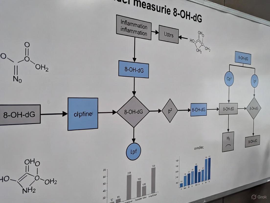
Visualizing the Experimental Workflow
The following diagram outlines the core decision points and steps in a robust 8-OHdG research study, from design to interpretation.
FAQs on 8-OHdG Measurement & Technical Challenges
FAQ 1: What are the primary methodological challenges when quantifying 8-OHdG in biological samples like exhaled breath condensate (EBC)?
The quantification of 8-OHdG in diluted biological matrices such as EBC is challenging due to the compound's very low concentration, often in the picomolar range, requiring highly sensitive analytical methods. Furthermore, the reliability of EBC collection itself is a concern, as factors during collection can lead to a loss of analyte, resulting in biomarker levels below the detection limit even with sensitive techniques. Precaution is needed when comparing literature results due to these methodological issues [7].
FAQ 2: What are the key differences between immunoassays and chromatographic methods for 8-OHdG measurement?
Two main analytical approaches are used: immunoassays and chemical analytical methods like liquid or gas chromatography coupled with mass spectrometry (LC-MS/GC-MS).
- Immunoassays: Widely used due to their simplicity and low cost. However, they can suffer from cross-reactivity with structurally similar isomers or interference from biological impurities, making them less specific and leading to potential discrepancies with other methods [7].
- Chromatography with MS Detection: Methods like LC-MS or GC-MS achieve superior specificity and accuracy by unequivocally identifying the molecule of interest. While GC-MS can require extensive manual sample preparation and derivatization, LC-MS is a powerful alternative that operates in liquid conditions and can allow for larger injection volumes, improving sensitivity [7].
FAQ 3: What specific factors during ELISA can lead to high background absorbance or unstable data?
When performing a competitive ELISA for 8-OHdG, several technical points are critical for reproducibility:
- Washing: Incomplete or improper washing can cause high background. Manual washing is often recommended over automated systems. The plate should be swung vigorously to discard reagent, and tapping on fresh paper towels helps remove water drops without letting wells dry [15].
- Pipetting Volume: Inaccuracy in pipetting samples and primary antibody reagent can cause errors [15].
- Temperature Control: Incubation must be carefully controlled at 37°C, as temperature fluctuations affect reproducibility [15].
- Cross-Contamination: The assay is sensitive to trace well-to-well contamination of the primary antibody, which can be prevented by careful pipetting and using fresh paper towels during washing [15].
FAQ 4: How is 8-OHdG standardized and reported in research?
In protocols like HPLC/EC (High-Performance Liquid Chromatography with Electrochemical Detection), the level of 8-OHdG is expressed relative to the amount of unmodified deoxyguanosine (dG) detected in the same sample. The results are typically expressed as the number of 8-OHdG adducts per 10⁶ deoxyguanosine bases (8-OHdG/10⁶ dG) [19].
Troubleshooting Guides
Guide 1: Addressing Low or Undetectable 8-OHdG in Exhaled Breath Condensate (EBC)
Problem: Despite using a sensitive LC-MS/MS method, 8-OHdG levels in EBC are below the limit of detection.
Potential Causes and Solutions:
- Cause: Analyte loss during EBC collection procedures.
- Solution: Review and validate the entire EBC collection protocol. Investigate factors such as condensing surface materials and temperature, and ensure consistency across samples. The collection device itself may adsorb the analyte [7].
- Cause: Insufficient sensitivity of the chemical analytical method for the sample volume.
- Solution: Use a highly sensitive method (e.g., LOD of 0.5 pg/mL for 8-OHdG). Consider methods that allow for larger sample injection volumes or pre-concentration steps to improve detection [7].
Guide 2: Troubleshooting High Background in 8-OHdG ELISA
Problem: High absorbance in blank wells, leading to unstable data and poor standard curves.
Potential Causes and Solutions:
- Cause: Inadequate washing, leading to residual primary or secondary antibody.
- Solution: Wash the plate manually and vigorously. After discarding reagent, hold the plate upside down and tap it firmly on new paper towels 5 times to remove residual liquid. Replace paper towels frequently to prevent contamination [15].
- Cause: Contamination of the blank well with primary antibody reagent.
- Solution: Ensure that the blank well receives PBS or wash buffer instead of the primary antibody. Take care not to touch the pipette tip to wells when dispensing to prevent well-to-well transfer of antibodies [15].
- Cause: Dried wells during the washing process.
- Solution: Do not let wells dry completely. The washing buffer should be added within 3 minutes after the previous step [15].
Guide 3: Managing Discrepancies Between Immunoassay and LC-MS Results
Problem: Measured 8-OHdG values from an immunoassay do not align with results from an LC-MS method.
Potential Causes and Solutions:
- Cause: Lack of specificity in the immunoassay due to antibody cross-reactivity.
- Solution: Validate the immunoassay against a reference chemical analytical method like LC-MS to check for overestimation. LC-MS methods can unequivocally identify 8-OHdG and are considered more specific [7].
- Cause: Matrix effects in the sample interfering with the immunoassay.
- Solution: For serum samples, remove proteins by ultra-filtration before the ELISA assay. For urine samples, centrifuge thawed samples to remove any insoluble materials [15].
Experimental Protocols
Protocol 1: Determination of 8-OHdG by HPLC with Electrochemical Detection (HPLC/EC)
This is a standard protocol for measuring oxidative DNA damage in cellular DNA [19].
- DNA Extraction: Isolate DNA from the biological sample (e.g., cells, tissue) using a standard phenol-chloroform method or a commercial kit.
- RNase Treatment: Treat the extracted DNA with RNase to remove any contaminating RNA.
- Enzymatic Hydrolysis: Digest the purified DNA into its constituent deoxynucleosides using an enzyme cocktail such as nuclease P1 and alkaline phosphatase.
- HPLC/EC Analysis:
- Inject the hydrolyzed DNA sample onto the HPLC system.
- Separate the deoxynucleosites using a reverse-phase column.
- Detect 8-OHdG using an electrochemical (EC) detector set at an oxidizing potential.
- Simultaneously, detect the canonical deoxyguanosine (dG) by its UV absorbance at 254 nm.
- Calculation: The level of oxidative damage is calculated as the ratio of 8-OHdG to dG, expressed as the number of 8-OHdG adducts per 10⁶ dG bases [19].
Protocol 2: Strand-Specific Quantification of 8-oxo-dG via Ligation-Dependent Probe Amplification (LPA)
This novel protocol allows for high-resolution, strand-specific mapping of oxidative DNA damage, providing insights into lesion formation and repair [20].
- DNA Extraction and Denaturation: Extract genomic DNA and denature it to obtain single-stranded DNA.
- Probe Ligation: Design specific DNA probes that are complementary to the target sequence on the DNA strand of interest. The probes will ligate only if the specific lesion (e.g., 8-oxo-dG) is present or absent at the target site.
- Ligation-Dependent Amplification: Amplify the successfully ligated probes using PCR. The amplification efficiency is directly related to the presence and quantity of the DNA lesion.
- Quantification and Analysis: Quantify the PCR products. The signal intensity allows for the strand-specific quantification of 8-oxo-dG lesions at single-nucleotide resolution [20].
Data Presentation: Methodological Challenges in 8-OHdG Quantification
Table 1: Common Methodological Challenges and Recommended Solutions in 8-OHdG Research
| Challenge | Description | Recommended Solution |
|---|---|---|
| Artifactual Oxidation | Oxidation of guanine during sample preparation (DNA extraction, hydrolysis) leading to overestimation. | Include antioxidants (e.g., deferoxamine) in buffers; minimize sample processing time [7]. |
| Low Analytical Sensitivity | Inability to detect 8-OHdG in samples with low concentration (e.g., EBC, plasma). | Employ highly sensitive methods like LC-MS/MS; use larger sample injection volumes [7]. |
| Sample Matrix Effects | Components in urine, serum, or EBC interfere with the detection system. | For ELISA, pre-treat samples (ultra-filtration for serum, centrifugation for urine) [15]. For LC-MS, use stable isotope-labeled internal standards [7]. |
| Lack of Specificity (Immunoassay) | Antibody cross-reactivity with structurally similar molecules or impurities. | Validate immunoassay results with a reference method like LC-MS; use MS-based methods for definitive identification [7]. |
| Data Normalization | Accounting for variable sample concentrations (e.g., urine dilution). | For urinary 8-OHdG, normalize to creatinine concentration. For cellular DNA, express as a ratio to total dG [19] [21]. |
Table 2: Comparison of Major Analytical Techniques for 8-OHdG Measurement
| Technique | Principle | Advantages | Disadvantages |
|---|---|---|---|
| ELISA | Competitive binding of 8-OHdG and an immobilized antigen for a specific antibody. | High throughput, low cost, technically simple, no expensive instrumentation required [7]. | Potential for cross-reactivity, less specific, can overestimate values, susceptible to matrix effects [7]. |
| HPLC/EC | Separation by HPLC, detection of 8-OHdG via electrochemical oxidation. | Good sensitivity, direct measurement without antibody use, provides ratio to native dG [19]. | Less specific than MS, potential for artifactual oxidation during sample prep, cannot distinguish from other isomers. |
| LC-MS/MS | Separation by LC, detection and identification by mass and fragmentation pattern. | High specificity and sensitivity, considered a "gold standard," can use internal standards for accuracy, can measure multiple biomarkers simultaneously [7]. | High instrument cost, requires specialized expertise, extensive method development. |
Visualization of Pathways and Workflows
Formation and Measurement of 8-OHdG
Experimental Workflow for 8-OHdG Analysis
The Scientist's Toolkit: Research Reagent Solutions
Table 3: Essential Reagents and Materials for 8-OHdG Research
| Item | Function/Application | Example & Notes |
|---|---|---|
| Stable Isotope-Labeled Internal Standard | Critical for accurate quantification in LC-MS/MS; corrects for sample loss and matrix effects. | [¹⁵N₅]-8-OHdG is used to spike samples before processing, allowing for precise relative quantification [7]. |
| DNA Digestion Enzymes | Hydrolyzes DNA into deoxynucleosides for HPLC or LC-MS analysis. | A combination of nuclease P1 and alkaline phosphatase is commonly used to digest DNA to deoxynucleosides [19]. |
| Antioxidants in Buffers | Prevents artifactual oxidation of guanine during sample preparation. | Deferoxamine (an iron chelator) or butylated hydroxytoluene (BHT) can be added to extraction buffers to minimize Fenton chemistry [7]. |
| Specific ELISA Kits | Immunoassay-based detection of 8-OHdG in various sample types. | Kits like the "New 8-OHdG Check" are designed for competitive ELISA. Requires careful attention to protocol details like temperature and washing [15]. |
| Solid-Phase Extraction (SPE) Cartridges | Purifies and concentrates samples before analysis, improving sensitivity. | Used in LC-MS protocols to remove interfering salts and matrix components, particularly for complex samples like urine or EBC. |
A Practical Guide to 8-OHdG Detection Methods: From HPLC to ELISA and Emerging Techniques
Within the context of a broader thesis on addressing technical challenges in 8-hydroxy-2'-deoxyguanosine (8-OH-dG) measurement research, the establishment of reliable analytical methods is paramount. The measurement of 8-OH-dG, a predominant biomarker of oxidative stress and DNA damage, is complicated by methodological artifacts and significant inter-laboratory discrepancies [22]. Accurate quantification is essential for understanding its role in mutagenesis, carcinogenesis, and aging [22]. This technical support center provides foundational principles, detailed protocols, and targeted troubleshooting guides for the two principal chromatographic techniques used in this field: High-Performance Liquid Chromatography with Electrochemical Detection (HPLC-ECD) and Liquid Chromatography with Tandem Mass Spectrometry (LC-MS/MS). The goal is to empower researchers to generate reproducible, accurate, and scientifically valid data, thereby advancing our understanding of oxidative DNA damage.
Core Principles and Technique Selection
Fundamental Operating Principles
- HPLC-ECD operates by separating compounds via liquid chromatography and then detecting electroactive species by measuring the electrical current generated when they undergo oxidation or reduction at a specific electrode potential. This makes it exquisitely sensitive to molecules like monoamines and the nucleoside 8-OH-dG [23].
- LC-MS/MS combines the separation power of liquid chromatography with the exquisite specificity of mass spectrometry. Molecules are identified based on their mass-to-charge (m/z) ratio and can be fragmented for further structural confirmation in the tandem MS stage, providing high confidence in analyte identification [23].
Technique Comparison and Selection Guide
Choosing the appropriate technique is a critical first step in experimental design. The table below summarizes the key characteristics of each method to guide this decision.
Table 1: Comparative Overview of HPLC-ECD and LC-MS/MS for 8-OH-dG Analysis
| Consideration | HPLC-ECD | LC-MS/MS |
|---|---|---|
| Fundamental Principle | Measures electrochemical redox current | Measures mass-to-charge (m/z) ratio |
| Sensitivity | High (pM / pg/µL range) [23] | Very High (often sub-pg/µL) [23] |
| Specificity/Selectivity | Excellent for electroactive compounds | Superior; provides structural confirmation and isomer differentiation [23] |
| Sample Preparation | Relatively simple (e.g., filtration) [23] | Complex (e.g., protein precipitation, solid-phase extraction) [23] |
| Typical Run Time | 5 - 30 minutes [23] | 15 - 45 minutes [23] |
| Instrument Cost | ~$45k – $80k [23] | ~$250k – $450k [23] |
| Operational Expertise | User-friendly, easier to train [23] | Requires significant technical expertise [23] |
| Ideal Use Case | Targeted, high-throughput analysis of known electroactive biomarkers like 8-OH-dG | Exploratory research, broad metabolite panels, complex matrices [23] |
The following decision pathway can help solidify your choice of technique:
Detailed Experimental Protocols
Protocol 1: Measurement of 8-OH-dG in DNA by LC-MS/MS
This protocol, adapted from a landmark study, details the measurement of 8-OH-dG using isotope-dilution mass spectrometry, which provides high accuracy and sensitivity [22] [24].
3.1.1 Research Reagent Solutions
Table 2: Essential Reagents for DNA Hydrolysis and 8-OH-dG Analysis via LC-MS/MS
| Reagent / Material | Function / Purpose | Source / Example |
|---|---|---|
| DNase I | Initiates DNA hydrolysis by cleaving into shorter oligonucleotides. | Sigma Chemical Company [22] |
| Phosphodiesterase I & II | Further hydrolyze oligonucleotides into mononucleotides. | Roche Diagnostics Corporation [22] |
| Alkaline Phosphatase | Converts mononucleotides into nucleosides (e.g., dGuo and 8-OH-dGuo) for analysis. | Roche Diagnostics Corporation [22] |
| Stable Isotope-Labeled 8-OH-dGuo (e.g., 8-OH-dGuo-18O) | Serves as an internal standard for Isotope-Dilution MS (IDMS), correcting for losses during sample prep and ionization variability. | Synthesized custom [22] |
| Calf Thymus DNA or Cell Line DNA | Used as a control or source for test DNA. | Sigma Chemical Company [22] |
| dGuo-15N5 | Labeled internal standard for deoxyguanosine. | Cambridge Isotope Laboratories [22] |
3.1.2 Sample Preparation Workflow
The multi-step process for preparing DNA samples for 8-OH-dG analysis is outlined below.
3.1.3 Key Steps and Parameters:
- DNA Hydrolysis: Digest 2-100 µg of DNA using the enzyme cocktail (DNase I, phosphodiesterases I and II, and alkaline phosphatase) in 35 mM phosphate buffer (pH 7.4) with 2 mM CaCl₂ [22].
- Internal Standard: A known amount of stable isotope-labeled 8-OH-dGuo (e.g., 8-OH-dGuo-18O) must be added prior to hydrolysis for accurate isotope-dilution quantification [22] [24].
- LC Separation: Critical for separating 8-OH-dGuo from other nucleosides in the hydrolysate to avoid mass spectral interferences.
- MS Detection: Uses atmospheric pressure electrospray ionization (ESI) with Selected Ion Monitoring (SIM). The ratio of the signal from the native 8-OH-dGuo to that of the added internal standard is used for quantification [22].
- Sensitivity: This method can detect approximately 5 lesions per 10⁶ DNA bases using as little as 2 µg of DNA, with potential for detection down to 1 lesion per 10⁶ bases with more DNA [22] [24].
Protocol 2: HPLC-ECD for Electroactive Analytes
While the search results focus on LC-MS for 8-OH-dG, HPLC-ECD is a widely used technique for this analyte and other neurotransmitters.
3.2.1 Core Principle and Workflow HPLC-ECD is ideal for routine, cost-effective analysis of electroactive compounds. The sample preparation is generally simpler than for LC-MS/MS, often involving only filtration or simple derivatization for non-electroactive targets [23].
Troubleshooting Guides and FAQs
Frequently Asked Questions (FAQs)
Q1: Our lab is new to 8-OH-dG research. Which technique should we invest in? A1: For labs primarily focused on targeted, high-throughput 8-OH-dG analysis, HPLC-ECD offers a lower-cost, user-friendly entry point with excellent sensitivity for this specific application. If your research scope is broader, requiring the analysis of multiple DNA lesions, metabolites, or structural confirmation, LC-MS/MS is the more powerful, albeit more expensive and complex, option [23]. A hybrid approach, using HPLC-ECD for routine work and accessing a core facility for LC-MS/MS validation, is also effective [23].
Q2: Why is there such variability in reported background levels of 8-OH-dG across different studies? A2: This is a well-documented challenge, which led to the establishment of the European Standards Committee on Oxidative DNA Damage (ESCODD). Variability arises from methodological artifacts during sample preparation (e.g., spurious oxidation during DNA hydrolysis) and differences in analytical techniques. The use of stable isotope-labeled internal standards added prior to DNA hydrolysis, as described in the LC-MS/MS protocol, is critical to minimize this variability and generate accurate data [22].
Q3: Can I use both HPLC-ECD and LC-MS/MS in the same study? A3: Absolutely. The techniques can be complementary. For instance, HPLC-ECD can be used for fast, cost-effective screening of a large number of samples, while LC-MS/MS can be employed to confirm the identity of chromatographic peaks or to perform a broader metabolomic profile on a subset of critical samples [23].
Troubleshooting Common Issues
Table 3: Troubleshooting Guide for HPLC-ECD and LC-MS/MS
| Problem | Potential Causes | Solutions |
|---|---|---|
| High Background Noise (ECD) | Contaminated mobile phase, degraded electrode, or voltage set too high. | Prepare fresh eluent buffers, polish or replace the electrode, and optimize the detection potential. |
| Low Sensitivity (Both) | ||
| - HPLC-ECD | Electrode fouling or suboptimal detector potential. | Implement a rigorous cleaning protocol and perform a hydrodynamic voltammetry experiment to re-optimize the voltage. |
| - LC-MS/MS | Ion source contamination or incorrect MS parameters. | Clean the ion source and ESI probe, and re-optimize MS parameters (e.g., collision energy) for 8-OH-dGuo. |
| Poor Chromatographic Separation (Both) | Degraded or contaminated HPLC column, incorrect mobile phase pH or composition. | Flush and regenerate the column or replace it. Ensure mobile phase is fresh and pH is accurately adjusted. |
| Artifactual Formation of 8-OH-dG | Oxidative damage during DNA isolation or hydrolysis. | Use chelating agents in buffers to sequester metal ions, add antioxidants where possible, and ensure the internal standard is added at the very beginning of sample preparation to correct for any artifactual formation [22]. |
| Inconsistent Results (MS) | Ion suppression from co-eluting matrix components. | Improve sample clean-up (e.g., solid-phase extraction) and enhance chromatographic separation to move the analyte away from the suppressing region. |
8-hydroxy-2'-deoxyguanosine (8-OHdG) is a widely recognized biomarker of oxidative stress, formed when reactive oxygen species attack guanine bases in DNA [1]. This oxidative lesion is significant because if not repaired by base excision repair pathways, it can lead to G-T transversion mutations, potentially contributing to various pathologies including cancer, neurodegeneration, and aging-related diseases [1]. The measurement of 8-OHdG provides valuable insights into oxidative DNA damage levels in physiological and pathological processes.
Enzyme-Linked Immunosorbent Assay (ELISA) represents a key methodological approach for quantifying 8-OHdG in biological samples. Unlike chromatographic methods like LC-MS/MS or HPLC that require expensive instrumentation and extensive sample preparation, ELISA offers advantages for high-throughput screening applications due to its relative simplicity, cost-effectiveness, and capacity to process multiple samples simultaneously [1]. This makes it particularly valuable for studies requiring large sample sizes, such as population studies, occupational exposure assessments, and therapeutic screening.
Within the broader context of technical challenges in 8-OHdG measurement research, ELISA-based methods must overcome several specific obstacles, including antibody specificity issues, matrix effects in different sample types, and the need for rigorous protocol standardization to ensure reproducible results across experiments and laboratories.
Comprehensive Troubleshooting Guide
Common ELISA Problems and Solutions
Table: Troubleshooting Common ELISA Issues for 8-OHdG Detection
| Problem | Possible Causes | Recommended Solutions |
|---|---|---|
| High Background | Insufficient washing; Contaminated buffers; Prolonged substrate incubation; Plate sealers reused [25] [26] | Increase wash steps and duration; Ensure proper draining; Use fresh plate sealers for each step; Prepare fresh buffers; Shorten substrate incubation time [9] [25] |
| Weak or No Signal | Reagents not at room temperature; Incorrect reagent storage; Expired reagents; Improper pipetting technique; Incomplete DNA digestion [25] [8] | Allow all reagents to warm for 15-20 minutes before use; Verify storage conditions (typically 2-8°C); Check expiration dates; Calibrate pipettes; Ensure complete DNA digestion with nuclease P1 [9] [8] |
| Poor Standard Curve | Improper standard reconstitution; Incorrect dilution series; Degraded standard; Inaccurate pipetting of viscous conjugate [26] | Reconstitute standard exactly as directed; Verify dilution calculations; Use standard within 1 hour of reconstitution; Allow viscous HRP conjugate to reach room temperature and wipe pipette tip before dispensing [26] |
| Poor Replicate Data | Inconsistent washing; Uneven plate coating; Temperature fluctuations; Cross-contamination between wells [25] [27] | Use consistent washing technique; Ensure even reagent distribution across wells; Maintain stable incubation temperature; Use fresh plate sealers and avoid tip reuse [9] [25] |
| Edge Effects | Uneven temperature distribution; Evaporation; Stacked plates during incubation [25] | Use tight plate sealers; Avoid stacking plates; Ensure incubator has uniform temperature distribution; Use water bath for more consistent temperature [9] |
| High Blank Well Absorbance | Incomplete washing leading to primary antibody contamination; Pipette tip contamination between wells [9] | Vigorously discard reagent after incubation; Tap plate forcefully on fresh paper towels 5 times; Replace paper towels when wet; Avoid touching wells with pipette tips during washing [9] |
Sample Preparation Issues and Solutions
Table: Sample-Specific Preparation Guidelines for 8-OHdG ELISA
| Sample Type | Preparation Requirements | Special Considerations |
|---|---|---|
| Cell and Tissue DNA | DNA extraction required; Digestion with nuclease P1 and alkaline phosphatase; Minimum 2 μg digested DNA per assay [8] | Complete digestion to single nucleosides critical; Confirm removal of RNA contamination; Store extracted DNA at -20°C to -80°C [8] |
| Urine | Can be used directly without DNA extraction; Centrifugation if insoluble particles present; Typically requires dilution (2-5 fold or higher) [8] | 8-OHdG is abundant in urine; Store at -80°C for long-term preservation; Perform preliminary dilution experiment to determine optimal dilution factor [9] [8] |
| Serum/Plasma | Can be used directly; Protein removal recommended for serum samples; Dilution often necessary [9] [8] | Remove proteins by ultra-filtration before assay; EDTA plasma is compatible; Expected values typically within standard curve range (0.078-20 ng/mL) [9] [8] |
| Exhaled Breath Condensate | Highly diluted matrix; Potential analyte loss during collection [7] | Methodological concerns regarding reliability; May require concentration step; Consider sample collection variability [7] |
Frequently Asked Questions (FAQs)
Sample Preparation FAQs
Q: Do I need to extract DNA for all sample types? A: No, DNA extraction is only required for cell and tissue samples. Body fluids including urine, plasma, serum, cerebrospinal fluid, and saliva can be used directly without DNA extraction, though they may require centrifugation if insoluble particles are present and typically need dilution [8].
Q: Why is complete DNA digestion critical and how can I achieve it? A: Complete digestion to single nucleosides is essential because even dinucleotides can affect results. Use nuclease P1 (e.g., Sigma #N8630) to digest DNA to single nucleotides, followed by alkaline phosphatase (e.g., Sigma #P5931) to convert nucleotides to nucleosides. The recommended amount of enzyme is sufficient for complete digestion when used according to protocols [8].
Q: How much DNA is needed per assay? A: A minimum of 2 μg of completely digested DNA is required per assay well. This requirement is based on the expected frequency of approximately one 8-OHdG site per 100,000 dG sites in genomic DNA and the detection sensitivity of the ELISA kit [8].
Q: Can I use DNA from any species? A: Yes, the 8-OHdG modification is universal across species, so the ELISA kit can be used with DNA from any biological source, including bacteria [8].
Assay Protocol FAQs
Q: Why does the 8-OHdG ELISA generate a reverse standard curve? A: The 8-OHdG ELISA is based on a competitive format. The 8-OHdG present in samples or standards competes with a pre-immobilized 8-OHdG conjugate on the plate for antibody binding. Higher 8-OHdG levels in the sample result in less antibody binding to the plate, producing lower optical density (OD) readings [8].
Q: Can I use part of the plate now and save the rest for later? A: Yes, most kits provide 96-well plates as strip well strips. Unused strips can be stored at 4°C for future use. However, fresh standard curves should be prepared for each experiment, and newly coated plates should be used for accurate results [8].
Q: What is the most critical step in the 8-OHdG ELISA procedure? A: The coating with the 8-OHdG conjugate is the most critical step. The conjugate is not stable once coated, so freshly coated plates should be used. The conjugate should be aliquoted and stored at -80°C, with multiple freeze-thaw cycles avoided [8].
Q: How do I know when to stop the substrate reaction? A: The substrate solution typically takes 2-30 minutes to develop. Focus on the standard curve wells, where you should observe a gradient of color from bright blue in the lowest concentration to very faint color in the highest concentration. Add stop solution when this gradient is clearly visible, using a multichannel pipettor to stop the reaction simultaneously across all wells [8].
Data Analysis FAQs
Q: How should I analyze my 8-OHdG ELISA data? A: The best approach is to use a 4-parameter curve fitting program. If this is unavailable, you can use Excel with the x-axis set to logarithmic scale to generate a linear or logarithmic trendline. The middle portion of the standard curve is most sensitive and should be used for quantifying samples. For linear trendlines, use the equation (OD value - b)/m; for logarithmic trendlines, use ln(x) = (OD value - b)/m [8].
Q: What are expected 8-OHdG levels in serum? A: Serum 8-OHdG levels typically fall within the standard curve range of 0.078-20 ng/mL, though this varies by individual sample. Occasionally, samples contain higher levels and require dilution. A preliminary experiment with a 1:5 dilution is recommended to determine optimal dilution factors [8].
Q: Why are my standard curve OD values consistently low? A: Low ODs across the standard curve suggest a problem with coating of the 8-OHdG conjugate onto the plate. Ensure the conjugate is properly aliquoted and stored at -80°C, use freshly coated plates, and verify that the substrate development time is adequate. The 0 ng/mL standard should yield the highest OD value [8].
Experimental Protocols and Workflows
Detailed DNA Digestion Protocol for Cellular Samples
Proper DNA digestion is crucial for accurate 8-OHdG measurement from cellular samples. The following protocol ensures complete digestion to single nucleosides:
DNA Extraction: Extract DNA using any commercial DNA extraction kit following manufacturer's instructions. Include RNase treatment step to remove contaminating RNA.
DNA Quantification: Precisely quantify DNA concentration using spectrophotometry. Adjust concentration to 1-5 mg/mL using ultrapure water.
Heat Denaturation: Convert double-stranded DNA to single-stranded DNA by heat denaturation at 95°C for 5 minutes, then immediately place on ice.
Nuclease P1 Digestion:
- Add 5-20 units of nuclease P1 (Sigma #N8630) per sample
- Incubate at 37°C for 30-60 minutes
- Buffer conditions: 20 mM sodium acetate, pH 5.2
Alkaline Phosphatase Treatment:
- Adjust pH by adding 100 mM Tris buffer (pH 8.0)
- Add 5-10 units of alkaline phosphatase (Sigma #P5931)
- Incubate at 37°C for 30-60 minutes
Storage: Digested DNA samples can be stored at -20°C or -80°C for up to one year without significant degradation of 8-OHdG [8].
Visual Workflow: Competitive ELISA Principle for 8-OHdG Detection
Visual Workflow: Sample Preparation Decision Tree
The Scientist's Toolkit: Essential Research Reagents
Table: Key Reagent Solutions for 8-OHdG ELISA Research
| Reagent/Category | Function/Purpose | Specific Examples/Considerations |
|---|---|---|
| DNA Digestion Enzymes | Complete digestion of DNA to single nucleosides for accurate detection | Nuclease P1 (Sigma #N8630); Alkaline phosphatase (Sigma #P5931) or Shrimp Alkaline Phosphatase [8] |
| ELISA Buffer Systems | Provide optimal conditions for antibody-antigen interactions | Cell Extraction Buffer (Thermo Fisher FNN0011) for lysate preparation; 5X Assay Buffer (Thermo Fisher CNB0011) for plate blocking and sample dilution [26] |
| Detection Components | Enable signal generation and quantification | Streptavidin-HRP conjugates (lot-specific for kits); TMB substrate solution; Stop solution [26] |
| Sample Preservation | Maintain 8-OHdG stability during storage | Protease inhibitor cocktails (e.g., Thermo Fisher 87786 with EDTA); PMSF (add fresh to 1 mM final concentration) [26] |
| Specialized Consumables | Ensure assay reproducibility and precision | ELISA plates (not tissue culture plates); Fresh plate sealers for each incubation step; Low-protein binding pipette tips [25] [27] |
Methodological Considerations for Different Research Applications
The application of 8-OHdG ELISA across different research domains requires consideration of methodological specifics:
Exercise Physiology Studies: Research indicates differential 8-OHdG responses based on exercise type and training status. Resistance exercise consistently increases circulating 8-OHdG levels in both trained and untrained individuals (SMD = 0.66, p < 0.001). For aerobic exercise, trained individuals show small increases (SMD = 0.42; p < 0.001), while untrained individuals demonstrate decreases, particularly after long-duration exercise (SMD = -1.16; p < 0.05) [28]. These findings suggest training status must be considered when interpreting 8-OHdG data in exercise studies.
Clinical Biomarker Applications: When using 8-OHdG as a diagnostic biomarker, method selection significantly impacts results. ELISA offers practical advantages for clinical studies with larger sample sizes, but researchers should be aware of potential cross-reactivity issues compared to more specific LC-MS/MS methods [1]. Consistent sample handling and processing protocols are essential for reliable clinical data.
Limitations and Complementary Methods: While ELISA provides high-throughput capability, technical challenges include antibody specificity and matrix effects. For critical applications requiring absolute quantification, mass spectrometry methods (LC-MS/MS) may be preferred despite higher cost and complexity [7] [1]. Method validation should always include recovery experiments and comparison with established protocols when possible.
FAQs: Troubleshooting Guides for 8-OH-dG Measurement Research
Solid-Phase Extraction (SPE) Troubleshooting
1. What are the common causes of poor analyte recovery in SPE and how can I fix them?
Poor recovery occurs when your target analytes, such as 8-OH-dG, are not effectively retained or eluted from the SPE sorbent. The following table outlines the common causes and their solutions [29] [30] [31].
| Problem Cause | Solution |
|---|---|
| Insufficient Retention | - Choose a sorbent with greater selectivity for your analytes. [29] [30]- Adjust the sample pH to increase analyte affinity for the sorbent. [29]- Reduce the loading flow rate to increase interaction time. [31] |
| Incomplete Elution | - Increase eluent strength (e.g., organic percentage) or volume. [29] [30]- Adjust elution solvent pH to ensure greater analyte affinity. [29]- Switch to a less retentive sorbent (e.g., C4 instead of C18). [30] [31] |
| Column Overload | - Decrease the sample volume or concentration. [29]- Use a cartridge with a larger amount of sorbent or higher capacity. [29] [30] |
| Sorbent Drying | - Do not let the sorbent bed dry out before sample loading. Re-condition if it does. [29] [30] |
2. How can I improve the reproducibility of my SPE results?
Inconsistent results often stem from variations in the extraction process [32] [30] [31].
- Ensure Proper Conditioning: Always condition and equilibrate the cartridge according to the manufacturer's recommendations, and do not let it dry before sample application [30] [31].
- Control Flow Rates: Use a consistent, controlled flow rate (typically 1-5 mL/min) during sample loading and elution. High or variable flow rates can lead to poor retention and irreproducibility [30] [31].
- Avoid Partial Elution: Ensure your wash solvent is not too strong, as it can accidentally elute some of your analyte. Incorporate soak steps to allow for proper solvent-sorbent equilibration [30] [31].
- Sample Pre-treatment: Follow a consistent sample preparation method. For complex matrices like biological fluids, pre-treatment such as filtration or centrifugation to remove particulate matter is crucial [29] [31].
3. My SPE extracts are not clean enough, with many interferences. What should I do?
Unsatisfactory cleanup means interfering compounds are co-eluting with your analytes [32] [30] [31].
- Optimize Wash Solvent: The wash solvent should have the maximum strength possible to elute impurities without displacing your target analyte [32] [31].
- Change Sorbent Selectivity: Consider using a more selective sorbent. Mixed-mode sorbents, which combine multiple retention mechanisms (e.g., reversed-phase and ion-exchange), can offer superior cleanup for analytes like 8-OH-dG that have both nonpolar and ionizable groups [32] [30].
- Pre-treat the Sample: Remove proteins, lipids, or salts prior to SPE using techniques like protein precipitation, liquid-liquid extraction, or ultrafiltration [32] [31].
Capillary Electrophoresis (CE) Troubleshooting
1. I am getting low or no signal for my 8-OH-dG samples in CE. What could be wrong?
Low signal intensity can originate from several parts of your workflow [33].
- Check Instrument and Reagents: Confirm that your internal size standard runs correctly. Ensure all reagents (e.g., polymer, buffer) are within their expiry dates and have been stored properly [33].
- Optimize Sample Preparation: Low signal may require optimization of the pre-CE reaction (e.g., PCR). Increasing template, primer concentration, or cycle number might be necessary. If the sample is visible on a gel but not in CE, the fluorescently labeled primer may be faulty and need re-synthesis [33].
- Verify Denaturation and Matrix: Use HiDi formamide as the sample matrix, not water, to ensure proper denaturation and sample stability. Confirm the denaturation step (95°C for 3 minutes) was performed correctly [33].
- Address Capillary Issues: A blocked capillary or air bubble can prevent sample injection. Running a size standard-only sample can help diagnose this. Centrifuging the plate before running can remove air bubbles [33].
2. Why are my CE peaks broad or off-scale?
Peak shape issues are often related to sample or instrument conditions [33].
- Off-Scale Peaks (Saturated Signal): This is caused by injecting too much sample. Reduce the sample concentration by diluting the PCR product further or decrease the injection time in the instrument run module [33].
- Broad Peaks: This can be caused by degraded polymer or buffer, a degraded capillary array, or sample degradation. Replace expired consumables. High salt concentration in the sample can also cause broad peaks; in this case, purifying the PCR product before injection is recommended [33].
The following workflow diagram summarizes the key steps for diagnosing and resolving common CE issues related to signal and peaks.
Method Selection and Cross-Technique Integration
1. Should I use immunoassay or chromatographic methods for measuring 8-OH-dG?
The choice of method involves a trade-off between throughput and specificity, which is critical for accurate 8-OH-dG measurement [7] [1].
| Method | Advantages | Disadvantages |
|---|---|---|
| Immunoassays(e.g., ELISA) | - Simplicity and low cost. [7]- High throughput. [1] | - Potential for cross-reactivity, leading to overestimation. [7]- Generally less specific and accurate than chemical methods. [7] [1] |
| Chemical Methods(e.g., LC-MS/MS, HPLC) | - High specificity and accuracy. [7] [1]- Considered a reference method for validation. [7] | - Requires expensive instrumentation. [7]- Can involve complex sample preparation. [7] |
| Capillary Electrophoresis(e.g., CZE-MS/MS) | - Minimal sample and solvent consumption. [34]- Can be a sustainable alternative to LC. [34] | - Less established for 8-OH-dG specifically.- May require method development. |
2. What are the major methodological challenges specific to measuring 8-OH-dG in exhaled breath condensate (EBC)?
EBC presents unique challenges due to its extreme dilution and the nature of the collection process [7].
- Extreme Dilution: EBC requires highly sensitive analytical methods capable of detecting biomarkers in the picomolar range. Even with sensitive LC-MS/MS methods, 8-OH-dG and 8-isoprostane levels can be below the limit of detection [7].
- Loss of Analyte: The EBC collection procedure itself may lead to the loss of analytes, affecting the reliability of quantification. Factors influencing collection efficiency are not fully resolved [7].
- Methodological Heterogeneity: Lack of standardization in EBC collection and analysis across studies makes it difficult to compare results. Robust, validated methods that account for all confounding factors are essential [7].
The Scientist's Toolkit: Essential Research Reagents and Materials
The following table lists key reagents and materials used in the sample preparation and analysis of 8-OH-dG, based on cited experimental protocols [7] [34].
| Item | Function in the Experiment |
|---|---|
| Solid-Phase Extraction Cartridges (e.g., C18, HLB, Mixed-mode) | To purify, concentrate, and extract 8-OH-dG from complex biological matrices like plasma, urine, or EBC prior to analysis. [31] |
| 8-OHdG Standard (≥98%) | Used as a reference standard for calibration curves to ensure accurate quantification of the target biomarker. [7] |
| Isotope-Labeled Internal Standard(e.g., [15N5]-8-OHdG) | Added to the sample at the start of preparation; corrects for analyte loss during sample workup and for matrix effects during MS analysis, improving accuracy. [7] |
| LC-MS Grade Solvents(e.g., Methanol, Acetonitrile) | High-purity solvents are required for mobile phases and sample preparation to avoid introducing contaminants that cause background noise or signal suppression in MS. [7] [35] |
| Background Electrolyte (BGE)(e.g., 20 mM NH₄HCO₃) | The conductive medium used for separation in Capillary Zone Electrophoresis (CZE). Its composition and pH are critical for achieving stable and reproducible separations. [34] |
| HiDi Formamide | Used as the sample matrix for capillary electrophoresis. It acts as a denaturant and provides sample stability during the heat denaturation step and electrophoresis run. [33] |
Experimental Protocol: Simultaneous Quantification of 8-OHdG and 8-Isoprostane in EBC by LC-MS
This detailed methodology is adapted from the research by Fijani et al. (2022) for the simultaneous measurement of key oxidative stress biomarkers [7].
1. Reagents and Standard Preparation
- Stock Solutions: Prepare 1 mg/mL stock solutions of 8-OHdG and 8-isoprostane in H₂O/MeOH (8:2, v/v). Store at -20°C.
- Working Solutions: Prepare intermediate stock solutions at 5000 ng/mL by dilution with ultrapure water. Store at 4°C.
- Internal Standards (IS): Prepare [15N5]-8-OHdG and 8-isoprostane-d4 at 500 ng/mL in ultrapure water. Store at 4°C. These stock solutions are stable for at least three months [7].
2. Sample Preparation (Solid-Phase Extraction)
- Conditioning: Condition the selected SPE sorbent (e.g., a reversed-phase cartridge) with methanol or acetonitrile followed by a solvent matching the sample solution pH.
- Loading: Acidify the EBC sample and load it onto the conditioned SPE cartridge at a controlled, slow flow rate (e.g., 1-2 mL/min) to ensure optimal retention [31].
- Washing: Wash the cartridge with a weak solvent or buffer to remove impurities without eluting the analytes. For reversed-phase, a common wash is 5% methanol in water [32].
- Elution: Elute the retained 8-OHdG and 8-isoprostane using a strong solvent, such as pure methanol or a mixture of methanol and acetonitrile. Ensure the elution volume is sufficient for complete recovery [29] [7]. Collect the eluate and evaporate to dryness under a gentle stream of nitrogen.
- Reconstitution: Reconstitute the dry extract in the initial mobile phase for LC-MS analysis.
3. LC-MS Analysis
- Chromatography: Use a suitable reversed-phase LC column (e.g., C18). The mobile phase typically consists of water (A) and methanol or acetonitrile (B), both modified with 0.1% formic acid to enhance ionization. A gradient elution is applied to separate the biomarkers.
- Mass Spectrometry: Operate the mass spectrometer in positive electrospray ionization (ESI+) mode. Monitor specific multiple reaction monitoring (MRM) transitions for each analyte and its corresponding internal standard for highly selective and sensitive detection [7].
The relationship between sample preparation, analytical separation, and detection in a comprehensive 8-OH-dG analysis workflow is illustrated below.
Within the framework of a broader thesis on overcoming technical challenges in 8-OH-dG measurement research, this guide addresses a foundational step: sample matrix selection. The quantification of 8-hydroxy-2'-deoxyguanosine (8-OHdG), a critical biomarker of oxidative DNA damage, is highly influenced by the choice of biological sample. This technical support center provides researchers, scientists, and drug development professionals with a detailed comparison of urine, serum, saliva, and tissue DNA, offering troubleshooting guides and FAQs to navigate the specific issues encountered during experimental work.
Sample Matrix Comparison Table
The table below summarizes the core characteristics, advantages, and challenges of each sample matrix for 8-OHdG analysis.
| Sample Matrix | Key Characteristics | Pros | Cons & Technical Challenges |
|---|---|---|---|
| Urine | - Represents total body oxidative stress & cumulative damage [36].- Contains 8-OHdG excised & excreted via repair processes. | - Non-invasive collection.- Suitable for large-scale/longitudinal studies [36].- Sample volume is typically not limiting. | - Concentration requires normalization to creatinine (e.g., ng/mg creatinine) [36].- Levels can be influenced by renal function.- Potential for artifact formation if samples are not processed/stored correctly. |
| Serum/Plasma | - Reflects circulating, cell-free oxidized DNA & instantaneous balance [36]. | - Offers a systemic view of oxidative stress.- Standardized collection protocols are widely available. | - Invasive collection procedure.- Generally has lower concentrations of 8-OHdG vs. saliva & urine [37].- Sensitive to hemolysis, which can interfere with assays. |
| Saliva | - Provides a localized view of oral & systemic conditions [37]. | - Non-invasive and easy to collect [37].- Studies show it can have the highest concentration of 8-OHdG among body fluids [37].- Ideal for oral disease research (e.g., periodontitis) [38]. | - Composition can be influenced by food, drink, or salivary flow rate.- Requires centrifugation to obtain clear supernatant for analysis [37].- May contain contaminants that inhibit downstream assays. |
| Tissue | - Provides direct measurement of DNA damage in a specific organ or lesion. | - Enables spatial localization of damage (e.g., tumor vs. adjacent tissue) [36].- Allows for mechanistic studies within a target organ. | - Invasive (biopsy/surgery required).- Requires DNA extraction,- introducing risk of artifactual oxidation during processing [1].- Results are highly dependent on the DNA extraction method used. |
Frequently Asked Questions (FAQs) and Troubleshooting
Q1: My urine 8-OHdG values are highly variable. How can I improve reliability?
- A: A primary step is to normalize your data. Report 8-OHdG values relative to urinary creatinine (e.g., ng/mg or nmol/mmol creatinine) to account for differences in urine concentration and hydration status [36]. Furthermore, control for diurnal variation by standardizing the time of collection; levels can fluctuate, with one study noting a range from 3.76 ng/mg in early morning to 6.46 ng/mg in the early afternoon [36].
- Troubleshooting Checklist:
- Have you centrifuged the urine sample to remove precipitates?
- Is the sample being stored at -80°C immediately after collection?
- Have you used a creatinine assay to normalize your 8-OHdG concentrations?
Q2: Why are my measured 8-OHdG levels in serum much lower than in saliva or urine?
- A: This is an expected finding. Research directly comparing matrices has confirmed that 8-OHdG concentration in serum is significantly lower than in saliva and urine [37]. Serum reflects a snapshot of the instantaneous balance between damage and repair in the circulation, whereas urine represents cumulative excretion over time [36] [37]. Saliva may concentrate the biomarker due to local production in the oral cavity [37]. If your serum levels are below the detection limit of your assay, you may need to switch to a more sensitive method (e.g., LC-MS/MS) or consider using urine or saliva for your study.
Q3: How can I prevent artificial oxidation of DNA during extraction from tissue samples?
- A: Artifactual oxidation is a major concern for tissue-based 8-OHdG measurement [1]. To minimize this:
- Use Chelating Agents: Include strong chelators like EDTA in your lysis buffers to sequester metal ions that catalyze oxidation.
- Add Antioxidants: Incorporate antioxidants such as butylated hydroxytoluene (BHT) into your extraction protocol [7].
- Choose Kits with Lysis: Select DNA extraction kits that utilize chaotropic salts (e.g., guanidine hydrochloride) in the lysis buffer, as these disrupt cellular activity and inhibit nucleases, thereby reducing artifactual oxidation [39].
- Validate Your Method: Test your entire workflow, from tissue homogenization to DNA elution, with a standard of known 8-OHdG content to gauge introduced artifacts.
Q4: My ELISA and LC-MS/MS results for the same sample are inconsistent. Which method should I trust?
- A: This is a common issue. Immunoassays like ELISA are susceptible to cross-reactivity with structurally similar molecules, which can lead to overestimation [7] [1]. Chromatographic methods coupled with mass spectrometry (LC-MS/MS) are generally considered the "gold standard" due to their high specificity, sensitivity, and ability to unequivocally identify the 8-OHdG molecule [7] [1]. If your research requires high accuracy and precision, LC-MS/MS is the recommended method. If using an ELISA kit for its throughput and lower cost, it is crucial to validate the method for your specific sample matrix against a reference LC-MS/MS technique.
Detailed Experimental Protocols
Protocol: Saliva and Serum 8-OHdG Analysis via ELISA
This protocol is adapted from a 2024 comparative study [37].
1. Sample Collection:
- Saliva: Collect approximately 4 ml of unstimulated saliva using the drooling method in the morning under quiet, resting conditions.
- Serum: Aspirate 3 ml of blood from the antecubital vein into a clot activator vial.
2. Sample Preparation:
- Allow blood to clot for 10-15 minutes at room temperature.
- Centrifuge both saliva and blood samples at 1000 RPM at 2–8°C for 15–20 minutes.
- Pipette 1.5 ml of the resultant supernatant (saliva or serum) into labelled Eppendorf tubes.
- Store samples at -80°C until analysis.
3. ELISA Procedure:
- Use a commercial 8-OHdG Biotinylated Detection Antibody ELISA Kit.
- Follow the manufacturer's instructions for pipetting standards and samples into the 96-well microplate.
- After adding the stop solution, read the optical density (OD) of each well at 450 nm using a microplate reader within 15 minutes.
- Calculate 8-OHdG concentrations from the standard curve.
Protocol: Key Considerations for Tissue DNA Extraction for 8-OHdG Analysis
The following workflow outlines the critical steps for preparing tissue samples to minimize artifactual oxidation and ensure accurate 8-OHdG measurement.
The Scientist's Toolkit: Essential Research Reagent Solutions
| Item | Function / Application in 8-OHdG Research |
|---|---|
| Chaotropic Salts (e.g., Guanidine HCl) | Denature proteins, inactivate nucleases, and enable DNA binding to silica in extraction kits, thereby minimizing artifactual oxidation during DNA isolation [39]. |
| DNA Extraction Kits (Silica-Membrane Based) | Provide a standardized, efficient method for purifying high-quality DNA from tissues or cells. Kits like the NucleoSpin Soil kit have been shown to provide high alpha diversity estimates in microbiota studies, indicating efficient lysis [40]. |
| Antioxidants (e.g., BHT) | Added to lysis buffers to prevent the formation of new oxidative damage (artifactual oxidation) during the DNA extraction process [7]. |
| Chelating Agents (e.g., EDTA) | Bind metal ions (e.g., Fe²⁺) that can catalyze Fenton reactions, which generate hydroxyl radicals and cause artifactual oxidation of DNA [39]. |
| Nuclease P1 & Alkaline Phosphatase | Enzymes used to digest purified DNA into individual nucleosides, a required step before analysis by HPLC or LC-MS/MS to quantify the 8-OHdG/dG ratio [1]. |
| Creatinine Assay Kit | Essential for normalizing 8-OHdG concentrations in urine samples to account for variations in urine dilution [36]. |
| LC-MS/MS System | The gold-standard analytical platform for specific and sensitive quantification of 8-OHdG, avoiding cross-reactivity issues associated with immunoassays [7] [1]. |
| SYBR Green I Dye | A fluorescent DNA intercalating dye used in real-time PCR and LAMP assays. Its concentration may require optimization when used in assays containing metal ions to avoid fluorescence quenching [41]. |
Method Selection and Workflow Diagram
Choosing the right analytical method is critical for generating reliable data. The following decision pathway guides you through this process based on your research requirements and sample type.
Solving Common Pitfalls: Strategies to Minimize Artifacts and Maximize Reproducibility in 8-OHdG Analysis
In the field of oxidative DNA damage research, particularly in studies measuring 8-hydroxy-2'-deoxyguanosine (8-OHdG), the accuracy of your results is fundamentally dependent on your sample handling procedures. The biomarker 8-OHdG is one of the most common oxidative DNA modifications, widely used in research and clinical diagnostics for risk assessment of various cancers and degenerative diseases [42]. However, this biomarker presents a unique challenge: it can be formed not only in vivo but also artifactually during sample isolation, hydrolysis, and analysis [6].
This technical support guide addresses the critical procedural challenges that can compromise DNA integrity during extraction and handling. When measuring 8-OHdG, researchers must contend with the paradox that the very processes required to isolate DNA can generate the oxidative damage they aim to measure. The indirect mechanism mediated by oxidative stress accounts for the majority, on the order of 60–70%, of the DNA damage induced by factors like ionizing radiation [43], and similar principles apply to artifactual oxidation during sample processing.
The following sections provide targeted troubleshooting guidance, detailed protocols, and preventative strategies to help you maintain DNA integrity from sample collection to analysis, ensuring your 8-OHdG measurements reflect biological reality rather than procedural artifacts.
Troubleshooting Guide: Common Scenarios and Solutions
Answer: Artifactual oxidation can arise at multiple stages of handling. The most common sources include:
- Sample Grinding and Homogenization: The process of tissue disruption generates heat and introduces oxygen, both of which can promote oxidative damage. When using a bead-based homogenizer like the Bead Ruptor Elite, ensure you optimize settings for speed, cycle duration, and temperature to minimize mechanical and thermal stress [44]. For particularly sensitive samples, using the cryo cooling unit can further reduce thermal damage.
- Inadequate Antioxidant Protection: Your extraction buffer may lack sufficient protective agents. The established CTAB-based extraction method for difficult plant species uses 0.3% (v/v) β-mercaptoethanol in the extraction buffer to combat phenolics and prevent oxidation [45]. For animal tissues, consider adding other antioxidants.
- Sample Storage Conditions: Storing samples at -20°C instead of -80°C can accelerate oxidative processes. Flash freezing in liquid nitrogen followed by storage at -80°C is considered the gold standard for maintaining DNA integrity by rapidly halting enzymatic activity [44].
- DNA Extraction Temperature: High incubation temperatures during lysis and protein digestion can dramatically increase oxidation rates. The click-code-seq methodology specifically warns that heat inactivation of enzymes and fragmenting gDNA by sonication before blocking background modifications can create artifactual oxidation [46].
Answer: Implement these specific protective measures during sample preparation:
- Immediate Stabilization: For tissue samples, immediately freeze in liquid nitrogen upon collection. Never allow samples to sit at room temperature. For biological fluids, consider chemical preservatives designed to stabilize nucleic acids [44].
- Optimized Homogenization in Cold Conditions: Always pre-chill your mortar and pestle with liquid nitrogen when grinding frozen tissue [45]. For bead-based homogenization, process samples in a cold room or using pre-chilled tubes.
- Antioxidant-Enriched Buffers: Prepare extraction buffers fresh and add reducing agents immediately before use. The standard CTAB protocol uses β-mercaptoethanol, but other options include ascorbate, dithiothreitol (DTT), or glutathione for particularly oxidation-prone samples.
- Control Sonication Artifacts: If you use sonication for DNA fragmentation, perform this step only after both blocking background modifications and labeling target DNA modifications to prevent artifactual oxidation [46].
FAQ 3: What quality control measures can help me identify artifactual oxidation in my DNA samples?
Answer: Implement these QC checkpoints to assess potential oxidation:
- UV Spectrophotometry: While A260/280 ratios of ~1.8 indicate protein-free DNA, this doesn't specifically detect oxidation. However, deviations can suggest general quality issues that may correlate with poor handling.
- Gel Electrophoresis: Visualize DNA on agarose gels looking for smearing rather than tight, high molecular weight bands, which can indicate degradation potentially linked to oxidative processes [45].
- Fragment Analysis: Provides detailed DNA size distribution, particularly helpful for degraded samples [44].
- Quantitative PCR (qPCR): Can assess both DNA concentration and amplification potential, as oxidized DNA templates often amplify poorly.
- Specific Oxidative Damage Assays: Techniques like click-fluoro-quant can rapidly quantify DNA breaks, AP sites, or guanine oxidation to specifically assess oxidative damage levels [46].
Research Reagent Solutions: Essential Materials for Oxidation Prevention
The table below summarizes key reagents and their critical functions in preventing artifactual oxidation during DNA isolation:
| Reagent/Chemical | Function in Preventing Oxidation | Example Usage & Concentration |
|---|---|---|
| β-mercaptoethanol | Reducing agent that prevents oxidation of phenolics and protects DNA | 0.3% (v/v) in CTAB extraction buffer [45] |
| EDTA (Ethylenediaminetetraacetic acid) | Chelates metal ions (e.g., Fe²⁺, Cu²⁺) that catalyze Fenton reactions generating ROS | Common component in DNA extraction buffers [47] |
| PVP (Polyvinylpyrrolidone) | Binds phenolics that could otherwise oxidize and damage DNA | Used in plant DNA extractions; effectiveness depends on protocol [45] |
| LBP (Lycium barbarum polysaccharide) | Antioxidant shown to protect stem cells during cryopreservation | 0.1-4 mg/mL in freezing medium [48] |
| Formamidopyrimidine DNA glycosylase (FPG) | Enzyme used to detect and quantify oxidized guanines in specific assays | Component of click-fluoro-quant assay [46] |
| Chaotropic salts | Promote protein denaturation and DNA binding to silica, reducing processing time | Used in silica-based extraction kits to minimize oxidation exposure [49] |
| RNAse A | Removes RNA to prevent inaccurate quantification and unnecessary nucleic acid content | 5 μL of (10 mg/mL) in standard protocols [45] |
The table below summarizes experimental findings on DNA oxidation levels under different handling conditions and stressors:
| Condition/Stress | Effect on DNA Oxidation | Magnitude of Change | Research Context |
|---|---|---|---|
| Resistance Exercise | Increased circulating 8-OHdG | SMD = 0.66 (medium effect) [18] | Acute response in trained and untrained adults |
| Aerobic Exercise (Trained) | Increased circulating 8-OHdG | SMD = 0.42 (small effect) [18] | Acute response in trained individuals |
| Aerobic Exercise (Untrained) | Decreased circulating 8-OHdG | SMD = -1.16 (large effect) [18] | Primarily after long-duration exercise |
| Computed Tomography (CT) | Increased DNA damage | 15% increase [43] | Associated with increased antioxidant enzyme activity |
| Heat Stress (43°C) | Induced DNA damage in SSCs | Varies with exposure duration [48] | 15-45 minute exposure model |
| Irofulven Chemotherapy | Preferentially induces depurination | Specific to ApA dimers and promoters [46] | Human cancer cell line study |
| Potassium Bromate Exposure | Induces guanine oxidation | Concentration-dependent [46] | Human cell line stress model |
Experimental Workflow: DNA Extraction with Oxidation Prevention
The following diagram illustrates a recommended workflow for DNA extraction that integrates key steps to prevent artifactual oxidation:
Advanced Methodologies: click-fluoro-quant for Oxidation Quantification
The click-fluoro-quant methodology provides a rapid (~3.5 hour), accessible fluorescence-based assay for quantifying DNA strand breaks, AP sites, and oxidative lesions, offering advantages over mass spectrometry [46]. This method can be implemented to establish baseline oxidation levels in your samples before proceeding with more extensive analyses.
Detailed Protocol:
Block Background Modifications: Begin with ddNTP blocking of background 3'-OH groups rather than dNTP repair, as this results in fewer unspecific 3'-OH groups [46].
Convert Modifications: Treat DNA with specific enzymes to convert different types of damage to detectable forms:
- For 8-oxoG: Use formamidopyrimidine DNA glycosylase (FPG) and T4 polynucleotide kinase (T4 PNK)
- For AP sites: Use endonuclease IV (ENDOIV)
Incorporate Propynyl Groups: Fill the resulting sites with 3'-(O-propargyl)-dNTPs (e.g., prop-dGTP for 8-oxoG) using Therminator IX DNA polymerase.
Fluorescent Labeling: Label with AF594 picolyl azide by copper(I)-catalyzed azide-alkyne cycloaddition (CuAAC).
Quantify: Measure fluorescence intensity, which correlates linearly with lesion concentration.
Critical Considerations:
- Avoid heat inactivation of enzymes during the process, as this promotes artifactual modifications [46].
- If DNA fragmentation is needed, perform sonication only after both ddNTP blocking and labeling of target DNA modifications are complete.
- Include appropriate controls with known modification concentrations to generate standard curves for accurate quantification.
Preventing artifactual oxidation during DNA isolation requires a comprehensive, systematic approach that begins at sample collection and continues through to final analysis. By implementing the protective strategies outlined in this guide—including immediate stabilization, antioxidant supplementation, temperature control, and rigorous quality assessment—researchers can significantly improve the reliability of 8-OHdG measurements and other oxidative DNA damage assessments.
The methodologies and troubleshooting approaches presented here, particularly the integration of oxidation-prevention steps into standard DNA extraction workflows and the application of specific quantification techniques like click-fluoro-quant, provide a foundation for obtaining biologically relevant results rather than measuring procedural artifacts. As research in this field advances, maintaining vigilance against technical artifacts remains paramount for generating meaningful data on oxidative DNA damage and its implications for human health and disease.
In the precise field of oxidative stress research, accurate measurement of biomarkers like 8-hydroxy-2'-deoxyguanosine (8-OHdG) is paramount. This oxidized guanosine derivative serves as a critical biomarker for oxidative DNA damage, with implications across numerous pathologies including cancer, neurodegenerative diseases, and aging [1] [50]. However, the accurate quantification of 8-OHdG is notoriously challenging, primarily due to issues of antibody cross-reactivity in immunoassay methods. Cross-reactivity occurs when an antibody raised against one specific antigen demonstrates affinity toward a different antigen that shares similar structural regions [51]. This phenomenon represents a significant threat to experimental validity and scientific reproducibility, potentially leading to false positives or substantial overestimation of analyte concentrations [52] [53]. Within 8-OHdG research, these challenges are particularly acute, as discrepancies of several hundred-fold have been observed between immunoassay and chromatographic methods [54]. This technical support center provides targeted guidance for researchers navigating these complex analytical challenges, offering troubleshooting strategies to ensure data integrity in 8-OHdG measurement and related applications.
FAQs: Understanding Antibody Cross-Reactivity
What exactly is antibody cross-reactivity and why does it occur?
Cross-reactivity is an immunological phenomenon where an antibody designed to recognize one specific antigen also binds to different antigens that share similar structural features or epitopes [51]. The binding occurs because the antibody's antigen-binding site (paratope) interacts with homologous regions on otherwise distinct molecules.
This occurs primarily because:
- Structural Similarity: Many biologically relevant molecules, including different isoprostanes or oxidized nucleotides, share conserved structural regions that antibodies may recognize [7] [51].
- Polyclonal Antibody Complexity: Polyclonal antibodies recognize multiple epitopes along the immunogen sequence, increasing the probability of recognizing similar structures on non-target molecules [51].
- Conserved Protein Domains: Across species or protein families, certain domains remain evolutionarily conserved, creating potential recognition sites for antibodies raised against related targets [55].
How does cross-reactivity specifically affect 8-OHdG measurement?
In 8-OHdG research, cross-reactivity presents a particularly significant problem, often leading to substantial overestimation of true values. Method comparison studies have demonstrated dramatic discrepancies:
| Measurement Method | 8-OHdG Concentration in Saliva | Notes |
|---|---|---|
| ELISA (Immunoassay) | 0.68-1.56 ng/mL | Affected by antibody cross-reactivity [54] |
| LC-MS/MS | 0.010 ± 0.007 ng/mL | Considered more specific [54] |
| HPLC-ECD | ~3.3 pg/mL (approximately 0.0033 ng/mL) | Similar to LC-MS/MS values [54] |
The cross-reactivity of 8-OHdG antibodies with structurally similar molecules like 8-hydroxyguanine (8-OHGua) or intact oxidized DNA fragments can lead to several hundred-fold overestimation of true 8-OHdG levels [54]. This discrepancy has profound implications for interpreting oxidative stress levels in clinical and research settings.
What is the difference between cross-reactivity and general interference?
While both can compromise assay results, these phenomena differ fundamentally:
Cross-reactivity: Specifically refers to antibody binding to non-target analytes with structural similarity to the intended target [51] [53]. For example, an 8-OHdG antibody might bind to 8-hydroxyguanosine due to their similar chemical structures.
Interference: A broader term describing any substance or factor that alters the correct measurement of an analyte, including:
How can I check if my antibody is likely to cross-react?
Several proactive strategies can help predict and identify cross-reactivity issues:
- Sequence Homology Analysis: Use NCBI-BLAST to compare the immunogen sequence against potential cross-reactants. Homology exceeding 60% suggests a strong likelihood of cross-reactivity, while over 75% makes it almost certain [51].
- Species Reactivity Verification: Check the antibody datasheet for tested species reactivity, or use BLAST to compare immunogen sequence with the target species protein sequence [51].
- Epitope Mapping: Determine whether the antibody recognizes a linear or conformational epitope, as linear epitopes may have higher cross-species reactivity [51].
Figure 1: Workflow for Assessing Potential Antibody Cross-Reactivity
Troubleshooting Guides
Guide 1: Validating Antibody Specificity for 8-OHdG Detection
Accurate measurement of 8-OHdG requires rigorous antibody validation to distinguish true signal from cross-reactive artifacts.
Step-by-Step Protocol:
Analyze Sequence Homology
Test Against Structurally Similar Molecules
- Spike samples with potential cross-reactants (8-hydroxyguanine, 8-hydroxyguanosine)
- Measure apparent "8-OHdG" levels in presence and absence of cross-reactants
- Calculate cross-reactivity percentage: (Apparent 8-OHdG/Cross-reactant concentration) × 100% [53]
Compare with Orthogonal Method
Perform Competitive Inhibition
- Pre-incubate antibody with potential cross-reactants
- Measure reduction in 8-OHdG signal
- Significant signal reduction indicates shared epitopes
Interpretation of Results:
- Acceptable: <5% cross-reactivity with major structurally similar molecules
- Questionable: 5-20% cross-reactivity requires standard curve correction
- Unacceptable: >20% cross-reactivity necessitates antibody replacement [53]
Guide 2: Minimizing Cross-Reactivity in Multiplexed Immunoassays
Multiplexed experiments present unique challenges for avoiding cross-reactivity between detection systems.
Primary Prevention Strategies:
Antibody Selection and Validation
Experimental Design Considerations
- Host species selection: Choose secondary antibodies from the same host (e.g., all donkey) to prevent inter-species recognition [55]
- Subtype differentiation: When using mouse monoclonals, select different IgG subtypes (IgG1, IgG2a, IgG2b, IgG3) that can be distinguished with subtype-specific secondaries [51]
- Sequential incubation: Avoid antibody cocktails; incubate primary and secondary antibodies sequentially to prevent complex formation [55]
Cross-Adsorption Implementation
- Select secondary antibodies specifically adsorbed against immunoglobulins from other species present in your experiment [55]
- For mouse tissue with rat primaries, use anti-rat IgG (min X Ms) to prevent endogenous mouse Ig detection [55]
- Balance specificity with signal: Overly aggressive cross-adsorption may diminish epitope recognition and signal strength [55]
Guide 3: Addressing Cross-Reactivity in Different Sample Matrices
Sample composition significantly influences cross-reactivity risk and must be considered during assay development.
Matrix-Specific Considerations:
| Sample Matrix | Cross-Reactivity Concerns | Mitigation Strategies |
|---|---|---|
| Exhaled Breath Condensate (EBC) | Highly diluted matrix requiring extreme sensitivity; potential interference from inhaled particles [7] | Concentrate samples; use LC-MS/MS instead of immunoassay; add antioxidant preservatives |
| Saliva | Multiple oxidized guanine species present; enzymatic activity; bacterial contamination [54] | Immediate freezing; protease inhibition; use HPLC-ECD for 8-OHGua instead of 8-OHdG |
| Urine | High salt concentration; metabolic variants; pH variability [1] [56] | pH adjustment; dilution optimization; solid-phase extraction |
| Plasma/Serum | Heterophilic antibodies; binding proteins; lipid content [52] [57] | Use heterophilic blocking reagents; additional purification; sample predilution |
Universal Matrix Interference Reduction Protocol:
Optimize Sample Dilution
Implement Blocking Strategies
Control for Matrix Effects
Research Reagent Solutions
Selecting appropriate reagents is fundamental to minimizing cross-reactivity and ensuring assay specificity.
Essential Reagents for Cross-Reactivity Management
| Reagent Category | Specific Recommendations | Purpose in Cross-Reactivity Management |
|---|---|---|
| Primary Antibodies | Monoclonal antibodies for target recognition [51] [52] | Single epitope specificity reduces cross-reactivity risk |
| Secondary Antibodies | Cross-adsorbed/Affinity-purified (min X varieties) [55] | Eliminates recognition of non-target species immunoglobulins |
| Blocking Reagents | Normal serum from host species [57], Commercial blockers (e.g., Scantibodies HBR) [52] | Saturates nonspecific binding sites without interfering with specific detection |
| Matrix Additives | BSA (1%), Casein, Host serum (10%) [57] | Reduces nonspecific binding in complex biological samples |
| Wash Buffers | TBST or PBST with 0.05% Tween-20 [57] | Removes loosely bound antibodies and potential cross-reactants |
| Validation Standards | Pure preparations of potential cross-reactants [54] | Enables specificity testing against structurally similar molecules |
Advanced Methodologies: Beyond Traditional Immunoassays
Orthogonal Method Validation for 8-OHdG Quantification
Given the significant cross-reactivity challenges in 8-OHdG immunoassays, confirmation with non-immunological methods is often essential for rigorous research.
Chromatographic Methods Comparison:
| Method | Sensitivity | Specificity | Sample Throughput | Cost Considerations |
|---|---|---|---|---|
| HPLC-ECD | ~0.2 ng/mL for 8-OHGua [54] | High - separates similar analytes | Medium | Moderate equipment cost, low per-sample cost |
| LC-MS/MS | 0.5 pg/mL for 8-OHdG [7] | Very high - mass identification | Low to medium | High equipment cost, moderate per-sample cost |
| GC-MS | Similar to LC-MS/MS [7] | High - requires derivatization | Low | High equipment cost, high technical expertise |
Implementation Protocol for Method Correlation:
Sample Preparation for Chromatographic Methods
Correlation Experiment Design
- Select 15-20 representative samples spanning expected concentration range
- Analyze by both immunoassay and chromatographic methods in random order
- Ensure sample integrity by minimizing freeze-thaw cycles
Data Analysis and Interpretation
- Perform Deming regression (accounts for both methods' error)
- Calculate Pearson correlation coefficient (target: R > 0.9)
- Analyze bias using Bland-Altman plots
- Investigate outliers for potential cross-reactivity patterns
Figure 2: Orthogonal Method Validation Decision Tree
Emerging Technologies and Future Directions
Novel approaches are continually being developed to address cross-reactivity challenges in immunoassays:
Microfluidic Immunoassay Platforms:
- Flow-Through Technology: Systems like Gyrolab use minimal contact times between reagents and samples, favoring specific high-affinity interactions while minimizing low-affinity cross-reactivity [52]
- Automated Processing: Reduces manual handling errors and improves reproducibility [52]
- Nanoliter Volumes: Substantially reduces reagent consumption and enables analysis of precious samples [52]
Advanced Antibody Engineering:
- Recombinant Antibodies: Offer superior lot-to-lot consistency and defined specificity profiles
- Nanobody Technology: Single-domain antibodies with potential for enhanced specificity in multiplexed applications [51]
- Cross-adsorption Innovations: Improved methods for removing cross-reactive antibodies while maintaining target affinity [55]
For researchers working with challenging samples like exhaled breath condensate, where 8-OHdG concentrations often fall below immunoassay detection limits even with highly sensitive LC-MS/MS methods (LOD = 0.5 pg/mL), these advanced approaches may offer solutions when traditional methods fail [7].
The accurate measurement of 8-hydroxy-2'-deoxyguanosine (8-OHdG) is paramount in oxidative stress research, serving as a key biomarker for DNA damage linked to cancer, neurodegenerative diseases, and aging [58] [59]. However, the reliability of 8-OHdG quantification is profoundly influenced by pre-analytical factors, with sample preparation representing the most significant source of variability. The inherent challenges of working with a low-abundance analyte in complex biological matrices, susceptibility to artifactual oxidation during processing, and the presence of interfering substances necessitate rigorously optimized protocols from sample collection to analysis [7] [60]. This guide addresses the core technical challenges in 8-OHdG research, providing evidence-based troubleshooting and standardized protocols to enhance data accuracy, reproducibility, and cross-study comparability.
Troubleshooting Guide: Common 8-OHdG Sample Preparation Challenges
Table 1: Troubleshooting Common Issues in 8-OHdG Sample Preparation
| Problem | Potential Causes | Recommended Solutions | Supporting Evidence |
|---|---|---|---|
| Low Analytical Sensitivity | • High matrix interference (e.g., salts, uric acid) [61] [7]• Inefficient analyte extraction [60]• Excessive sample dilution | • Implement lyophilization for pre-concentration and salt removal [60].• Use selective extraction solvents like isopropanol [60].• For electrochemical sensors, functionalize with PVP to enrich analyte via H-bond complexation [61]. | |
| Artifactual Oxidation | • Pro-oxidant contaminants in reagents [7]• Sample exposure to metals or light during processing• Inappropriate storage temperature or duration | • Use mass spectrometry-grade or higher-purity reagents [7] [60].• Add antioxidants like butylated hydroxytoluene (BHT) to collection tubes where validated [7].• Standardize freezing at -80°C immediately after collection [60]. | |
| Poor Method Reproducibility (High CV%) | • Inconsistent sample handling (e.g., vortexing, centrifugation) [60]• Variable recovery from solid-phase extraction (SPE) [7] [60]• Lack of internal standard for normalization | • Adopt a detailed, step-by-step Standard Operating Procedure (SOP).• Transition to lyophilization-based methods to replace multi-step SPE, reducing variability [60].• Use a stable isotope-labeled internal standard (e.g., 15N5-8-OHdG) [7] [60]. | |
| Uric Acid Interference (in Urine Analysis) | • Uric acid concentration ~100x that of 8-OHdG [61] | • Introduce uricase (enzyme) to the sample preparation or sensing system to eliminate uric acid [61].• Ensure chromatographic or sensing method has sufficient resolution to separate 8-OHdG from uric acid and its metabolites. |
Frequently Asked Questions (FAQs) on 8-OHdG Protocols
FAQ 1: What is the optimal method for urine sample pre-treatment prior to LC-MS/MS analysis to maximize sensitivity and minimize matrix effects?
For LC-MS/MS analysis, a lyophilization-based pre-concentration method is highly effective. This protocol involves adding an internal standard to urine, freeze-drying the sample for 24 hours, and then extracting the dried residue with isopropanol in a cooled ultrasonic bath for 15 minutes [60]. This method eliminates salts and organic interferences, achieving a limit of quantification (LOQ) as low as 0.05 μg/L without the need for laborious solid-phase extraction (SPE), which can introduce variability [60]. Simple dilution methods are not recommended for low-concentration samples due to signal suppression and lower sensitivity [7] [60].
FAQ 2: How can I prevent the artificial generation of 8-OHdG during the sample preparation process?
Artifactual oxidation is a major concern. Key preventive measures include:
- Reagent Quality: Use high-purity solvents and reagents (e.g., MS-grade) to avoid pro-oxidant contaminants [7] [60].
- Chelating Agents: Consider incorporating metal chelators (e.g., EDTA) in buffers to sequester redox-active metal ions.
- Controlled Environment: Minimize sample exposure to light and air during processing. Using amber vials and reducing vortexing time can help.
- Validated Kits: If using ELISA, be aware that cross-reactivity with similar molecules can lead to overestimation; mass spectrometry is considered the gold standard for specificity [7] [59].
FAQ 3: What are the best practices for long-term storage of biological samples for 8-OHdG analysis?
The consensus for long-term storage is to maintain samples at -80°C [60]. Studies on urine and placenta samples show that immediate freezing at -80°C after collection and avoidance of freeze-thaw cycles are critical for preserving analyte integrity. Storage at -20°C is not sufficient for long-term stability. Samples should be aliquoted to prevent repeated thawing of the original material [60].
FAQ 4: Are there innovative approaches to detect 8-oxo-dG directly in genomic DNA without complex sample digestion?
Yes, emerging sequencing technologies are addressing this. Nanopore sequencing now allows for the direct detection of 8-oxo-dG in DNA without enzymatic digestion or chemical hydrolysis [62]. This method uses a deep-learning model trained on synthetic DNA molecules to identify 8-oxo-dG from raw electrical signals with single-nucleotide resolution. It enables simultaneous mapping of 8-oxo-dG and other epigenetic marks like 5-methylcytosine across the genome, providing spatial context that bulk digestion-based methods (like LC-MS/MS or ELISA) cannot offer [62].
Standard Operating Procedure: Lyophilization-Based Extraction of 8-OHdG from Urine for LC-MS/MS
This protocol, adapted from current research, offers a robust and sensitive method for urine sample preparation [60].
Table 2: Key Reagents and Materials for Lyophilization-Based Extraction
| Item Name | Function / Description | Example / Specification |
|---|---|---|
| Internal Standard | Corrects for procedural losses & matrix effects | 15N5-8-hydroxy-2'-deoxyguanosine (15N5-8-OHdG) [7] [60] |
| Lyophilizer | Removes water & volatile interferents via freeze-drying | Freeze-drier (e.g., L10-55P) [60] |
| Extraction Solvent | Dissolves & extracts 8-OHdG from lyophilized residue | Isopropanol (MS grade) [60] |
| Centrifuge | Pellet insoluble matrix post-extraction | Capable of 12,000×g at 10°C [60] |
| Nitrogen Evaporator | Gently concentrates the final extract | Controlled stream of nitrogen gas [60] |
Procedure:
- Sample Preparation: Thaw urine samples at room temperature and vortex thoroughly to homogenize.
- Internal Standard Addition: Pipette 0.5 mL of urine into a 2 mL vial. Add 10 μL of internal standard working solution (e.g., 15N5-8-OHdG at 1 μg/mL in 0.1% formic acid). Vortex well.
- Freezing & Lyophilization: Gradually freeze the samples at -20°C, then -80°C overnight. Subsequently, lyophilize the completely frozen samples for approximately 24 hours.
- Analyte Extraction: Add 0.5 mL of isopropanol to the lyophilized dry residue. Extract the analyte by sonicating in a cooled ultrasonic bath for 15 minutes.
- Insoluble Material Removal: Centrifuge the samples at 12,000×g for 10 minutes at 10°C to pellet insoluble material.
- Concentration & Reconstitution: Transfer 350 μL of the supernatant to a clean glass vial. Evaporate to complete dryness under a gentle stream of nitrogen. Reconstitute the dry residue in a suitable volume (e.g., 50-100 μL) of the initial mobile phase used for your LC-MS/MS analysis (e.g., 0.1% formic acid in water). Vortex thoroughly and transfer to an autosampler vial for analysis.
Workflow Visualization: 8-OHdG Analysis Pathways
The following diagram illustrates the two primary technical pathways for 8-OHdG analysis, highlighting the sample preparation steps discussed in this guide.
The accurate measurement of 8-hydroxy-2'-deoxyguanosine (8-OHdG) and its ratio to 2'-deoxyguanosine (2-dG) is fundamental for assessing oxidative DNA damage in biomedical research. The 8-OHdG/dG ratio represents a critical biomarker reflecting the balance between oxidative insult and cellular repair mechanisms. Proper normalization of this ratio is essential for generating reliable, comparable data across studies, particularly in clinical and translational research focused on central nervous system disorders, cancer, and other pathologies linked to oxidative stress [63] [1].
Several analytical platforms are employed for 8-OHdG detection, each with distinct advantages and limitations. Chromatographic methods like HPLC-ECD (High-Performance Liquid Chromatography with Electrochemical Detection) and UPLC-MS/MS (Ultra-Performance Liquid Chromatography with Tandem Mass Spectrometry) are considered gold standards due to their high specificity and sensitivity, enabling precise quantification of the 8-OHdG/dG ratio in DNA hydrolysates [63] [54]. Immunoassays, such as Enzyme-Linked Immunosorbent Assay (ELISA), offer a more accessible and high-throughput alternative but may suffer from issues of antibody cross-reactivity, potentially leading to an overestimation of 8-OHdG levels [63] [54] [1]. The choice of methodology significantly impacts the resulting data, making understanding their differences crucial for experimental design and data interpretation.
Method Comparison and Data Normalization Strategies
Comparative Analysis of Detection Methods
Table 1: Comparison of Major Analytical Methods for 8-OHdG Quantification
| Method | Sensitivity | Key Advantages | Key Limitations | Typical Sample Requirements |
|---|---|---|---|---|
| HPLC-ECD | High (pg/mL) | High specificity for redox-active compounds; avoids antibody cross-reactivity [54] | Requires extensive sample preparation; potential for artifactual oxidation during workup [63] | Digested DNA (for 8-OHdG/dG ratio); saliva; urine [54] |
| LC-MS/MS | Very High (femtomolar) | Unambiguous analyte identification; high accuracy with stable isotope internal standardization; considered a gold standard [63] [7] [1] | Expensive instrumentation; requires technical expertise; dynamic range must cover 6 logs for 8-OHdG/dG ratio [63] [1] | Digested DNA; tissue hydrolysates; EBC; urine [63] [7] |
| ELISA | Moderate (~100 pg/mL) | High-throughput; lower cost; simpler protocol [8] [1] | Antibody cross-reactivity can cause overestimation (e.g., with 8-OHG); less specific [63] [54] [1] | Minimum 2 µg digested DNA/well; urine; serum (with pretreatment) [8] [64] |
Data Normalization Approaches for Biomarker Research
Biological variance across cohorts can challenge the validation of models. Data-driven normalization methods are powerful tools to mitigate inter-sample variance. A comparative analysis of normalization techniques in metabolomics found that Probabilistic Quotient Normalization (PQN), Median Ratio Normalization (MRN), and Variance Stabilizing Normalization (VSN) demonstrated superior diagnostic quality in Orthogonal Partial Least Squares (OPLS) models. Specifically, the OPLS model based on VSN showed superior performance with 86% sensitivity and 77% specificity [65]. These methods help correct for technical and biological variations unrelated to the biological condition under investigation.
For urinary 8-OHdG, normalization to creatinine is a common strategy to account for urine concentration variations. However, this correction is unsuitable for studies investigating the effects of exercise, as creatinine concentration itself increases with physical activity. In such cases, reporting 8-OHdG excretion per unit of time (e.g., ng/hour) may be more appropriate [64].
Table 2: Data Normalization Methods and Their Applications
| Normalization Method | Principle | Best Suited For | Considerations |
|---|---|---|---|
| Creatinine Correction | Accounts for urine concentration by normalizing 8-OHdG levels to creatinine [64] | Urinary 8-OHdG in steady-state conditions | Not suitable for exercise studies; subject to individual variation [64] |
| Variance Stabilizing Normalization (VSN) | Uses glog transformation to reduce signal intensity variation relative to the mean [65] | Large-scale and cross-study metabolomic investigations; multimodal biomarker studies | Uniquely highlighted pathways related to fatty acid oxidation and purine metabolism in one study [65] |
| Probabilistic Quotient Normalization (PQN) | Applies a correction factor based on the median relative signal intensity to a reference sample [65] | Correcting batch effects in metabolic profiles | Demonstrates high diagnostic quality in OPLS models [65] |
| Reporting 8-OHdG/dG Ratio | Expresses 8-OHdG lesions relative to the total dG content in DNA [63] | Quantifying oxidative damage in extracted DNA from cells or tissues | Requires complete DNA digestion to single nucleosides; avoids artifacts from varying DNA concentrations [63] [8] |
Troubleshooting Guides and FAQs
Pre-Analytical Phase: Sample Collection and Preparation
Q: What are the critical steps for collecting and storing urine samples for 8-OHdG analysis? A: 8-OHdG is chemically stable in urine. Samples can be stored for 3 days at room temperature and 7 days at 4°C, but precautions should be taken to prevent bacterial contamination. For long-term storage, keep samples at -80°C, where they remain stable for years. Avoid repeated freeze-thaw cycles. If insoluble materials form upon thawing, remove them by centrifugation before analysis [64].
Q: How should I prepare DNA samples for an 8-OHdG ELISA? A: DNA must be completely digested to single nucleosides to ensure accurate results. The recommended protocol is:
- Heat denaturation: Convert double-stranded DNA to single-stranded DNA.
- Enzymatic digestion: Use nuclease P1 (e.g., Sigma #N8630) to digest DNA to single nucleotides.
- Conversion to nucleosides: Treat with alkaline phosphatase (e.g., Sigma #P5931) to convert nucleotides to nucleosides. A minimum of 2 µg of digested DNA is required per ELISA well to reliably detect the expected low frequency of 8-OHdG lesions (approximately 1 per 10^5-10^6 dG bases) [8].
Analytical Phase: Assay Execution
Q: My ELISA standard curve shows low optical density (OD) values. What could be wrong? A: Low ODs across the standard curve often indicate a problem with the coating of the 8-OHdG conjugate onto the plate. Ensure that you are using freshly coated plates and that the 8-OHdG conjugate aliquot has not undergone multiple freeze-thaw cycles. Always store the conjugate at -80°C in aliquots [8].
Q: The absorbance in my blank well is too high. How can I resolve this? A: A high blank value is frequently caused by inadequate washing, which leads to non-specific binding of antibodies. The 8-OHdG ELISA is a competitive assay and is highly sensitive to trace contamination of the primary antibody between wells. Manually wash the plate vigorously, tap it firmly on fresh paper towels after each wash to remove residual liquid and prevent cross-contamination via pipette tips [15].
Q: Why does the 8-OHdG ELISA generate a reverse standard curve? A: This is expected behavior for a competitive ELISA. In this format, the 8-OHdG in your samples competes with the 8-OHdG conjugate pre-coated on the plate for binding to a limited amount of antibody. Therefore, a high concentration of 8-OHdG in the sample results in less antibody binding to the plate and a lower final OD signal [8].
Post-Analytical Phase: Data Analysis and Interpretation
Q: How should I analyze the data from a competitive ELISA? A: The best practice is to use a 4-parameter curve-fitting program. If using Excel, set the x-axis (concentration) to a logarithmic scale and generate a logarithmic trendline. Use the equation of the trendline to calculate the concentration of unknown samples. The middle part of the standard curve is the most sensitive and reliable for quantification [8].
Q: We see significant variability in urinary 8-OHdG levels from random samples. How can we improve consistency? A: The concentration of 8-OHdG in random urine samples is inherently variable due to diurnal rhythms, hydration status, and individual factors. For more stable data, normalize urinary 8-OHdG levels to creatinine concentration. This correction accounts for fluctuations in urine dilution and is a standard practice in the field, though it is not suitable for exercise studies as noted previously [64].
Detailed Experimental Protocols
Protocol for DNA Digestion and 8-OHdG/dG Ratio Analysis by UPLC-MS/MS
This protocol is adapted from a validated method for determining the 8-OHdG/2-dG ratio in brain tissue samples [63].
Principle: DNA is isolated and enzymatically hydrolyzed to deoxyribonucleosides. The nucleoside mixture is then analyzed via UPLC-MS/MS, with the 8-OHdG/dG ratio calculated to express the extent of oxidative DNA damage.
Reagents and Materials:
- Nuclease P1 (from Penicillium citrinum, Sigma-Aldrich)
- Alkaline Phosphatase (from bovine intestinal mucosa, Sigma-Aldrich P5931)
- Internal Standard: 8-oxo-2′-deoxyguanosine-13C–15N2 (Toronto Research Chemicals)
- Ultrapure water (e.g., from a Milli-Q system)
- Sodium acetate buffer (pH 5.0, for nuclease P1 digestion)
- Tris-HCl buffer (pH 7.5, for alkaline phosphatase digestion)
- UPLC-MS/MS system equipped with an electrospray ionization (ESI) source
Procedure:
- DNA Isolation: Isolate DNA from tissues (e.g., brain) using a phenol-chloroform extraction method to minimize artifactual oxidation during extraction. Avoid commercial kits that may lead to significant DNA loss.
- DNA Digestion: a. Resuspend the purified DNA in ultrapure water at a concentration of 1-5 mg/mL. b. Add sodium acetate buffer (pH 5.0) and nuclease P1 (5-20 units). Incubate at 37°C for 30-60 minutes to digest DNA to deoxynucleoside 5'-monophosphates. c. Add Tris-HCl buffer (pH 7.5) and alkaline phosphatase (5-10 units). Incubate at 37°C for 30-60 minutes to convert deoxynucleoside 5'-monophosphates to deoxynucleosides.
- Sample Analysis: a. Inject the digested DNA hydrolysate into the UPLC-MS/MS system. b. Use a C18 reverse-phase column for chromatographic separation. c. Monitor the transitions for 2-dG, 8-OHdG, and the stable isotope-labeled internal standard using multiple reaction monitoring (MRM).
- Calculation: The 8-OHdG/2-dG ratio is calculated based on the peak areas of 8-OHdG and 2-dG, often normalized to the internal standard for maximum accuracy. This ratio expresses the number of 8-OHdG lesions per 10^5 or 10^6 deoxyguanosines [63].
Protocol for Salivary 8-OHGua Analysis by HPLC-ECD
Saliva is an ideal non-invasive sample source. Measuring 8-hydroxyguanine (8-OHGua), the base moiety of 8-OHdG, is advantageous because its concentration is several hundred-fold higher than 8-OHdG, facilitating accurate analysis [54].
Principle: Saliva is digested with proteinase K, purified, and analyzed using a column-switching HPLC system with electrochemical detection (ECD), which is highly sensitive for redox-active compounds.
Reagents and Materials:
- Proteinase K (e.g., Wako Chemical, 20 mg/mL solution)
- Ultrafiltration device (e.g., Amicon Ultra, 10K MWCO)
- HPLC-ECD system with a column-switching valve, two columns (HPLC-1: guard column; HPLC-2: analytical C18 column), and an electrochemical detector.
- Solvent A: 2% acetonitrile in 0.3 mM sulfuric acid.
- Solvent B: 9 mM K₂HPO₄, 25 mM KH₂PO₄, 0.5 mM EDTA•2Na, 2.5% acetonitrile.
Procedure:
- Sample Collection: Collect passive drool (approximately 5 mL) from subjects after rinsing the mouth with water. Store samples at -20°C until analysis.
- Sample Pretreatment: a. Digest 0.6 mL of saliva with 30 µL of proteinase K (20 mg/mL) at 37°C for 1 hour. b. Evaporate the digested sample to dryness using a vacuum centrifuge. c. Reconstitute the residue in 300 µL of diluent (1.8% acetonitrile, 62 mM NaOAc pH 4.5, 0.01 mM H₂SO₄). d. Perform ultrafiltration with a 10K centrifugal filter.
- HPLC-ECD Analysis: a. Inject a 20 µL aliquot of the filtrate into the HPLC-1 column (guard column) with Solvent A. b. The column-switching valve directs the 8-OHGua fraction, based on retention time, to the HPLC-2 analytical column (C18) with Solvent B. c. Detect 8-OHGua using an ECD detector with an applied voltage of 550 mV. d. After each run, the guard column is automatically reverse-cleaned with 0.5 M ammonium sulfate in 30% acetonitrile.
- Quantification: Calculate the concentration of 8-OHGua in the sample by comparing the peak area to a calibration curve prepared with known standards. Normalize the results to the total protein content in the saliva sample if necessary [54].
The Scientist's Toolkit: Research Reagent Solutions
Table 3: Essential Reagents and Kits for 8-OHdG Research
| Reagent / Kit | Function | Application Notes |
|---|---|---|
| Nuclease P1 | Digests DNA to deoxynucleoside 5'-monophosphates during sample preparation for 8-OHdG analysis [8] | Critical for complete DNA digestion; recommended source: Sigma-Aldrich #N8630 [8] |
| Alkaline Phosphatase | Converts deoxynucleoside 5'-monophosphates to deoxynucleosides for chromatographic or ELISA analysis [8] | Use after nuclease P1 treatment; recommended source: Sigma-Aldrich #P5931 [8] |
| 8-OHdG ELISA Kits (e.g., "New 8-OHdG Check", "Highly Sensitive 8-OHdG Check") | Immunoassay-based quantification of 8-OHdG in urine, serum, plasma, and digested DNA [15] [64] | "Highly Sensitive" version is recommended for serum and tissue samples [64]. |
| Stable Isotope-Labeled Internal Standard (e.g., 8-oxo-2′-deoxyguanosine-13C–15N2) | Serves as an internal control for MS-based analyses, correcting for sample loss and matrix effects [63] [7] | Essential for achieving high accuracy and precision in LC-MS/MS methods [63]. |
| DNA Extraction Kit (NaI-based) | Isolates DNA from tissues or cells with minimal artifactual oxidation [64] | NaI method is recommended to prevent oxidative artifacts during DNA isolation [64]. |
Workflow and Pathway Diagrams
Pathway of 8-OHdG Formation, Repair, and Measurement
Sample Preparation Workflow for Different Matrices
Benchmarking Performance: A Critical Comparison of 8-OHdG Assays for Rigorous Validation
FAQ: Comparing Methodologies for 8-OHdG Quantification
What are the key methods for quantifying 8-OHdG, and how do their sensitivities compare?
The primary methods for quantifying 8-OHdG are Enzyme-Linked Immunosorbent Assay (ELISA), High-Performance Liquid Chromatography (HPLC), Liquid Chromatography with Tandem Mass Spectrometry (LC-MS/MS), and emerging techniques like nanopore sequencing. Their sensitivities and dynamic ranges vary significantly, as summarized in the table below.
Table 1: Head-to-Head Comparison of 8-OHdG Quantification Methods
| Method | Typical Sensitivity | Dynamic Range | Key Advantages | Key Limitations / Challenges |
|---|---|---|---|---|
| ELISA | ~100 pg/mL to 20 ng/mL [66] | Defined by standard curve (e.g., 0-20 ng/mL) [66] | Low cost; high-throughput; simple protocol [1] | Cross-reactivity with structurally similar compounds can reduce specificity and accuracy [67] [1]. |
| LC-MS/MS | Femtomolar levels [1]; 0.5 pg/mL for 8-OHdG in EBC [67] | >6 orders of magnitude required for 8-OHdG/dG ratio [1] | High specificity and precision; considered a reference method [67] [1] | Requires extensive sample preparation; high cost; complex instrumentation [67]. |
| HPLC | Similar to LC-MS/MS [1] | Similar to LC-MS/MS [1] | Precise quantitation [1] | Requires DNA isolation and digestion; technically demanding [1]. |
| Immunoaffinity Chromatography + ELISA | 1 lesion per 10^5 nucleosides using 100 μg DNA [68] | Not specified | Good sensitivity for monitoring relative oxidative damage [68] | Values can be significantly higher (e.g., 6x) than those from HPLC, potentially due to detected oligonucleotides or cross-reactivity [68]. |
| Nanopore Sequencing | Designed for rare modifications (1-100 lesions per 10^6 G) [62] | Single-molecule resolution [62] | Maps location of 8-oxo-dG in genome; can detect concurrently with other modifications (e.g., 5-mC) [62] | Requires specialized deep-learning models; high false-positive rate risk for rare modifications [62]. |
How does the specificity of immunoassays like ELISA compare to chromatographic methods?
Immunoassays and chromatographic methods differ greatly in specificity.
- Immunoassays (ELISA): A major limitation is antibody cross-reactivity. Antibodies may bind to structurally similar molecules like guanosine, 8-mercaptoguanosine, or 8-bromoguanosine, which can lead to overestimation of 8-OHdG levels [68]. This can cause large discrepancies when compared to methods like LC-MS/MS [67].
- Chromatographic Methods (LC-MS/MS): These methods provide high specificity by physically separating 8-OHdG from other compounds in the sample before detection, allowing for unequivocal identification [67] [1].
What are the critical protocol steps for an ELISA-based measurement of 8-OHdG in blood serum?
A typical protocol for quantifying 8-OHdG in serum via ELISA, as used in clinical studies, involves the following key steps [66]:
- Sample Collection & Preparation: Blood is collected into vacuum tubes without anticoagulant. The sample is centrifuged (e.g., at 3000× g for 10 minutes) within 10-15 minutes of collection. The supernatant serum is aliquoted and stored at -80°C until analysis.
- ELISA Plate Coating: A 96-well plate is coated with an 8-OHdG/BSA conjugate (e.g., 100 µL of a 1 µg/mL solution) and incubated overnight at 4°C.
- Blocking: The coating solution is discarded, the plate is washed, and a blocking agent (e.g., 200 µL of assay diluent) is added to each well. The plate is incubated for 1 hour at room temperature.
- Standard and Sample Loading: Serial dilutions of an 8-OHdG standard (e.g., covering 0 ng/mL to 20 ng/mL) and the prepared patient samples are loaded onto the pre-coated plate.
- Detection and Quantification: The assay is performed according to the manufacturer's instructions. The concentration of 8-OHdG in the samples is determined by comparing their signal to the standard curve.
What advanced method allows for mapping the genomic location of 8-oxo-dG?
Nanopore sequencing is an emerging technique that enables direct detection of 8-oxo-dG with single-nucleotide resolution across the genome [62]. The workflow involves:
Diagram 1: Nanopore sequencing workflow for 8-oxo-dG.
This method revealed that genomic 8-oxo-dG distribution is uneven and found a local depletion of DNA methylation (5-mC) around 8-oxo-dG sites [62].
My ELISA results are higher than expected. What could be causing this?
Overestimation in ELISA is a common challenge, primarily due to:
- Antibody Cross-reactivity: The antibody may be detecting other guanosine derivatives or damaged bases present in the sample [68] [1].
- Matrix Effects: Components in your sample matrix (e.g., serum, plasma) may interfere with the antibody binding, leading to non-specific signal [67].
- Sample Preparation Artifacts: The process of DNA isolation and digestion for cellular 8-OHdG analysis can inadvertently introduce oxidative damage, artificially increasing the measured levels [1].
Troubleshooting Steps:
- Validate with a Reference Method: If possible, cross-check a subset of samples using a more specific method like LC-MS/MS [67] [1].
- Use a Blocking Buffer: Employ appropriate blocking buffers during the immunoassay to minimize non-specific binding.
- Optimize Sample Preparation: Minimize sample processing time and use antioxidants in buffers to prevent artifactual oxidation during DNA extraction [1].
The Scientist's Toolkit: Key Research Reagent Solutions
Table 2: Essential Reagents for 8-OHdG Research
| Reagent / Kit | Function / Description | Example Use Case |
|---|---|---|
| OxiSelect Oxidative DNA Damage ELISA Kit | A commercial competitive ELISA kit for quantifying 8-OHdG in biological fluids [66]. | Measuring 8-OHdG levels in blood serum or plasma samples from clinical cohorts [66]. |
| 8-OHdG Monoclonal Antibodies (e.g., 1F7, 1F11) | Antibodies with characterized specificity and inhibition profiles for 8-OHdG [68]. | Used for immunoaffinity chromatography to isolate 8-OHdG from complex DNA hydrolyzates prior to detection [68]. |
| Stable Isotope-Labeled Internal Standards (e.g., [¹⁵N₅]-8-OHdG) | A chemically identical form of 8-OHdG with a heavier mass used in mass spectrometry [67]. | Added to samples at the start of processing for LC-MS/MS to correct for analyte loss and matrix effects, improving accuracy and precision [67]. |
| Butylated Hydroxytoluene (BHT) | An antioxidant reagent [67]. | Added to samples and buffers during collection and preparation to prevent ex vivo oxidation of guanine, preserving the true in vivo levels of 8-OHdG [67]. |
| Fc Receptor Blocking Buffer | A solution to block non-specific binding to Fc receptors on immune cells. | Used prior to staining in flow cytometry or other antibody-based applications to reduce background and false-positive signals. |
| Brilliant Stain Buffer | A buffer designed to prevent non-specific interaction between polymer-based fluorochromes. | Essential for flow cytometry panels using "Brilliant" dye conjugates (e.g., BV421) to avoid aberrant staining patterns [69]. |
How can I visualize the logical process of selecting a quantification method?
The following decision diagram outlines the selection criteria based on your research goals and resources.
Diagram 2: Method selection logic for 8-OH-dG analysis.
Core Concepts: 8-OHdG, 8-oxodG, and 8-OHGuo
What are the key biomarkers of oxidative nucleic acid damage, and how do they differ?
Researchers must distinguish between several structurally similar biomarkers of oxidative stress. The following table summarizes the core analogues frequently encountered in this field.
Table 1: Key Biomarkers of Oxidative Nucleic Acid Damage
| Biomarker | Full Name | Description | Primary Source |
|---|---|---|---|
| 8-OHdG / 8-oxodG [70] [71] | 8-Hydroxy-2'-deoxyguanosine / 8-oxo-7,8-dihydro-2'-deoxyguanosine | A modified nucleoside representing oxidative damage to DNA. It is excised during DNA repair and excreted in urine. | Oxidized guanine base in DNA. |
| 8-OHGua [72] | 8-hydroxyguanine | The oxidized guanine base itself, which can be a product of DNA or RNA repair. | Excision from oxidized DNA or RNA. |
| 8-OHGuo [73] | 8-hydroxyguanosine | A modified nucleoside representing oxidative damage to RNA. | Oxidized guanine base in RNA. |
The critical distinction lies in their origin: 8-OHdG is a specific marker for DNA damage, whereas 8-OHGuo is a specific marker for RNA damage [73]. Due to its single-stranded nature and lack of protective histones, RNA is more susceptible to oxidative damage than DNA. Consequently, 8-OHGuo may serve as a more sensitive indicator of acute oxidative stress in the earliest stages of disease or after exposure to toxins [73].
Methodological Challenges and Cross-Reactivity
What are the primary methodological sources of cross-reactivity and inaccuracy in measuring 8-OHdG?
The accurate quantification of 8-OHdG is notoriously challenging, with cross-reactivity and artifactual oxidation being significant concerns. The selection of an appropriate method is critical.
Table 2: Comparison of Analytical Methods for 8-OHdG and Analogues
| Method | Principle | Risk of Cross-Reactivity | Key Limitations and Advantages |
|---|---|---|---|
| Enzyme-Linked Immunosorbent Assay (ELISA) | Uses antibodies to bind specifically to the target analyte. | High. Antibodies may cross-react with structurally similar molecules, including 8-OHGua, 8-oxo-GTP, and other oxidized nucleosides [7]. | Advantage: Simple, low-cost, high-throughput. Limitation: Lower specificity and accuracy; requires validation with a chemical analytical method [7]. |
| Liquid/Gas Chromatography with Mass Spectrometry (LC-MS/GC-MS) | Separates analytes based on mass and charge. | Low. High specificity due to separation by chromatography and detection by precise mass-to-charge ratio [7]. | Advantage: High specificity, sensitivity, and accuracy; considered the "gold standard." Limitation: Expensive, complex operation, requires specialized expertise [7]. |
| High-Performance Liquid Chromatography with Electrochemical Detection (HPLC-ECD) | Separates analytes via chromatography and detects them based on electrochemical properties. | Moderate. Good specificity, though not as definitive as MS. Less prone to immunological cross-reactivity. | Advantage: A widely used and validated method for 8-OHdG analysis [74]. Limitation: Can be susceptible to matrix interference. |
A major challenge with immunoassays is their cross-reactivity. As noted in a 2022 methodological study, "These discrepancies [between methods] are related to the cross-reactivity between isoprostane and structurally similar isomers or biological impurities interfering with antibody binding" [7]. This lack of specificity makes validation of ELISA results with a reference LC-MS or GC-MS method essential for rigorous research [7].
Furthermore, the sample collection and DNA isolation process itself is a source of error. During DNA extraction, the use of phenol or other aggressive methods can cause artifactual oxidation of guanine, leading to falsely elevated 8-OHdG levels. Pronase digestion and ethanol precipitation methods are recommended to minimize this artificial formation [75].
Diagram 1: Method Selection Workflow for Accurate 8-OHdG Measurement.
Troubleshooting FAQs and Experimental Protocols
FAQ 1: Our ELISA results for urinary 8-OHdG are consistently higher than expected. What could be the cause and how can we verify them?
Answer: This is a common issue, most often stemming from antibody cross-reactivity. We recommend the following troubleshooting protocol:
- Confirm with a Reference Method: Validate your results using a more specific chemical analytical method. The gold-standard protocol is as follows:
- Sample Preparation: Isolate the analyte from urine using solid-phase extraction (SPE) to purify and concentrate the sample [7].
- Internal Standard: Use a stable isotope-labeled internal standard (e.g., [¹⁵N₅]-8-OHdG) to correct for recovery and matrix effects during analysis [7].
- Analysis by LC-MS/MS:
- Chromatography: Separate the extract using a reverse-phase C18 column with a mobile phase of water and acetonitrile, both containing a volatile buffer like ammonium acetate [7].
- Detection: Use a tandem mass spectrometer in Multiple Reaction Monitoring (MRM) mode. This detects specific fragments of 8-OHdG, providing high specificity and sensitivity at the picomolar level [7].
- Check for RNA Oxidation Interference: Ensure your ELISA antibody is not cross-reacting with 8-OHGuo, the RNA oxidation marker. Running a parallel analysis for 8-OHGuo can help identify this interference [73].
- Review Sample Handling: Although less likely to cause consistent overestimation, ensure urine samples are processed promptly or stored at -80°C to prevent degradation.
FAQ 2: We are detecting 8-OHdG in exhaled breath condensate (EBC), but levels are near the detection limit. How can we improve sensitivity and reliability?
Answer: EBC is an extremely diluted matrix, making biomarker quantification challenging. A 2022 study found that even with a highly sensitive LC-MS method (LOD = 0.5 pg/mL), 8-OHdG was often below the detection limit [7]. Consider these steps:
- Increase Sample Volume: Concentrate a larger volume of EBC (e.g., 1-2 mL) using vacuum centrifugation or solid-phase extraction prior to analysis [7].
- Optimize the Analytical Method: Use LC-MS/MS with an electrospray ionization (ESI) source in positive ion mode. Inject a substantially larger volume of the reconstituted extract (e.g., 10-20 µL) to improve the signal [7].
- Assess Collection Efficiency: The lack of detection may be due to analyte loss during the EBC collection procedure itself. This is a known methodological hurdle that may be difficult to overcome without optimizing the collection hardware [7].
FAQ 3: How should we handle urine samples to avoid pre-analytical variability in 8-OHdG measurement?
Answer: Pre-analytical variability is a critical factor. Adhere to this protocol:
- Collection: Collect spot urine or 24-hour urine samples in containers with antioxidants like butylated hydroxytoluene (BHT) to prevent ex vivo oxidation [7].
- Normalization: Always normalize urinary 8-OHdG levels to creatinine concentration to account for variations in urine dilution [73] [72].
- Timing: Be aware of diurnal variation. Levels can fluctuate throughout the day. For consistency, collect samples at a fixed time, such as first-morning void [73].
- Storage: Centrifuge samples to remove debris, aliquot the supernatant, and store at ≤ -80°C until analysis. Avoid multiple freeze-thaw cycles.
The Scientist's Toolkit: Essential Research Reagents
Table 3: Key Reagents for 8-OHdG and 8-OHGuo Analysis
| Reagent / Material | Function | Example & Notes |
|---|---|---|
| Stable Isotope-Labeled Internal Standard | Corrects for analyte loss during sample preparation and for matrix effects during MS analysis. | [¹⁵N₅]-8-OHdG [7]. Crucial for achieving accurate quantification in LC-MS/MS. |
| Solid-Phase Extraction (SPE) Cartridges | Purify and concentrate analytes from complex biological matrices like urine or EBC. | Reverse-phase C18 cartridges are commonly used [7]. |
| Antioxidants for Sample Preservation | Prevent ex vivo oxidation of samples during collection and processing. | Butylated Hydroxytoluene (BHT) [7]. |
| Specific ELISA Kits | Immunoassay-based detection of 8-OHdG. | Use with caution. Select kits with validated cross-reactivity profiles and confirm results with MS if possible [7]. |
| Enzymatic DNA Digestion Kit | Gently release 8-OHdG from genomic DNA for cellular level assessment. | Kits using pronase and other enzymes are preferred over phenol-based methods to minimize artifactual oxidation [75]. |
| HPLC-ECD System | For specific detection of 8-OHdG without requiring mass spectrometry. | A well-established system for laboratories without access to MS instrumentation [74]. |
Frequently Asked Questions (FAQs)
Q1: What are the primary methods available for measuring 8-OHdG, and how do their costs and technical demands compare?
The primary methods are Immunoassays (like ELISA), Chromatographic techniques (HPLC, LC-MS/MS), and Immunohistochemistry/Immunofluorescence. ELISA is generally the most cost-effective and has the highest throughput but can suffer from issues like antibody cross-reactivity, potentially leading to overestimation of 8-OHdG. Chromatographic methods are considered the gold standard for accuracy and sensitivity but require expensive instrumentation and significant technical expertise, resulting in lower throughput. Immunohistochemistry provides spatial localization within tissues but is less quantitative [4] [76] [1].
Q2: Our ELISA results show high background or high absorbance in the blank well. What could be the cause and how can we resolve this?
This is a common issue in competitive ELISAs for 8-OHdG and is often related to the washing process. Inadequate washing can lead to residual primary antibody remaining in the well, causing high background signals.
- Solution: Ensure vigorous manual washing. After discarding the reagent by inverting the plate, tap the plate onto fresh paper towels 5 times to remove water drops. Avoid automatic plate washers as they can be unsuitable for this specific assay. Take care not to touch the inside of the wells with pipette tips during washing to prevent well-to-well cross-contamination of antibodies [15].
Q3: Why might our 8-OHdG measurements differ significantly from established reference ranges, and what pre-analytical factors should we control?
Differences can arise from the measurement method itself (e.g., ELISA vs. LC-MS/MS), sample type, and pre-analytical handling.
- Sample Pretreatment: For urine samples, remove insoluble materials by centrifugation after thawing. For serum samples, remove proteins by ultra-filtration before the assay [15].
- Biological Variability: Levels can fluctuate throughout the day and are influenced by factors like smoking, BMI, and exercise. It is crucial to align your sample collection and handling protocols with the literature you are comparing against and to report the method used [4] [77] [18].
Q4: The absorbance values in our ELISA are too high, making the standard curve difficult to interpret. What steps can we take?
Excessively high absorbance can often be managed by adjusting the substrate reaction time.
- Solution: Shorten the substrate incubation time. If the room temperature is high (>25°C), consider reducing the reaction time from 15 minutes to 10-13 minutes. Additionally, confirm that you are not prolonging the primary and secondary antibody reactions beyond 70 minutes [15].
Q5: How does the choice of sample type (urine vs. plasma vs. tissue) impact the interpretation of 8-OHdG results?
The sample type dictates what the biomarker level represents.
- Urinary 8-OHdG: Reflects the total body burden of oxidative DNA damage over time and is a non-invasive measure of systemic oxidative stress. It is often normalized to creatinine concentration to account for urine dilution [4] [77].
- Plasma/Serum 8-OHdG: Represents an instantaneous balance between ongoing damage and repair, or damage from cell turnover [77].
- Tissue/Cellular 8-OHdG: Provides a direct measure of DNA damage within a specific tissue or cell type and allows for spatial localization via immunostaining, but requires invasive collection methods [78] [1].
Method Comparison and Selection Guide
The following table summarizes the key characteristics of the major 8-OHdG detection methodologies to aid in cost-benefit analysis.
Table 1: Comparison of Primary 8-OHdG Detection Methods
| Method | Typical Throughput | Technical Expertise Required | Instrumentation Requirements | Relative Cost | Key Advantages | Key Limitations |
|---|---|---|---|---|---|---|
| ELISA | High | Moderate | Microplate reader | Low | High throughput, easy to use, low cost | Potential for antibody cross-reactivity, less specific [76] [1] |
| HPLC-ECD | Low | High | HPLC system with electrochemical detector | Medium-High | High accuracy and sensitivity, good specificity | Requires DNA hydrolysis, skilled operator, lower throughput [76] [1] |
| LC-MS/MS | Low | High | Liquid chromatograph with tandem mass spectrometer | High | Gold standard for specificity and sensitivity, can be multiplexed | Very expensive equipment, highly skilled operator, complex sample prep [4] [1] |
| Immunofluorescence/Immunohistochemistry | Medium | High | Fluorescence/confocal microscope | Medium | Provides spatial localization in cells/tissues | Semi-quantitative, requires specialized analysis software [78] [1] |
Experimental Protocols for Key Methodologies
Protocol 1: Detailed Workflow for 8-OHdG Measurement by ELISA
This protocol is adapted from common procedures for competitive ELISAs, such as the "New 8-OHdG Check ELISA" [15].
Principle: The assay is based on competitive binding between 8-OHdG in the sample and 8-OHdG coated on the microplate well for a fixed amount of specific primary antibody.
Procedure:
- Reagent and Plate Preparation: Bring all kit components to room temperature. Remove the desiccant from the pre-coated microplate. If not using the entire plate, remove unused strips and store at 4°C.
- Sample and Standard Application: Pre-dilute samples and standards as required. Precisely pipette 50 µL of samples and standards into respective wells. Accuracy is critical here, as pipetting errors directly affect results.
- Primary Antibody Reaction: Add 50 µL of prepared primary antibody reagent to all wells except the blank well. To the blank well, add 100 µL of PBS or wash buffer. Seal the plate tightly and incubate for 60 minutes (50-70 min range) in a water bath or incubator precisely maintained at 37°C. Uniform temperature control is vital for reproducibility.
- Washing (Critical Step): Manually discard the liquid by inverting the plate. Tap the plate vigorously on fresh paper towels 5 times to remove residual liquid. Add 250 µL of wash buffer to each well within 3 minutes to prevent wells from drying. Repeat the wash process for a total of 3 cycles.
- Secondary Antibody Reaction: Add 100 µL of enzyme-conjugated secondary antibody reagent to all wells. Seal and incubate again at 37°C for 60 minutes.
- Washing: Repeat the manual washing process as in step 4, using fresh paper towels.
- Substrate Reaction: Add 100 µL of substrate solution to each well. Incubate for exactly 15 minutes at room temperature in the dark. Troubleshooting Tip: If absorbance is too high, shorten this incubation time to 10-13 minutes.
- Stop Reaction and Reading: Add 100 µL of stop solution to each well. Measure the absorbance at 450 nm using a microplate reader within 30 minutes.
- Data Analysis: Generate a standard curve by plotting the log of standard concentrations against their absorbance. Calculate the concentration of 8-OHdG in unknown samples by interpolation from the standard curve.
This protocol outlines the process for detecting 8-OHdG in fixed samples, enabling high-throughput automated microscopy [78].
Principle: A highly specific anti-8-OHdG antibody is used to bind the antigen in fixed cells or tissue sections, which is then visualized using a fluorescently-labeled secondary antibody.
Procedure:
- Sample Preparation and Fixation: Culture cells on glass coverslips or collect tissue specimens. Fix cells/tissues with appropriate fixative (e.g., 4% formaldehyde) to preserve structure and antigenicity.
- Permeabilization: Treat samples with a permeabilization agent (e.g., Triton X-100, Saponin, or ice-cold methanol) to allow antibody access to the nucleus. Note: The choice of permeabilization method can significantly impact the signal and requires optimization [79].
- Blocking: Incubate samples with a blocking solution (e.g., Bovine Serum Albumin or serum from the secondary antibody host) to minimize non-specific antibody binding.
- Primary Antibody Incubation: Apply the specific anti-8-OHdG monoclonal or polyclonal antibody (e.g., the self-developed antibody described in [78]) and incubate overnight at 4°C or for a shorter period at room temperature.
- Washing: Wash thoroughly with buffer (e.g., PBS) to remove unbound primary antibody.
- Secondary Antibody Incubation: Apply a fluorophore-conjugated secondary antibody (e.g., Alexa Fluor-labeled) specific to the host species of the primary antibody. Incubate in the dark.
- Counterstaining and Mounting: Stain DNA with a dye like DAPI to visualize nuclei. Mount coverslips onto slides with an anti-fading mounting medium.
- Image Acquisition and Analysis: Acquire images using a fluorescence or confocal microscope. For high-throughput analysis, use automated microscopy systems and software to quantify fluorescence intensity and/or count 8-OHdG foci within nuclei [78].
Experimental Workflow and Decision Pathway
The following diagram illustrates the logical decision-making process for selecting an appropriate 8-OHdG detection method based on key research parameters.
The Scientist's Toolkit: Key Research Reagent Solutions
Table 2: Essential Materials and Reagents for 8-OHdG Research
| Item | Function in Research | Key Considerations |
|---|---|---|
| Anti-8-OHdG Antibody | The primary reagent for specific detection in ELISA, IF, IHC. | Critical to select a highly specific monoclonal antibody to minimize cross-reactivity with other guanine derivatives [78] [1]. |
| 8-OHdG ELISA Kit | Provides a complete, optimized system for quantitative detection in various samples. | Kits are for research use only. Choose a kit validated for your sample type (urine, serum, tissue extract) [15]. |
| Pre-coated Microplate | Solid phase for the antigen-antibody reaction in ELISA. | Typically supplied in kits. Handle with care to avoid scratching the coated well surface [15]. |
| Proteinase K | Digests proteins in complex samples like saliva or tissue homogenates prior to analysis. | Used in sample pre-treatment for chromatographic methods or DNA extraction [76]. |
| DNA Extraction Kit | Isolates DNA from cells or tissues for direct measurement of 8-OHdG/DNA ratio. | Required for methods like LC-MS/MS or ELISA that analyze hydrolyzed DNA [1]. |
| Fluorophore-conjugated Secondary Antibody | Binds to the primary antibody to enable detection in fluorescence-based methods. | Select a bright, photostable fluorophore (e.g., Alexa Fluor dyes) compatible with your microscope's filters [78] [79]. |
| Cell Fixation and Permeabilization Reagents | Preserve cellular architecture and allow antibody penetration for intracellular staining. | Common fixatives: Formaldehyde. Common permeabilizers: Triton X-100, Saponin, Methanol. Method requires optimization [79]. |
Frequently Asked Questions
What are the primary technical challenges when measuring 8-OH-dG? The main challenges include the low abundance of 8-OH-dG in biological samples, the potential for artifactual oxidation during sample preparation, and the lack of specificity in some common assays, particularly antibody-based methods like ELISA, which can lead to overestimation [62] [76] [80].
Which sample type—saliva, urine, or serum—is best for my research? The choice depends on your research goals. Saliva is excellent for non-invasive, localized oxidative stress measurement (e.g., oral health). Urine is ideal for assessing systemic, whole-body oxidative stress over time. Serum provides a snapshot of circulating levels but is generally less concentrated [37] [76].
Why do my ELISA results show much higher 8-OH-dG concentrations than my HPLC-ECD results? This is a common issue due to the cross-reactivity of antibodies used in ELISA kits. Compounds in the sample matrix, such as urea, can interfere with the antibody, leading to false positives and overestimation. Chromatographic methods like HPLC-ECD or LC-MS/MS are more specific and considered the gold standard [76] [80].
Can I use nanopore sequencing to detect 8-oxo-dG in genomic DNA? Yes. Emerging methods using nanopore sequencing now allow for the direct detection of 8-oxo-dG in DNA with single-nucleotide resolution. This approach can simultaneously map 8-oxo-dG and other base modifications, like 5-mC, across the genome, providing insights into their interplay [62].
Troubleshooting Guides
Problem: Inconsistent results between laboratories using the same ELISA protocol.
- Possible Cause: High inter-laboratory variability, even with standardized kits.
- Solution: Implement a standardized sample pre-treatment protocol. The use of solid-phase extraction (SPE) before ELISA has been shown to significantly improve inter-laboratory agreement and correlation with LC-MS/MS data [80].
Problem: Undetectable or very low levels of 8-OHdG in saliva when using HPLC-ECD.
- Possible Cause: The concentration of 8-OHdG in saliva is inherently very low (in the picogram per milliliter range), making detection challenging.
- Solution: Consider measuring 8-hydroxyguanine (8-OHGua) instead. The concentration of 8-OHGua in saliva is several hundred-fold higher than 8-OHdG, making it easier to detect accurately with HPLC-ECD without the need for complex pre-concentration steps [76].
Problem: Nanopore sequencing detects 8-oxo-dG, but with a high false positive rate.
- Possible Cause: 8-oxo-dG is a rare modification relative to guanine. A model with low specificity will be overwhelmed by false signals.
- Solution: Use a deep learning model trained on a ground truth dataset of synthetic DNA molecules containing 8-oxo-dG in known positions. This approach specifically addresses the class imbalance between modified and unmodified bases to achieve high specificity [62].
Data Presentation: Comparing Sample Types and Methods
Table 1: Average Concentrations of Oxidative Stress Biomarkers in Different Sample Types from Healthy Subjects
| Sample Type | Biomarker | Analytical Method | Average Concentration | Key Advantage |
|---|---|---|---|---|
| Saliva | 8-OHGua | HPLC-ECD | 3.80 ng/mL [76] | Non-invasive; high biomarker levels |
| Saliva | 8-OHdG | HPLC-ECD | 3.3 pg/mL [76] | Non-invasive |
| Saliva | 8-OHdG | ELISA | 1.69 ng/mL [37] | Non-invasive; easy workflow |
| Urine | 8-OHdG | ELISA | 1.32 ng/mL [37] | Assesses systemic oxidative load |
| Serum | 8-OHdG | ELISA | 0.64 ng/mL [37] | Snapshot of circulating levels |
Table 2: Comparison of Major Analytical Methods for 8-OH-dG Measurement
| Method | Principle | Sensitivity | Specificity | Throughput | Key Challenge |
|---|---|---|---|---|---|
| LC-MS/MS | Chromatographic separation with mass detection | Very High | Very High (Gold Standard) | Medium | High cost and operational expertise |
| HPLC-ECD | Chromatographic separation with electrochemical detection | High | High | Medium | Method development and optimization |
| ELISA | Antibody-based colorimetric detection | Medium | Low to Medium (Subject to cross-reactivity) [76] [80] | High | Overestimation due to matrix interference |
| Nanopore Sequencing | Direct electrical current measurement of DNA bases | Context-dependent [62] | High (with tailored models) [62] | High | Requires specialized training data and models [62] |
Experimental Protocols
Protocol 1: Quantification of Salivary 8-OHGua using HPLC-ECD [76] This protocol is optimized for the more abundant 8-OHGua, simplifying analysis.
- Sample Collection: Collect approximately 5 mL of unstimulated, passive drool saliva in the morning. Centrifuge at 1000 RPM for 15-20 minutes at 2-8°C. Collect the supernatant and store at -80°C.
- Digestion: Mix 0.6 mL of saliva with 30 μL of proteinase K solution (20 mg/mL). Incubate at 37°C for 1 hour.
- Sample Preparation: Evaporate the digested sample to dryness using a vacuum centrifuge. Reconstitute the residue in 300 μL of diluent (1.8% acetonitrile, 62 mM NaOAc pH 4.5, 0.01 mM H2SO4).
- Filtration: Perform ultrafiltration using a 10 kDa molecular weight cut-off centrifugal filter.
- HPLC-ECD Analysis:
- Injection Volume: 20 μL.
- Columns: Use a column-switching system. The first column (HPLC-1) is a guard column for initial clean-up and trapping of the analyte. The second column (HPLC-2) is a C18 analytical column for separation.
- Detection: Use an electrochemical detector with an applied voltage of +550 mV.
- Mobile Phases: Solvent A (for HPLC-1): 2% acetonitrile in 0.3 mM sulfuric acid. Solvent B (for HPLC-2): 25 mM phosphate buffer with 2.5% acetonitrile.
Protocol 2: Improved ELISA for Urinary 8-oxodG with Solid-Phase Extraction [80] This protocol enhances the specificity and reliability of ELISA.
- Sample Pre-treatment: Pre-treat urine samples using a solid-phase extraction (SPE) protocol. This step removes interfering compounds like urea and salts.
- Antibody Incubation: Follow the manufacturer's instructions for the commercial ELISA kit, but modify the incubation step by performing it at 4°C instead of room temperature. This improves antibody binding specificity.
- Detection: Read the absorbance of the plate at 450 nm.
The Scientist's Toolkit: Research Reagent Solutions
Table 3: Essential Materials for 8-OH-dG Research
| Item | Function | Example & Notes |
|---|---|---|
| Competitive ELISA Kit | Immunoassay for 8-OHdG quantification | Commercial kits available (e.g., from JaICA [80]). Note: Requires validation against a gold standard method. |
| SPE Cartridges | Sample clean-up to remove interferents | Used prior to ELISA to significantly improve accuracy and inter-laboratory agreement [80]. |
| Proteinase K | Digests proteins in sample matrices | Used in saliva pre-treatment to release biomarkers and prevent clogging of chromatography columns [76]. |
| Synthetic Oligonucleotides with 8-oxo-dG | Ground truth for method development | Essential for training and validating novel detection models like nanopore sequencing [62]. |
| HPLC Columns | Chromatographic separation of analytes | C18 columns are standard. A column-switching system can automate sample clean-up [76]. |
Experimental Workflow Visualization
Conclusion
The accurate measurement of 8-OHdG remains a cornerstone in oxidative stress research, yet it is fraught with technical challenges that can compromise data integrity. Success hinges on a deep understanding of the biomarker's biology, a critical selection of the appropriate methodology—whether gold-standard chromatographic techniques or high-throughput immunoassays—and a rigorous application of protocols designed to prevent artifacts. As we look to the future, the field demands continued development of more robust, accessible, and standardized assays. Such advancements will be crucial for validating 8-OHdG as a reliable diagnostic and prognostic tool in clinical settings, ultimately unlocking its full potential for personalized medicine and therapeutic monitoring in a wide range of oxidative stress-related diseases.
References
- 1. Detection of 8 - OHdG as a diagnostic biomarker - Korkmaz - Journal of... [https://jlpm.amegroups.org/article/view/4605/html]
- 2. DNA oxidation - Wikipedia [https://en.wikipedia.org/wiki/DNA_oxidation]
- 3. 8-Hydroxydeoxyguanosine: Not mere biomarker for oxidative ... [https://pmc.ncbi.nlm.nih.gov/articles/PMC3261525/]
- 4. Urinary 8-OHdG as a Biomarker for Oxidative Stress [https://pmc.ncbi.nlm.nih.gov/articles/PMC7313038/]
- 5. -Hydroxy-2'-deoxyguanosine as a marker of oxidative DNA damage... [https://pubmed.ncbi.nlm.nih.gov/16685565/]
- 6. Why and how should we measure oxidative DNA damage ... [https://www.sciencedirect.com/science/article/pii/S0002916523068417]
- 7. Challenges in Quantifying 8-OHdG and 8-Isoprostane in ... [https://pmc.ncbi.nlm.nih.gov/articles/PMC9138069/]
- 8. FAQ: DNA Damage ELISA Kit (8-OHdG) [https://www.cellbiolabs.com/faq/oxidative-stress-faq/8-ohdg]
- 9. Technical tips for 8-OHdG ELISA [https://www.genox.com/8ohdg_elisa_procedure_pc.html]
- 10. Mechanism of genotoxicity induced by targeted ... [https://www.nature.com/articles/6605888]
- 11. 8-OHdG Test: Measuring DNA Damage and Oxidative Stress [https://www.jinfiniti.com/8-ohdg-test/?srsltid=AfmBOopiTuLSgAYrUGSw-wjWakIggsM36U5cku7EsJoE9eToFBQrhup9]
- 12. A Novel Platform for 8-Hydroxy-2'-deoxyguanosine ... [https://pubmed.ncbi.nlm.nih.gov/40893028/]
- 13. 2.4.2. 8-Hydroxy-2′-deoxyguanosine (8OHdG) Assay [https://bio-protocol.org/exchange/minidetail?id=10559438&type=30]
- 14. 8-OHdG Test: Measuring DNA Damage and Oxidative Stress [https://www.jinfiniti.com/8-ohdg-test/?srsltid=AfmBOoqxqZXR2hDBEoFhmbrYm5AlTh0wFv89dnumtIpuuCKE0HtDsPua]
- 15. Technical tips for 8-OHdG ELISA [https://www.jaica.com/e/8ohdg_elisa_procedure.html]
- 16. 8-Hydroxyguanosine - an overview [https://www.sciencedirect.com/topics/biochemistry-genetics-and-molecular-biology/8-hydroxyguanosine]
- 17. Interpreting Results of Clinical Trials: A Conceptual ... [https://pmc.ncbi.nlm.nih.gov/articles/PMC4571159/]
- 18. DNA oxidation after exercise: a systematic review and meta ... [https://pmc.ncbi.nlm.nih.gov/articles/PMC10644354/]
- 19. 2.3. Measurement of 8-OH-dG [https://bio-protocol.org/exchange/minidetail?id=3705914&type=30]
- 20. Strand-specific quantification of 8-oxo-dG and apurinic ... [https://www.sciencedirect.com/science/article/pii/S2213231725003556]
- 21. 8-OHdG Test: Measuring DNA Damage and Oxidative Stress [https://www.jinfiniti.com/8-ohdg-test/?srsltid=AfmBOor5sTQXUVjOpiWNh7HrHMArTiWZyQ1WoYd_tjHJcNTmQnvBJYF0]
- 22. Measurement of 8-hydroxy-2′-deoxyguanosine in DNA by ... [https://pmc.ncbi.nlm.nih.gov/articles/PMC30413/]
- 23. Neurotransmitter Analysis: HPLC-ECD vs. LC-MS/MS [https://www.amuzainc.com/blog/hplc-ecd-vs-lc-ms-neurotransmitter-analysis/?srsltid=AfmBOoqwpz6l-9dq5HWDRzIK1Vhey4MfcR-SOhaMGm8JXNZLjrmuHZTX]
- 24. Measurement of 8-Hydroxy-2'-Deoxyguanosine in DNA by ... [https://www.nist.gov/publications/measurement-8-hydroxy-2-deoxyguanosine-dna-high-performance-liquid-chromatography-mass]
- 25. ELISA Troubleshooting Guide [https://www.thermofisher.com/us/en/home/life-science/protein-biology/protein-biology-learning-center/protein-biology-resource-library/pierce-protein-methods/overview-elisa/elisa-troubleshooting-guide.html]
- 26. ELISA Kits and Antibody Pairs Support—Troubleshooting [https://www.thermofisher.com/us/en/home/technical-resources/technical-reference-library/antibodies-immunoassays-support-center/elisa-kits-antibody-pairs-support/elisa-kits-antibody-pairs-support-troubleshooting.html]
- 27. Troubleshooting Guide: ELISA [https://www.rndsystems.com/resources/troubleshooting-guide-elisa-development]
- 28. DNA oxidation after exercise: a systematic review and meta ... [https://www.frontiersin.org/journals/physiology/articles/10.3389/fphys.2023.1275867/full]
- 29. Chromatography Troubleshooting Guides-Solid Phase ... [https://www.thermofisher.com/us/en/home/industrial/chromatography/chromatography-learning-center/chromatography-consumables-resources/chromatography-troubleshooting-guides/chromatography-troubleshooting-guides-spe.html]
- 30. Common Problems in Solid-Phase Extraction: A Practical ... [https://www.welch-us.com/blogs/knowleage-base/common-problems-in-solid-phase-extraction-a-practical-troubleshooting-guide]
- 31. How to Solve Common Challenges in Solid-Phase Extraction [https://www.silicycle.com/articles/how-to-solve-common-challenges-in-solid-phase-extraction/]
- 32. Three Common SPE Problems [https://www.chromatographyonline.com/view/three-common-spe-problems-0]
- 33. Fragment Analysis Support—Troubleshooting [https://www.thermofisher.com/us/en/home/technical-resources/technical-reference-library/capillary-electrophoresis-applications-support-center/fragment-analysis-support/fragment-analysis-support-troubleshooting.html]
- 34. Development and Validation of a Capillary Zone ... [https://pmc.ncbi.nlm.nih.gov/articles/PMC11054939/]
- 35. 7 Horrible Mistakes You're Making with Solid Phase ... [https://www.biotage.com/blog/7-horrible-mistakes-youre-making-with-solid-phase-extraction]
- 36. 8-OHdG Test: Measuring DNA Damage and Oxidative Stress [https://www.jinfiniti.com/8-ohdg-test/?srsltid=AfmBOor61LOSrBDI6VD-22iTOBRQZ4Z22rYywuOiAmdQL063gmuPvV_H]
- 37. Comparative evaluation of salivary, serum and urinary 8 ... [https://pmc.ncbi.nlm.nih.gov/articles/PMC11126255/]
- 38. Using 8-Hydroxy-2′-Deoxiguanosine (8-OHdG) as a ... [https://www.mdpi.com/1422-0067/25/3/1425]
- 39. DNA Purification | DNA Extraction Methods [https://www.promega.com/resources/guides/nucleic-acid-analysis/dna-purification/]
- 40. Comparison of DNA extraction methods on different ... [https://www.nature.com/articles/s41598-024-59086-4]
- 41. DNA extraction from complex matrices using magnetic ... [https://www.sciencedirect.com/science/article/abs/pii/S0003267025010256]
- 42. Automated determination of 8-OHdG in cells and tissue via ... [https://www.sciencedirect.com/science/article/pii/S2215017X24000067]
- 43. Oxidative Stress-Mediated DNA Damage Induced by Ionizing ... [https://pmc.ncbi.nlm.nih.gov/articles/PMC12466383/]
- 44. How to Work with Challenging or Limited Genomic ... [https://blog.omni-inc.com/blog/how-to-work-with-challenging-or-limited-genomic-samples-updated-for-2025]
- 45. Protocol: a simple method for extracting next-generation sequencing quality genomic DNA from recalcitrant plant species | Plant Methods | Full Text [https://plantmethods.biomedcentral.com/articles/10.1186/1746-4811-10-21]
- 46. Click-code-seq reveals strand biases of DNA oxidation and depurination in human genome | Nature Chemical Biology [https://www.nature.com/articles/s41589-025-02052-6]
- 47. DNA extraction - Wikipedia [https://en.wikipedia.org/wiki/DNA_extraction]
- 48. Non-invasive assessment of stem cell DNA integrity in ... [https://www.nature.com/articles/s41598-025-00701-3]
- 49. DNA Extraction and Purification [https://www.labome.com/method/DNA-Extraction-and-Purification.html]
- 50. 8 Hydroxydeoxyguanosine - an overview [https://www.sciencedirect.com/topics/neuroscience/8-hydroxydeoxyguanosine]
- 51. How do I know if the antibody will cross-react? [https://www.ptglab.com/news/blog/how-do-i-know-if-the-antibody-will-cross-react/?srsltid=AfmBOoozOosjslQKLLptFBroly4ynzIiYX0kRQLkENAkFo9eU_hiM0Mg]
- 52. Solutions to immunoassay interference, cross reactivity and ... [https://www.gyrosproteintechnologies.com/spinblog/immunoassay-interference-crossreactivity-challenges]
- 53. Immunoassay Cross-reactivity: The Important of Accurate ... [https://blog.averhealth.com/immunoassay-cross-reactivity-the-important-of-accurate-interpretation]
- 54. Measurement of 8-hydroxyguanine as an oxidative stress ... [https://pmc.ncbi.nlm.nih.gov/articles/PMC5883350/]
- 55. Cross-adsorbed secondary antibodies and cross-reactivity [https://www.jacksonimmuno.com/secondary-antibody-resource/technical-tips/cross-adsorbed-and-cross-reactivity/]
- 56. 8-Hydroxy-2-Deoxyguanosine as Oxidative DNA Damage ... [https://pmc.ncbi.nlm.nih.gov/articles/PMC8236100/]
- 57. Immunoassay Methods - Assay Guidance Manual - NCBI - NIH [https://www.ncbi.nlm.nih.gov/books/NBK92434/]
- 58. 8-Oxoguanine: from oxidative damage to epigenetic and ... [https://www.nature.com/articles/s12276-022-00822-z]
- 59. 8-OHdG DNA Damage ELISA Kit | SKT-120 [https://www.stressmarq.com/products/assay-kits/elisa-kits/dna-damage-8-ohdg-elisa-kit-skt-120/?srsltid=AfmBOormZCOTRfsN4D7LHwiAjlpSOOw3t9DvojJ-FIjTrFQfNwkPTX69]
- 60. Rapid extraction and analysis of oxidative stress and DNA ... [https://pmc.ncbi.nlm.nih.gov/articles/PMC10186372/]
- 61. High-sensitivity 8-hydroxy-2′-deoxyguanosine micro ... [https://www.sciencedirect.com/science/article/abs/pii/S0026265X24019957]
- 62. Direct detection of 8-oxo-dG using nanopore sequencing [https://www.nature.com/articles/s41467-025-60391-3]
- 63. 7,8-hydroxy-2′-deoxyguanosine/2′-deoxiguanosine ... [https://www.sciencedirect.com/science/article/abs/pii/S0039914017303673]
- 64. Technical Information page 2 of 4 for Oxidative stress ... [https://www.jaica.com/e/technical_forum_8ohdg.html]
- 65. A Comparative Analysis of Data Normalization Approaches ... [https://www.mdpi.com/2079-3197/12/7/137]
- 66. The Predictive Value of 8-Hydroxy-Deoxyguanosine ... [https://pmc.ncbi.nlm.nih.gov/articles/PMC10813052/]
- 67. Challenges in Quantifying 8-OHdG and 8-Isoprostane in ... [https://www.mdpi.com/2076-3921/11/5/830]
- 68. Determination of 8-hydroxydeoxyguanosine by an ... - PubMed [https://pubmed.ncbi.nlm.nih.gov/7628728/]
- 69. Spectral Panel Design [https://fluorofinder.com/spectral-panel-design-2/]
- 70. 8-OHdG Test: Measuring DNA Damage and Oxidative Stress [https://www.jinfiniti.com/8-ohdg-test/?srsltid=AfmBOooc0T1QuhngAt4IWPxKkSt6Lm2bzr2n0XhA3SdbnU8pT44ca164]
- 71. 8-OxodG: A Potential Biomarker for Chronic Oxidative ... [https://www.mdpi.com/2673-8856/4/3/15]
- 72. 8-oxoGua and 8-oxodG as molecular markers of cancer [https://pmc.ncbi.nlm.nih.gov/articles/PMC3539537/]
- 73. Diurnal and daily fluctuations in levels of the urinary oxidative ... [https://genesenvironment.biomedcentral.com/articles/10.1186/s41021-025-00324-0]
- 74. Analysis of -Hydroxy-2′-Deoxyguanosine as a Marker of... | SpringerLink [https://link.springer.com/chapter/10.1007/978-0-387-72974-9_14]
- 75. Improvements in the analytical method for -hydroxydeoxyguanosine in... [https://pubmed.ncbi.nlm.nih.gov/7859356/]
- 76. Measurement of 8-hydroxyguanine as an oxidative stress ... [https://genesenvironment.biomedcentral.com/articles/10.1186/s41021-018-0095-2]
- 77. 8-OHdG Test: Measuring DNA Damage and Oxidative Stress [https://www.jinfiniti.com/8-ohdg-test/?srsltid=AfmBOooMX399bFAXePrPhNcXgtCwlzEr6m80J4bKB3Wfzghl44cAYgLn]
- 78. Automated determination of 8-OHdG in cells and tissue via ... [https://pmc.ncbi.nlm.nih.gov/articles/PMC11211097/]
- 79. Flow Cytometry Troubleshooting Guide [https://www.cellsignal.com/learn-and-support/troubleshooting/flow-cytometry-troubleshooting-guide?srsltid=AfmBOoqYAMn82geGjW8Ml_6r3lbp89qeTwqipAZfoN6TP86WGur29bzk]
- 80. Urinary 8-oxo-7,8-dihydro-2′-deoxyguanosine analysis ... [https://www.sciencedirect.com/science/article/abs/pii/S0891584916001222]