Nuclear Fraction Isolation for NF-κB Translocation Assays: A Comprehensive Guide from Basics to Advanced Applications
This article provides researchers, scientists, and drug development professionals with a comprehensive guide to nuclear fraction isolation techniques specifically optimized for NF-κB translocation assays.
Nuclear Fraction Isolation for NF-κB Translocation Assays: A Comprehensive Guide from Basics to Advanced Applications
Abstract
This article provides researchers, scientists, and drug development professionals with a comprehensive guide to nuclear fraction isolation techniques specifically optimized for NF-κB translocation assays. Covering foundational principles through advanced applications, we detail traditional fractionation protocols and cutting-edge imaging methods that enable precise quantification of NF-κB subcellular localization. The content addresses critical troubleshooting scenarios for common isolation challenges and offers systematic validation approaches to ensure assay reliability. By comparing method advantages and limitations across different research contexts, this resource serves as an essential reference for studying NF-κB signaling in immune response, inflammation, and disease mechanisms, with direct relevance to drug discovery and therapeutic development.
Understanding NF-κB Biology and Translocation Fundamentals
Nuclear Factor kappa B (NF-κB) represents a family of transcription factors that serve as master regulators of inducible gene expression in eukaryotic cells. First identified in 1986 as a nuclear protein binding to the immunoglobulin kappa light chain enhancer in B cells, NF-κB has since been established as a pivotal controller of diverse cellular processes including immune responses, inflammation, cell proliferation, differentiation, and survival [1] [2]. The NF-κB pathway is characterized by its rapid-response capability, allowing cells to quickly adapt to environmental stimuli such as pathogens, stress signals, and inflammatory cytokines without requiring new protein synthesis [1].
NF-κB signaling has direct screening applications for drug discovery across multiple therapeutic areas, most notably inflammatory diseases and cancer [3]. In pathological conditions including rheumatoid arthritis, hematological malignancies, and triple-negative breast cancer, dysregulated NF-κB activation drives disease progression through persistent expression of pro-inflammatory mediators and anti-apoptotic factors [2] [4]. Consequently, measuring NF-κB activation, particularly through nuclear translocation assays, has become an essential methodology for both basic research and pharmaceutical development.
Structural Diversity of NF-κB Family Members
Classification and Domain Architecture
The mammalian NF-κB family comprises five structurally related proteins categorized into two distinct classes based on their structural features and processing requirements [1] [5]:
Class I: Precursor Proteins
- NF-κB1 (p105/p50): Synthesized as the p105 precursor that undergoes proteasomal processing to generate the mature p50 subunit.
- NF-κB2 (p100/p52): Synthesized as the p100 precursor that undergoes regulated processing to produce the mature p52 subunit.
Class II: Transactivating Subunits
- RelA (p65): Contains a C-terminal transactivation domain.
- RelB: Contains a C-terminal transactivation domain and requires an N-terminal leucine zipper for full activity.
- c-Rel: Contains a C-terminal transactivation domain.
All NF-κB family members share a conserved approximately 300-amino acid Rel homology domain (RHD) at their N-terminus [5] [6]. This domain mediates several critical functions including DNA binding, dimerization between family members, interaction with inhibitory IκB proteins, and contains the nuclear localization signal (NLS) that controls subcellular trafficking [5]. The structural diversity beyond the RHD determines the functional specificity of each subunit, particularly regarding their transcriptional activation potential.
Comparative Structural and Functional Features
Table 1: Structural and Functional Properties of NF-κB Family Members
| Protein | Precursor | Transactivation Domain | DNA Binding | Key Dimerization Partners | Functional Role |
|---|---|---|---|---|---|
| p50 | p105 | No | Yes | p65, c-Rel, p50 | Transcriptional repression as homodimer; activation as heterodimer |
| p52 | p100 | No | Yes | RelB, p65 | Lymphoid organ development; B-cell maturation |
| RelA (p65) | None | Yes | Yes | p50, p52, c-Rel | Primary transcriptional activator; inflammatory responses |
| RelB | None | Yes | Yes | p50, p52 | Non-canonical pathway; lymphoid organogenesis |
| c-Rel | None | Yes | Yes | p50, p65 | Immune cell proliferation; anti-apoptotic genes |
The Class II subunits (RelA, RelB, and c-Rel) contain C-terminal transactivation domains (TADs) that enable them to directly activate transcription of target genes [5] [6]. In contrast, the Class I subunits (p50 and p52) lack intrinsic transactivation capacity and primarily function as DNA-binding components that must heterodimerize with TAD-containing partners to activate transcription [1]. When forming homodimers, p50 and p52 can actually repress transcription by competing for κB binding sites or recruiting transcriptional repressors [5].
The NF-κB dimerization repertoire is extensive, with up to 15 different homo- and heterodimeric combinations possible [5]. The p50-RelA heterodimer represents the most abundant and well-characterized combination, found in almost all cell types and often referred to as "canonical" NF-κB [5]. Different dimer combinations exhibit preferences for specific κB binding sites and regulate distinct subsets of genes, providing a layer of regulatory specificity to the NF-κB response [5].
NF-κB Signaling Pathways
NF-κB activation occurs primarily through two distinct signaling cascades—the canonical and non-canonical pathways—that respond to different extracellular stimuli and regulate specific aspects of immune function and development [2] [6]. Both pathways ultimately control the subcellular localization of NF-κB dimers by regulating their release from inhibitory proteins.
The Canonical NF-κB Pathway
The canonical pathway is activated by a diverse array of stimuli including pro-inflammatory cytokines (TNF-α, IL-1), pathogen-associated molecular patterns (LPS, viral RNA), and antigen receptor engagement [2] [6]. This pathway centers on the IκB kinase (IKK) complex, composed of two catalytic subunits (IKKα and IKKβ) and a regulatory subunit NEMO/IKKγ [1] [2].
The activation mechanism involves:
- Receptor Proximal Signaling: Ligand binding to receptors (TNFR, TLR, TCR/BCR) recruits adaptor proteins (TRADD, TRAF, CARD) that ultimately activate the kinase TAK1 [2].
- IKK Complex Activation: TAK1 phosphorylates IKKβ within the IKK complex, enhancing its kinase activity [2].
- IκB Phosphorylation and Degradation: Activated IKK phosphorylates IκBα at specific serine residues (Ser32/Ser36), targeting it for K48-linked polyubiquitination and proteasomal degradation [1] [2].
- Nuclear Translocation: Degradation of IκBα exposes the nuclear localization sequence on NF-κB dimers (typically p50-RelA), allowing their import into the nucleus via the importin-α/β system [2].
- Target Gene Activation: Nuclear NF-κB dimers bind to κB enhancer elements (consensus: 5′-GGGRNWYYCC-3′) and recruit co-activators such as CBP/p300 to initiate transcription of target genes [2].
The canonical pathway operates rapidly, with NF-κB nuclear translocation typically occurring within minutes of stimulation [3]. This pathway primarily activates p50-RelA heterodimers and regulates genes involved in immediate inflammatory and immune responses [6].
The Non-Canonical NF-κB Pathway
The non-canonical pathway responds to a more limited set of stimuli including lymphotoxin-β, B-cell activating factor (BAFF), and CD40 ligand [6]. This pathway functions independently of NEMO/IKKγ and instead relies on NF-κB inducing kinase (NIK) and IKKα homodimers [2] [6].
Key steps in non-canonical activation include:
- NIK Stabilization: Receptor engagement prevents NIK degradation, allowing its accumulation.
- IKKα Activation: NIK phosphorylates and activates IKKα homodimers.
- p100 Processing: Activated IKKα phosphorylates p100, triggering its partial proteasomal processing to p52.
- Nuclear Translocation: The processing liberates RelB-p52 heterodimers for nuclear translocation.
The non-canonical pathway operates with slower kinetics (hours rather than minutes) and is particularly important for lymphoid organ development, B-cell maturation, and adaptive immunity [2] [6].
Methodologies for NF-κB Translocation Analysis
The critical step of NF-κB activation—nuclear translocation—can be quantified using multiple methodological approaches. The choice of technique depends on the specific research requirements, including throughput needs, quantitative precision, and available instrumentation.
Comparative Analysis of NF-κB Translocation Assays
Table 2: Methodologies for Assessing NF-κB Nuclear Translocation
| Method | Principle | Key Output Parameters | Throughput | Advantages | Limitations |
|---|---|---|---|---|---|
| Immuno-fluorescence Microscopy | Fluorescent antibody staining of p65 and nuclear counterstain | Nuclear-to-cytoplasmic ratio; Translocation difference | Medium | Single-cell resolution; Accessible instrumentation | Lower throughput; Operator-dependent analysis |
| High-Content Screening (HCS) | Automated fluorescence microscopy with computational image analysis | Multiple translocation metrics; Morphological parameters | High | High-content data; Statistical robustness | Specialized equipment; Complex optimization |
| ImageStream Cytometry | Flow cytometry with imaging capability | Similarity score (pixel correlation coefficient) | High | Large cell numbers; Objective quantification | Specialized instrument requirement |
| Western Blot (Nuclear Fractions) | Subcellular fractionation with immunodetection | Nuclear p65 protein levels | Low | Protein size confirmation; Familiar technique | No single-cell resolution; Population average only |
| Gel Shift Assay (EMSA) | Electrophoretic mobility shift of nuclear extracts | DNA-binding activity | Low | Functional assessment of DNA binding | Radioactive materials; Technical complexity |
Detailed Protocol: Quantitative NF-κB Translocation Assay Using Immunofluorescence and Image Analysis
This protocol adapts methodology from multiple sources [3] [7] [8] to provide a robust approach for quantifying NF-κB nuclear translocation in adherent cell cultures.
Cell Culture and Stimulation
- Cell Preparation: Plate appropriate cell type (e.g., primary macrophages, HeLa cells) on glass coverslips at 2×10^5 cells/coverslip and culture until 60-80% confluent.
- Stimulation: Treat cells with NF-κB activator (e.g., TNF-α at 10-50 ng/mL, IL-1α at 10-50 ng/mL, or LPS at 100 ng/mL - 1 μg/mL) for predetermined optimal time (typically 15-45 minutes).
- Inhibition Controls: Include control samples pre-treated with NF-κB pathway inhibitors (e.g., IKK inhibitors, proteasome inhibitors) for 1-2 hours prior to stimulation.
- Fixation: Immediately fix cells with 3.7-4% paraformaldehyde in PBS for 15 minutes at room temperature.
Immunofluorescence Staining
- Permeabilization: Incubate fixed cells with 0.1-0.5% Triton X-100 in PBS for 10 minutes.
- Blocking: Apply blocking buffer (3-10% serum in PBS) for 30-60 minutes to reduce non-specific binding.
- Primary Antibody Incubation: Incubate with anti-RelA/p65 antibody (e.g., Santa Cruz C-20, 1:50-1:200 dilution) in blocking buffer overnight at 4°C.
- Secondary Antibody Incubation: Apply fluorophore-conjugated secondary antibody (e.g., Alexa Fluor 488, 546, or 633, 1:200-1:1000 dilution) for 45-60 minutes at room temperature, protected from light.
- Nuclear Counterstaining: Incubate with DNA stain (DAPI, Hoechst, or DRAQ5) for 5-10 minutes.
- Mounting: Mount coverslips on glass slides using anti-fade mounting medium.
Image Acquisition and Analysis
- Image Capture: Acquire fluorescence images using confocal or widefield fluorescence microscopy with consistent exposure settings across samples.
- Nuclear Segmentation: Create binary mask from nuclear stain channel to define nuclear regions of interest (ROI).
- Cytoplasmic Definition: Subtract nuclear mask from cell mask (defined by p65 staining or brightfield) to generate cytoplasmic ROI.
- Intensity Quantification: Measure mean fluorescence intensity in nuclear and cytoplasmic ROIs for each cell.
- Translocation Calculation: Compute translocation metrics:
- Nuclear-to-cytoplasmic ratio (Nuc/Cyt Ratio)
- Difference metric (Cyto-Nuc Difference)
- Similarity score (pixel correlation coefficient between p65 and nuclear stain)
Validation and Quality Control
- Positive Controls: Include TNF-α-stimulated cells as positive translocation control.
- Negative Controls: Include unstimulated cells and isotype control antibodies.
- Specificity Controls: Verify specificity with siRNA knockdown or inhibitor treatments.
- Statistical Analysis: Analyze minimum of 200-500 cells per condition across multiple biological replicates.
The Scientist's Toolkit: Essential Reagents for NF-κB Translocation Research
Table 3: Key Research Reagent Solutions for NF-κB Translocation Studies
| Reagent Category | Specific Examples | Function/Application | Notes |
|---|---|---|---|
| Activation Stimuli | TNF-α, IL-1α/β, LPS, Pam3CSK4 | Induce canonical NF-κB pathway activation | Titrate concentration for optimal response |
| Pathway Inhibitors | IKK inhibitors (e.g., BAY-11-7082), Proteasome inhibitors (e.g., MG-132) | Block NF-κB activation; Negative controls | Confirm specificity for interpretation |
| Primary Antibodies | Anti-RelA/p65 (e.g., Santa Cruz C-20), Anti-p50, Anti-phospho-p65 | Detect NF-κB subunits and activation states | Validate for immunofluorescence applications |
| Secondary Antibodies | Fluorophore-conjugated (e.g., Alexa Fluor series) | Signal amplification and detection | Match to microscope filter sets |
| Nuclear Stains | DAPI, Hoechst 33342, DRAQ5 | Nuclear segmentation and localization reference | Consider compatibility with fixation |
| Cell Lines | HeLa, THP-1, Primary macrophages | Model systems for NF-κB studies | Primary cells may show donor variability |
Advanced Applications and Therapeutic Targeting
The quantitative assessment of NF-κB nuclear translocation has become increasingly important in drug discovery and development, particularly for diseases characterized by dysregulated NF-κB signaling.
High-Throughput Screening Applications
Advanced image analysis algorithms enable high-content screening for modulators of NF-κB translocation [3] [9]. These systems can automatically extract multiple translocation parameters including:
- Number of translocation peaks within a time course
- Time to reach peak nuclear localization
- Amplitude of each translocation event
- Oscillation kinetics in single cells
Such detailed kinetic profiling allows identification of compounds that subtly modulate NF-κB dynamics rather than simply inhibiting or activating the pathway, providing more sophisticated intervention strategies [9].
Therapeutic Targeting of NF-κB Nuclear Translocation
Recent advances in NF-κB targeted therapies include the development of specific inhibitors that prevent nuclear translocation rather than upstream signaling events. For example, the small molecule CRL1101 was designed to bind specifically to RelA and disrupt its nuclear localization signal, thereby sequestering NF-κB in the cytoplasm [4]. This approach has shown promising results in triple-negative breast cancer models, where constitutive NF-κB activation drives tumor growth and therapy resistance [4].
The strategy of blocking nuclear translocation of transcription factors represents a novel approach in cancer drug development that may overcome limitations associated with upstream pathway inhibitors while maintaining specificity compared to broad-acting anti-inflammatory agents.
The NF-κB transcription factor family exemplifies the sophisticated integration of structural diversity and functional specialization in cellular signaling systems. The distinct composition of NF-κB dimers, coupled with their regulation through multiple activation pathways, enables precise control over diverse gene expression programs fundamental to immune function, inflammation, and cell survival.
Methodologies for quantifying NF-κB nuclear translocation, particularly advanced imaging approaches, continue to evolve toward higher throughput, greater quantitative precision, and single-cell resolution. These technical advances, combined with growing understanding of NF-κB structural biology, are enabling new therapeutic strategies that target specific aspects of NF-κB regulation. As research continues to unravel the complexity of NF-κB signaling networks, the measurement of nuclear translocation remains a cornerstone methodology for both basic research and drug discovery applications focused on this pivotal transcriptional regulatory system.
The nuclear factor kappa B (NF-κB) family of transcription factors serves as a critical regulator of immune responses, inflammation, cell survival, and proliferation. The canonical (or classical) NF-κB pathway is a prototypical proinflammatory signaling pathway activated by diverse stimuli including proinflammatory cytokines such as tumor necrosis factor-alpha (TNFα) and interleukin-1 (IL-1), pathogen-associated molecular patterns (PAMPs) like bacterial lipopolysaccharide (LPS), and T-cell receptor (TCR) engagement [10] [11] [12]. This pathway is characterized by the rapid, inducible nuclear translocation of specific NF-κB dimers, predominantly the p50/RelA heterodimer, which controls the expression of genes encoding cytokines, chemokines, adhesion molecules, and anti-apoptotic factors [10] [11]. Given its central role in inflammation and immunity, precise measurement of NF-κB nuclear translocation through nuclear fraction isolation is fundamental for research in immunology, cancer biology, and drug development.
The following diagram illustrates the key stages of the canonical NF-κB activation pathway, from receptor stimulation to target gene transcription:
Key Molecular Events in Canonical NF-κB Activation
Cytoplasmic Sequestration and IKK Complex Activation
In unstimulated cells, NF-κB dimers (primarily p50/RelA) are sequestered in the cytoplasm through interaction with inhibitory proteins of the IκB family, with IκBα being the major inhibitor [10] [11]. The IκB proteins mask the nuclear localization signals (NLS) of NF-κB proteins through their ankyrin repeat domains, preventing nuclear translocation and maintaining the transcription factor in an inactive state [1] [10]. Upon cellular stimulation through receptors such as TNFR, IL-1R, or TLRs, a signaling cascade is triggered that converges on activation of the IκB kinase (IKK) complex [11] [12]. This complex consists of two catalytic subunits, IKKα (IKK1) and IKKβ (IKK2), and a regulatory subunit IKKγ (NEMO) [10] [11]. For canonical signaling, IKKβ and NEMO are essential, while IKKα plays a minor role [11].
IκBα Phosphorylation, Ubiquitination, and Degradation
Activated IKK complex phosphorylates IκBα at two critical N-terminal serine residues (Ser32 and Ser36 in human IκBα) [10] [11]. This phosphorylation event serves as a recognition signal for E3 ubiquitin ligases, which then conjugate lysine-linked polyubiquitin chains to IκBα [1]. The ubiquitinated IκBα is subsequently targeted for degradation by the 26S proteasome [10] [11]. This degradation process is rapid and typically occurs within minutes of stimulation, representing a critical point of regulation in the canonical pathway.
NF-κB Nuclear Translocation and DNA Binding
With the degradation of IκBα, the NF-κB dimers are freed from cytoplasmic retention and their nuclear localization signals are exposed. The liberated NF-κB complexes, predominantly p50/RelA, then translocate to the nucleus through the nuclear pore complex [13] [1]. Once in the nucleus, these dimers bind to specific DNA sequences known as κB enhancer elements (with a consensus sequence of 5'-GGGRNWYYCC-3') in the regulatory regions of target genes [14]. The p50 subunit is responsible for DNA binding, while the RelA subunit contains a transactivation domain that recruits transcriptional co-activators and the basal transcription machinery to initiate gene expression [1] [10].
Quantitative Data of Canonical NF-κB Activation
Table 1: Key Temporal and Structural Parameters of Canonical NF-κB Activation
| Parameter | Value/Range | Biological Context | Measurement Method |
|---|---|---|---|
| IκBα Degradation Onset | 5-10 minutes | Macrophages stimulated with LPS [13] | Western Blot |
| NF-κB Nuclear Translocation | 15-40 minutes | TNFα-stimulated fibroblasts [15] | Immunofluorescence |
| Peak Nuclear NF-κB | 30-60 minutes | TLR-activated macrophages [13] | High-content imaging |
| NF-κB-DNA Binding Affinity | ~10-100 nM Kd | p50/RelA heterodimer to consensus κB site [14] | EMSA |
| IκBα Resynthesis | 60-120 minutes | Negative feedback loop [1] [14] | Western Blot, qPCR |
| Estimated κB Sites in Human Genome | ~300,000 | Only fraction occupied in specific contexts [14] | ChIP-seq, ENCODE |
Table 2: NF-κB Family Members and Their Roles in Canonical Signaling
| NF-κB Subunit | Precursor | Transactivation Domain | Primary Dimers in Canonical Pathway | Function in Canonical Pathway |
|---|---|---|---|---|
| RelA (p65) | None | Yes | p50/RelA, p65/p65 | Primary transcriptional activator; recruits coactivators |
| p50 | p105 | No | p50/RelA, p50/p50 | DNA binding; nuclear localization |
| c-Rel | None | Yes | p50/c-Rel | Alternative transcriptional activator |
| IκBα | None | N/A | N/A | Major cytoplasmic inhibitor; negative feedback |
Research Reagent Solutions for NF-κB Translocation Studies
Table 3: Essential Research Reagents for NF-κB Nuclear Translocation Assays
| Reagent Category | Specific Examples | Function/Application | Experimental Notes |
|---|---|---|---|
| Cell Lines | RAW 264.7 G9 (macrophages), HeLa, HEK293, primary dermal fibroblasts | Model systems for studying NF-κB activation | RAW G9 cells stably express GFP-RelA for live imaging [13] [15] |
| Activation Stimuli | LPS (TLR4 agonist), TNFα, IL-1β | Induce canonical NF-κB pathway activation | Typical concentrations: LPS 10-100 ng/mL, TNFα 10-20 ng/mL [13] [15] |
| Nuclear Stains | Hoechst 33342, DAPI, NucBlue Live | Nuclear counterstain for imaging and normalization | Essential for defining nuclear region in image analysis [13] [15] |
| Fixation Reagents | 4% Paraformaldehyde (PFA) | Preserve cellular architecture and protein localization | Freshly prepared PFA recommended for best results [13] |
| Permeabilization Agents | 0.1% Triton X-100, 0.1% Saponin, 0.05% NP-40 | Enable antibody access to intracellular epitopes | Concentration critical for membrane integrity [15] [16] |
| NF-κB Antibodies | Anti-RelA/p65 (Santa Cruz sc-109), Anti-acetylated NF-κB (CST 3045) | Detect endogenous NF-κB for immunostaining | Validate for specific applications (WB, ICC, ChIP) [13] [15] |
| Fractionation Detergents | NP-40 (0.1-0.5%), Tween-20 | Selective membrane permeabilization for fractionation | Lower concentrations (0.1%) preserve nuclear integrity [16] |
Experimental Protocols for Monitoring NF-κB Translocation
Image-Based Measurement of NF-κB Nuclear Translocation
The following protocol describes a method for quantifying NF-κB activation through nuclear translocation in macrophages, adaptable to other adherent cell types, using either stable expression of GFP-tagged RelA or immunodetection of endogenous RelA [13] [15].
Materials Required:
- Cells: RAW 264.7 G9 macrophages stably expressing RelA-GFP or primary cells
- Culture medium appropriate for cell type
- Activation stimulus: LPS, TNFα, or other relevant activator
- Fixation: 4% paraformaldehyde in PBS
- Nuclear stain: Hoechst 33342 or DAPI
- Permeabilization buffer: PBS with 0.1% Triton X-100 and 0.1% saponin
- Blocking solution: 5% BSA in PBS
- Primary antibody: Anti-RelA/p65 (e.g., Santa Cruz sc-109)
- Secondary antibody: Fluorescently-labeled (e.g., Alexa Fluor 488)
- Clear-bottom 96-well or 384-well plates for imaging
- High-content imaging system or fluorescence microscope
Procedure:
Cell Seeding and Culture:
- Seed 10,000 cells per well in a clear-bottom black 96-well plate in 100 μL culture medium.
- Incubate overnight at 37°C, 5% CO₂ to reach 80-90% confluence.
Cell Stimulation:
- Prepare fresh dilutions of stimulus (e.g., LPS at 110 ng/mL in culture medium).
- Remove culture medium and add stimulus-containing medium.
- Incubate for appropriate time points (typically 15-60 minutes) at 37°C, 5% CO₂.
Cell Fixation:
- Add 200 μL of pre-warmed 4% PFA directly to wells for a final concentration of approximately 2%.
- Incubate at 37°C for 10 minutes.
- Aspirate PFA and wash once with 500 μL PBS.
Immunostaining (for endogenous NF-κB):
- Permeabilize cells with 200 μL permeabilization buffer for 10 minutes at room temperature.
- Aspirate and add blocking solution (5% BSA) for 30-60 minutes.
- Incubate with primary antibody diluted in blocking solution for 2 hours at room temperature or overnight at 4°C.
- Wash 3 times with PBS.
- Incubate with fluorescently-labeled secondary antibody for 1 hour at room temperature.
- Wash 3 times with PBS.
Nuclear Staining and Imaging:
- Add nuclear stain (Hoechst or DAPI) according to manufacturer's instructions.
- Seal plate with clear film and proceed to imaging.
- Acquire images using 20× or 40× objective on high-content imager or fluorescence microscope.
- For GFP-RelA expressing cells, skip immunostaining steps and proceed directly to fixation and nuclear staining.
Image Analysis:
- Use image analysis software to identify nuclei based on nuclear stain.
- Define cytoplasmic ring around each nucleus.
- Measure mean fluorescence intensity of NF-κB signal (GFP or immunostain) in both nuclear and cytoplasmic compartments.
- Calculate nuclear-to-cytoplasmic ratio for each cell: N/C Ratio = Mean Nuclear Intensity / Mean Cytoplasmic Intensity.
- Analyze at least 100-200 cells per condition for statistical power.
The following workflow diagram illustrates the key experimental steps from cell preparation to data analysis:
REAP Method for Rapid Nuclear-Cytoplasmic Fractionation
The REAP (Rapid, Efficient, And Practical) method is a quick, non-ionic detergent-based technique for subcellular fractionation that minimizes protein degradation and maintains protein interactions, ideal for tracking rapid changes in NF-κB localization [16].
Materials:
- Ice-cold PBS
- 0.1% NP-40 in PBS (ice-cold)
- Tabletop centrifuge
- Microcentrifuge tubes
- Laemmli sample buffer
Procedure:
- Wash cells in ice-cold PBS and scrape from culture dishes on ice.
- Collect cells in 1 mL PBS in 1.5 mL microcentrifuge tube.
- Centrifuge for 10 seconds at maximum speed in tabletop microcentrifuge.
- Resuspend cell pellet in 900 μL ice-cold 0.1% NP-40 in PBS.
- Triturate 5 times using p1000 micropipette.
- Remove 300 μL as "whole cell lysate" and add 100 μL 4× Laemmli buffer.
- Centrifuge remaining 600 μL for 10 seconds.
- Remove 300 μL supernatant as "cytosolic fraction" and add 100 μL 4× Laemmli buffer.
- Discard remaining supernatant and wash pellet with 1 mL 0.1% NP-40 in PBS.
- Centrifuge for 10 seconds, discard supernatant.
- Resuspend pellet in 180 μL 1× Laemmli buffer as "nuclear fraction."
- Sonicate nuclear fraction and whole cell lysate twice for 5 seconds each.
- Boil all samples for 1 minute before Western blot analysis.
Western Blot Analysis:
- Use antibodies against NF-κB p65/RelA for tracking translocation.
- Verify fraction purity with markers: lamin A/C or nucleoporin for nuclear fraction; pyruvate kinase or α-tubulin for cytoplasmic fraction.
- Typical results show NF-κB predominantly in cytoplasmic fraction of unstimulated cells, shifting to nuclear fraction after stimulation.
Technical Considerations and Applications in Drug Development
The methods described for monitoring NF-κB nuclear translocation have significant applications in basic research and drug development. When implementing these protocols, several technical considerations are essential for obtaining reliable results. The choice of detection method depends on the specific research question: high-content imaging provides single-cell resolution and reveals population heterogeneity, while biochemical fractionation offers quantitative protein data suitable for subsequent analysis like Western blotting [13] [16] [14]. For screening applications, the GFP-RelA system in a multi-well format enables higher throughput [13].
In drug development, these assays are invaluable for screening therapeutic compounds that modulate NF-κB activity, particularly for inflammatory diseases, autoimmune disorders, and cancer [10] [11]. Many natural products and synthetic drugs with anti-inflammatory and anti-cancer properties have been shown to inhibit NF-κB signaling [10]. When interpreting results, researchers should consider the dynamic and oscillatory nature of NF-κB activation, which can exhibit different activation states including transient "high-ON" states in acute inflammation and persistent "low-ON" states in chronic inflammation and cancer [14]. The integration of NF-κB translocation assays with other methods such as electrophoretic mobility shift assays (EMSA), chromatin immunoprecipitation (ChIP), and transcriptome analysis provides a comprehensive understanding of NF-κB activity from translocation to target gene regulation [13] [14].
Nuclear Factor kappa B (NF-κB) represents a family of inducible transcription factors that serve as pivotal regulators of immunity, inflammation, and cell survival. First identified in 1986 by Ranjan Sen and David Baltimore as a nuclear protein binding to the immunoglobulin κ light-chain enhancer in B cells, NF-κB has since been recognized as a master coordinator of gene expression programs in response to diverse immunological challenges [2] [17]. The NF-κB system operates through sophisticated molecular mechanisms that allow cells to mount precise transcriptional responses to environmental stimuli, including pathogens, cytokines, and cellular stress signals. When dysregulated, NF-κB signaling contributes to the pathogenesis of numerous inflammatory diseases, autoimmune disorders, and cancers, making it a compelling therapeutic target for drug development [18] [10] [17].
The mammalian NF-κB transcription factor family comprises five structurally related members: RelA (p65), RelB, c-Rel, NF-κB1 (p105/p50), and NF-κB2 (p100/p52). These proteins share a conserved Rel homology domain (RHD) that mediates DNA binding, dimerization, and interaction with inhibitory proteins [2] [10]. The subunits form various homo- and heterodimers, with the p50/RelA heterodimer representing the most abundant and extensively studied combination. RelA, RelB, and c-Rel contain C-terminal transactivation domains that enable them to directly stimulate transcription, while p50 and p52 homodimers lacking these domains can function as transcriptional repressors [10] [17]. This combinatorial complexity allows NF-κB to regulate diverse gene sets with precise context-dependent specificity.
NF-κB Signaling Pathways
Cells activate NF-κB through two principal signaling routes: the canonical (or classical) and non-canonical (or alternative) pathways. These pathways differ in their activation mechanisms, kinetics, and biological functions, yet they exhibit significant crosstalk that enables integrated cellular responses [18] [12].
The Canonical NF-κB Pathway
The canonical NF-κB pathway responds to a broad range of stimuli, including ligands of cytokine receptors (e.g., TNFR, IL-1R), pattern-recognition receptors (e.g., TLRs), antigen receptors (TCR, BCR), and various stress signals [18] [2]. This pathway mediates rapid but transient NF-κB activation, typically within minutes of stimulation. In resting cells, canonical NF-κB dimers (primarily p50/RelA and p50/c-Rel) are sequestered in the cytoplasm through interaction with inhibitory IκB proteins, particularly IκBα [18] [10].
Upon cell stimulation, a signaling cascade converges on the IκB kinase (IKK) complex, composed of two catalytic subunits (IKKα and IKKβ) and a regulatory subunit (NEMO/IKKγ). Activation of the IKK complex, particularly through phosphorylation of IKKβ by upstream kinases such as TAK1, leads to site-specific phosphorylation of IκBα [2] [17]. Phosphorylated IκBα undergoes K48-linked polyubiquitination and subsequent degradation by the 26S proteasome, liberating the NF-κB dimers [2]. The exposed nuclear localization sequences enable NF-κB nuclear translocation, DNA binding to κB enhancer elements, and transcriptional activation of target genes [2] [10].
The canonical pathway regulates the expression of numerous pro-inflammatory mediators, including cytokines (TNF-α, IL-1β, IL-6), chemokines (IL-8, MCP-1), adhesion molecules, and enzymes such as cyclooxygenase-2 (COX-2) [18]. This pathway also stimulates the resynthesis of IκBα, creating an auto-regulatory feedback loop that terminates NF-κB activity and restores cellular homeostasis [10].
The Non-Canonical NF-κB Pathway
The non-canonical NF-κB pathway responds to a more limited set of stimuli, primarily ligands of specific TNF receptor superfamily members including BAFFR, CD40, LTβR, and RANK [18] [10]. This pathway operates with slower kinetics (hours rather than minutes) and depends on the inducible processing of the NF-κB2 precursor protein p100 into mature p52 [18].
Activation of the non-canonical pathway centers on NF-κB-inducing kinase (NIK), which phosphorylates and activates IKKα homodimers [2] [10]. Activated IKKα then phosphorylates p100, triggering its partial ubiquitination and proteasomal processing to p52. This processing event removes the C-terminal IκB-like domain of p100, allowing nuclear translocation of the predominant non-canonical dimer p52/RelB [18] [10]. The non-canonical pathway plays crucial roles in lymphoid organ development, B cell maturation, and adaptive immunity [2] [12].
Diagram 1: NF-κB Signaling Pathways. The canonical pathway (top) responds to diverse stimuli and activates IKK complex-mediated IκBα degradation, leading to p50/RelA nuclear translocation. The non-canonical pathway (bottom) responds to specific TNF receptor family members and involves NIK-mediated p100 processing to p52, enabling p52/RelB nuclear translocation.
NF-κB in Immune Regulation and Inflammation
NF-κB in Innate Immunity and Inflammation
NF-κB serves as a master regulator of innate immune responses, particularly in myeloid cells such as macrophages and dendritic cells. These cells express pattern-recognition receptors (PRRs) that detect pathogen-associated molecular patterns (PAMPs) and damage-associated molecular patterns (DAMPs), leading to NF-κB activation and subsequent inflammatory gene expression [18]. In macrophages, NF-κB activation through Toll-like receptors (TLRs) drives polarization toward the pro-inflammatory M1 phenotype, characterized by production of cytokines (TNF-α, IL-1β, IL-6, IL-12), chemokines, and additional inflammatory mediators [18].
The critical role of NF-κB in coordinating inflammatory responses is exemplified by its regulation of numerous genes encoding pro-inflammatory molecules. TLR4 activation by bacterial lipopolysaccharide (LPS) initiates signaling through both MyD88- and TRIF-dependent pathways, both converging on NF-κB activation [18]. The MyD88-dependent pathway involves IRAK kinases and TRAF6, leading to TAK1 activation and subsequent IKK stimulation. The TRIF-dependent pathway activates NF-κB through RIP1, providing an alternative route to inflammation [18]. This sophisticated regulatory network ensures robust inflammatory responses to diverse microbial challenges while maintaining signaling specificity.
NF-κB in Adaptive Immunity
In adaptive immunity, NF-κB regulates multiple aspects of T cell and B cell biology. Canonical NF-κB members, particularly RelA and c-Rel, play central roles in mediating T cell receptor (TCR) signaling and naive T cell activation [18] [12]. NF-κB activation in T cells involves the CBM signalosome (composed of CARMA1, BCL10, and MALT1), which recruits TRAF6 and TAK1 to activate the IKK complex [12].
NF-κB signaling influences T helper cell differentiation, particularly the development of inflammatory Th1 and Th17 subsets, and regulates T cell survival through induction of anti-apoptotic genes [18] [12]. In B cells, NF-κB activation is essential for development, survival, and antibody responses. The non-canonical pathway specifically regulates B cell maturation and lymphoid organogenesis through receptors such as BAFFR [10] [12]. Dysregulated NF-κB activation in lymphocytes contributes to autoimmune and inflammatory diseases by promoting aberrant T cell activation and inflammatory cytokine production [18].
NF-κB in Human Disease
Dysregulated NF-κB activation is a hallmark of numerous pathological conditions, including chronic inflammatory diseases, autoimmune disorders, and cancer. The pathogenic mechanisms involve constitutive NF-κB activation that perpetuates inflammatory responses, promotes cell survival, and drives pathological tissue remodeling [18] [10].
In rheumatoid arthritis, persistent NF-κB activation in synovial fibroblasts and immune cells drives production of pro-inflammatory cytokines (TNF-α, IL-1, IL-6), matrix metalloproteinases, and adhesion molecules that collectively mediate joint destruction [2] [10]. In inflammatory bowel diseases, NF-κB activation in intestinal epithelial and immune cells disrupts mucosal barrier function and amplifies inflammatory cascades [10]. Psoriasis represents another NF-κB-driven disorder, with recent research identifying the c-Rel subunit as a critical regulator of TLR7-induced inflammation in dendritic cells that exacerbates disease pathology [19].
In cancer, NF-κB contributes to multiple aspects of tumorigenesis, including cancer cell proliferation, evasion of apoptosis, angiogenesis, metastasis, and therapy resistance [10] [17]. Constitutively active NF-κB is observed in various hematological malignancies, including Hodgkin's lymphoma and diffuse large B cell lymphoma, as well as in solid tumors [2] [10]. The transcription factor promotes tumor development through induction of anti-apoptotic genes (e.g., BCL-2, XIAP), cell cycle regulators, and pro-angiogenic factors [2].
Table 1: NF-κB in Human Disease Pathogenesis
| Disease Category | Specific Disorders | NF-κB Involvement | Key Mechanisms |
|---|---|---|---|
| Chronic Inflammatory Diseases | Rheumatoid Arthritis | Constitutive activation in synovium [2] [10] | Production of pro-inflammatory cytokines (TNF-α, IL-1, IL-6), matrix metalloproteinases [2] |
| Inflammatory Bowel Disease | Activation in epithelial and immune cells [10] | Disruption of mucosal barrier, amplification of inflammation [10] | |
| Psoriasis | c-Rel-dependent inflammation [19] | TLR7-mediated activation in dendritic cells, skin inflammation [19] | |
| Autoimmune Diseases | Multiple Sclerosis | Dysregulated activation [10] | Inflammation, demyelination [10] |
| Cancer | Hematological Malignancies | Constitutive activation [2] [10] | Promotion of cell survival, proliferation, chemoresistance [2] |
| Solid Tumors | Aberrant activation [10] [17] | Anti-apoptotic gene expression, angiogenesis, metastasis [10] | |
| Metabolic Diseases | Atherosclerosis | Chronic activation [10] | Vascular inflammation, plaque formation [10] |
Quantitative Assessment of NF-κB Activation
Accurate measurement of NF-κB activation is essential for both basic research and drug discovery. The subcellular localization of NF-κB subunits, particularly the nuclear translocation of RelA/p65, serves as a key indicator of pathway activation [3] [7] [8]. Various methodological approaches have been developed to quantify this translocation event, each with distinct advantages and limitations.
Immunofluorescence microscopy combined with image analysis provides a sensitive approach for quantifying NF-κB nuclear translocation at the single-cell level. This method typically involves immunostaining for NF-κB subunits (e.g., p65) combined with nuclear counterstains (e.g., DAPI, Hoechst), followed by computational analysis to determine nuclear-to-cytoplasmic distribution [3] [7]. Automated image analysis algorithms calculate translocation metrics such as the nuclear-to-cytoplasmic ratio or difference in intensity, enabling quantitative assessment of NF-κB activation [3]. This approach has been successfully applied in primary human macrophages, revealing LPS-induced NF-κB translocation with precise temporal resolution [7].
ImageStream cytometry combines the high-content information of microscopy with the statistical power of flow cytometry, allowing quantitative analysis of NF-κB translocation in thousands of individual cells [8]. This technology uses an algorithm that calculates a "similarity score" based on the pixel intensity correlation between NF-κB and nuclear staining, providing a robust metric for nuclear translocation that correlates well with Western blot and microscopy data [8]. This method is particularly valuable for detecting heterogeneity in NF-κB activation within cell populations and for analyzing suspension cells such as leukemic cell lines [8].
Table 2: Methods for Assessing NF-κB Nuclear Translocation
| Method | Key Features | Advantages | Limitations |
|---|---|---|---|
| Immunofluorescence Microscopy + Image Analysis | Quantifies nuclear vs. cytoplasmic intensity using algorithms [3] [7] | Single-cell resolution, accessible image analysis software (e.g., ImageJ) [7] | Lower throughput, limited statistical power with small cell numbers [8] |
| ImageStream Cytometry | Flow cytometry with imaging capabilities; calculates similarity score [8] | High-throughput, statistical robustness, detects population heterogeneity [8] | Requires specialized instrumentation [8] |
| Western Blot of Nuclear Fractions | Detects NF-κB subunits in nuclear extracts [8] | Standard molecular biology technique | No single-cell information, masks population heterogeneity [8] |
| Electrophoretic Mobility Shift Assay (EMSA) | Measures DNA binding activity in nuclear extracts [7] | Direct assessment of functional activity | Low sensitivity, requires large cell numbers, radioactive materials [7] |
Protocol: NF-κB Translocation Assay in Human Macrophages
This protocol describes a quantitative immunofluorescence method for assessing NF-κB nuclear translocation in primary human macrophages, adapted from established methodologies [7]. The approach utilizes confocal microscopy and ImageJ software analysis, providing researchers with an accessible method for quantifying this key signaling event.
Sample Preparation and Stimulation
- Isolation and differentiation of primary human macrophages: Isolate peripheral blood mononuclear cells (PBMCs) from healthy donors by density-gradient centrifugation. Seed PBMCs on 13mm glass coverslips at 2×10⁵ cells/coverslip and allow monocytes to adhere for 1 hour. Remove non-adherent cells and culture adherent monocytes for 6 days in RPMI 1640 medium supplemented with 10% human serum and 20 ng/mL M-CSF to generate macrophages [7].
- Cell stimulation: Stimulate macrophages with ultrapure LPS (10-100 ng/mL) or other NF-κB agonists (e.g., Pam3CSK4, TNF-α) for appropriate time periods (typically 15-60 minutes). Include unstimulated controls and specific inhibitors (e.g., polymyxin B for LPS neutralization) as experimental controls [7].
- Cell fixation: Fix cells with 3.7% paraformaldehyde for 15 minutes at room temperature. Permeabilize with 0.2% Triton X-100 for 10 minutes [7].
Immunofluorescence Staining
- Antibody staining: Incubate fixed cells with blocking buffer (10% normal goat serum) for 30 minutes. Apply primary antibody against RelA/p65 (e.g., rabbit polyclonal C-20 antibody, 2 μg/mL) diluted in blocking buffer and incubate overnight at 4°C. After washing, apply fluorescent secondary antibody (e.g., Alexa Fluor 633-conjugated F(ab')₂ goat anti-rabbit IgG, 4 μg/mL) for 1 hour at room temperature [7].
- Nuclear counterstaining: Counterstain nuclei with DAPI (2 μg/mL) for 5 minutes. Mount coverslips using hard-set mounting medium [7].
- Image acquisition: Acquire fluorescence images using a confocal microscope with appropriate laser lines and emission filters. Capture 5 or more high-power fields per condition, selecting fields based on DAPI staining to avoid bias. Use consistent acquisition settings (laser power, gain, offset) across all samples [7].
Image Analysis with ImageJ
- Image processing: Open images in ImageJ and split channels. Apply a median filter (3×3 pixel radius) to reduce noise. Create binary masks for nuclear (DAPI) and NF-κB staining using automatic thresholding (Isodata algorithm) [7].
- Region of interest (ROI) definition: Use the DAPI mask to define nuclear ROIs. Subtract the nuclear mask from the NF-κB mask to create cytoplasmic ROIs. Apply these ROIs to the original NF-κB images to separate nuclear and cytoplasmic signals [7].
- Quantification: Measure mean fluorescence intensity for NF-κB in nuclear and cytoplasmic compartments. Calculate translocation metrics: Nuclear-to-Cytoplasmic Ratio = Mean Nuclear Intensity / Mean Cytoplasmic Intensity; or Cyto-Nuc Difference = Mean Cytoplasmic Intensity - Mean Nuclear Intensity [3] [7]. Analyze at least 500 cells per condition to ensure statistical power [7].
Diagram 2: NF-κB Translocation Assay Workflow. The experimental procedure encompasses cell stimulation, fixation, immunofluorescence staining, image acquisition, and quantitative image analysis to determine NF-κB nuclear translocation.
Research Reagent Solutions
Table 3: Essential Reagents for NF-κB Translocation Studies
| Reagent Category | Specific Examples | Function/Application |
|---|---|---|
| Cell Models | Primary human monocyte-derived macrophages [7] | Physiologically relevant model for innate immunity studies |
| THP-1 human monocytic cell line [20] | Differentiable to macrophage-like state with PMA/TPA treatment | |
| NF-κB Activators | Ultrapure LPS (TLR4 ligand) [7] [20] | Canonical pathway activation via TLR4 |
| TNF-α (cytokine) [8] | Canonical pathway activation via TNFR1 | |
| CD40L, BAFF (cytokines) [18] [10] | Non-canonical pathway activation | |
| Detection Antibodies | Anti-RelA/p65 (C-20) antibody [7] | Immunofluorescence detection of primary NF-κB subunit |
| Alexa Fluor-conjugated secondary antibodies [7] [8] | Fluorescent detection for microscopy/imaging | |
| Nuclear Stains | DAPI, Hoechst, DRAQ5 [3] [8] | Nuclear counterstaining for segmentation |
| Inhibitors | IKK inhibitors (e.g., BMS-345541) [17] | Specific pathway inhibition for mechanistic studies |
| Proteasome inhibitors (e.g., MG-132) [17] | Block IκBα degradation | |
| Natural compounds (e.g., Pinostrobin) [20] | Experimental anti-inflammatory agents |
Therapeutic Targeting of NF-κB
The central role of NF-κB in inflammation and cancer has motivated extensive drug discovery efforts targeting this pathway. Therapeutic strategies include small molecule inhibitors, biological agents, and natural compounds that interfere with specific steps in NF-κB activation [17].
IKK inhibitors represent a direct approach to suppressing NF-κB signaling by preventing IκB phosphorylation and degradation. However, the broad physiological functions of NF-κB necessitate careful consideration of therapeutic windows and potential immunosuppressive side effects [17]. Alternative strategies include proteasome inhibitors that prevent IκB degradation, nuclear translocation inhibitors that block NF-κB nuclear import, and compounds that interfere with NF-κB DNA binding [17].
Natural products with NF-κB inhibitory activity offer promising therapeutic candidates with potentially favorable safety profiles. Recent research has identified pinostrobin, a natural flavonoid, as an effective NF-κB inhibitor that blocks IκBα phosphorylation and degradation, thereby preventing NF-κB nuclear translocation and suppressing production of pro-inflammatory cytokines (IL-6, TNF-α) and chemokines (IL-8, MCP-1, CXCL10) in human macrophages [20]. Such compounds may provide templates for developing targeted anti-inflammatory therapies with reduced side effects.
Biologics targeting extracellular NF-κB activators, particularly TNF-α inhibitors, have revolutionized the treatment of autoimmune diseases such as rheumatoid arthritis and inflammatory bowel disease [10] [17]. Additional approaches include receptor-blocking antibodies, antisense oligonucleotides, and gene therapy strategies designed to modulate specific NF-κB components [17]. The future of NF-κB-targeted therapy likely lies in context-specific inhibition, pathway modulation rather than complete blockade, and combination strategies that enhance efficacy while minimizing toxicity.
Nuclear Translocation as a Key Regulatory Checkpoint in NF-κB Activation
Nuclear Factor Kappa-B (NF-κB) represents a family of transcription factors that function as master regulators of immune and inflammatory responses, controlling the expression of numerous genes encoding cytokines, chemokines, adhesion molecules, and regulators of cell survival and proliferation [13] [1]. The NF-κB family comprises five structurally related proteins: RelA (p65), RelB, c-Rel, NF-κB1 (p50), and NF-κB2 (p52), which form various transcriptionally active homo- and heterodimers [13] [1]. Among these, the RelA-p50 heterodimer is considered the prototypical NF-κB complex and has been most extensively studied [13]. In unstimulated cells, NF-κB dimers are sequestered in the cytoplasm through interaction with inhibitory proteins of the IκB family, which mask the nuclear localization signals of NF-κB proteins and prevent their nuclear import [13] [1]. Upon cellular activation by diverse stimuli including pathogens, cytokines, and stress signals, a well-orchestrated activation cascade is triggered, culminating in the critical regulatory checkpoint of NF-κB nuclear translocation [13] [21].
The translocation of NF-κB from the cytoplasm to the nucleus represents a decisive step in coupling extracellular stimuli to specific genomic responses, serving as a fundamental control point in the inflammatory signaling cascade [3]. This process is tightly regulated through multiple mechanisms, including stimulus-dependent inhibitor degradation, post-translational modifications, and feedback control systems [13] [22]. The nuclear translocation checkpoint ensures that NF-κB-dependent gene expression occurs only when appropriate signals are received, thereby preventing aberrant inflammatory responses and maintaining cellular homeostasis. Understanding the dynamics and regulation of this process provides critical insights into both physiological immune responses and pathological conditions involving NF-κB dysregulation, including autoimmune diseases, cancer, and chronic inflammatory disorders [13] [3].
Molecular Mechanisms of NF-κB Activation and Nuclear Import
The Canonical NF-κB Signaling Pathway
The canonical NF-κB activation pathway begins with the recognition of extracellular stimuli through specific cell surface receptors, including Toll-like receptors (TLRs), cytokine receptors (e.g., TNFR, IL-1R), and antigen receptors [3] [1]. Receptor engagement triggers the activation of the IκB kinase (IKK) complex, composed of two catalytic subunits (IKKα and IKKβ) and a regulatory subunit (NEMO/IKKγ) [1]. The activated IKK complex phosphorylates IκB proteins at specific N-terminal serine residues, marking them for ubiquitination and subsequent degradation by the 26S proteasome [13] [1]. This degradation process liberates the NF-κB dimer from its cytoplasmic sequestration, exposing its nuclear localization signal and enabling its interaction with the nuclear import machinery [1].
The translocation process is not merely a passive diffusion but an active transport mechanism involving importin proteins that recognize the exposed nuclear localization signals on NF-κB subunits [1]. Once in the nucleus, NF-κB dimers bind to specific κB DNA sequences in the regulatory regions of target genes, recruiting transcriptional co-activators or co-repressors to initiate or suppress gene expression [13]. The activation process is subject to multiple layers of regulation, including post-translational modifications of NF-κB subunits (such as phosphorylation, acetylation, and ubiquitination) that modulate DNA binding affinity, transcriptional activity, and nuclear retention [13] [23].
Feedback Regulation and Termination Mechanisms
The NF-κB signaling pathway incorporates sophisticated feedback mechanisms to ensure appropriate termination of the inflammatory response. One of the primary negative feedback regulators is IκBα, which is itself an NF-κB target gene [22] [1]. Newly synthesized IκBα enters the nucleus, where it binds to NF-κB dimers, displacing them from DNA and facilitating their export back to the cytoplasm through nuclear export sequences, thereby re-establishing the latent state of the pathway [3] [1]. Additional negative regulators include A20/TNFAIP3, a deubiquitinating enzyme that inhibits IKK activation, and various other proteins that interfere at different steps of the signaling cascade [22].
The dynamic balance between positive and negative regulators results in complex temporal patterns of NF-κB nuclear localization, which can exhibit monophasic, oscillatory, or sustained activation profiles depending on cell type, stimulus, and cellular context [22]. In macrophages activated through TLR pathways, NF-κB typically translocates to the nucleus within 20-40 minutes after stimulation, showing nuclear persistence rather than oscillations, which contrasts with the oscillatory behavior observed in fibroblasts in response to TNF-α [13] [22]. These cell-type-specific dynamics contribute to the precise control of gene expression programs appropriate for specific physiological contexts.
Methodological Approaches for Studying NF-κB Nuclear Translocation
Multiple experimental techniques have been developed to monitor and quantify NF-κB activation, each with distinct advantages, limitations, and applications [13]. These methods target different stages of the NF-κB activation cascade, from initial IκB degradation to final transcriptional activation, providing complementary insights into the regulation and dynamics of this critical signaling pathway.
Table 1: Methodological Approaches for Assessing NF-κB Activation
| Method Category | Specific Techniques | Measured Parameter | Key Advantages | Key Limitations |
|---|---|---|---|---|
| Nuclear Translocation Assays | Fluorescence microscopy (live-cell or fixed) [13] [21] | Subcellular localization (cytoplasm to nucleus) | Single-cell resolution, spatial information, kinetic analysis | Requires specialized equipment, image analysis expertise |
| Cell fractionation + Western blot [13] | NF-κB protein in nuclear vs. cytoplasmic fractions | Population average, standard laboratory technique | No single-cell resolution, potential cross-contamination | |
| DNA Binding assays | Electrophoretic Mobility Shift Assay (EMSA) [13] | Protein-DNA binding in vitro | Direct measure of DNA binding activity | Radioactive labels, semi-quantitative, technically challenging |
| No-Shift assay (ELISA format) [13] | Protein-DNA binding in vitro | Quantitative, higher throughput | May not reflect cellular context | |
| Transcriptional Activity | Reporter gene assays (luciferase) [13] | Promoter-driven reporter expression | Functional readout of transcriptional activity | Transfection artifacts, artificial promoter context |
| Chromatin Immunoprecipitation (ChIP) [13] | Endogenous DNA binding in vivo | Genome-wide mapping (ChIP-seq), endogenous context | Technically demanding, population average | |
| Post-translational Modifications | Western blot with phospho-specific antibodies [13] | Phosphorylation, acetylation status | Information on regulatory modifications | Population average, may not correlate with functional activity |
Image-Based Translocation Assays: Principles and Quantification
Image-based assays for NF-κB nuclear translocation have emerged as particularly powerful approaches due to their ability to provide single-cell resolution, spatial information, and dynamic kinetic data [13] [3]. These techniques typically utilize either stable expression of fluorescent protein-tagged NF-κB subunits (e.g., GFP-RelA) or immunocytochemical detection of endogenous NF-κB proteins combined with fluorescent dyes for nuclear counterstaining (e.g., Hoechst 33342, DAPI) [13] [21].
The quantitative analysis of NF-κB translocation relies on computational algorithms that define nuclear and cytoplasmic compartments based on the nuclear stain, then measure the intensity and distribution of the NF-κB signal within these defined regions [3]. Common quantitative parameters include:
- Nuclear-to-Cytoplasmic Ratio (Nuc/Cyt Ratio): The ratio of average NF-κB intensity in the nuclear region to the average intensity in the cytoplasmic region [3].
- Cytoplasmic-to-Nuclear Difference (Cyto-Nuc Difference): The absolute difference in NF-κB intensity between cytoplasmic and nuclear compartments [3].
- Pearson's Correlation Coefficient: A measure of pixel intensity correlation between the NF-κB signal and nuclear stain, with values ≥0.6 typically indicating positive translocation [21].
- Percent Translocation-Positive Cells: The percentage of cells in a population exhibiting translocation above a defined threshold [21].
Advanced high-content screening platforms and automated image analysis have enabled the application of these assays to drug discovery and chemical genomics, allowing for the identification of novel modulators of NF-κB signaling [3] [24]. Furthermore, technological improvements such as image deconvolution algorithms have significantly enhanced the accuracy and statistical power of translocation measurements by reducing out-of-focus light artifacts that can lead to false positives or negatives in standard widefield microscopy [21].
Detailed Experimental Protocols
Protocol 1: Live-Cell Imaging of NF-κB Translocation Using GFP-RelA
This protocol describes a method for monitoring NF-κB nuclear translocation dynamics in live macrophages expressing a GFP-tagged RelA fusion protein, enabling real-time kinetic analysis of pathway activation in response to inflammatory stimuli [13].
Materials and Reagents:
- Raw 264.7 G9 cells stably expressing RelA-GFP [13]
- Culture medium: DMEM containing 10% FCS, 20 mM HEPES buffer, 4 mM L-glutamine, penicillin, and streptomycin [13]
- Non-treated tissue culture flasks [13]
- Phosphate Buffered Saline (PBS)
- 2 mM EDTA in PBS
- TLR ligands: Ultrapure LPS (1 mg/ml stock) [13]
- Clear bottom, black-walled 96-well or 384-well microplates [13]
- Nuclear stain: Hoechst 33342 or similar cell-permeable DNA dye
- Live-cell imaging system with environmental control (temperature, CO₂)
Procedure:
- Cell Culture and Preparation:
- Grow RAW264.7 G9 cells in culture medium in non-treated tissue culture flasks at 37°C in a humidified incubator with 5% CO₂.
- Allow cells to reach 80-90% confluence prior to experimentation.
Cell Seeding:
- Aspirate medium and detach cells by adding cold 2 mM EDTA in PBS for 5 minutes.
- Gently pipette up and down at least 5 times to generate a single-cell suspension.
- Collect cells in a centrifuge tube and add an equivalent volume of culture medium.
- Centrifuge for 5 minutes at 400 × g.
- Resuspend pellet in fresh growth medium and count cells.
- Seed 10,000 cells per well in black, clear-bottom 96-well plates in a total volume of 100 μl.
- Incubate overnight to allow cell attachment and recovery.
Stimulus Preparation and Treatment:
- The following day, prepare working dilutions of TLR ligands in culture medium.
- For LPS stimulation, dilute stock solution (1 mg/ml) to 110 ng/ml in culture medium.
- Replace medium in experimental wells with ligand-containing medium.
Live-Cell Imaging:
- Place microplate in pre-warmed live-cell imaging system maintained at 37°C with 5% CO₂.
- Define imaging parameters: acquire images using 20× or 40× objective at multiple sites per well.
- Set appropriate exposure times for GFP and nuclear stain channels.
- Establish imaging time course with frequent intervals (e.g., every 5-10 minutes) for several hours to capture translocation dynamics.
Image Analysis:
- Use translocation analysis module in image analysis software.
- Identify nuclei based on Hoechst staining and create nuclear masks.
- Define cytoplasmic region as a ring around the nuclear mask.
- Measure GFP-RelA intensity in both nuclear and cytoplasmic compartments for each cell.
- Calculate translocation parameters (Nuc/Cyt ratio, Cyto-Nuc difference) for each time point.
- Export data for statistical analysis and visualization.
Protocol 2: Fixed-Cell Immunofluorescence Detection of Endogenous NF-κB
This protocol describes the detection and quantification of endogenous NF-κB nuclear translocation using immunostaining in fixed cells, applicable to various cell types including primary macrophages and HeLa cells [21].
Materials and Reagents:
- HeLa cells or primary macrophages
- Appropriate cell culture medium and supplements
- 96-well black wall, clear bottom microplates
- Fixative: 4% formaldehyde in PBS
- Blocking/permeabilization buffer: PBS + 5% donkey serum + 0.2% Triton X-100 [21]
- Antibody dilution buffer: PBS + 1% donkey serum + 0.02% Triton X-100 [21]
- Stimuli: TNF-α (1500 ng/ml), IL-1α, or other NF-κB inducers
- Primary antibody: Anti-NF-κB RelA/p65 (e.g., ProteinTech cat. #10745-1-AP) [21]
- Secondary antibody: Fluorescently-labeled anti-rabbit IgG (e.g., Alexa Fluor 488)
- Nuclear counterstain: Hoechst 33342, DAPI, or DRAQ5
- Optional: Phalloidin conjugate for actin staining
Procedure:
- Cell Seeding and Stimulation:
- Plate cells into black-wall, clear bottom 96-well plates at appropriate density (e.g., 5,000 cells/well for HeLa cells).
- Incubate at 37°C, 5% CO₂ for 24 hours to allow cell attachment and growth.
- Treat cells with NF-κB stimuli (e.g., TNF-α at 1500 ng/ml) for appropriate time points (0, 15, 30, 45, 60 minutes).
- Include unstimulated controls and inhibitor treatments as experimental design requires.
Cell Fixation and Permeabilization:
- After stimulation, wash cells twice with PBS.
- Fix cells with 4% formaldehyde for 10 minutes at room temperature.
- Wash cells three times with PBS.
- Incubate with blocking/permeabilization buffer for 60 minutes.
Immunostaining:
- Incubate cells with primary antibody (anti-RelA, 1:200 dilution) in antibody dilution buffer overnight at 4°C.
- Wash cells three times with PBS.
- Incubate with fluorescent secondary antibody (1:500 dilution) in antibody dilution buffer for 2 hours at room temperature.
- During the last 30 minutes of secondary antibody incubation, add nuclear stain (Hoechst 33342 at 1 μL/mL).
- Wash cells three times with PBS prior to imaging.
Image Acquisition and Analysis:
- Acquire images using high-content imaging system or fluorescence microscope.
- For improved resolution, use image deconvolution if available [21].
- Acquire multiple fields per well to ensure adequate cell numbers for statistical analysis.
- Analyze images using translocation analysis algorithm as described in Protocol 1.
- Calculate percentage of translocation-positive cells based on Pearson's correlation coefficient (threshold ≥0.6) or Nuc/Cyt ratio.
Quantitative Data and Kinetic Parameters
Temporal Dynamics of NF-κB Nuclear Translocation
The kinetics of NF-κB nuclear translocation vary significantly depending on cell type, stimulus, and specific experimental conditions. Understanding these temporal dynamics is essential for proper experimental design and interpretation of results.
Table 2: Kinetic Parameters of NF-κB Nuclear Translocation in Different Cell Models
| Cell Type | Stimulus | Time to Initial Detection | Peak Translocation | Translocation Half-time | Duration | Dynamic Pattern |
|---|---|---|---|---|---|---|
| RAW264.7 Macrophages [13] | LPS (TLR4) | 10-15 min | 30-45 min | ~20 min | 1-3 hours | Monophasic, sustained |
| HeLa Cells [21] | TNF-α (1500 ng/ml) | 5-10 min | 30-45 min | 7-8 min [24] | 45-60 min | Transient, returns to baseline by 60 min |
| Human Chondrocytes [24] | TNF-α or IL-1 | 10-15 min | 30-45 min | 12-13 min [24] | 1-2 hours | Monophasic |
| A549 Epithelial Cells [22] | TNF-α (30 ng/ml) | 10-20 min | 30-60 min | ~15 min | Variable | Monophasic or damped oscillations |
Quantification Metrics and Statistical Analysis
The quantitative analysis of NF-κB translocation generates multiple parameters that can be used to compare experimental conditions and assess statistical significance. Different calculation methods offer complementary information about the translocation process.
Table 3: Quantitative Parameters for NF-κB Nuclear Translocation Analysis
| Parameter | Calculation Method | Typical Baseline Values (Unstimulated) | Typical Activated Values | Applications |
|---|---|---|---|---|
| Nuclear-to-Cytoplasmic Ratio | Mean nuclear intensity / Mean cytoplasmic intensity | 0.5 - 0.8 | 1.5 - 3.0 | Standard measure of translocation extent |
| Cyto-Nuc Difference | Mean cytoplasmic intensity - Mean nuclear intensity | Positive values | Negative values | Alternative metric for translocation |
| Percent Positive Cells | % cells with Nuc/Cyt ratio > threshold (e.g., 1.2) | 5-15% | 60-90% | Population response assessment |
| Pearson's Correlation Coefficient | Pixel intensity correlation between NF-κB and nuclear stain | <0.3 | ≥0.6 [21] | Objectively defined translocation events |
| Total Nuclear Intensity | Integrated intensity of NF-κB in nucleus | Low | High | Measure of nuclear accumulation |
| Translocation Rate | Slope of Nuc/Cyt ratio over time | Near zero | Peak 0.1-0.3 min⁻¹ | Kinetic analysis of activation speed |
Advanced imaging and analysis techniques such as image deconvolution have been shown to significantly improve the statistical quality of translocation data. Studies comparing standard widefield microscopy with deconvolution approaches demonstrate that the latter reduces standard deviations between replicates by 30-50%, thereby increasing statistical power and assay robustness [21]. This improvement is particularly important for detecting subtle phenotypic changes in screening applications or when comparing the efficacy of pharmacological inhibitors.
Successful investigation of NF-κB nuclear translocation requires careful selection of appropriate biological tools, detection reagents, and experimental conditions. The following table summarizes key resources for implementing the described methodologies.
Table 4: Research Reagent Solutions for NF-κB Translocation Studies
| Reagent Category | Specific Examples | Function/Application | Considerations |
|---|---|---|---|
| Cell Models | RAW264.7 G9 (GFP-RelA) [13] | Stably expresses GFP-RelA fusion protein | Enables live-cell imaging without staining |
| HeLa cells [21] | Commonly used model for translocation studies | Well-characterized NF-κB response to TNF-α | |
| Primary macrophages [13] | Physiologically relevant innate immune cells | Requires isolation, more variable response | |
| Activation Stimuli | Ultrapure LPS [13] | TLR4 agonist for macrophage activation | Concentration range: 10-100 ng/ml |
| TNF-α [21] | Pro-inflammatory cytokine | Potent NF-κB inducer in most cell types | |
| IL-1α/IL-1β [3] | Pro-inflammatory cytokine | Alternative stimulus for NF-κB pathway | |
| Detection Reagents | Anti-RelA/p65 antibodies [21] | Detection of endogenous NF-κB | Multiple commercial sources available |
| Fluorescent secondary antibodies | Signal amplification and detection | Match to microscope filter sets | |
| Hoechst 33342, DAPI [13] [21] | Nuclear counterstain | Cell-permeable (Hoechst) or fixed-cell (DAPI) | |
| Inhibitors/Controls | IKK inhibitors (e.g., BAY-11) | Negative control for inhibition | Confirm specificity of translocation |
| Proteasome inhibitors (e.g., MG-132) [24] | Blocks IκB degradation | Prevent NF-κB activation | |
| Kinase inhibitors (e.g., K252a) [24] | Broad-spectrum kinase inhibition | IC₅₀ ~0.4 μM for NF-κB inhibition | |
| Instrumentation | High-content imaging systems [3] | Automated image acquisition and analysis | Essential for screening applications |
| Image deconvolution software [21] | Enhanced image resolution | Improves assay accuracy and statistical power |
Technical Considerations and Optimization Strategies
Methodological Challenges and Solutions
The accurate measurement of NF-κB nuclear translocation presents several technical challenges that require careful experimental design and appropriate controls. A common issue in image-based assays is the potential for out-of-focus light in widefield microscopy, which can lead to inaccurate quantification of nuclear localization [21]. This problem is particularly pronounced in thicker cells or when using high magnification objectives. Implementation of image deconvolution algorithms can significantly mitigate this issue by computationally removing out-of-focus light, resulting in improved image resolution and more accurate translocation measurements [21]. Comparative studies have demonstrated that deconvolution approaches reduce false-positive and false-negative translocation calls and decrease variability between replicates, thereby enhancing statistical significance [21].
Another significant consideration is the substantial cell-to-cell variability in NF-κB translocation dynamics, even within clonal cell populations [22]. This heterogeneity arises from multiple sources, including differences in cellular geometry, initial concentrations of signaling components, variable IκBα translation rates, and stochastic biochemical events [22]. To address this variability, researchers should ensure adequate sample sizes (typically hundreds of cells per condition) and utilize single-cell analytical approaches rather than relying solely on population averages. Bayesian inference methods incorporating quantitative measurements of cellular geometry and NF-κB concentration have been successfully employed to estimate biophysically realistic parameters and understand the sources of cell-to-cell heterogeneity [22].
Validation and Quality Control
Rigorous validation of NF-κB translocation assays is essential for generating reliable and reproducible data. Key validation steps include:
- Time Course Experiments: Preliminary time course studies should be performed for each new cell type or stimulus to establish optimal measurement time points that capture the peak and dynamics of NF-κB translocation [21].
- Inhibitor Controls: Specific pathway inhibitors (e.g., IKK inhibitors, proteasome inhibitors) should be used to confirm that observed nuclear translocation is dependent on canonical NF-κB activation mechanisms [24].
- Antibody Validation: For immunodetection approaches, antibodies should be validated using appropriate controls including knockout cells, competing peptides, or comparison with alternative detection methods.
- Threshold Determination: Objective thresholds for defining translocation-positive cells should be established based on unstimulated control populations rather than arbitrary values [3].
- Cell Health Assessment: Concurrent assessment of cell viability and morphology should be included to exclude potential confounding effects of cytotoxicity or stress responses.
Statistical quality metrics such as Z'-factor calculations are recommended for assay validation, particularly in screening applications. For NF-κB translocation assays, Z'-factors >0.5 are typically achievable, indicating robust assays suitable for compound screening [3]. Additionally, the use of internal controls on each experimental plate (e.g., unstimulated and fully stimulated wells) facilitates normalization and comparison across multiple experiments.
NF-κB nuclear translocation represents a critical regulatory checkpoint in the inflammatory signaling cascade, serving as a decisive control point that connects extracellular stimuli to specific genomic responses. The methodologies described in this application note, particularly image-based approaches with single-cell resolution, provide powerful tools for investigating this fundamental biological process with high temporal and spatial precision. The quantitative parameters and kinetic data presented here establish benchmarks for experimental design and interpretation across different cellular models and stimulation conditions.
The continued refinement of these methodologies, including advances in live-cell imaging, image analysis algorithms, and deconvolution techniques, promises to further enhance our understanding of NF-κB biology and its regulation. These technical advances, combined with appropriate validation and quality control measures, ensure that NF-κB translocation assays remain invaluable tools for both basic research and drug discovery applications aimed at modulating inflammatory responses in health and disease.
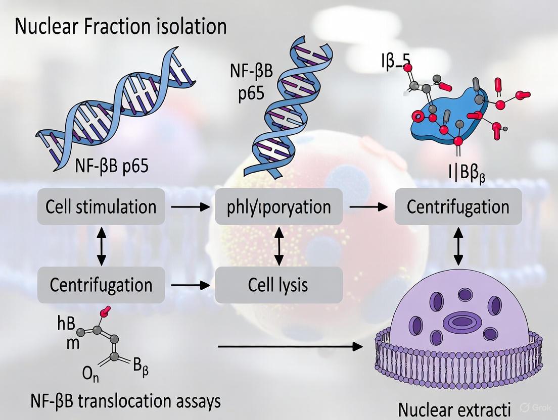
Within the intricate landscape of the eukaryotic cell, the compartmentalization of function between the nucleus and cytoplasm is a fundamental biological principle. The nucleus serves as the command center, housing the genetic material and coordinating vital processes such as DNA replication, RNA synthesis, and gene regulation. The physical separation of the nucleus from the cytoplasm allows for sophisticated control of cellular activities, enabling precise spatial and temporal regulation of signaling events. Isolating nuclear fractions is therefore a critical methodological approach in molecular cell biology, providing a powerful means to study nuclear-specific processes, transcription factor dynamics, and gene regulatory mechanisms free from the obscuring background of the total cellular proteome. This application note details the rationale and protocols for nuclear fractionation, with a specific focus on its indispensable role in investigating the activation dynamics of the Nuclear Factor Kappa B (NF-κB) transcription factor, a key regulator of immune and inflammatory responses [13] [7].
The NF-κB pathway exemplifies why nuclear fractionation is a cornerstone of cellular research. In unstimulated cells, NF-κB is sequestered in the cytoplasm in an inactive complex bound to inhibitory proteins known as IκBs [1]. Upon receiving an activating signal—such as from pro-inflammatory cytokines like TNFα or bacterial components like LPS—a conserved signaling cascade is triggered. This leads to the phosphorylation and degradation of IκB, unmasking NF-κB's nuclear localization signal (NLS) and allowing its rapid translocation into the nucleus [3] [1]. Once in the nucleus, NF-κB binds to specific κB DNA sequences and initiates the transcription of target genes involved in inflammation, cell survival, and proliferation [13]. Consequently, the translocation of NF-κB from the cytoplasm to the nucleus is a critical, defining step in its activation, and isolating a pure nuclear fraction is essential for accurately measuring this event and understanding the resulting gene expression programs.
The Biological Imperative for Nuclear Isolation
The Nucleus as a Distinct Cellular Compartment
The nucleus is delineated from the cytoplasm by a double-membrane nuclear envelope, which is perforated by nuclear pore complexes that regulate the exchange of macromolecules. This structural segregation means that the nuclear compartment possesses a unique protein and nucleic acid composition distinct from the cytosol or other organelles. Key nuclear components include:
- Chromatin: The complex of DNA and histone proteins.
- Nucleoli: Sites of ribosomal RNA synthesis and assembly.
- The Nuclear Matrix: A proteinaceous scaffold that provides structural organization.
- Transcription Factors and Regulatory Complexes: Proteins that control gene expression.
Analyzing these components in isolation allows researchers to obtain a clear picture of nuclear events without interference from the abundant cytoplasmic proteins, which can constitute over 50% of the total cellular protein content. This is particularly crucial for studying processes like gene activation, where a transcription factor's presence and binding activity within the nucleus is the functionally relevant metric, not its total cellular abundance [7].
Rationale in the Context of NF-κB Signaling
For the NF-κB pathway, the rationale for nuclear fractionation is threefold:
- Defining Activation Status: The mere presence of NF-κB in the nucleus is a direct indicator of pathway activation. Biochemical confirmation of this translocation is a gold-standard endpoint in signaling studies [13] [3].
- Quantifying Kinetics and Magnitude: Fractionation enables researchers to track the time course of NF-κB nuclear accumulation and the subsequent re-accumulation of its inhibitor, IκBα, which eventually leads to NF-κB's export back to the cytoplasm [3]. This dynamic process is obscured in whole-cell lysates.
- Downstream Analysis: Isolated nuclear fractions can be used for a wide array of downstream applications to understand the functional consequences of NF-κB translocation, including measuring its binding to DNA target sequences (e.g., via Electrophoretic Mobility Shift Assay or ELISA) and analyzing its role in regulating histone modifications and chromatin structure (e.g., via Chromatin Immunoprecipitation or ChIP) [13].
Quantitative Comparison of Nuclear Fractionation Methods
The choice of nuclear isolation method depends heavily on experimental goals, requiring a balance between speed, purity, and compatibility with downstream applications. The table below summarizes key quantitative and qualitative attributes of several common methods.
Table 1: Comparison of Nuclear Fractionation Method Characteristics
| Method | Processing Time | Key Principle | Purity vs. Yield | Key Advantages | Primary Limitations | Best Suited For |
|---|---|---|---|---|---|---|
| REAP Protocol [16] | ~2 minutes | Rapid lysis with 0.1% NP-40 detergent; brief centrifugation. | High purity, minimal cross-contamination shown in validation. | Extreme speed; minimal protein degradation/deactivation; uses basic lab equipment. | Not optimized for tissue samples; may not preserve nuclear structure perfectly. | Tracking rapid, dynamic protein translocation (e.g., NF-κB). |
| Differential Centrifugation [25] | ~1 hour | Series of centrifugation steps at increasing speeds in hypotonic buffer. | Good purity, but risk of cytoplasmic contamination if not optimized. | Well-established; robust for many cell types; good yield. | Time-consuming; requires careful optimization; hyperosmotic buffers can cause leakage. | General studies of nuclear proteins and transcription factors. |
| Sucrose Density Gradient [16] | ~20+ minutes | Centrifugation through a dense sucrose cushion to purify nuclei. | Very high purity, as contaminants are left in less dense layers. | Excellent separation purity; isolates intact nuclei. | Lengthy protocol; specialized reagents; low yield; non-physiological osmotic stress. | Applications demanding the highest nuclear purity. |
| Commercial Kits | Varies by kit | Pre-optimized buffers and spin columns. | Typically high purity and consistency. | Convenience and reliability; minimal optimization needed. | High cost; proprietary buffer compositions. | Standardized assays and high-throughput workflows. |
Detailed Experimental Protocols
Protocol 1: The REAP Method for Rapid NF-κB Translocation Assay
The REAP (Rapid, Efficient, And Practical) method is ideal for capturing rapid changes in subcellular localization, such as the TNFα-induced translocation of NF-κB, with minimal protein degradation or complex disruption [16].
Materials:
- Ice-cold Phosphate Buffered Saline (PBS)
- Lysis Buffer: 0.1% NP-40 in ice-cold PBS
- 4x Laemmli Sample Buffer
- Table-top microcentrifuge
- Micropipette with p1000 tips
Procedure:
- Cell Culture and Stimulation: Grow adherent cells (e.g., HeLa, HCT116, MEFs) in 10 cm culture dishes. Stimulate with an appropriate NF-κB activator, such as 1 ng/mL TNFα, for the desired time (e.g., 15 minutes) [16].
- Harvesting: Place dishes on ice. Aspirate media and wash cells twice with ice-cold PBS. Scrape cells into 1 mL of ice-cold PBS and transfer to a 1.5 mL microcentrifuge tube.
- Centrifugation: Pellet cells with a quick "pop-spin" (10 seconds) in a microcentrifuge. Remove the supernatant completely.
- Lysis: Resuspend the cell pellet in 900 μL of ice-cold 0.1% NP-40/PBS lysis buffer. Triturate exactly 5 times using a p1000 micropipette to lyse the cell membranes without disrupting nuclei.
- Whole-Cell Lysate (Optional): Immediately remove 300 μL of the lysate to a new tube as the "whole cell lysate" fraction. Add 100 μL of 4x Laemmli buffer, and keep it on ice.
- Fraction Separation: Centrifuge the remaining 600 μL of lysate for 10 seconds.
- Cytosolic Fraction: Transfer 300 μL of the supernatant to a new tube. This is the "cytosolic fraction." Add 100 μL of 4x Laemmli buffer and boil for 1 minute.
- Nuclear Wash and Fraction: Carefully remove and discard the remaining supernatant. Wash the pellet (containing nuclei) by resuspending it in 1 mL of ice-cold 0.1% NP-40/PBS and centrifuging for 10 seconds. Discard the supernatant.
- Nuclear Fraction Solubilization: Resuspend the final nuclear pellet in 180 μL of 1x Laemmli sample buffer. Sonicate briefly (e.g., 2 pulses of 5 seconds at low power) to shear genomic DNA. Boil for 1 minute.
- Analysis: Analyze all fractions by SDS-PAGE and Western blotting.
Validation:
- Western Blotting: Probe blots with antibodies against:
- Nuclear Markers: Lamin A/C or Nucleoporin to confirm purity of the nuclear fraction (should be absent from cytosol).
- Cytoplasmic Markers: α-Tubulin or Pyruvate Kinase to confirm purity of the cytoplasmic fraction (should be absent from nucleus).
- Protein of Interest: NF-κB p65. Upon TNFα stimulation, a clear increase in p65 should be visible in the nuclear fraction with a corresponding decrease in the cytoplasmic fraction [16].
Protocol 2: Conventional Centrifugation-Based Nuclear Extraction
This method, adapted from Abcam's protocol, provides a robust approach for nuclear protein isolation and is compatible with a wider range of downstream applications, including proteomics and enzyme activity assays [25].
Materials:
- Fractionation Buffer: 20 mM HEPES (pH 7.4), 10 mM KCl, 2 mM MgCl₂, 1 mM EDTA, 1 mM EGTA, 1 mM DTT, and Protease Inhibitor Cocktail (added fresh) [25].
- Ice-cold PBS
- 1 mL syringe with 27-gauge needle
- Refrigerated centrifuge
Procedure:
- Harvesting: Wash and scrape cells in ice-cold PBS. Pellet cells by centrifugation at 500 g for 5 minutes at 4°C.
- Initial Lysis: Resuspend the cell pellet in 500 μL of Fractionation Buffer and incubate on ice for 15 minutes.
- Mechanical Lysis: Using a 1 mL syringe fitted with a 27-gauge needle, pass the cell suspension through the needle 10 times (or until >90% of cells are lysed, as verified by trypan blue staining).
- Incubation: Incubate the lysate on ice for another 20 minutes.
- Nuclear Pellet Isolation: Centrifuge the lysate at 720 g for 5 minutes at 4°C.
- The resulting supernatant (S1) contains the cytoplasmic, membrane, and mitochondrial fractions.
- The pellet (P1) contains the crude nuclear fraction.
- Nuclear Wash: Resuspend the nuclear pellet in 500 μL of Fractionation Buffer. Disperse the pellet by pipetting and pass it through a 25-gauge needle 10 times. Centrifuge again at 720 g for 10 minutes at 4°C. Discard the supernatant.
- Nuclear Protein Extraction: Resuspend the final purified nuclear pellet in an appropriate buffer for downstream analysis (e.g., TBS with 0.1% SDS or a high-salt nuclear extraction buffer). Briefly sonicate the suspension on ice to shear genomic DNA and homogenize the lysate.
- Clarification: Centrifuge at high speed (e.g., >14,000 g) for 10 minutes to remove insoluble debris. The supernatant is the soluble nuclear extract.
Visualization of Workflows and Signaling Pathways
NF-κB Activation and Nuclear Translocation Pathway
The following diagram illustrates the canonical NF-κB signaling pathway, from receptor activation to nuclear translocation and gene activation, highlighting the step where nuclear fractionation is critical for measurement.
Nuclear Fractionation Experimental Workflow
This diagram outlines the general decision-making and procedural workflow for isolating nuclear fractions, from cell preparation to downstream analysis.
Successful nuclear fractionation and subsequent analysis require a set of key reagents. The following table details essential components for a typical workflow centered on NF-κB research.
Table 2: Key Research Reagent Solutions for NF-κB Nuclear Translocation Assays
| Reagent Category | Specific Examples | Function & Rationale |
|---|---|---|
| Inhibitors | Protease Inhibitor Cocktails, Phosphatase Inhibitors | Preserve protein integrity, phosphorylation status, and protein-complex interactions during the fractionation process [25]. |
| Lysis Buffers | Hypotonic Buffers, NP-40 (0.1-0.5%), Triton X-100 | Selectively disrupt the plasma membrane while keeping nuclear integrity intact. Concentration is critical to avoid nuclear lysis [25] [16]. |
| Centrifugation Aids | Sucrose Cushions (for density gradients) | Provide a dense medium through which only intact nuclei can pellet, improving purity by separating nuclei from lighter cellular debris [16]. |
| Antibodies for Validation | Anti-Lamin A/C, Anti-Nucleoporin, Anti-Histone H3 | Specific markers for the nuclear fraction. Their absence in the cytoplasmic fraction indicates purity [16]. |
| Anti-α-Tubulin, Anti-GAPDH, Anti-Pyruvate Kinase | Specific markers for the cytoplasmic fraction. Their absence in the nuclear fraction indicates purity [16]. | |
| Antibodies for Analysis | Anti-NF-κB p65 (RelA), Anti-p50, Anti-phospho-NF-κB | Detect the protein of interest (NF-κB) and its activated, post-translationally modified forms in the different fractions [13] [7] [16]. |
| Detection Reagents | HRP-conjugated Secondary Antibodies, ECL Substrate | Enable visualization and quantification of target proteins via Western blotting after SDS-PAGE separation of fractions. |
The isolation of nuclear fractions remains a foundational technique for dissecting the spatial regulation of cellular signaling. In the context of NF-κB research, it provides an unambiguous biochemical measure of transcription factor activation that is directly linked to functional outcomes in gene regulation. The protocols and comparisons detailed in this application note—from the ultra-rapid REAP method to more rigorous centrifugation-based approaches—provide researchers with a clear framework for selecting and implementing the optimal strategy. By enabling precise analysis of proteins within their relevant cellular compartments, nuclear fractionation continues to be an indispensable tool for advancing our understanding of gene expression, immune signaling, and the development of novel therapeutic interventions.
Practical Protocols: From Traditional Fractionation to Advanced Imaging Techniques
Nuclear protein extraction is a foundational technique in molecular biology, essential for studying gene regulation and intracellular signaling. Within the context of nuclear factor kappa B (NF-κB) translocation research, the isolation of pure nuclear fractions is particularly critical. NF-κB, a key mediator of inflammatory and immune responses, resides in the cytoplasm in an inactive state until cellular activation prompts its translocation to the nucleus, where it binds DNA and regulates target gene expression [3] [26]. Accurate assessment of this translocation event requires high-quality nuclear extracts free from cytoplasmic contamination, making the extraction methodology paramount for reliable data in drug discovery and basic research.
This protocol outlines a standardized centrifugation-based approach for nuclear extraction, optimized for subsequent NF-κB translocation assays such as electrophoretic mobility shift assays (EMSAs), enzyme-linked immunosorbent assays (ELISAs), and transcription factor activity kits.
Theoretical Framework: NF-κB Signaling and Nuclear Translocation
The NF-κB signaling pathway is a complex system regulating gene expression in response to diverse stimuli. The core components include five transcription factor proteins (p65/RELA, p50, p52, c-REL, and RELB) that form various homo- and heterodimers, and inhibitor of κB (IκB) proteins that sequester NF-κB in the cytoplasm [26]. The canonical activation pathway, triggered by pro-inflammatory cytokines like TNF-α and IL-1, involves IκB phosphorylation and degradation, exposing nuclear localization signals on NF-κB dimers and enabling their active transport into the nucleus [3] [26].
The following diagram illustrates this key signaling pathway and the conceptual role of nuclear extraction in its analysis:
Materials and Reagent Solutions
The Scientist's Toolkit: Essential Reagents for Nuclear Extraction
The following table catalogues the critical reagents required for successful nuclear fraction isolation, with particular emphasis on ensuring protein stability and fraction purity.
| Reagent/Buffer | Key Components | Primary Function |
|---|---|---|
| Cell Extraction Buffer [27] | 10 mM Tris (pH 7.4), 100 mM NaCl, 1 mM EDTA, 1 mM EGTA, 1% Triton X-100, 10% glycerol, 0.1% SDS | Lyses plasma membrane and solubilizes cytoplasmic proteins. |
| Hypotonic Buffer [27] | 20 mM Tris-HCl (pH 7.4), 10 mM NaCl, 3 mM MgCl₂ | Swells cells, weakening the plasma membrane prior to lysis. |
| Protease Inhibitors [27] | PMSF, proprietary cocktail (e.g., Halt Protease Inhibitor Cocktail) | Prevents proteolytic degradation of proteins during extraction. |
| Phosphatase Inhibitors [27] | NaF, Na₄P₂O₇, Na₃VO₄ | Preserves protein phosphorylation states, crucial for signaling studies. |
| Sucrose Buffer [28] | 0.3 M Sucrose, 10 mM HEPES-NaOH (pH 7.9), 2 mM MgOAc | Provides density for centrifugation and helps maintain organelle integrity. |
| Glycerol Buffer [28] | 25% Glycerol, 10 mM HEPES-NaOH (pH 7.9), 0.1 mM EDTA, 5 mM MgOAc | Stabilizes the isolated nuclear fraction. |
| NP-40 Detergent [27] | Nonyl phenoxypolyethoxylethanol, 10% solution | Non-ionic detergent used for gentle membrane permeabilization. |
Detailed Centrifugation Protocol
This protocol synthesizes steps from established commercial and peer-reviewed methods [27] [28], emphasizing critical centrifugation parameters.
Step 1: Cell Harvesting and Washing
- Culture Processing: Grow and treat cells (e.g., HeLa, HUVECs) as required for the experiment. For NF-κB activation, common stimuli include 20 ng/mL TNF-α for 30 minutes [29] or IL-1α [3].
- Harvesting:
- Adherent cells: Place the culture vessel on ice. Remove and discard the medium. Wash cells gently with ice-cold Phosphate-Buffered Saline (PBS). Scrape the cells into a fresh volume of ice-cold PBS using a cold cell scraper [27] [28].
- Suspension cells: Transfer the cell suspension to a conical tube and collect by centrifugation [27].
- Washing: Transfer the cell suspension to a pre-chilled 15 mL conical tube. Centrifuge at 300 × g for 7 minutes at 4°C. Carefully aspirate the supernatant without disturbing the cell pellet. Resuspend the pellet in 10 mL of ice-cold PBS and repeat the centrifugation step. Aspirate the final PBS supernatant completely [27].
Step 2: Cell Membrane Lysis and Cytoplasmic Extraction
This step separates the cytoplasmic fraction from the intact nuclei.
- Resuspension: Resuspend the cell pellet in a pre-chilled hypotonic buffer (e.g., 500 μL per 5 × 10⁶ cells) [27] or a scrapping buffer containing 0.1% Triton-X100 [28]. Pipette up and down gently to mix.
- Incubation: Incubate the suspension on ice for 15 minutes to allow for cell swelling.
- Detergent Addition: Add a volume of 10% NP-40 detergent (e.g., 25 μL per 500 μL of homogenate) [27]. Vortex the mixture vigorously for 10 seconds.
- Centrifugation for Cytoplasmic Fraction: Centrifuge the homogenate at 3,000 × g for 10 minutes at 4°C [27].
- Supernatant Collection: Immediately after centrifugation, carefully transfer the supernatant to a new, pre-chilled microcentrifuge tube. This is the cytoplasmic fraction. Store it on ice or at –80°C.
Step 3: Nuclear Pellet Washing
The pellet contains the crude nuclear fraction. Washing removes adherent cytoplasmic material.
- Wash: Resuspend the nuclear pellet in 1 mL of a sucrose buffer (0.3 M sucrose, 10 mM HEPES, 1% Triton-X100, 2 mM MgOAc, 1 mM PMSF) [28].
- Centrifugation for Washing: Centrifuge the suspension at 3,200 × g for 10 minutes at 4°C [28].
- Repeat: Aspirate the supernatant. For higher purity, this wash step can be repeated [28].
Step 4: Nuclear Protein Solubilization
- Lysis: Resuspend the washed nuclear pellet in a volume of complete cell extraction buffer containing protease and phosphatase inhibitors (e.g., 50 μL per 5 × 10⁶ cells) [27]. The high-salt and detergent conditions in this buffer disrupt nuclear membranes and solubilize nuclear proteins.
- Incubation: Incubate the suspension on ice for 30 minutes, vortexing briefly at 10-minute intervals to keep the nuclei in suspension and promote efficient lysis [27].
- Clarification: Centrifuge the lysate at high speed (14,000 × g for 30 minutes at 4°C) to pellet insoluble debris such as DNA and nuclear membranes [27].
- Collection: Transfer the clarified supernatant (the nuclear extract) to a new pre-chilled microcentrifuge tube.
The complete workflow, with an emphasis on the key centrifugation steps, is summarized below:
The following table consolidates the critical centrifugation forces, times, and temperatures required for each phase of the protocol, enabling reproducibility and standardization across laboratories.
| Protocol Step | Centrifugation Force | Time | Temperature | Fraction Obtained |
|---|---|---|---|---|
| Cell Harvesting [27] | 300 × g | 7 min | 4°C | Cell pellet |
| Cytoplasmic Fraction Separation [27] | 3,000 × g | 10 min | 4°C | Cytoplasmic supernatant |
| Nuclear Pellet Washing [28] | 3,200 × g | 10 min | 4°C | Washed nuclear pellet |
| Nuclear Extract Clarification [27] | 14,000 × g | 30 min | 4°C | Clarified nuclear extract supernatant |
Method Optimization and Troubleshooting
Comparison of Nuclear Isolation Techniques
A recent study compared the simplified centrifugation method against other common techniques, evaluating them based on yield, purity, and suitability for downstream applications like Co-IP and Ch-IP [28]. The key findings are summarized below:
| Method | Relative Purity | Relative Speed | Key Characteristics | Ideal for... |
|---|---|---|---|---|
| Simplified Centrifugation [28] | High | High | Fewer organelles/cytoplasm; no homogenization | High-purity needs (Ch-IP, Co-IP) |
| Sucrose Centrifugation [28] | High | Medium | Traditional, reliable; requires homogenization | High-purity needs |
| Homogenization [28] | Medium | Very High | Potential for cytoplasmic contamination | Rapid processing |
| Commercial Kits (e.g., NE-PER) [27] [28] | Medium | High | User-friendly; cost factor | Standardized workflows |
Critical Considerations for NF-κB Research
- Inhibitors are Non-Negotiable: Protease and phosphatase inhibitors must be added fresh to all buffers just before use to preserve the integrity and activation states of signaling proteins. PMSF is highly unstable in aqueous solutions and requires particular attention [27].
- Temperature Control: All steps must be performed on ice or at 4°C to slow enzymatic degradation and maintain protein stability.
- Avoiding Freeze-Thaw Cycles: Aliquot clarified extracts and store at –80°C. Repeated freezing and thawing can degrade proteins and compromise assay results [27].
- Handling the Nuclear Pellet: After the initial lysis step, the nuclear pellet is often gelatinous and difficult to resuspend. Careful and patient pipetting is required to avoid loss of material.
- Downstream Compatibility: For ELISA-based transcription factor assays like the NF-κB p65 kit, nuclear extracts typically require dilution by a factor of at least 1:10 in a specific diluent buffer before analysis [27] [29].
Application in NF-κB Translocation Analysis
The nuclear extracts prepared using this protocol are directly compatible with a range of functional assays to quantify NF-κB activation. The Transcription Factor Assay Kit (e.g., ab133112) provides a specific example: the NF-κB p65 in the nuclear extract binds to an immobilized NF-κB response element on a 96-well plate, which is then detected colorimetrically via an antibody specific to the p65 subunit [29]. This method specifically measures the DNA-binding capacity of the translocated transcription factor, providing a direct readout of pathway activation.
Differential Centrifugation and Buffer Optimization for Compartment Separation
The isolation of pure cellular compartments via differential centrifugation is a cornerstone technique in molecular cell biology. Its critical application lies in studying processes like the nuclear translocation of the Nuclear Factor Kappa B (NF-κB) transcription factor, a pivotal event in immune responses, inflammation, and cancer [3]. The reliability of such studies is entirely dependent on the purity of the isolated fractions, which is a function of both the centrifugation protocol and the buffer systems employed. This application note provides a detailed, optimized framework for separating nuclear, cytoplasmic, and membrane fractions, specifically tailored for subsequent analysis of NF-κB translocation. We summarize key centrifugation parameters and buffer formulations in structured tables and provide visual workflows to guide researchers and drug development professionals in achieving highly reproducible and meaningful results.
The Scientific Context: NF-κB Translocation
NF-κB is a transcription factor that typically resides in the cytoplasm in an inactive state, complexed with its inhibitory protein, IκB. Upon cellular activation by stimuli such as pro-inflammatory cytokines (e.g., TNF-α, IL-1), bacterial lipopolysaccharide (LPS), or other stressors, a signaling cascade is triggered [3]. This leads to the phosphorylation and degradation of IκB, which unmasks the nuclear localization sequence (NLS) of NF-κB. The transcription factor subsequently translocates to the nucleus, where it binds DNA and regulates the expression of target genes [3] [7].
Quantifying this translocation from the cytoplasm to the nucleus is a fundamental assay for understanding cellular activation and screening for potential anti-inflammatory or anti-cancer drugs [3]. This quantification can be achieved through various methods, including immunofluorescence microscopy [7] [30] and analysis of fractionated cell lysates by western blotting. The protocol described herein for cellular fractionation is the critical first step for the latter approach.
Experimental Workflow for Subcellular Fractionation
The following diagram and protocol outline the optimized steps for the sequential separation of cytoplasmic, mitochondrial, nuclear, and membrane fractions from cultured mammalian cells using differential centrifugation.
Principle: Cellular compartments are separated based on their size and density through a series of centrifugations at progressively higher forces.
Materials:
- Pre-chilled centrifuges (standard and ultracentrifuge)
- Swinging-bucket rotor (e.g., SW 32 Ti)
- Refrigerated microcentrifuge
- 1 mL syringes and 27-gauge needles
- Ice
- Subcellular Fractionation Buffer (See Section 4.1)
Method:
- Cell Harvest and Lysis: Culture and harvest adherent cells (e.g., HeLa, primary macrophages) from a 10 cm plate by scraping into 500 μL of ice-cold Fractionation Buffer. Incubate the suspension on ice for 15 minutes. Pass the suspension through a 27-gauge needle attached to a 1 mL syringe 10 times to ensure complete cell lysis. Incubate on ice for another 20 minutes [31].
- Nuclear Pellet Isolation: Centrifuge the lysate at 720 x g for 5 minutes at 4°C.
- The resulting pellet (P1) contains the crude nuclear fraction.
- The supernatant (S1) contains the cytosol, mitochondria, and other membrane components. Transfer S1 to a fresh tube and keep on ice.
- Nuclear Fraction Washing: Wash the nuclear pellet (P1) to remove cytoplasmic contamination. Resuspend it in 500 μL of Fractionation Buffer by pipetting and pass it through a 25-gauge needle 10 times. Centrifuge again at 720 x g for 10 minutes at 4°C. Discard the supernatant. The final pellet is the purified nuclear fraction. Resuspend it in an appropriate buffer (e.g., TBS with 0.1% SDS) and briefly sonicate on ice to shear DNA and homogenize the sample [31] [25].
- Mitochondrial Pellet Isolation: Take the supernatant (S1) and centrifuge it at 10,000 x g for 5 minutes at 4°C.
- The resulting pellet (P2) contains the mitochondrial fraction.
- The supernatant (S2) contains the cytosol and microsomal membranes. Transfer S2 to a fresh tube (e.g., ultracentrifuge tube).
- Membrane and Cytosolic Separation: To separate the soluble cytosolic proteins from cellular membranes, centrifuge supernatant (S2) in an ultracentrifuge at 100,000 x g for 1 hour at 4°C.
- The resulting pellet (P3) is the membrane fraction. Wash this pellet by resuspending in fractionation buffer and re-centrifuging at 100,000 x g for 45 minutes to improve purity.
- The final supernatant (S3) is the pure cytosolic fraction [31].
Table 1: Summary of Differential Centrifugation Steps for Organelle Separation.
| Fraction | Centrifugation Force & Time | Cellular Components Isolated | Key Considerations |
|---|---|---|---|
| Nuclei | 720 x g for 5 min | Nuclei, large debris | Wash pellet to reduce cytoplasmic contamination [31]. |
| Mitochondria | 10,000 x g for 5 min | Mitochondria, peroxisomes, lysosomes | Pellet is often a mixed organelle fraction. |
| Membranes | 100,000 x g for 1 h | Plasma membrane, microsomes, ER | Requires an ultracentrifuge; yields pure membrane proteins [31]. |
| Cytosol | Supernatant after 100,000 x g | Soluble cytoplasmic proteins | Can be concentrated using centrifugal filter units [31]. |
Buffer Optimization for Fractionation
The composition of the lysis and fractionation buffer is critical for maintaining organelle integrity, preventing protein degradation, and ensuring minimal cross-contamination between fractions.
Table 2: Composition and Function of a Standard Fractionation Buffer.
| Component | Final Concentration | Molecular Function | Role in Fractionation |
|---|---|---|---|
| HEPES (pH 7.4) | 20 mM | Good buffer capacity | Maintains physiological pH during the procedure. |
| KCl | 10 mM | Salt, osmotic regulator | Provides ionic strength and helps maintain osmotic balance. |
| MgCl₂ | 2 mM | Divalent cation, co-factor | Stabilizes nuclear and membrane structures. |
| EDTA | 1 mM | Chelates Ca²⁺ and Mg²⁺ | Inhibits metalloproteases that degrade proteins. |
| EGTA | 1 mM | Chelates Ca²⁺ specifically | More specific calcium chelation, inhibiting calcium-dependent proteases. |
| DTT (add fresh) | 1 mM | Reducing agent | Prevents oxidation of cysteine residues in proteins. |
| Protease Inhibitors | 1X | Inhibits serine, cysteine proteases | Prevents protein degradation during the fractionation process. |
Phosphate Buffer System for Downstream Applications
For downstream analyses like HPLC that may follow fractionation, a well-prepared phosphate buffer is essential. Its low UV cutoff makes it ideal for spectroscopic detection.
Preparation of 0.2 M Phosphate Buffer (PB), pH 7.4 [32]:
- Stock Solution A (0.2 M Na₂HPO₄): Accurately weigh 28.392 g of Na₂HPO₄ and dissolve in 1 L of water.
- Stock Solution B (0.2 M NaH₂PO₄): Accurately weigh 23.996 g of NaH₂PO₄ and dissolve in 1 L of water.
- Working Buffer: To prepare 100 mL of 0.2 M PB at pH 7.4, combine 81 mL of Stock Solution A with 19 mL of Stock Solution B and mix thoroughly.
- Dilution: This 0.2 M stock can be diluted with water to create commonly used concentrations like 0.1 M, 0.02 M, or 0.01 M PB, pH 7.4 [32].
Optimization Tip: For precise and reproducible pH adjustment when preparing mobile phases, use lower concentration (e.g., 0.1 M) acid/base solutions. This introduces smaller volumetric errors and significantly improves the reproducibility of analyses sensitive to pH fluctuations, such as HPLC [32].
The Scientist's Toolkit: Key Research Reagent Solutions
Table 3: Essential Reagents and Kits for Subcellular Fractionation and Analysis.
| Reagent / Kit Name | Supplier Example | Function / Application | Key Feature |
|---|---|---|---|
| Cell Fractionation Kit - Standard | Abcam (ab109719) | Isolation of mitochondrial, cytoplasmic, and nuclear fractions from mammalian cells. | All-inclusive kit with required reagents for standard fractionation [31]. |
| Nuclear/Cytosol Fractionation Kit | Abcam (ab289882) | Isolation of nuclear and cytosol fractions. | Does not require an ultracentrifuge, simplifying the protocol [31]. |
| Fraction-PREP Cell Fractionation Kit | Abcam (ab288085) | Extraction of four subcellular fractions (cytosol, nucleus, membrane, cytoskeleton). | High reproducibility from a single sample [31]. |
| Protease Inhibitor Cocktail | Various | Added to buffers before use. | Broad-spectrum inhibition of proteases to preserve protein integrity. |
| Phosphatase Inhibitor Cocktail | Various | Added to buffers before use. | Prevents dephosphorylation of proteins, crucial for phospho-protein assays like p-NF-κB. |
| Anti-NF-κB p65 Antibody | Santa Cruz (C-20) | Immunodetection of NF-κB in fractions or by immunofluorescence [7]. | Used for validating translocation via western blot or imaging. |
| Anti-Lamin B1 Antibody | PTM BIO | Western blot loading control for the nuclear fraction [33]. | Confirms nuclear fraction purity. |
| Anti-β-Actin Antibody | Various | Western blot loading control for the cytoplasmic fraction. | Confirms cytoplasmic fraction purity and equal loading. |
Validation and Application in NF-κB Research
Validating Fraction Purity and Assay Success
After fractionation, it is imperative to validate the purity of each fraction using western blot analysis with compartment-specific markers.
- Nuclear Fraction Purity: Probe for Lamin B1 [33] or transcription factors. The absence of cytoplasmic markers like β-Actin confirms a clean separation.
- Cytoplasmic Fraction Purity: Probe for β-Actin. The absence of strong nuclear marker signals indicates minimal nuclear leakage.
- Assay Validation: Successful NF-κB translocation is confirmed by a time-dependent increase in NF-κB p65 signal in the nuclear fraction and a corresponding decrease in the cytoplasmic fraction upon stimulation with an agonist like LPS or TNF-α [30].
Important Considerations for Primary Cells
Research indicates that the basal state of primary cells can differ significantly from immortalized cell lines. For example, native primary macrophages have an elevated pre-activation basal level of nuclear NF-κB (25-35% of total cellular NF-κB) compared to transformed RAW 264.7 cells (5-10%) [30]. This pre-activated state allows for a much faster translocation response to low concentrations of LPS. Therefore, when isolating nuclei from such primary cells, a higher basal level of NF-κB in the nuclear fraction is expected and should not be misinterpreted as assay failure. This underscores the importance of including proper stimulated and unstimulated controls in every experiment.
Mastering differential centrifugation and buffer optimization is a prerequisite for obtaining high-quality subcellular fractions. The protocols and guidelines provided here offer a robust foundation for isolating nuclear, cytoplasmic, and membrane compartments with minimal cross-contamination. When applied within the context of NF-κB research, this methodology enables the accurate detection and quantification of its activation status, a critical parameter in fundamental immunology research and drug discovery pipelines. By adhering to these detailed protocols, researchers can generate reliable, reproducible, and biologically meaningful data on the subcellular localization of NF-κB and other important regulatory proteins.
This application note provides a detailed methodological framework for optimizing digitonin-based permeabilization, a critical step in nuclear fraction isolation for NF-κB translocation assays. We present standardized protocols and quantitative data for achieving selective plasma membrane permeabilization while preserving nuclear envelope integrity across various cell types. The guidelines enable researchers to obtain clean subcellular fractions for accurate assessment of transcription factor translocation, essential for drug development studies of inflammatory signaling and nuclear transport mechanisms.
Selective plasma membrane permeabilization using digitonin provides a critical foundation for nuclear fractionation and the study of nucleocytoplasmic transport mechanisms, including NF-κB translocation. This steroid glycoside detergent exploits the differential cholesterol content between cellular membranes—typically higher in the plasma membrane than in intracellular membranes—to create permeability gradients that can be precisely controlled through concentration and temperature optimization [34] [35]. When properly optimized, digitonin permeabilization enables researchers to access cytoplasmic contents while maintaining nuclear envelope integrity, thereby allowing for the separation of nuclear and cytoplasmic fractions for downstream analysis of transcription factor localization [34].
Within the context of NF-κB research, this technique enables precise tracking of the transcription factor's movement from cytoplasm to nucleus following activation stimuli. The cholesterol-dependent selectivity of digitonin makes it particularly valuable for preparing fractions that accurately reflect subcellular localization without cross-contamination, providing cleaner results than complete lysis methods [35]. This application note establishes evidence-based protocols for digitonin use specifically tailored to nuclear fraction isolation requirements.
Theoretical Foundation: Digitonin Selectivity Mechanism
Digitonin achieves selective membrane permeabilization through specific interactions with membrane cholesterol. The plasma membrane typically contains significantly higher cholesterol concentrations (approximately 30-50% of lipid content) compared to intracellular membranes such as the nuclear envelope (less than 5% cholesterol content) [35] [36]. This cholesterol gradient enables digitonin to form large pores exclusively in the plasma membrane at optimized concentrations, while leaving nuclear membranes intact for subsequent fractionation procedures.
The molecular basis of selectivity:
- Cholesterol binding: Digitonin molecules specifically complex with membrane cholesterol through its steroid moiety
- Pore formation: At critical concentrations, digitonin-cholesterol complexes reorganize into membrane pores approximately 8-12 nm in diameter
- Selective accessibility: These pores allow passage of cytoplasmic components and antibodies while retaining nuclear contents
- Concentration dependence: Higher digitonin concentrations progressively affect intracellular membranes, compromising nuclear integrity
This selective permeability enables researchers to selectively extract cytoplasmic components while maintaining nuclear compartmentalization, providing a fundamental advantage for NF-κB translocation assays that require precise separation of nuclear and cytoplasmic fractions.
Diagram Title: Digitonin Permeabilization Workflow for NF-κB Assays
Quantitative Optimization Parameters
Concentration Ranges by Cell Type
Table 1: Optimal Digitonin Concentration Ranges for Various Cell Types
| Cell Line | Origin | Recommended Digitonin Concentration | Incubation Conditions | Primary Assessment Method |
|---|---|---|---|---|
| HeLa S3 | Human cervical carcinoma | 25-50 µg/mL | 3-5 min, RT | Lamin B immunostaining [35] |
| LLC-PK1 | Porcine kidney epithelium | 40-60 µg/mL | 5-7 min, RT | GFP-β-galactosidase exclusion [35] |
| NIH/3T3 | Mouse fibroblast | 30-55 µg/mL | 3-5 min, RT | Nuclear import assays [34] [35] |
| Primary hepatocytes | Various species | 35-70 µg/mL | 5-10 min, 4°C | Mitochondrial function assays [37] |
Temperature and Temporal Parameters
Table 2: Temperature and Time Optimization for Nuclear Fractionation
| Temperature Condition | Optimal Incubation Time | Permeabilization Efficiency | Nuclear Envelope Integrity | Recommended Applications |
|---|---|---|---|---|
| 4°C | 8-12 minutes | Gradual, controlled | Excellent preservation | Primary cells, delicate nuclei [35] |
| Room Temperature (22-25°C) | 3-7 minutes | Rapid, uniform | Good preservation | Cell lines, high-throughput [34] |
| 37°C | 1-3 minutes | Very rapid | Risk of compromise | Not recommended for nuclear isolation |
Experimental Protocols
Protocol 1: Basic Nuclear Fraction Isolation for NF-κB Assays
Purpose: To isolate clean nuclear and cytoplasmic fractions for NF-κB translocation analysis from cultured mammalian cells.
Reagents and Solutions:
- Digitonin stock solution: 50 mg/mL in DMSO (store at -20°C)
- Buffer B: 20 mM HEPES (pH 7.3), 110 mM potassium acetate, 5 mM sodium acetate, 2 mM magnesium acetate, 1 mM EGTA [35]
- Protease inhibitor cocktail (add fresh: 2 mM DTT, 1 μg/mL each of aprotinin, leupeptin, pepstatin) [35]
- Phosphate-buffered saline (PBS), pH 7.4
- Formaldehyde, 4% solution in PBS
- Triton X-100, 0.1% solution in PBS/BSA
Procedure:
- Cell Culture and Stimulation: Grow cells to 70-80% confluence on poly-lysine coated plates or coverslips. Apply NF-κB activation stimulus (e.g., TNF-α) according to experimental design.
- Prewash and Preparation: Wash cells twice with ice-cold PBS. Gently aspirate completely.
- Digitonin Working Solution: Prepare digitonin working solution in Buffer B at the predetermined optimal concentration for your cell type (refer to Table 1).
- Selective Permeabilization: Add digitonin working solution to cover cells (approximately 100 μL per 1×10^6 cells). Incubate at optimized temperature and time (refer to Table 2).
- Cytoplasmic Fraction Collection: Carefully collect the supernatant—this represents the cytoplasmic fraction. Centrifuge at 800 × g for 5 minutes to remove any intact cells or debris.
- Nuclear Fraction Isolation: Wash the permeabilized cells once with Buffer B. Lyse nuclei with high-salt RIPA buffer (containing 0.5M NaCl and 0.5% NP-40) for 15 minutes on ice with occasional vortexing.
- Clarification: Centrifuge nuclear lysate at 12,000 × g for 15 minutes at 4°C. Collect supernatant as nuclear fraction.
- Quality Assessment: Verify fraction purity by immunoblotting for compartment-specific markers (e.g., GAPDH for cytoplasm, Lamin B for nucleus).
Critical Notes:
- Always include a no-stimulus control and a total lysate control for proper normalization
- Perform digitonin titration for each new cell type or growth condition
- Avoid excessive incubation times which can compromise nuclear integrity
Protocol 2: Validation of Permeabilization Efficiency and Nuclear Integrity
Purpose: To confirm successful plasma membrane permeabilization while maintaining nuclear envelope integrity.
Reagents:
- Primary antibodies: anti-Lamin B (nuclear envelope marker), anti-tubulin (cytoplasmic marker)
- Fluorescently-labeled secondary antibodies
- DAPI solution (1 μg/mL)
- Blocking solution: PBS with 2 mg/mL BSA and 1 mM NaN₃ [35]
- Formaldehyde, 4% solution in PBS
Validation Procedure:
- Fixation and Permeabilization: Following digitonin treatment, immediately fix cells with 4% formaldehyde for 25 minutes at room temperature.
- Immunostaining:
- Block fixed cells with blocking solution for 1 hour
- Incubate with primary antibodies diluted in blocking solution overnight at 4°C
- Wash 3×10 minutes with blocking solution
- Incubate with fluorescent secondary antibodies for 2 hours at room temperature
- Nuclear Counterstaining: Incubate with DAPI solution for 2 minutes to visualize nuclei.
- Microscopy and Analysis:
- Image using confocal microscopy with 63× objective
- Assess cytoplasmic antibody access while verifying nuclear envelope preservation through continuous Lamin B staining
- Alternative Validation: For functional assessment of nuclear integrity, utilize GFP-β-galactosidase exclusion assay as described [35]
Interpretation Guidelines:
- Successful permeabilization: Cytoplasmic markers accessible to antibodies, nuclear interior remains inaccessible to large proteins
- Optimal conditions: Continuous nuclear envelope staining, no DAPI leakage into cytoplasm
- Over-permeabilization: Discontinuous or absent nuclear envelope staining, diffusion of nuclear content
The Scientist's Toolkit: Essential Research Reagents
Table 3: Key Research Reagent Solutions for Digitonin Permeabilization
| Reagent | Function/Application | Recommended Concentrations | Special Considerations |
|---|---|---|---|
| Digitonin (Sigma) | Selective plasma membrane permeabilization | 25-100 μg/mL in respiration/buffer B | Prepare fresh in DMSO; cholesterol content affects efficacy [35] [37] |
| Buffer B (Transport Buffer) | Physiological ionic environment for permeabilization | Standard formulation: See Section 4.1 | Maintain at pH 7.3; add fresh DTT and protease inhibitors [34] [35] |
| Protease Inhibitor Cocktail | Prevents protein degradation during fractionation | 1 μg/mL each of aprotinin, leupeptin, pepstatin | Add immediately before use; avoid freeze-thaw cycles [35] |
| Lamin B Antibodies | Nuclear envelope integrity marker | 0.5 μg/mL for immunofluorescence | Validated for nuclear envelope-specific staining [35] |
| GFP-β-galactosidase | Nuclear integrity reporter | Expression vector transfection | Large size (≈470 kDa) excludes intact nuclei [35] |
| Triton X-100 | Complete membrane permeabilization (control) | 0.1% in PBS/BSA | Use as positive control for antibody access [35] [38] |
Troubleshooting and Technical Considerations
Common Optimization Challenges
Incomplete cytoplasmic extraction:
- Cause: Suboptimal digitonin concentration or incubation time
- Solution: Perform digitonin titration (20-100 μg/mL) with incremental time points
- Validation: Monitor cytoplasmic marker release (e.g., LDH assay or GAPDH western blot)
Nuclear envelope compromise:
- Cause: Excessive digitonin concentration, prolonged incubation, or inappropriate temperature
- Solution: Reduce concentration by 25-40% and implement colder temperatures (4°C)
- Validation: Assess nuclear retention of transcription factors in unstimulated controls
Cell-type specific variability:
- Cause: Differential cholesterol content in plasma membranes
- Solution: Always perform initial optimization for new cell types using Table 1 as starting point
- Validation: Combine morphological assessment with biochemical fractionation quality controls
Advanced Applications: Integration with Proximity Labeling
Recent advancements combine digitonin permeabilization with TurboID-based proximity labeling to map organelle membrane protein interactomes [39]. This innovative approach leverages digitonin's selective permeabilization to remove cytosolic proteins before proximity labeling, thereby enhancing spatial resolution in proteomic analysis. For NF-κB research, this methodology could be adapted to study the dynamic protein interactions at the nuclear envelope during translocation events.
Diagram Title: NF-κB Signaling and Detection Workflow
Optimized digitonin permeabilization represents a powerful technique for nuclear fraction isolation in NF-κB translocation studies. The concentration and temperature parameters provided in this application note serve as a robust foundation for researchers to establish reproducible subcellular fractionation protocols. By adhering to the cell-type specific recommendations and validation methods outlined herein, scientists can achieve highly pure nuclear and cytoplasmic fractions with preserved protein interactions and transcriptional competence. The continued refinement of these permeabilization strategies will further enhance our understanding of nuclear transport mechanisms and their implications in disease and drug development.
Nuclear fractionation is a critical preparatory step for investigating transcription factor dynamics, such as NF-κB translocation, which serves as an early marker of immune cell activation [40]. However, the integrity of isolated nuclear fractions is often compromised by the leakage of small proteins and nucleoplasmic components, leading to experimental inaccuracies. This application note details a optimized protocol utilizing Wheat Germ Agglutinin (WGA), a lectin that binds nuclear pore complexes (NPCs), to effectively seal nucleoporins and prevent passive diffusion of sub-100 kDa proteins. We provide a standardized methodology, validation data, and practical tools to enhance the reliability of nuclear translocation assays for research and drug development applications.
The nuclear envelope forms a selective barrier regulated by nuclear pore complexes (NPCs), which permit the regulated transport of macromolecules. During nuclear fractionation, the isolation process can render these pores susceptible to passive diffusion, resulting in the leakage of critical small proteins and confounding the analysis of transcription factor localization. NF-κB is a key transcription factor involved in immune responses; its nuclear translocation is a pivotal event studied in immune activation [40]. Accurate measurement of this translocation via imaging flow cytometry or other techniques requires high-purity nuclear fractions [40].
Wheat Germ Agglutinin (WGA) binds specifically to N-acetylglucosamine residues on nucleoporin proteins (Nups) within the NPC. This binding provides a steric and chemical seal that significantly reduces passive leakage without inhibiting active, importin-mediated transport. This protocol outlines the application of WGA to stabilize nuclear fractions post-isolation, ensuring the reliable retention of proteins like the NF-κB p65 subunit for subsequent analysis.
The following tables summarize quantitative data from experiments validating the efficacy of WGA treatment in nuclear fractionation protocols.
Table 1: Efficacy of WGA in Preventing Leakage of Marker Proteins from Isolated Nuclei
| Cellular Fraction | Treatment | Lamin B1 (Nuclear Envelope) Recovery (%) | Histone H3 (Nucleoplasm) Recovery (%) | LDH (Cytosol) Contamination (%) |
|---|---|---|---|---|
| Nuclear Fraction | Untreated | 100 ± 5 | 62 ± 8 | 4.5 ± 1.2 |
| Nuclear Fraction | + WGA (50 µg/mL) | 98 ± 4 | 95 ± 3 | 1.8 ± 0.7 |
| Cytosolic Fraction | Untreated | 2 ± 1 | 35 ± 6 | 100 ± 5 |
| Cytosolic Fraction | + WGA (50 µg/mL) | 1 ± 1 | 4 ± 2 | 99 ± 4 |
Table 2: Impact of WGA Treatment on NF-κB p65 Translocation Assay Readout
| Experimental Condition | NF-κB Nuclear Localization (Fold Increase over Control) | Inter-Assay CV (%) | Signal-to-Noise Ratio |
|---|---|---|---|
| Untreated Nuclei | 4.5 ± 0.9 | 22.5 | 8.1 |
| WGA-Treated Nuclei | 7.2 ± 0.6 | 9.8 | 15.4 |
Experimental Protocol
Reagent Preparation
- WGA Stock Solution (5 mg/mL): Dissolve 5 mg of WGA (Vector Laboratories, #L-1020) in 1 mL of sterile, nuclease-free PBS. Gently vortex until fully dissolved. Aliquot and store at -20°C.
- Cell Lysis Buffer: 10 mM HEPES (pH 7.9), 1.5 mM MgCl₂, 10 mM KCl, 0.5 mM DTT, 0.1% IGEPAL CA-630, and 1x Halt Protease Inhibitor Cocktail [41].
- Nuclear Resuspension Buffer: 20 mM HEPES (pH 7.9), 1.5 mM MgCl₂, 0.42 M NaCl, 0.2 mM EDTA, 25% (v/v) Glycerol, 0.5 mM DTT, and 1x Halt Protease Inhibitor Cocktail [41].
Step-by-Step WGA Treatment Workflow
Procedure:
- Cell Harvesting: Grow and treat cells (e.g., NK cells, HeLa) as required. Collect cells by gentle scraping or trypsinization and pellet by centrifugation (300 g for 5 min).
- Wash: Wash the cell pellet twice with ice-cold PBS.
- Hypotonic Lysis: Resuspend the cell pellet in 5 volumes of pre-chilled Cell Lysis Buffer. Vortex briefly and incubate on ice for 15 minutes.
- Nuclear Pellet Formation: Centrifuge the lysate at 1,500 g for 5 minutes at 4°C.
- Cytosolic Fraction Collection: Transfer the supernatant to a fresh tube. This is the cytosolic fraction.
- Wash Nuclei: Gently resuspend the pellet (crude nuclei) in 1 mL of Cell Lysis Buffer (without IGEPAL) and centrifuge again at 1,500 g for 5 minutes. Discard the supernatant.
- WGA Treatment: Resuspend the purified nuclear pellet in Nuclear Resuspension Buffer.
- Add WGA Stock Solution to a final concentration of 50 µg/mL.
- For the control sample, add an equal volume of PBS.
- Incubation: Incubate the nuclei on ice for 30 minutes with gentle inversion every 10 minutes.
- Post-Treatment Centrifugation: Pellet the WGA-stabilized nuclei by centrifugation at 5,000 g for 10 minutes at 4°C. The resulting nuclear pellet is now stabilized and ready for downstream protein extraction or imaging.
Validation via Immunoblotting
Validate fraction purity and WGA efficacy by immunoblotting:
- Nuclear Markers: Lamin B1 or Histone H3 should be enriched in the nuclear fraction and depleted in the cytosolic fraction.
- Cytosolic Marker: LDH or GAPDH should be enriched in the cytosolic fraction and depleted in the nuclear fraction.
- Compare the intensity of nuclear markers in the cytosolic fraction of treated vs. untreated samples to visualize the reduction in leakage.
The Scientist's Toolkit
Table 3: Essential Research Reagent Solutions
| Item | Function / Rationale | Example Source / Catalog # |
|---|---|---|
| Wheat Germ Agglutinin (WGA) | Binds NPCs to seal nucleoporins and prevent passive leakage of sub-100 kDa proteins. | Vector Labs, #L-1020 |
| Protease Inhibitor Cocktail | Prevents proteolytic degradation of proteins during fractionation. | Thermo Scientific, #87785 [41] |
| HEPES Buffer | Maintains stable physiological pH during the isolation process. | Various suppliers |
| IGEPAL CA-630 Detergent | Non-ionic detergent for cell membrane lysis while keeping nuclear membranes intact. | Various suppliers |
| Anti-NF-kB p65 Antibody | For detection and quantification of the NF-κB subunit in translocation assays. | Various suppliers |
| Anti-Lamin B1 Antibody | Marker for the nuclear envelope; validates nuclear fraction integrity. | Various suppliers |
| Anti-Histone H3 Antibody | Marker for the nucleoplasm; assesses leakage of internal nuclear components. | Various suppliers |
| Anti-GAPDH Antibody | Common cytosolic contamination marker; assesses fraction purity. | Various suppliers |
Mechanism of Action
The following diagram illustrates how WGA functions at the molecular level to prevent small protein leakage from isolated nuclei.
Integrating WGA treatment into nuclear fractionation protocols provides a simple, robust, and highly effective method for safeguarding nuclear integrity. By mitigating the leakage of nucleoplasmic proteins, this technique significantly enhances the accuracy, sensitivity, and reproducibility of downstream assays, including the measurement of NF-κB nuclear translocation—a critical parameter in immunology, oncology, and drug discovery research [40]. This protocol ensures that observed changes in nuclear protein localization genuinely reflect cellular activation states rather than experimental artifact.
Within the broader research on nuclear fraction isolation for studying NF-κB translocation, image-based methods provide a powerful alternative by enabling quantitative, single-cell analysis of this critical cellular event. Nuclear Factor kappa-B (NF-κB) is a key transcription factor regulating the innate immune inflammatory response in activated macrophages and other immune cells [13]. Its activation through translocation from the cytoplasm to the nucleus constitutes a hallmark of cellular stimulation by pathogens, cytokines, and other stressors [3]. While traditional biochemical methods like nuclear fractionation and western blotting provide population-average data, high-content imaging captures heterogeneity within cell populations and offers spatial resolution at the single-cell level [7] [8]. This application note details two principal high-content imaging approaches for quantifying NF-κB nuclear translocation: using GFP-tagged RelA fusion proteins and immunofluorescence detection of endogenous proteins, providing researchers with robust methodologies to advance their investigations into NF-κB signaling dynamics.
Technical Comparison of Methodologies
The selection between GFP-tagged reporters and immunofluorescence detection depends on specific research requirements, including need for live-cell analysis, quantification precision, and model system relevance. The table below summarizes the key characteristics of each approach.
Table 1: Comparison of GFP-Tagged RelA and Immunofluorescence Methods for NF-κB Translocation Assays
| Feature | GFP-Tagged RelA | Immunofluorescence Detection |
|---|---|---|
| Cell Preparation | Live or fixed cells | Fixed cells only |
| Sample Throughput | High (amenable to 96-well and 384-well formats) [13] | Moderate to high |
| Temporal Resolution | Excellent (kinetics in live cells) [13] | Limited (single time point per sample) |
| Experimental Model | Requires engineered cell lines [13] | Compatible with primary cells and unmodified cell lines [7] |
| Key Reagents | GFP-RelA expressing cell line (e.g., RAW264.7 G9) [13] | Primary anti-RelA antibody, fluorescent secondary antibody [7] |
| Quantification Methods | Nuclear-to-cytoplasmic ratio, difference metrics [3] | Similarity score, nuclear-to-cytoplasmic ratio [8] |
| Key Advantages | Enables live-cell dynamic studies; no antibody required | Applicable to primary cells and tissues; endogenous protein detection |
| Main Limitations | Potential overexpression artifacts; requires genetic modification | Fixed endpoint only; antibody validation critical |
Quantitative Analysis of NF-κB Translocation
Accurate quantification of NF-κB translocation requires specific image analysis algorithms that measure the redistribution of the protein from cytoplasm to nucleus. The fundamental principle involves identifying cellular compartments and calculating intensity-based metrics.
Table 2: Key Quantitative Parameters for NF-κB Nuclear Translocation Analysis
| Parameter | Calculation | Biological Interpretation | Typical Values with Stimulation |
|---|---|---|---|
| Nuclear-to-Cytoplasmic (N/C) Ratio | Mean nuclear intensity / Mean cytoplasmic intensity | Measures enrichment of NF-κB in nucleus | Increases from ~0.5 to >2.0 [3] |
| Cyto-Nuc Difference | Mean cytoplasmic intensity - Mean nuclear intensity | Measures depletion from cytoplasm | Decreases with activation [3] |
| Similarity Score | Pearson's correlation between NF-κB and nuclear stain images [8] | Quantifies colocalization with nucleus | Increases from low values (~1-2) to higher values (>5) [8] |
| Translocation Index | (Nuclear - Cytoplasmic) / (Nuclear + Cytoplasmic) | Normalized measure of distribution | Ranges from -1 (cytoplasmic) to +1 (nuclear) |
| % Cells with Nuclear Translocation | (Cells with N/C ratio > threshold / Total cells) × 100 | Population response heterogeneity | Can range from partial (30-70%) to full (>80%) response [7] |
Experimental Protocols
Protocol for GFP-Tagged RelA Translocation Assay
This protocol utilizes RAW264.7 macrophage cells stably expressing a RelA-GFP fusion protein (RAW G9 cells) for quantitative measurement of NF-κB translocation in response to Toll-Like Receptor (TLR) activation [13].
Materials:
- RAW264.7 G9 cells stably expressing RelA-GFP [13]
- Culture medium: DMEM with 10% FCS, 20 mM HEPES, 4 mM L-glutamine, penicillin/streptomycin [13]
- TLR ligands: Ultra-pure LPS (1 mg/ml stock) [13]
- 96-well clear bottom black-walled plates [13]
- 4% Paraformaldehyde in PBS (freshly prepared) [13]
- Nuclear stain: Hoechst 33342 or similar [13]
- High-content imaging system with environmental control
Procedure:
- Cell Culture and Seeding: Maintain RAW264.7 G9 cells in non-treated tissue culture flasks at 37°C with 5% CO₂. Grow to 80-90% confluence. Detach cells using cold 2 mM EDTA in PBS. Pipette vigorously to generate single-cell suspension. Count cells and seed at 10,000 cells per well in 96-well plates in 100 μl culture medium. Incubate overnight [13].
Stimulation: Prepare working solution of LPS at 110 ng/ml in culture medium. Remove culture medium from wells and replace with LPS-containing medium or control medium. Incubate at 37°C for appropriate time points (typically 30-90 minutes) based on kinetic requirements [13].
Fixation and Staining: At experimental endpoint, add equal volume of 4% PFA directly to culture medium to achieve final concentration of 2% PFA. Fix for 15 minutes at room temperature. Alternatively, replace medium with 4% PFA for 15 minutes. Wash twice with PBS. Add nuclear stain (e.g., Hoechst 33342) diluted in PBS according to manufacturer's instructions. Incubate 15-30 minutes at room temperature [13].
Image Acquisition: Acquire images using 20× or 40× objective on high-content imaging system. Capture multiple fields per well (typically 9-25) to ensure adequate cell numbers (≥500 cells per condition). Use appropriate filter sets for GFP (excitation/emission: 488/510 nm) and nuclear stain (excitation/emission: 350/461 nm for Hoechst) [13].
Image Analysis:
- Segment nuclei based on nuclear stain signal.
- Create cytoplasmic mask by expanding nuclear boundary or using whole-cell segmentation.
- Measure mean GFP intensity in nuclear and cytoplasmic regions for each cell.
- Calculate translocation metrics (N/C ratio, Cyto-Nuc difference) for each cell.
- Export data for statistical analysis and visualization.
Protocol for Immunofluorescence Detection of Endogenous NF-κB
This protocol describes immunofluorescence staining and analysis of endogenous NF-κB RelA in primary human macrophages, adaptable to other primary cells and cell lines [7].
Materials:
- Primary cells (e.g., human monocyte-derived macrophages) or cell lines [7]
- Anti-RelA primary antibody (e.g., Santa Cruz Biotechnology, sc-109) [13]
- Fluorescently labeled secondary antibody (e.g., Alexa Fluor 633 goat anti-rabbit) [7]
- Blocking solution: 5% BSA, 0.05% Tween 20 in PBS [13]
- Permeabilization solution: 0.2% Triton X-100 in PBS [7]
- Nuclear stain: DAPI or similar
- Mounting medium
Procedure:
- Cell Culture and Stimulation: Seed cells on appropriate substrate (coverslips, glass-bottom plates). For primary human macrophages, differentiate monocytes for 6 days with M-CSF before stimulation [7]. Stimulate with appropriate NF-κB activator (LPS, TNF-α, etc.) for optimized time course.
Fixation and Permeabilization: Aspirate medium and wash cells with PBS. Fix with 3.7% PFA for 15 minutes at room temperature. Wash twice with PBS. Permeabilize with 0.2% Triton X-100 for 10 minutes at room temperature [7].
Immunostaining: Block with 5% BSA, 0.05% Tween 20 in PBS for 30 minutes at room temperature. Incubate with primary antibody diluted in blocking solution overnight at 4°C. Wash three times with PBS. Incubate with fluorescent secondary antibody diluted in blocking solution for 1 hour at room temperature in the dark. Wash three times with PBS [7].
Nuclear Counterstaining and Mounting: Incubate with DAPI (2 μg/ml) for 5 minutes at room temperature. Wash twice with PBS. If using coverslips, mount on slides with appropriate mounting medium. For microplates, can image directly in PBS or add sealing film to prevent evaporation [7].
Image Acquisition and Analysis: Acquire images using fluorescence microscope or high-content imager. For quantitative analysis using ImageJ [7]:
- Apply median filter (3×3 pixel) to reduce noise.
- Create binary masks using automatic thresholding (Isodata algorithm).
- Use nuclear mask to define nuclear ROI.
- Create cytoplasmic ROI by subtracting nuclear mask from RelA mask.
- Measure mean intensities in each compartment.
- Calculate translocation metrics.
Signaling Pathways and Experimental Workflows
The NF-κB activation pathway involves a coordinated series of molecular events culminating in nuclear translocation. The following diagrams illustrate the signaling pathway and experimental workflows for both detection methods.
Diagram 1: NF-κB Activation Signaling Pathway. External stimuli trigger receptor activation, leading to IKK complex activation, IκB degradation, NF-κB release, nuclear translocation, and target gene transcription [13] [3].
Diagram 2: Experimental Workflow for NF-κB Translocation Assays. The workflow encompasses cell preparation, selection of detection method (GFP-RelA or immunofluorescence), sample processing, image acquisition, and quantitative analysis [13] [7].
The Scientist's Toolkit: Research Reagent Solutions
Successful implementation of NF-κB translocation assays requires specific reagents and tools optimized for each method. The following table details essential materials and their applications.
Table 3: Essential Research Reagents for NF-κB Translocation Assays
| Reagent Category | Specific Examples | Application Notes |
|---|---|---|
| Cell Models | RAW264.7 G9 (GFP-RelA) [13], Primary human macrophages [7], HeLa, HEK293 | GFP-tagged lines enable live-cell imaging; primary cells maintain physiological relevance |
| NF-κB Antibodies | Rabbit anti-RelA (Santa Cruz, sc-109) [13], Anti-acetylated NF-κB (CST #3045) [13] | Validate for immunofluorescence; phospho-specific antibodies assess activation status |
| Secondary Antibodies | Alexa Fluor conjugates (488, 555, 647) [42] | Direct conjugates reduce cross-reactivity; Alexa Fluors offer brightness and photostability |
| Nuclear Stains | Hoechst 33342 [13], DAPI [7], DRAQ5 [8] | Hoechst for live-cell; DAPI for fixed cells; DRAQ5 for far-red channel multiplexing |
| TLR Ligands | Ultra-pure LPS (TLR4) [13], Pam3CSK4 (TLR2) [7] | Use ultrapure preparations to minimize non-specific activation |
| Fixation Reagents | 4% Paraformaldehyde [13] [7] | Freshly prepared PFA preserves morphology and antigenicity |
| Permeabilization Agents | 0.1-0.5% Triton X-100 [7], 0.05% Tween-20 [13] | Concentration optimization needed for different cell types |
| Blocking Solutions | 5% BSA, 10% normal serum [7] | Serum should match secondary antibody host species |
Advanced Applications and Multiplexing Strategies
Advanced imaging applications enable researchers to extract more comprehensive information from NF-κB translocation assays through multiplexing and dynamic analysis.
Multiplexed Detection with CycIF
Cyclic Immunofluorescence (CycIF) is a public domain method for highly multiplexed immunofluorescence imaging that enables measurement of NF-κB translocation alongside other signaling events in the same cells [42]. This method uses sequential rounds of staining, imaging, and fluorophore inactivation to overcome the spectral limitations of conventional microscopy. For NF-κB studies, this enables correlation of nuclear translocation with phosphorylation events, cell cycle markers, and other pathway components. The technique employs directly conjugated antibodies to Alexa Fluor dyes (488, 555, 647) and chemical inactivation between cycles, preserving cell morphology through multiple rounds [42].
ImageStream Platform for Suspension Cells
The ImageStream platform combines flow cytometry with high-resolution imaging, enabling quantitative NF-κB translocation analysis in suspension cells like leukemic cell lines that are challenging for traditional microscopy [8]. This approach uses similarity scoring (a log-transformed Pearson's correlation coefficient) to quantify the degree of colocalization between NF-κB and nuclear stains, providing statistically robust data from thousands of cells while maintaining single-cell resolution [8]. This method correlates well with western blot analysis and enables detection of heterogeneity in cellular responses that would be masked in population-average measurements [8].
Troubleshooting and Optimization Considerations
Successful implementation of NF-κB translocation assays requires attention to potential technical challenges and appropriate optimization.
Low Signal-to-Noise Ratio: For immunofluorescence, consider signal amplification methods such as tyramide signal amplification for low-abundance targets [43]. For GFP-tagged proteins, verify expression levels and consider alternative cell lines if signal is weak.
High Background Staining: Optimize antibody concentrations and include appropriate controls (no primary antibody, isotype controls). Increase blocking time or try different blocking agents (BSA, serum, commercial blocking buffers).
Poor Nuclear Segmentation: Test different nuclear stains and concentrations. Adjust segmentation parameters in analysis software. Consider using cytoplasmic markers for whole-cell segmentation when calculating cytoplasmic intensities.
Variable Response to Stimulation: Use fresh ligand preparations and validate activity. Perform time course experiments to identify optimal stimulation duration. Consider cell density effects on responsiveness.
Multiplexing Challenges: When designing multiplexed panels, verify spectral compatibility and include controls for cross-talk. For sequential staining methods, validate complete fluorophore inactivation between cycles [42].
These high-content imaging methods for quantifying NF-κB translocation provide powerful alternatives to traditional biochemical approaches, offering single-cell resolution, spatial context, and the ability to capture cellular heterogeneity in response to pathogenic stimuli and therapeutic interventions.
Nuclear Factor kappa B (NF-κB) represents a family of transcription factors present in all eukaryotic cells that regulate inducible expression of wide-ranging genes involved in immune responses and cell-cycle regulation [7]. In immune cells, NF-κB is most abundant as a heteromeric complex of two components, p65 (Rel A) and p50, or as a p65/p65 homodimer, with the p65 component containing the main transactivating domain responsible for transcription factor function [7]. Activation of NF-κB provides a key molecular switch relevant to many aspects of cellular immunology research, making its accurate measurement essential for understanding immune function and developing therapeutic interventions [7].
Regulation of NF-κB activity is dependent upon cytoplasmic sequestration in association with an inhibitory molecule, IκBα [7]. As a consequence of intracellular kinase signaling cascades triggered by stimuli such as bacterial lipopolysaccharide (LPS), IκBα is phosphorylated, leading to its degradation and subsequent nuclear translocation of p65/Rel A [7] [13]. This translocation from cytoplasm to nucleus represents the crucial activation step that enables NF-κB to regulate transcription of target genes involved in immune responses [3] [13].
The nuclear fraction isolation and analysis of NF-κB translocation presents unique challenges that vary significantly between cell types, particularly when comparing immortalized cell lines to primary cells. This application note examines the critical considerations for selecting appropriate cellular models and optimizing protocols for NF-κB translocation studies in macrophage systems, which are pivotal in innate immunity and inflammatory responses [44].
NF-κB Activation Pathway and Technical Approaches
The canonical NF-κB activation pathway involves a coordinated series of molecular events that ultimately lead to nuclear translocation and gene activation. The pathway begins with extracellular stimuli such as LPS, TNF-α, or IL-1β activating their respective receptors, which then trigger intracellular signaling cascades [3] [13]. This leads to activation of the IκB kinase (IKK) complex, which phosphorylates IκB proteins, targeting them for ubiquitination and proteasomal degradation [13] [45]. With IκB degraded, the nuclear localization sequence on NF-κB becomes exposed, allowing the transcription factor to translocate to the nucleus where it binds to specific κB sites in regulatory regions of target genes and initiates transcription [3] [13].
Figure 1: Canonical NF-κB Activation Pathway. This diagram illustrates the key molecular events from receptor activation to nuclear translocation and gene transcription.
Several technical approaches have been developed to measure NF-κB activation, each with distinct advantages and limitations. Traditional methods include electrophoretic mobility shift assays (EMSAs) that measure DNA binding capacity, reporter gene assays that assess transcriptional activity, and western blotting of nuclear fractions that quantifies nuclear translocation [7] [13]. More advanced image-based methods utilizing immunofluorescence microscopy or live-cell imaging with fluorescently tagged NF-κB proteins enable quantitative measurement of translocation dynamics in individual cells [7] [13] [45]. High-content screening platforms have further automated these image-based approaches, allowing for high-throughput analysis of NF-κB translocation kinetics in response to various stimuli or inhibitory compounds [3] [45].
Critical Differences Between Primary Macrophages and Cell Lines
Functional and Physiological Distinctions
Substantial functional and physiological differences exist between primary macrophages and immortalized cell lines that significantly impact NF-κB signaling dynamics. Primary macrophages exhibit tissue-specific programming and maintain more authentic physiological responses, while cell lines offer convenience and genetic manipulability but may display altered signaling pathways due to immortalization [44].
Primary macrophages originate from distinct developmental pathways, including tissue-resident macrophages derived from yolk-sac progenitors and monocyte-derived macrophages from hematopoietic stem cells [44]. This ontogeny contributes to their functional diversity and authentic responsiveness to inflammatory stimuli. In contrast, immortalized macrophage cell lines like RAW 264.7 represent homogenous populations that may have lost important physiological characteristics during the immortalization process [44] [30].
Research demonstrates that primary bone marrow-derived macrophages (BMMφ) exhibit significantly faster NF-κB activation kinetics compared to RAW 264.7 cells, with peak nuclear translocation occurring at 10 minutes versus 30 minutes post-LPS stimulation [30]. This accelerated response in primary cells enables more rapid immune reactions to pathogen detection, particularly important at low agonist concentrations near activation thresholds [30].
Pre-Activation States and Basal NF-κB Levels
A critical difference between primary macrophages and cell lines lies in their pre-activation states and basal nuclear NF-κB levels, which dramatically impact response kinetics and sensitivity. Primary macrophages naturally maintain elevated basal NF-κB in their nuclei, with approximately 25-35% of total cellular NF-κB constitutively present in the nuclear compartment prior to stimulation [30]. This pre-activated state significantly accelerates the initial rate of NF-κB translocation in response to low concentration stimuli [30].
In contrast, transformed macrophage cell lines like RAW 264.7 exhibit substantially lower baseline nuclear NF-κB levels, typically comprising only 5-10% of total cellular NF-κB [30]. This fundamental difference in basal activation states means that primary macrophages can respond more rapidly and sensitively to low levels of stimulation, a crucial adaptation for their role in early pathogen detection and immune activation [30].
Table 1: Comparative Analysis of Primary Macrophages vs. Immortalized Cell Lines for NF-κB Studies
| Parameter | Primary Macrophages | Immortalized Cell Lines (e.g., RAW 264.7) |
|---|---|---|
| Basal Nuclear NF-κB | 25-35% of total cellular NF-κB [30] | 5-10% of total cellular NF-κB [30] |
| Peak Translocation Time | 10 minutes post-LPS stimulation [30] | 30 minutes post-LPS stimulation [30] |
| Physiological Relevance | High, maintains tissue-specific functions [44] | Moderate, altered by immortalization [44] [30] |
| Heterogeneity | High, multiple subpopulations [44] [46] | Low, homogeneous population [44] |
| Genetic Manipulation | Challenging, requires viral transduction [46] | Relatively easy, stable lines possible [13] |
| Availability & Cost | Limited, expensive to isolate [7] [44] | Readily available, cost-effective [44] [13] |
| Response to Low LPS | Fast and sensitive [30] | Slower and less sensitive [30] |
Experimental Protocols for NF-κB Translocation Assays
Protocol 1: Quantitative Imaging Assay for Primary Human Macrophages
This protocol describes a method for quantitative detection of NF-κB Rel A nuclear translocation in primary human macrophages using immunofluorescence microscopy and ImageJ software, providing a sensitive and cost-effective approach suitable for primary cells [7].
Cell Culture and Stimulation
- Isolate peripheral blood mononuclear cells (PBMCs) from healthy donors by density-gradient centrifugation using Lymphoprep [7]
- Resuspend PBMCs in RPMI 1640 with 5% heat-inactivated human serum and seed onto 13mm glass coverslips at 2×10⁵ cells/coverslip [7]
- After 1 hour at 37°C, remove non-adherent cells by washing with HBSS [7]
- Culture adherent monocytes in medium containing 10% autologous serum supplemented with 20 ng/mL M-CSF for 3 days [7]
- Replace medium without additional M-CSF and culture for 6 days total to obtain adherent macrophages with <5% lymphocyte contamination [7]
- Stimulate with ultra-pure LPS (typically 10-100 ng/mL) for appropriate time points (time course from 5-120 minutes recommended) [7]
Immunofluorescence Staining
- Fix cells with 3.7% paraformaldehyde for 15 minutes at room temperature [7]
- Permeabilize with 0.2% Triton-X100 for 10 minutes [7]
- Block with 10% normal goat serum for 30 minutes [7]
- Incubate with primary antibody (rabbit anti-Rel A, 2 μg/mL) overnight at 4°C [7]
- Incubate with secondary antibody (Alexa-Fluor 633-conjugated goat anti-rabbit IgG, 4 μg/mL) for 1 hour at room temperature [7]
- Counterstain nuclei with DAPI (2 μg/mL) for 5 minutes [7]
- Mount coverslips using hard-set mounting media [7]
Image Acquisition and Analysis
- Capture fluorescence images on a confocal microscope using sequential acquisition for DAPI (excitation 405 nm) and AF633 (excitation 633 nm) [7]
- Select five high-power fields by viewing nuclear (DAPI) staining only to avoid bias [7]
- Create binary image masks using ImageJ: apply median filter (3×3 pixel radius) and automatic thresholding with Isodata algorithm [7]
- Use DAPI mask to define nuclear region of interest (ROI) [7]
- Subtract DAPI mask from Rel A mask to create cytoplasmic ROI [7]
- Apply ROI masks to original Rel A images to separate nuclear and cytoplasmic staining [7]
- Calculate nuclear-to-cytoplasmic ratio of Rel A fluorescence for quantitative analysis [7]
Protocol 2: High-Content Screening Assay for NF-κB Translocation
This protocol utilizes automated fluorescent microscopy and computer-assisted image analysis for high-throughput quantification of NF-κB translocation, suitable for compound screening and detailed kinetic analysis [3] [13].
Cell Preparation and Stimulation
- Use RAW264.7 G9 cells stably expressing RelA-GFP or alternative macrophage models [13]
- Culture cells in DMEM containing 10% FCS, 20 mM HEPES, 4 mM L-glutamine, and antibiotics [13]
- Maintain in non-treated tissue culture flasks at 37°C in 5% CO₂ [13]
- Detach cells using cold 2 mM EDTA in PBS for 5 minutes [13]
- Seed 10,000 cells per well in black, clear-bottom 96-well plates and incubate overnight [13]
- Stimulate with LPS (typically 100 ng/mL) or test compounds for appropriate time points [13]
Image Acquisition and Analysis
- Fix cells with 4% paraformaldehyde if using immunostaining [13]
- Stain nuclei with Hoechst 33342 or DAPI [13]
- Image using high-content screening platform such as CellInsight NXT [13]
- For endogenous NF-κB staining: block with 5% BSA, 0.05% Tween 20, incubate with anti-NF-κB primary antibody followed by fluorescent-labeled secondary antibody [13]
Automated Image Analysis Algorithm
- Segment nuclear regions using watershed masked clustering (WMC) or similar approach [45]
- Define cytoplasmic regions using best-fit ellipse of Voronoi cell (BEVC) method [45]
- Calculate key features: NF-κB intensity in nucleus, NF-κB intensity in cytoplasm [3]
- Compute translocation metrics: Cyto-Nuc Difference or Nuc/Cyt Ratio [3]
- Extract dynamic parameters: number of peaks, time to reach each peak, amplitude of each peak [45]
Figure 2: Experimental Workflow for NF-κB Translocation Assays. The diagram outlines key steps from cell preparation to quantitative analysis.
The Scientist's Toolkit: Essential Research Reagents and Materials
Table 2: Essential Research Reagents for NF-κB Translocation Studies in Macrophages
| Reagent/Material | Function/Application | Examples/Specifications |
|---|---|---|
| Cell Models | Experimental system for NF-κB studies | Primary human macrophages, RAW264.7, Bone marrow-derived macrophages [7] [13] [30] |
| Stimulation Agents | Activate NF-κB pathway | Ultra-pure LPS, Pam3CSK4, TNF-α, IL-1α [7] [3] [13] |
| Fixation Reagent | Preserve cellular architecture | 3.7-4% paraformaldehyde in PBS [7] [13] |
| Permeabilization Agent | Enable antibody access to intracellular epitopes | 0.2% Triton-X100 [7] |
| Blocking Solution | Reduce non-specific antibody binding | 5% BSA with 0.05% Tween 20 or 10% normal serum [7] [13] |
| Primary Antibodies | Detect NF-κB subunits | Rabbit anti-Rel A (e.g., Santa Cruz C-20) [7] [13] |
| Secondary Antibodies | Fluorescent detection of primary antibodies | Alexa-Fluor conjugated species-specific F(ab')₂ fragments [7] [13] |
| Nuclear Stains | Identify nuclear regions | DAPI, Hoechst 33342, DRAQ5 [7] [3] [13] |
| Imaging Platform | Image acquisition and analysis | Confocal microscope, High-content screening systems [7] [3] [13] |
| Analysis Software | Quantitative assessment of translocation | ImageJ, CellInsight NXT, Custom algorithms [7] [3] [45] |
Applications in Disease Research and Drug Discovery
The study of NF-κB translocation in macrophages has significant implications for understanding disease mechanisms and developing therapeutic interventions. In inflammatory bowel disease (IBD) research, dynamic measurements of NF-κB activation in patient-derived macrophages have demonstrated potential for patient stratification [46]. Studies reveal that macrophages from Crohn's disease patients exhibit hyper-responsive NF-κB activation, while those from ulcerative colitis patients show shorter duration of NF-κB nuclear localization compared to healthy controls [46].
NF-κB activation assays also provide valuable platforms for drug discovery, particularly for identifying compounds with immunomodulatory activity [3]. High-content screening approaches enable the evaluation of compound effects on NF-κB translocation dynamics, allowing identification of inhibitors that might interfere with pathological NF-κB activation while preserving physiological immune functions [3] [45]. The ability to measure oscillatory patterns and heterogeneous responses at single-cell resolution makes these assays particularly valuable for understanding complex drug effects in heterogeneous cell populations [45] [46].
Furthermore, research demonstrates that macrophage-released signals co-secreted with TNF-α and IL-1β can potentiate NF-κB translocation and endothelial cell activation beyond levels achieved with recombinant cytokines alone [47]. This highlights the importance of studying NF-κB signaling in physiologically relevant contexts that include the complex mixture of factors present in authentic inflammatory environments.
The selection of appropriate cellular models for NF-κB translocation studies requires careful consideration of experimental goals and limitations. Primary macrophages provide superior physiological relevance, particularly for studies of rapid response kinetics and sensitivity to low-level stimuli, while immortalized cell lines offer practical advantages for high-throughput screening and genetic manipulation [44] [30]. The recognition that primary macrophages maintain elevated basal nuclear NF-κB levels fundamentally changes our understanding of their activation threshold and response capabilities [30].
Future directions in NF-κB research will likely include increased utilization of patient-derived primary cells for personalized medicine approaches, further development of live-cell imaging to capture dynamic single-cell responses, and integration of NF-κB translocation data with other signaling pathways and functional outputs [45] [46]. The growing appreciation of macrophage heterogeneity and the development of methods to account for this diversity in experimental design will continue to enhance the physiological relevance and translational potential of NF-κB research in macrophage biology and immune function.
Solving Common Challenges in Nuclear Fractionation and Translocation Quantification
Within the context of nuclear factor kappa B (NF-κB) translocation research, the isolation of a pure nuclear fraction is a critical prerequisite for generating reliable and interpretable data. NF-κB is a transcription factor that, upon cellular stimulation, translocates from the cytoplasm to the nucleus to regulate gene expression. The accurate measurement of this process, whether by high-content screening, biochemical fractionation, or immunofluorescence, is entirely dependent on the purity of the nuclear isolate. Cytoplasmic contamination, specifically the inadvertent presence of proteins like IκBα (the inhibitory protein of NF-κB) or other cytoplasmic constituents in the nuclear fraction, can lead to significant experimental artifacts, false negatives, or a misinterpretation of the degree of NF-κB activation [3] [7]. This application note details robust methods and protocols for verifying nuclear fraction purity and assessing cytoplasmic contamination, providing a vital framework for scientists engaged in drug discovery and cellular immunology research.
The Critical Role of Purity in NF-κB Research
NF-κB transcription factors, such as the p65/RelA subunit, are typically sequestered in the cytoplasm in an inactive complex with IκBα. Upon activation by stimuli such as pro-inflammatory cytokines (e.g., IL-1α, TNF-α) or bacterial lipopolysaccharide (LPS), a signaling cascade leads to the phosphorylation and degradation of IκBα. This degradation unmasks the nuclear localization signals on NF-κB, allowing its rapid translocation into the nucleus to bind DNA and initiate transcription [3] [7]. The core principle of many NF-κB activation assays is to quantify the amount of NF-κB that has moved from the cytoplasm to the nucleus.
If a nuclear fraction is contaminated with cytoplasm, several critical errors can occur:
- Underestimation of Translocation: The measured nuclear intensity of NF-κB will be artificially lowered if the cytoplasmic background signal is high.
- False Negatives: In inhibitor screens, a compound that truly blocks translocation may appear ineffective if the nuclear fraction is contaminated with cytoplasmic NF-κB.
- Misleading Kinetics: The time course of translocation may appear skewed or delayed due to persistent cytoplasmic signal in the nuclear fraction.
Therefore, rigorous assessment of nuclear purity is not a supplementary step but an integral component of any quantitative NF-κB translocation assay [7] [48].
Experimental Protocols for Nuclear Isolation and Purity Assessment
Optimized Nuclear Extraction and Fractionation Protocol
The following protocol, adapted from established methods, is designed to minimize cytoplasmic contamination during nuclear extraction [25] [49]. All steps should be performed on ice or at 4°C using pre-chilled buffers.
Reagents:
- Hypotonic Buffer: 20 mM HEPES (pH 7.4), 10 mM KCl, 2 mM MgCl₂, 1 mM EDTA, 1 mM EGTA. Add 1 mM DTT and a protease inhibitor cocktail just before use.
- Lysis Buffer: Hypotonic Buffer supplemented with 0.1% - 0.5% non-ionic detergent (NP-40 or Digitonin).
- Wash Buffer: Hypotonic Buffer without detergent.
- Nuclear Lysis Buffer: Tris-buffered saline (TBS) with 0.1% SDS or a RIPA-type buffer for downstream applications.
Procedure:
- Cell Harvesting: Harvest and wash cells (e.g., HeLa, HUVECs, or primary macrophages) with PBS. Pellet cells by centrifugation at 500 x g for 5 minutes.
- Hypotonic Swelling: Resuspend the cell pellet in 500 μL of Hypotonic Buffer and incubate on ice for 15 minutes. This step swells the cells, making them more susceptible to gentle lysis.
- Cytoplasmic Lysis: Add Lysis Buffer (containing 0.1% NP-40) and mix thoroughly. For effective lysis, pass the suspension through a 27-gauge needle 10-20 times using a syringe.
- Nuclei Pelletation: Centrifuge the lysate at 720 x g for 5 minutes. The supernatant (S1) contains the cytoplasmic fraction. Transfer S1 to a new tube and re-centrifuge at 15,000 x g for 3 minutes to pellet any residual debris; the resulting supernatant is the clean cytoplasmic fraction.
- Nuclear Washing: The pellet from Step 4 contains the crude nuclei. Resuspend this pellet in 500 μL of Wash Buffer and pass it through a 25-gauge needle 10 times. Centrifuge again at 720 x g for 10 minutes. Discard the supernatant.
- Nuclear Protein Extraction: Resuspend the final purified nuclear pellet in Nuclear Lysis Buffer. Briefly sonicate the suspension on ice to shear genomic DNA and homogenize the lysate.
- Storage: Aliquot and store fractions at -80°C for downstream analysis.
Verification of Fraction Purity by Western Blotting
The gold-standard method for assessing fraction purity is the immunodetection of compartment-specific protein markers.
Procedure:
- Prepare samples from the whole cell lysate, cytoplasmic fraction (from Step 4), and nuclear fraction (from Step 6).
- Separate equal protein amounts from each fraction by SDS-PAGE.
- Transfer to a membrane and perform western blotting using the following panel of antibodies:
Table 1: Key Protein Markers for Assessing Subcellular Fraction Purity
| Cellular Compartment | Marker Protein | Expected Localization | Function and Significance in Purity Assessment |
|---|---|---|---|
| Cytoplasm | GAPDH | Cytoplasmic fraction only | Glycolytic enzyme. Its absence in the nuclear fraction indicates no cytoplasmic contamination. |
| Cytoplasm/Membranes | Na+/K+ ATPase | Cytoplasmic fraction only | Plasma membrane protein. A pure nuclear fraction will be devoid of this marker. |
| Endoplasmic Reticulum | ERp29 | Cytoplasmic fraction only | ER lumen protein. Its presence in the nuclear fraction indicates contamination with ER membranes. |
| Mitochondria | Cytochrome c | Cytoplasmic fraction only | Mitochondrial intermembrane space protein. Its detection in the nucleus suggests gross contamination. |
| Nucleus | Lamin B1 / Lamin A/C | Nuclear fraction only | Constituents of the nuclear lamina. Their absence in the cytoplasmic fraction confirms intact nuclei during isolation. |
| Nucleus (Histone) | H2AX | Nuclear fraction only | Core histone protein. A highly specific marker for the nuclear compartment. |
Interpretation: A high-purity preparation will show a clear segregation of markers: cytoplasmic markers (GAPDH, Na+/K+ ATPase) should be entirely absent from the nuclear fraction, and nuclear markers (Lamin B1, H2AX) should be exclusive to the nuclear fraction with no "leakage" into the cytoplasmic fraction [49].
Quantitative and Imaging-Based Assessment Methods
Beyond western blotting, several quantitative techniques can be employed to validate nuclear purity and function.
High-Content Imaging and Analysis
This method is particularly powerful for NF-κB translocation assays as it provides single-cell data and visual confirmation of purity.
- Cell Seeding and Stimulation: Seed cells (e.g., HUVECs or primary macrophages) on glass coverslips or in 384-well plates. Treat with stimulus (e.g., TNF-α, LPS) or vehicle control.
- Immunofluorescence Staining: Fix cells with paraformaldehyde, permeabilize with Triton X-100, and block. Stain for NF-κB p65 using a primary antibody (e.g., mouse anti-p65) and a fluorescent secondary antibody (e.g., Alexa Fluor 488). Counterstain nuclei with DAPI or Hoechst.
- Image Acquisition: Capture images using a high-content imager or confocal microscope. Acquire images in both the nuclear (DAPI) and NF-κB (FITC) channels.
- Image Analysis: Use software (e.g., MetaXpress, ImageJ) to identify individual nuclei based on the DAPI signal. Create a secondary mask to define the cytoplasmic ring surrounding each nucleus. Quantify the average fluorescence intensity of NF-κB in both the nuclear and cytoplasmic regions for each cell.
- Purity and Translocation Metrics:
- Translocation Index: Calculate the ratio of nuclear to cytoplasmic NF-κB intensity (Nuc/Cyt Ratio) or the difference (Cyto-Nuc Difference) [3] [48].
- Purity Assessment: In unstimulated cells, a low Nuc/Cyt Ratio indicates that the NF-κB signal is correctly localized to the cytoplasm, which indirectly validates the lack of nuclear contamination by cytoplasmic proteins. Visually, the NF-κB staining in control cells should be exclusively cytoplasmic, forming a ring around the DAPI-stained nucleus.
Spectrophotometric Analysis of Fraction Purity
While more common for nucleic acid quantification, UV spectrophotometry can provide a rapid, initial assessment of fraction contamination by examining absorbance ratios.
Table 2: Spectrophotometric Ratios for Assessing Nucleic Acid and Contaminant Presence
| Absorbance Ratio | Target Contaminant | Ideal Value (Pure RNA/DNA) | Deviation in Nuclear Fraction |
|---|---|---|---|
| A260/A280 | Protein | ~1.8 (DNA), ~2.0 (RNA) [50] | A lower ratio in the nuclear fraction suggests protein contamination from the cytoplasm. |
| A260/A230 | Salts, Solvents (Guanidine, Phenol, EDTA) | >1.8 - 2.0 [50] [51] | A lower ratio indicates carry-over of salts or organic solvents from the isolation buffers. |
- Dilute a small aliquot of the nuclear fraction in the same buffer used for the blank.
- Measure the absorbance at 230 nm, 260 nm, and 280 nm.
- Calculate the A260/A280 and A260/A230 ratios.
Limitation: This method is non-specific and cannot differentiate between protein types. It should be used as a quick quality check alongside the more specific western blot analysis.
The Scientist's Toolkit: Essential Research Reagents
Table 3: Key Research Reagent Solutions for Nuclear Fractionation and Purity Analysis
| Reagent / Material | Function / Application | Example |
|---|---|---|
| Non-Ionic Detergents | Selective lysis of the plasma membrane while leaving the nuclear envelope intact. | NP-40, Digitonin [49] |
| Protease Inhibitor Cocktails | Prevent degradation of proteins (e.g., NF-κB, IκBα, markers) during the fractionation process. | Commercial cocktails (e.g., EDTA-free for metal-dependent proteases) [25] |
| Compartment-Specific Antibodies | Critical for validating fraction purity via western blot. | Anti-GAPDH (cytosol), Anti-Lamin B1 (nucleus), Anti-Na+/K+ ATPase (membrane) [49] |
| NF-κB Antibodies | Primary antibody for detecting the transcription factor in imaging and biochemical assays. | Anti-NF-κB p65 (e.g., Santa Cruz sc-8008) [48] |
| Fluorescent Dyes & Secondary Antibodies | For nuclear counterstaining and detection of primary antibodies in imaging assays. | DAPI (nuclear stain), Alexa Fluor-conjugated secondary antibodies [7] [48] |
| Hypotonic Lysis Buffers | To swell cells, facilitating more efficient and gentle rupture of the plasma membrane. | HEPES-based buffers with KCl, MgCl₂, and EDTA/EGTA [25] |
Workflow and Data Interpretation Diagrams
Nuclear Fractionation and Purity Verification Workflow
Data Interpretation Logic for Purity Assessment
The fidelity of conclusions drawn from NF-κB translocation assays is inextricably linked to the quality of the underlying subcellular fractionation. Cytoplasmic contamination represents a significant, yet addressable, source of experimental error. By implementing the detailed protocols for nuclear isolation, rigorously verifying purity through western blot analysis of compartment-specific markers, and utilizing complementary quantitative and imaging techniques, researchers can ensure the integrity of their data. This systematic approach to purity assessment is fundamental for advancing our understanding of NF-κB biology and for the robust identification and characterization of novel immunomodulatory therapeutics in drug development.
The Nuclear Factor kappa B (NF-κB) signaling pathway is a pivotal regulator of immune responses, inflammation, and cell survival, making it a major focus in drug discovery for conditions ranging from inflammatory diseases to cancer [3] [17]. A critical event in its activation is the translocation of the NF-κB protein complex (often the p65/p50 heterodimer) from the cytoplasm into the nucleus, where it initiates the transcription of target genes [3]. Accurately measuring this translocation through nuclear fractionation and subsequent analysis is therefore a cornerstone of research in this field.
The integrity of proteins throughout this isolation process is paramount. Protein degradation during sample preparation can generate misleading data, obscuring the true subcellular localization and abundance of NF-κB and its inhibitory proteins, IκBs [52]. This article details essential protocols and application notes, framed within NF-κB translocation research, for preventing protein degradation through the strategic use of protease inhibitors and stringent temperature control.
Scientific Background: NF-κB Pathway and the Need for Precise Fractionation
NF-κB transcription factors are typically sequestered in the cytoplasm by inhibitory proteins known as IκBs [17]. Upon cellular stimulation by agents such as Tumor Necrosis Factor-alpha (TNF-α) or interleukin-1 (IL-1), a signaling cascade is triggered, leading to the phosphorylation and ubiquitin-dependent proteasomal degradation of IκB [3] [17]. This degradation unmasks the nuclear localization signal of NF-κB, allowing it to translocate to the nucleus.
The following diagram illustrates the key stages of the canonical NF-κB pathway and highlights where proper nuclear fractionation is critical for analysis:
This process underscores why preventing non-physiological protein degradation is crucial. Artifactual degradation of IκB during cell lysis could be misinterpreted as pathway activation, while degradation of nuclear NF-κB could lead to underestimation of its translocation [52]. Furthermore, novel mechanisms of NF-κB activation, such as through the aggregation and sequestration of IκBα into insoluble cytoplasmic complexes, further highlight the need for precise fractionation to distinguish between different molecular states [52].
Core Methodologies: Protocols for Nuclear Fractionation
The following protocol is adapted from established methods for nuclear extraction [25] [27], optimized for NF-κB translocation studies with an emphasis on preserving protein integrity.
Protocol: Subcellular Fractionation for NF-κB Translocation Assay
Principle: Differential centrifugation and buffer conditions are used to sequentially separate cytoplasmic and nuclear components, enabling independent analysis of NF-κB in each compartment.
Sample Preparation and Cell Lysis
- Cell Stimulation: Stimulate cells (e.g., HeLa cells) with an NF-κB inducer like TNF-α (e.g., 1500 ng/mL) for a predetermined time (e.g., 15-45 minutes) [21].
- Harvesting: Collect cells by centrifugation (for suspension cells) or scraping (for adherent cells). Perform all subsequent steps on ice or at 4°C.
- Washing: Wash cell pellet with ice-cold Phosphate-Buffered Saline (PBS).
- Hypotonic Lysis: Resuspend the cell pellet in a Hypotonic Buffer (e.g., containing 20 mM Tris-HCl pH 7.4, 10 mM NaCl, 3 mM MgCl₂) and incubate on ice for 15 minutes to swell cells [27].
- Detergent Lysis: Add a non-ionic detergent like NP-40 (e.g., 0.5% final concentration) and vortex vigorously for 10 seconds.
- Centrifugation: Centrifuge the homogenate at 3,000 x g for 10 minutes at 4°C [27].
- Cytoplasmic Fraction Collection: Carefully transfer the supernatant to a new, pre-chilled tube. This is the cytoplasmic fraction.
Nuclear Extraction
- Nuclear Pellet Washing: Wash the pellet (crude nuclei) with the Hypotonic Buffer to remove residual cytoplasmic contamination.
- Nuclear Lysis: Resuspend the nuclear pellet in a complete Cell Extraction Buffer (e.g., containing 10 mM Tris pH 7.4, 100 mM NaCl, 1% Triton X-100, 10% glycerol, 0.1% SDS) [27].
- Incubation: Incubate the suspension on ice for 30 minutes with intermittent vortexing every 10 minutes to solubilize nuclear proteins.
- Clarification: Centrifuge at 14,000 x g for 30 minutes at 4°C [27].
- Nuclear Fraction Collection: Transfer the supernatant to a clean tube. This is the nuclear fraction.
- Storage: Aliquot all fractions and store at -80°C. Avoid repeated freeze-thaw cycles.
The workflow for this protocol, highlighting critical control points, is as follows:
The Scientist's Toolkit: Research Reagent Solutions
The following table details essential reagents and their critical functions in preventing protein degradation during nuclear fractionation.
Table 1: Essential Reagents for Protein Degradation Prevention in Nuclear Fractionation
| Reagent | Function & Role in Degradation Prevention | Example Formulation / Notes |
|---|---|---|
| Protease Inhibitor Cocktail | Broad-spectrum inhibition of serine, cysteine, aspartic, and metalloproteases released during cell lysis. Crucial for preserving full-length NF-κB, IκB, and other proteins. | Use commercial cocktails (e.g., Halt Protease Inhibitor Cocktail). Add to all buffers immediately before use [27]. |
| PMSF (Phenylmethylsulfonyl fluoride) | Serine protease inhibitor. It is unstable in aqueous solution and must be added fresh. | Typically used at 1 mM final concentration from a 0.3 M stock in DMSO [27]. |
| EDTA/EGTA | Chelating agents that bind metal ions, inhibiting metalloproteases. Also used in buffer formulations for NF-κB translocation assays [25] [27]. | Commonly used at 1 mM concentration [25] [27]. |
| DTT (Dithiothreitol) | Reducing agent that helps maintain a reducing environment, preventing artificial disulfide bond formation and inhibiting some proteases. | Added to fractionation buffers just before use, e.g., at 1 mM [25]. |
| NP-40 / Triton X-100 | Non-ionic detergents used for cell membrane lysis while keeping nuclear membranes intact, enabling clean fractionation. | NP-40 is used for initial cytoplasmic lysis [27]. Triton X-100 is a component of cell extraction buffers [27]. |
| SDS / Deoxycholate | Ionic detergents used in nuclear extraction buffers to efficiently solubilize nuclear proteins and inhibit protease activity. | Component of complete cell extraction buffer [27]. |
Data Presentation: Quantitative Impact of Pre-analytical Variables
Proper technique directly impacts data quality. The following table summarizes key parameters and the quantitative effects of poor degradation control.
Table 2: Quantitative Guide to Degradation Control Parameters and Effects
| Parameter | Optimal Condition / Observation | Effect of Deviation / Poor Control |
|---|---|---|
| Temperature | Maintained at 4°C or on ice throughout the procedure [25] [27]. | Significant increase in non-specific protein degradation, leading to loss of signal, appearance of lower molecular weight bands on Western blots, and increased experimental variability. |
| Protease Inhibitor Addition | Added to all buffers immediately before use due to instability (e.g., PMSF half-life ~30-110 min in aqueous solution) [27]. | Rapid proteolysis of native proteins, potentially resulting in false-negative data (degraded NF-κB) or false-positive data (degraded IκB mimicking activation). |
| Centrifugation Speed & Time | Cytoplasmic fraction: 3,000 x g for 10 min [27].Nuclear clarification: ~14,000 x g for 10-30 min [25] [27]. | Lower speeds may fail to pellet nuclei, causing cytoplasmic contamination. Excessive speeds or times may damage nuclei, leading to nuclear protein leakage. |
| Assay Window (Z-factor) | A high-quality NF-κB translocation assay should have a Z-factor >0.5, indicating an excellent assay window [3]. | Poor protein integrity increases variability, reducing the Z-factor and the statistical power and reliability of the screening assay. |
| Translocation Quantification | In a well-optimized imaging assay, a clear time-dependent translocation can be observed, peaking around 30-45 minutes post-TNF-α stimulation [21]. | Degradation can blur compartmental distinctions, reducing the measured "Nuc/Cyt Ratio" or "Cyto-Nuc Difference" and obscuring kinetic profiles [3]. |
Advanced Applications and Troubleshooting
Integration with Downstream Assays
High-quality nuclear and cytoplasmic extracts are compatible with a wide range of downstream applications essential for NF-κB research:
- Western Blotting: To quantify the levels of NF-κB subunits (e.g., p65), IκB proteins, and histone or tubulin loading controls in each fraction.
- Electrophoretic Mobility Shift Assay (EMSA): To assess the DNA-binding activity of NF-κB in nuclear extracts.
- Enzyme-Linked Immunosorbent Assay (ELISA): To quantitatively measure specific phosphorylated or total NF-κB proteins [27].
- High-Content Screening (HCS) / Imaging: Fixed cells from translocation assays can be imaged, and analysis algorithms can quantify the movement of NF-κB from the cytoplasm to the nucleus, often reported as a nuclear to cytoplasmic ratio [3] [21].
Troubleshooting Common Issues
Table 3: Troubleshooting Protein Degradation and Fractionation Problems
| Problem | Potential Cause | Solution |
|---|---|---|
| Smearing or loss of signal on Western Blot | Protease inhibitors omitted, outdated, or added incorrectly; samples not kept cold. | Prepare fresh buffers with inhibitors. Keep samples on ice at all times. Confirm inhibitor stability. |
| High background or cross-contamination | Incomplete separation of cytoplasm and nuclei; insufficient washing. | Optimize detergent concentration and lysis time. Ensure correct centrifugation speed. Include a nuclear wash step. |
| Low nuclear yield | Inefficient nuclear lysis; over-lysed nuclei during first step. | Ensure nuclear extraction buffer contains ionic detergents (SDS). Vortex nuclear pellet intermittently during extraction. |
| Poor assay window in screening | High variability from degradation or contamination. | Validate the Z-factor of the assay [3]. Use image deconvolution in HCS to improve accuracy [21]. Standardize the fractionation protocol rigorously. |
In the context of nuclear fraction isolation for NF-κB translocation assays, complete and controlled cellular permeabilization is a critical prerequisite. Incomplete permeabilization presents a major technical hurdle, leading to inefficient separation of cytoplasmic and nuclear components and consequently, inaccurate assessment of transcription factor localization [53] [49]. The NF-κB pathway's function relies on the rapid translocation of the transcription factor from the cytoplasm to the nucleus, and studying this dynamic process requires high-quality subcellular fractions free from cross-contamination [53]. This Application Note details optimized methodologies combining mechanical forces and detergent chemistry to overcome permeabilization challenges, ensuring reproducible and high-quality samples for downstream drug discovery applications.
Background and Significance
The NF-κB Pathway and the Imperative for Pure Fractions
The NF-κB transcription factor is a central regulator of immune response, inflammation, and cell survival, making it a significant target in drug development. In its inactive state, NF-κB is sequestered in the cytoplasm. Upon cellular activation, it rapidly translocates into the nucleus to regulate gene expression [53]. High-Content Screening (HCS) translocation assays are a powerful tool for quantifying this event, providing a better understanding of novel drug targets by examining the sub-cellular spatial distribution of proteins [53]. The accuracy of these assays is entirely dependent on the quality of the underlying biochemistry; incomplete permeabilization during fractionation results in cytoplasmic contamination of the nuclear fraction or vice versa, directly leading to false-negative or false-positive results in compound screening campaigns.
The Core Challenge: Incomplete Permeabilization
Incomplete permeabilization occurs when the cell membrane is not sufficiently disrupted to release all cytoplasmic content, or when the permeabilization method compromises the nuclear envelope. This imbalance is often caused by:
- Sub-optimal Detergent Concentration: Too little detergent fails to fully lyse the plasma membrane, while too much can disrupt nuclear integrity, leading to the loss of nuclear proteins and contamination of the cytoplasmic fraction [54] [49].
- Inefficient Mechanical Disruption: Relying on a single mechanical method without calibration for specific cell types can leave a significant proportion of cells unlysed.
- Lack of Integration: Using mechanical and chemical methods in isolation, rather than as a synergistic, optimized system.
The diagram below illustrates the NF-κB signaling pathway and the critical point where high-quality fractionation is essential for accurate measurement.
Quantitative Optimization Data
Selecting the appropriate detergent and mechanical method is highly dependent on the cell type and experimental goal. The following tables summarize key optimization data from established protocols.
Table 1: Optimized Detergent Concentrations for Subcellular Fractionation
| Detergent | Typical Working Concentration | Primary Function | Considerations | Applicable Model System |
|---|---|---|---|---|
| NP-40 | 0.1% (v/v) [49] | Lyses plasma membrane and organelles; used in stepwise lysis and wash protocols. | Effective for mammalian cells; concentration may need adjustment for different cell lines. [49] | Mammalian Cell Lines (HeLa, Caov-4) [49] |
| Triton X-100 | 0.1-0.25% (v/v) [54] | Lyses chloroplasts and organelle membranes in plant tissues. | Crucial for separating nuclei from chloroplasts in green tissue; high concentrations can disrupt nuclear membrane. [54] | Plant Leaf Tissues (Tobacco, Potato, Apple) [54] |
| Digitonin | 0.1% (w/v) [49] | Permeabilizes the plasma membrane by complexing with cholesterol. | Can be used as an alternative to NP-40; efficiency may vary. [49] | Mammalian Cell Lines (Comparative Studies) [49] |
Table 2: Comparison of Mechanical Disruption Methods
| Method | Principle | Throughput | Efficiency / Yield | Key Applications |
|---|---|---|---|---|
| Syringe & Needle (Gauge: 27-25) [25] | Shear forces from passing cell suspension through a narrow needle. | Medium (Manual) | High for cultured mammalian cells. [25] | Standard nuclear extraction from cultured cells. [25] |
| Dounce Homogenizer [49] | Shear forces from pestle clearance; iterations are controllable. | Low (Manual) | High; requires optimization of iteration count. [49] | Delicate fractionation where organelle integrity is key. [49] |
| Column-Based (Detergent-Free) [55] | Physical filtration and separation. | High (Rapid, 20 minutes) | Efficient for soft tissues (e.g., zebrafish brain). [55] | High-throughput transcriptome/epigenome profiling from tissue. [55] |
Recommended Protocols
The L&W ("Lyse-and-Wash") Protocol for Mammalian Cells
This protocol, adapted from published methods [49], uses a stepwise approach with NP-40 to ensure complete permeabilization while preserving nuclear integrity. It is validated for both normal and apoptotic cells.
Reagents:
- Hypotonic Solution: 10 mM HEPES, 10 mM NaCl, 10 mM KCl, 2.5 mM EDTA, 250 mM sucrose, pH 7.4 [25] [49]
- Lysis Buffer: Hypotonic Solution + 0.1% NP-40 + 1 mM DTT + Protease Inhibitors
- Wash Buffer: Hypotonic Solution + 0.01-0.05% NP-40 (adjust concentration per cell line) + Protease Inhibitors [49]
- Storage Buffer: TBS + 0.1% SDS [25]
Procedure:
- Harvest and Wash: Collect cells (e.g., HeLa) by trypsinization and wash with ice-cold PBS.
- Hypotonic Incubation: Resuspend cell pellet in 1 mL of Hypotonic Solution and incubate on ice for 3 minutes. This swells cells, making them more susceptible to lysis. [49]
- Primary Lysis: Add NP-40 to a final concentration of 0.1%. Vortex briefly and incubate on ice for 3-5 minutes.
- Mechanical Disruption: Pass the lysate through a 27-gauge needle 10-20 times until the solution becomes viscous and clear. This mechanical action ensures complete lysis of resilient cells. [25] [49]
- Pellet Nuclei: Centrifuge at 720 x g for 5 minutes at 4°C. The supernatant (S1) is the cytoplasmic fraction. Transfer S1 to a new tube and re-centrifuge at 15,000 x g for 3 minutes to pellet debris. [49]
- Wash Nuclei: Resuspend the nuclear pellet in 500 μL of Wash Buffer (with lower NP-40). Disperse the pellet by pipetting and pass through a 25-gauge needle 10 times. This critical wash step removes adhering cytoplasmic contaminants. [25] [49]
- Final Pellet: Centrifuge again at 720 x g for 10 minutes. Discard the supernatant.
- Solubilize Nuclear Proteins: Resuspend the final nuclear pellet in Storage Buffer. Briefly sonicate on ice (e.g., 3 seconds at low power) to shear genomic DNA and homogenize the lysate. [25]
The complete workflow, integrating these steps, is visualized below.
Optimized Protocol for Plant Leaf Tissues
Isolating nuclei from plant material requires additional steps to deal with rigid cell walls and contaminating organelles like chloroplasts [54].
Reagents:
- Nuclei Isolation Buffer (NIB): 10 mM MES-KOH (pH 5.4), 10 mM NaCl, 10 mM KCl, 2.5 mM EDTA, 250 mM sucrose, 0.1 mM spermine, 0.5 mM spermidine, 1 mM DTT [54].
- Triton X-100
- Percoll solution (60%)
Procedure:
- Homogenize: Grind leaf tissue to a fine powder in liquid nitrogen. Transfer the powder to NIB containing 0.1-0.25% Triton X-100 and homogenize with an UltraTurrax homogenizer.
- Filter: Filter the homogenate through a layer of Miracloth or cheesecloth to remove debris.
- Detergent Treatment: Incubate the filtrate with Triton X-100 to lyse chloroplasts and other organelle membranes. The concentration and time must be optimized to avoid nuclear lysis. [54]
- Density Gradient Purification: Layer the lysate over a 60% Percoll cushion and centrifuge at low speed. Intact nuclei will form a pellet, while most cellular debris remains at the interface. [54]
- Wash and Resuspend: Wash the nuclear pellet with NIB and resuspend for downstream applications.
The Scientist's Toolkit
Table 3: Essential Research Reagent Solutions
| Reagent / Material | Function / Role | Specific Example |
|---|---|---|
| Non-Ionic Detergents (NP-40, Triton X-100) | Solubilize lipid membranes of the cell and organelles while preserving protein-protein interactions and nuclear integrity. [25] [54] [49] | 0.1% NP-40 for mammalian cell lysis; 0.25% Triton X-100 for plant chloroplast lysis. [54] [49] |
| Protease Inhibitor Cocktail | Prevents proteolytic degradation of proteins during the isolation process, crucial for maintaining protein integrity for western blot analysis. [25] [54] | Added fresh to the lysis and wash buffers. [25] |
| Dithiothreitol (DTT) | A reducing agent that maintains cysteine residues in reduced form, preventing unwanted protein oxidation and aggregation. [54] | Used at 1 mM in Nuclei Isolation Buffer. [54] |
| Polyamines (Spermine, Spermidine) | Stabilize nuclear chromatin structure and prevent aggregation of nuclei, which is essential for obtaining a clean nuclear pellet. [54] | Included in plant nuclear isolation buffers (e.g., 0.1 mM spermine). [54] |
| Percoll / Sucrose Gradient | Provides a density medium for the purification of nuclei away from lighter cellular debris and organelles via centrifugation. [54] | 60% Percoll layer for purifying nuclei from plant leaf extracts. [54] |
Troubleshooting and Validation
Common Issues and Solutions
- Low Nuclear Yield: Ensure thorough mechanical disruption (sufficient needle passes). Increase cell starting material. Verify that detergent concentration is not too high, as this can dissolve nuclei. [25] [49]
- Cytoplasmic Contamination in Nuclear Fraction: Repeat the wash step with Wash Buffer. Optimize the NP-40 concentration in the Wash Buffer for your specific cell line. Ensure centrifugation speed and duration are correct. [49]
- Protein Degradation: Always keep samples on ice. Ensure protease inhibitors are fresh and added to all buffers. [25]
- Poor Western Blot Signal: After fractionation, briefly sonicate the nuclear lysate in SDS-containing buffer to shear genomic DNA and homogenize the sample. [25]
Quality Control and Validation
The success of the fractionation protocol must be validated before use in NF-κB assays.
- Western Blot Analysis: Probe fractions with antibodies against compartment-specific markers.
- Confocal Microscopy: Can be used to assess the purity and integrity of isolated nuclei, confirming the absence of cytoplasmic remnants. [49]
Nuclear Factor Kappa B (NF-κB) is a pivotal transcription factor regulating expression of numerous genes involved in immune responses, inflammation, and cell survival [13]. In resting cells, NF-κB is sequestered in the cytoplasm through interaction with inhibitory proteins known as IκB (Inhibitor of kappa B) [3]. Upon cellular stimulation by pathogen-associated molecular patterns (PAMPs) such as bacterial lipopolysaccharide (LPS), or pro-inflammatory cytokines like TNF-α, a signaling cascade is triggered leading to IκB phosphorylation and subsequent degradation [13]. This process exposes nuclear localization signals on NF-κB, facilitating its translocation from the cytoplasm to the nucleus where it binds specific DNA sequences and regulates target gene transcription [3].
Accurate assessment of NF-κB activation through nuclear translocation is crucial for understanding inflammatory disease mechanisms and screening potential therapeutic compounds [3]. However, the validity of these assays is highly dependent on the purity of subcellular fractions. Cross-contamination between cytoplasmic and nuclear compartments can yield misleading results, making optimized fractionation protocols essential for reliable data interpretation in drug development research.
Table 1: Key NF-κB Family Subunits
| Subunit Name | Alternative Name | Transactivation Domain | Key Characteristics |
|---|---|---|---|
| RelA | p65 | Yes | Prototypical subunit; part of most abundant heterodimer |
| RelB | - | Yes | Activated via alternative pathway |
| c-Rel | - | Yes | Important in immune cell function |
| NF-κB1 | p50/p105 | No | Derived from p105 precursor |
| NF-κB2 | p52/p100 | No | Derived from p100 precursor |
NF-κB Activation Signaling Pathway
The canonical NF-κB activation pathway begins when extracellular stimuli such as LPS or TNF-α engage specific cell surface receptors [13]. This interaction triggers intracellular signaling events that activate the IκB kinase (IKK) complex, which subsequently phosphorylates IκB proteins [3]. Phosphorylated IκB is targeted for ubiquitination and proteasomal degradation, releasing the NF-κB dimer (typically composed of p50 and p65 subunits) from cytoplasmic sequestration [13]. The exposed nuclear localization signals enable NF-κB to translocate to the nucleus via the nuclear pore complex [3]. Within the nucleus, NF-κB binds to specific κB enhancer motifs in promoter regions of target genes, recruiting transcriptional co-activators to initiate gene expression of pro-inflammatory mediators including cytokines, chemokines, and adhesion molecules [13].
Diagram 1: NF-κB Canonical Activation Pathway. This diagram illustrates the sequential process from extracellular stimulation to target gene transcription.
Nuclear Fractionation Protocol for NF-κB Translocation Assays
The following detailed protocol for subcellular fractionation has been optimized to minimize cross-contamination between cytoplasmic and nuclear compartments, specifically tailored for NF-κB translocation studies.
Reagents and Solutions Preparation
- Hypotonic Lysis Buffer: 10 mM HEPES (pH 7.5), 10 mM KCl, 0.1 mM EDTA, 1 mM dithiothreitol (DTT), 0.5% Nonidet P-40, plus protease inhibitors [56]
- Nuclear Extraction Buffer: 20 mM HEPES (pH 7.5), 400 mM NaCl, 1 mM EDTA, 1 mM DTT, plus protease inhibitors [56]
- PIPES-Triton Buffer (for nuclear isolation): 10 mM PIPES, 0.1 M sodium chloride, 2 mM magnesium chloride, 0.1% Triton X-100 [57]
- Protease Inhibitor Cocktail: Include comprehensive protease inhibitors to prevent protein degradation during fractionation
- Phosphate Buffered Saline (PBS): Ice-cold, for washing steps
Cell Culture and Stimulation
- Culture appropriate cell line (e.g., macrophages, HeLa, or primary cells) to 80-90% confluence [13]
- Serum-starve cells overnight if required for specific experimental conditions [56]
- Stimulate cells with NF-κB activator (e.g., 5 μM LPA or 20 ng/ml TNF-α) for appropriate time course (0.5, 1, and 2 hours) [56]
- Include unstimulated control cells for baseline comparison
Cytoplasmic and Nuclear Fractionation
Cell Harvesting:
Initial Low-Speed Centrifugation:
- Centrifuge lysate at 12,000 × g for 10 minutes at 4°C [56]
- Carefully transfer supernatant to a new tube - this represents the cytoplasmic fraction
- Retain the pellet containing nuclei and cellular debris
Nuclear Wash Steps (Critical for Reducing Cross-Contamination):
- Wash nuclear pellet three times with hypotonic lysis buffer [56]
- For each wash, resuspend pellet thoroughly but gently, then centrifuge at 12,000 × g for 10 minutes at 4°C
- After final wash, carefully discard supernatant completely
Nuclear Protein Extraction:
Table 2: Critical Centrifugation Parameters for Subcellular Fractionation
| Fractionation Step | Relative Centrifugal Force (RCF) | Duration | Temperature | Purpose |
|---|---|---|---|---|
| Cytoplasmic Fraction Collection | 12,000 × g | 10 minutes | 4°C | Separate nuclei from cytosol |
| Nuclear Washes | 12,000 × g | 10 minutes | 4°C | Remove cytoplasmic contaminants |
| Nuclear Extract Clarification | 12,000 × g | 15 minutes | 4°C | Remove nuclear debris |
Downstream Analysis
- Analyze cytoplasmic and nuclear fractions by Western blotting for NF-κB subunits (e.g., p65) [56]
- Use compartment-specific markers to validate fraction purity:
- Nuclear markers: Lamin A/C, Histone H3
- Cytoplasmic markers: GAPDH, α-tubulin
- Alternative detection methods include ELISA-based transcription factor assays [29] or DNA binding studies
Experimental Workflow Visualization
Diagram 2: Nuclear Fractionation Workflow. This diagram outlines the complete procedure for obtaining cytoplasmic and nuclear fractions with minimal cross-contamination.
The Scientist's Toolkit: Essential Research Reagents
Table 3: Key Research Reagent Solutions for NF-κB Translocation Studies
| Reagent/Category | Specific Examples | Function/Application |
|---|---|---|
| Cell Lines | RAW264.7 G9 (macrophage), HeLa 57A | Stably express GFP-RelA or NF-κB-luciferase reporters [13] |
| Stimulation Agents | LPS (100 ng/mL), TNF-α (20 ng/mL) | Activate NF-κB pathway via TLR or cytokine signaling [13] |
| Fractionation Buffers | Hypotonic HEPES buffer, Nuclear extraction buffer | Cell lysis and compartment-specific protein extraction [56] |
| Detection Antibodies | Anti-RelA/p65, Anti-acetylated NF-κB | Detect NF-κB in fractions or intact cells [13] |
| Nuclear Stains | Hoechst 33342, DAPI, DRAQ5 | Identify and mask nuclei in imaging assays [3] |
| Protease Inhibitors | PMSF, Complete Mini Cocktail | Prevent protein degradation during fractionation |
| Kinase Inhibitors | IKK inhibitors, BAY 11-7082 | Negative controls for NF-κB inhibition [3] |
Troubleshooting Cross-Contamination Issues
Optimization Strategies
- Detergent Concentration Optimization: Titrate Nonidet P-40 concentration (typically 0.5-1%) to ensure complete plasma membrane lysis while maintaining nuclear envelope integrity [56]
- Centrifugation Force Validation: Verify that 12,000 × g effectively pellets nuclei without causing rupture; adjust if necessary for different cell types
- Wash Buffer Composition: Ensure wash buffers contain appropriate salt concentrations to minimize protein aggregation and non-specific binding
- Inhibition of Nuclear Export: Consider adding leptomycin B to prevent nuclear export during fractionation if studying dynamic translocation
Quality Control Measures
- Fraction Purity Validation: Always include Western blots for compartment-specific markers in each experiment
- Cross-Contamination Assessment: Quantify the percentage of cytoplasmic marker in nuclear fractions and vice versa; aim for <5% cross-contamination
- Alternative Method Correlation: Validate fractionation results with complementary methods such as immunofluorescence microscopy [13] or flow cytometry of isolated nuclei [57]
Method Comparison for NF-κB Translocation Assessment
Table 4: Alternative Methods for Assessing NF-κB Activation
| Method | Principle | Advantages | Limitations |
|---|---|---|---|
| Immunofluorescence Microscopy | Image-based tracking of NF-κB localization | Single-cell resolution, visual validation | Requires specialized equipment, lower throughput [13] |
| Transcription Factor ELISA | DNA binding activity in nuclear extracts | Quantitative, medium throughput | Requires nuclear extraction, no single-cell data [29] |
| Reporter Gene Assay | Luciferase expression under NF-κB promoter | Highly sensitive, functional readout | Indirect measurement, transfection required [58] |
| Flow Cytometry of Nuclei | Antibody staining of isolated nuclei | Quantitative, medium throughput | Nuclear isolation required [57] |
| Nuclear Fractionation + Western | Physical separation of compartments | Direct measurement, protein visualization | Potential for cross-contamination [56] |
Optimized centrifugation parameters and strategic wash steps are fundamental for obtaining high-purity subcellular fractions in NF-κB translocation studies. The protocol detailed herein, employing serial centrifugation at 12,000 × g combined with multiple wash steps, effectively minimizes cross-contamination between cytoplasmic and nuclear compartments. Implementation of these rigorous fractionation techniques ensures reliable assessment of NF-κB activation status, providing robust data for both basic research and drug discovery applications focused on inflammatory pathways. Regular validation of fraction purity through compartment-specific markers remains essential for maintaining experimental integrity in NF-κB research.
In the study of cellular signaling pathways, such as the nuclear factor kappa-B (NF-κB) pathway, a quintessential method for quantifying activation involves imaging the transcription factor's translocation from the cytoplasm to the nucleus [13] [23]. However, a significant technical challenge in widefield fluorescence microscopy is the presence of fluorescent blurring from out-of-focus light, which can obscure critical details and introduce artifacts into quantitative analyses [21]. This artifact can compromise the accuracy of data interpretation, particularly for precise measurements like nuclear translocation. Image deconvolution techniques present a powerful solution to this problem by computationally reversing the blurring inherent in widefield images, thereby enhancing resolution and contrast without altering the physical sample [59] [21]. This application note details the use of image deconvolution to improve the resolution and statistical significance of NF-κB nuclear translocation assays, a key methodology in immunology and drug development research.
The NF-κB Signaling Pathway and Its Analysis
NF-κB is a key transcription factor regulating the innate immune inflammatory response in activated macrophages and other cell types [13] [23]. It functions as a homo- or hetero-dimer, with the RelA-p50 complex being the most common. In resting cells, NF-κB is sequestered in the cytoplasm by inhibitory proteins (IκB). Upon cellular stimulation via Toll-Like Receptors (TLRs) or cytokines like TNF-α, the IκB kinase (IKK) complex is activated, leading to IκB phosphorylation and degradation. This releases NF-κB, allowing its translocation into the nucleus to bind DNA and regulate target gene expression [13] [23] [21].
A hallmark of NF-κB activation is its movement from the cytoplasm to the nucleus [13]. Image-based tracking of this translocation, using either fluorescent protein tags or immunocytochemistry, is a popular and effective method to measure NF-κB activity in single cells [13] [23]. The workflow below illustrates the key stages in the NF-κB signaling cascade and its analysis.
The Artifact Problem: Out-of-Focus Light in Widefield Microscopy
Widefield microscopy, while a powerful tool, suffers from an inherent limitation: it collects light not only from the focal plane but also from areas above and below it. This out-of-focus light results in a blurred image with reduced contrast and resolution, an effect characterized by the microscope's point spread function (PSF) [59] [21]. For an assay as precise as quantifying nuclear translocation, this blur can have several detrimental effects:
- Reduced Statistical Significance: Analysis of standard widefield images can yield higher standard deviations between replicates, reducing the confidence in the results [21].
- False Positives and Negatives: Out-of-focus light from the cytoplasm can lead to the misidentification of false-positive translocation events, where cytoplasmic signal is incorrectly attributed to the nucleus. Conversely, low contrast can mask genuine translocation, leading to false negatives [21].
- Inaccurate Quantification: The decreased signal-to-noise ratio makes it difficult to precisely define the boundaries between cellular compartments and accurately measure the concentration of fluorescent probes within them.
Deconvolution as a Solution
Deconvolution is a computational image processing technique designed to reverse the optical blur introduced by the microscope. It uses a mathematical model of the PSF to reassign out-of-focus light back to its point of origin [59]. The result is a sharper image with improved resolution and signal-to-noise ratio, allowing for more accurate quantitative analysis.
Comparative Analysis: Widefield vs. Deconvolution
The table below summarizes a direct comparison between standard widefield imaging and deconvolution for an NF-κB translocation assay, based on experimental data [21].
Table 1: Quantitative Comparison of Widefield and Deconvolution Imaging in an NF-κB Translocation Assay
| Feature | Standard Widefield | Image Deconvolution | Impact on NF-κB Assay |
|---|---|---|---|
| Image Resolution | Lower, with significant out-of-focus blur | Enhanced, with reduced blur and sharper detail [21] | Enables precise localization of NF-κB within subcellular compartments. |
| Signal-to-Noise Ratio | Lower, with high background signal | Higher, with reduced background [21] | Improves accuracy of distinguishing nuclear from cytoplasmic signal. |
| Translocation Positive Cells | Inflated counts due to false positives (e.g., at 45-60 min) [21] | More accurate counts by eliminating artifactual signal [21] | Provides a truer measure of pathway activation kinetics. |
| Data Variability (Std. Dev.) | Higher standard deviation between replicates [21] | Significantly lower standard deviation [21] | Increases statistical power and reliability of experimental results. |
| Identification of Translocation Events | Less accurate due to low contrast [21] | More accurate identification of true positives and negatives [21] | Reduces both Type I and Type II errors in the assay. |
Types of Deconvolution
Several deconvolution approaches are available, ranging from classical methods to modern deep-learning-based techniques:
- Classical Image Restoration Algorithms: These are iterative techniques that operate on every pixel in a 3D image stack to reverse the blur caused by the PSF. While powerful, they are computationally intensive and require accurate modeling of the PSF, which can be challenging [59].
- 2D On-the-Fly Deconvolution: This method, often described as "Digital Confocal," is a practical and faster deblurring algorithm applied to 2D images during or immediately after acquisition. It effectively removes out-of-focus light from a single plane, significantly improving image clarity for many applications without the need for complex PSF measurement [21].
- Deep Learning (AI) Deconvolution: Newer models based on deep convolutional neural networks (e.g., Residual Channel Attention Networks) are highly effective for image restoration. These models are trained to transform blurred input images into sharp outputs, often requiring less processing power and user input for parameter optimization compared to classical methods [59].
Experimental Protocol: NF-κB Translocation Assay with Image Deconvolution
This protocol outlines the steps for measuring stimulus-dependent NF-κB nuclear translocation in HeLa cells using immunocytochemistry and image deconvolution [21]. The workflow integrates deconvolution as a critical step to ensure high-quality, quantifiable data.
Research Reagent Solutions
Table 2: Essential Materials and Reagents for NF-κB Translocation Assay
| Item | Function / Description | Example (Supplier Catalog Number) |
|---|---|---|
| Cell Line | Model system for the assay. | HeLa cells [21] or RAW264.7 G9 (for macrophages) [13]. |
| Microplate | Vessel for cell culture and imaging. | 96-well black wall, clear bottom microplate (Corning, #3904) [21]. |
| Stimulant | Activates the NF-κB pathway. | TNF-α (Millipore Sigma, #H8916) [21] or LPS (for TLR activation) [13]. |
| Fixative | Preserves cellular morphology and protein location. | 4% Formaldehyde in PBS [13] [21]. |
| Permeabilization Buffer | Allows antibodies to access intracellular targets. | PBS + 0.2% TritonX-100 [21]. |
| Blocking Buffer | Reduces non-specific antibody binding. | PBS + 5% donkey serum [21]. |
| Primary Antibody | Binds specifically to NF-κB (RelA). | Anti-RelA antibody (ProteinTech, #10745-1-AP) [21]. |
| Secondary Antibody | Fluorescently-labeled detector for primary antibody. | AlexaFluor 488 donkey anti-rabbit IgG (ThermoFisher, #R37118) [21]. |
| Nuclear Stain | Labels all nuclei for segmentation. | Hoechst 33342 (ThermoFisher) [13] [21]. |
| Actin Stain | Labels cell cytoplasm for morphology. | AlexaFluor 594 phalloidin (ThermoFisher, #A12381) [21]. |
| Imaging System | Automated microscope for data acquisition. | ImageXpress Pico system or equivalent [21]. |
| Analysis Software | Software for running translocation algorithms. | CellReporterXpress software or equivalent [21]. |
Step-by-Step Methodology
Part A: Cell Stimulation and Immunostaining
- Cell Seeding: Plate HeLa cells into a 96-well microplate at a density of 5,000 cells/well in complete culture medium. Incubate at 37°C with 5% CO₂ for 24 hours to allow for cell attachment [21].
- Stimulation: Prepare a dilution series of the stimulant (e.g., TNF-α at 1500 ng/ml or LPS at 110 ng/ml). Treat cells for varying time points (e.g., 0, 15, 30, 45, 60 minutes) to capture activation kinetics. Include replicate wells for each condition [13] [21].
- Fixation: Aspirate the medium and wash wells twice with PBS. Fix cells by adding 4% formaldehyde in PBS for 10 minutes at room temperature [21].
- Permeabilization and Blocking: Wash wells three times with PBS. Incubate cells with Block and Permeabilization Buffer (PBS + 5% serum + 0.2% Triton X-100) for 60 minutes [21].
- Immunolabeling:
- Incubate cells with primary antibody (RelA, 1:200 dilution) in Antibody Dilution Buffer (PBS + 1% serum + 0.02% Triton X-100) overnight at 4°C [21].
- The next day, wash wells and incubate with secondary antibody (e.g., AlexaFluor 488, 1:500 dilution) and actin stain (e.g., AlexaFluor 594 phalloidin, 1:50) for 2 hours at room temperature [21].
- For the final 30 minutes, add nuclear stain (Hoechst 33342) [21].
- Final Wash: Perform three final washes with PBS before imaging. The plate can be stored in PBS at 4°C protected from light.
Part B: Image Acquisition and Deconvolution
- Microscope Setup: Use an automated imaging system with a 40x or higher objective. Set up channels for DAPI (nuclei), FITC (NF-κB-RelA), and Texas Red (actin) [21].
- Acquisition with Deconvolution: Acquire images in two modes for comparison:
- Standard Widefield Mode: Capture images with standard settings.
- Deconvolution Mode: Enable the 2D on-the-fly deconvolution (Digital Confocal) feature during acquisition [21].
- Image Stitching: Use a stitching acquisition protocol to create a larger field of view from multiple imaged sites per well [21].
The following workflow diagram summarizes the key experimental and analytical steps.
Part C: Image Analysis for Nuclear Translocation
- Use Translocation Analysis Module: In analysis software (e.g., CellReporterXpress), use the preconfigured translocation module [21].
- Define Compartments: Select the Hoechst-stained nuclei as the "compartments." The software will define a nuclear region and a surrounding ring of cytoplasm for each cell [21].
- Define Probe: Designate the NF-κB (RelA) channel as the "probe" whose translocation is being measured [21].
- Set Threshold: Use a metric like Pearson's correlation coefficient to define positive translocation. This measures the pixel-intensity correlation between the NF-κB signal and the nuclear stain within the nucleus. A value of 1 indicates perfect overlap. Cells with a correlation ≥ 0.6 are typically deemed positive for translocation [21].
- Quantify and Export: The software will calculate the percentage of translocation-positive cells for each well and experimental condition. Data from deconvolved and widefield images can be compared for statistical robustness.
Image deconvolution is a highly effective method for mitigating artifacts caused by out-of-focus light in widefield fluorescence microscopy. When applied to NF-κB nuclear translocation assays, it significantly enhances image resolution, improves the accuracy of identifying true translocation events, and increases the statistical significance of the resulting data by reducing variability [21]. By integrating deconvolution into the standard protocol, researchers can obtain more reliable and reproducible quantitative data, which is crucial for both basic research into inflammatory pathways and for drug screening applications where NF-κB is a target.
Nuclear Factor Kappa B (NF-κB) is a pivotal transcription factor that regulates genes critical for immune responses, inflammation, and cell survival [17]. Its activation is characterized by translocation from the cytoplasm to the nucleus, making this spatial redistribution a key metric for researchers studying cellular signaling pathways in drug development and basic research [13] [3]. The reliability of these measurements hinges on robust normalization methods and reference standards that ensure consistency across experiments. Quantification consistency is particularly crucial when comparing results across different laboratories, evaluating potential drug candidates, or validating research findings for translational applications. Without standardized approaches, technical variability can obscure biological significance, leading to irreproducible results and costly research delays.
Within the broader context of nuclear fraction isolation for NF-κB research, normalization serves as a critical control mechanism that accounts for experimental variability in sample processing, detection efficiency, and cellular heterogeneity. This application note examines the current methodologies for achieving quantification consistency, providing researchers with practical protocols and standards for reliable NF-κB translocation analysis.
Normalization Strategies for NF-κB Translocation Assays
Image-Based Normalization Approaches
High-content imaging enables single-cell analysis of NF-κB translocation by measuring subcellular distribution of NF-κB proteins, typically the p65/RelA subunit [13] [3]. The following normalization approaches are commonly employed:
- Nuclear to Cytoplasmic Ratio: This method calculates the ratio of NF-κB intensity in the nucleus to that in the cytoplasm (Nuc/Cyt Ratio) [3]. Following cell stimulation, NF-κB translocates to the nucleus within 40 minutes in macrophages, increasing this ratio significantly [13]. The ratio approach inherently normalizes for expression variability between cells.
- Difference Method: This calculates the difference in NF-κB intensity between cytoplasmic and nuclear regions (Cyto-Nuc Difference) [3]. This method is particularly effective for detecting complete translocation events.
- Single-Cell Normalization: Image-based platforms analyze hundreds to thousands of individual cells, enabling statistical power and identification of heterogeneous responses within cell populations [60]. This approach normalizes for cell-to-cell variability by treating each cell as its own control.
Biochemical Fractionation Normalization Methods
Biochemical fractionation separates cellular compartments followed by Western blot analysis, requiring different normalization strategies:
- Marker Protein Normalization: Purity of fractions is verified using specific markers, with cytoplasmic markers (pyruvate kinase, α-tubulin) and nuclear markers (lamin A, nucleoporin) ensuring no cross-contamination [16]. This quality control step is essential before quantitative analysis.
- Loading Control Normalization: After fraction purity is established, protein loading is normalized using housekeeping proteins such as β-actin or GAPDH, though these must be validated for subcellular fractionation experiments [25].
- Whole-Cell Reference Normalization: In the REAP method, whole cell lysates are collected alongside fractions, enabling direct comparison between fractional amounts and total cellular content [16].
Table 1: Comparison of NF-κB Translocation Quantification Methods
| Method | Normalization Approach | Key Metrics | Advantages | Limitations |
|---|---|---|---|---|
| Image-Based Analysis [13] [3] | Nuclear to cytoplasmic ratio; Single-cell analysis | Nuc/Cyt Ratio; Cyto-Nuc Difference | Single-cell resolution; High content data; Preserves spatial information | Requires specialized equipment; Antibody validation needed |
| Biochemical Fractionation [61] [16] | Fraction purity markers; Loading controls; Whole-cell reference | Relative intensity in fractions; Purity validation | Compatible with standard lab equipment; No specialized instrumentation | Population average only; Potential cross-contamination |
| REAP Method [16] | Cross-contamination checks; Whole-cell reference | Fraction purity; Relative distribution | Rapid processing (2 minutes); Minimal protein degradation | Limited to nuclear/cytoplasmic separation |
Experimental Protocols for Consistent Quantification
Image-Based NF-κB Translocation Assay Protocol
This protocol utilizes high-content screening (HCS) platforms for quantitative measurement of NF-κB translocation in macrophages, adaptable to other cell types [13]:
- Cell Preparation and Stimulation: Seed RAW264.7 G9 cells stably expressing RelA-GFP at 10,000 cells per well in black, clear-bottom 96-well plates and incubate overnight. For endogenous NF-κB detection, use untransfected cells. Prepare TLR ligands such as LPS at working concentration (e.g., 110 ng/mL in culture medium). Stimulate cells for time course (e.g., 15 min to 5 h) to capture translocation kinetics [13] [3].
- Fixation and Staining: Fix cells with freshly prepared 4% paraformaldehyde in PBS for 15 minutes at room temperature. For endogenous NF-κB staining, permeabilize cells with 0.05% Tween 20 in PBS for 10 minutes, block with 5% BSA for 1 hour, incubate with anti-NF-κB primary antibody (e.g., Santa Cruz Biotechnology, sc-109) for 2 hours, followed by fluorescent-labeled secondary antibody (e.g., Alexa Fluor 488) for 1 hour [13]. Add nuclear stain (Hoechst 33342 or DAPI) following manufacturer's protocol.
- Image Acquisition and Analysis: Acquire images using a high-content screening platform or fluorescence microscope with appropriate filters. Use automated image analysis software to identify nuclei based on nuclear stain, create a cytoplasmic mask (typically a ring region around the nucleus), and measure NF-κB intensity in both compartments. Calculate translocation metrics (Nuc/Cyt Ratio or Cyto-Nuc Difference) for each cell [3].
REAP Fractionation Protocol for NF-κB Studies
The Rapid Efficient And Practical (REAP) method enables quick separation of nuclear and cytoplasmic fractions in just two minutes, ideal for capturing rapid translocation events [16]:
- Cell Harvesting: Grow cells in 10 cm diameter dishes until 80-90% confluent. Wash cells with ice-cold PBS and scrape from culture dishes on ice using a plastic cell scraper. Collect cells in 1.5 mL micro-centrifuge tubes in 1 mL of ice-cold PBS. Centrifuge for 10 seconds in a tabletop microfuge and remove supernatant [16].
- Fraction Separation: Resuspend cell pellet in 900 μL of ice-cold 0.1% NP40 in PBS and triturate 5 times using a p1000 micropipette. Remove 300 μL of the lysate as "whole cell lysate" and add 100 μL of 4× Laemmli sample buffer. Centrifuge the remaining 600 μL for 10 seconds and transfer 300 μL of supernatant as the "cytosolic fraction." Add 100 μL of 4× Laemmli sample buffer to this fraction and boil for 1 minute. Wash the remaining pellet with 1 mL of ice-cold 0.1% NP40 in PBS, centrifuge for 10 seconds, discard supernatant, and resuspend the pellet in 180 μL of 1× Laemmli sample buffer as "nuclear fraction." Sonicate nuclear fractions and whole cell lysates using microprobes at level 2, twice for 5 seconds each, followed by boiling for 1 minute [16].
- Validation and Analysis: Validate fraction purity by Western blotting using cytoplasmic markers (pyruvate kinase, α-tubulin) and nuclear markers (lamin A, nucleoporin). Assess NF-κB distribution using anti-NF-κB p65 antibody. Compare stimulated vs. unstimulated conditions to quantify translocation [16].
Table 2: Research Reagent Solutions for NF-κB Translocation Assays
| Reagent/Category | Specific Examples | Function/Application | Considerations |
|---|---|---|---|
| Cell Lines | RAW264.7 G9 (RelA-GFP); THP-1; HeLa; MEFs | Model systems for NF-κB studies | Select based on research question; consider stable GFP-tagged lines for live-cell imaging |
| Detection Methods | Anti-RelA/p65 antibodies; GFP-tagged RelA; Fluorescent secondary antibodies | Visualizing and quantifying NF-κB | Validate antibodies for specific applications; consider species compatibility |
| Nuclear Stains | Hoechst 33342; DAPI; DRAQ5; NucBlue Live | Nuclear segmentation and normalization | Match to fixation method and imaging equipment filters |
| Stimuli/Activators | LPS; TNF-α; IL-1α | Inducing NF-κB translocation | Optimize concentration and time course for specific cell type |
| Inhibitors | Curcumin; IKK inhibitors; Leptomycin B | Blocking translocation for controls | Use appropriate controls for specificity and toxicity |
| Fractionation Reagents | NP-40 detergent; HEPES buffer; Protease inhibitors | Cell lysis and fraction preparation | Maintain cold temperatures; prepare fresh inhibitors |
NF-κB Signaling Pathway and Assay Workflow
The canonical NF-κB activation pathway begins with stimulation through various receptors including Toll-like receptors (TLRs) and cytokine receptors such as TNFR [62] [17]. Upon ligand binding, intracellular signaling cascades activate the IκB kinase (IKK) complex, which phosphorylates IκB proteins, targeting them for ubiquitination and proteasomal degradation [17]. This process releases the NF-κB dimer (typically p65-p50), exposing its nuclear localization sequence and enabling rapid translocation to the nucleus [13] [3]. Once in the nucleus, NF-κB binds to specific κB sites in promoter and enhancer regions, initiating transcription of target genes involved in inflammation, immunity, and cell survival [17].
Quantification consistency in NF-κB translocation assays depends heavily on appropriate normalization methods and rigorous reference standards. Both image-based analysis and biochemical fractionation approaches offer distinct advantages, with the choice of method depending on research objectives, available equipment, and required throughput. The key to reliable quantification lies in consistent application of normalization controls, validation of fraction purity where applicable, and implementation of appropriate reference standards across experiments. By adhering to the protocols and standards outlined in this application note, researchers can achieve the quantification consistency necessary for robust, reproducible NF-κB research that advances both basic scientific knowledge and therapeutic development.
Method Validation, Performance Assessment, and Technique Selection Guidance
The reliability of data obtained from subcellular fractionation, a cornerstone of molecular biology research, hinges entirely on the purity of the isolated compartments. In the context of studying nuclear translocations, such as that of the transcription factor NF-κB, rigorous validation of fraction purity is not merely a preliminary step but a fundamental requirement for credible results. NF-κB activation, a critical event in immune responses, inflammation, and cancer, involves the rapid translocation of its subunits (e.g., p65) from the cytoplasm to the nucleus following degradation of its inhibitory protein, IκBα [3] [17] [1]. This application note provides a detailed framework for using specific marker proteins in Western blotting to validate nuclear and cytoplasmic fractions, ensuring the integrity of NF-κB translocation assays.
The Critical Role of Fraction Purity in NF-κB Research
NF-κB is a transcription factor family that is sequestered in the cytoplasm of most cells in an inactive state, bound to inhibitors known as IκB proteins [17] [1]. Upon cellular stimulation by a diverse range of agents—including cytokines like TNF-α, bacterial lipopolysaccharides (LPS), or viral products—a signaling cascade is triggered, leading to the phosphorylation and degradation of IκB [17]. This degradation unmasks the nuclear localization signals (NLS) on NF-κB subunits, allowing the transcription factor to translocate into the nucleus, bind DNA, and regulate the expression of target genes [3] [63]. The canonical NF-κB complex, a p65-p50 heterodimer, is a primary actor in this process [17].
The entire premise of measuring NF-κB activation via translocation assays rests on the ability to cleanly separate the cytoplasmic and nuclear contents. Cross-contamination between fractions can lead to severe misinterpretation; for instance, the presence of a cytoplasmic marker in the nuclear fraction could falsely suggest incomplete NF-κB activation, while residual nuclear material in the cytoplasmic fraction might obscure the quantification of translocated protein. Therefore, employing a panel of well-characterized marker proteins is essential to demonstrate successful fractionation.
Recommended Marker Proteins for Fraction Validation
The following table summarizes the essential marker proteins used to assess the purity and identity of subcellular fractions. These markers are selected based on their exclusive or predominant localization to a specific cellular compartment.
Table 1: Essential Marker Proteins for Validating Subcellular Fractions
| Cellular Compartment | Marker Protein | Molecular Weight (Approx.) | Function and Significance as a Marker |
|---|---|---|---|
| Cytoplasm | α-Tubulin | 55 kDa | A major component of the microtubule cytoskeleton. Its presence should be exclusive to the cytoplasmic fraction [64]. |
| Nucleus | Lamin B | 60-70 kDa | A key structural protein of the nuclear lamina, underlying the inner nuclear membrane. A robust marker for the nuclear fraction [64]. |
| Nucleus / Chromatin | Histone H3 | 15 kDa | A core protein involved in DNA packaging into chromatin. Its detection strongly indicates a successful nuclear and chromatin enrichment [64]. |
Process-Specific Controls for NF-κB Translocation
Beyond static compartment markers, analyzing proteins whose subcellular localization dynamically changes during NF-κB activation provides an internal control for the biological relevance of your fractionation.
Table 2: Dynamic Protein Controls for NF-κB Assays
| Protein | Localization in Resting Cells | Localization upon Activation | Role in NF-κB Pathway |
|---|---|---|---|
| NF-κB p65 | Cytoplasm [3] [1] | Nucleus [3] [65] | The major transactivating subunit; its nuclear shift is the primary readout of activation. |
| IκBα | Cytoplasm (bound to NF-κB) | Degraded upon activation [17] | The inhibitory subunit; its degradation in the cytoplasm is a prerequisite for p65 translocation [17]. |
The accompanying diagram below illustrates the logical relationship and dynamic shift of these markers and controls during the NF-κB activation process.
Detailed Experimental Protocol for Subcellular Fractionation and Western Blot Validation
This protocol is optimized for cultured mammalian cells and combines methodologies from several technical sources [25] [64] [66].
Stage 1: Cytoplasmic and Nuclear Fractionation
Principle: Utilizes a hypotonic lysis buffer with mild detergent to disrupt the plasma membrane while leaving nuclei intact, followed by differential centrifugation to separate the fractions [25].
Reagents and Buffers:
- Phosphate-Buffered Saline (PBS), ice-cold
- Cytoplasmic Extraction (CE) Buffer: 10 mM HEPES-KOH (pH 7.5), 60 mM KCl, 1 mM EDTA, 0.075% (v/v) NP-40, 1 mM DTT, supplemented with protease inhibitors immediately before use [25] [66].
- Nuclear Extraction (NE) Buffer: 20 mM Tris-HCl (pH 8.0), 400 mM NaCl, 1.5 mM MgCl₂, 0.2 mM EDTA, 25% (v/v) glycerol, supplemented with protease inhibitors [66].
Procedure:
- Cell Harvesting: Grow and treat cells (e.g., HeLa or Jurkat) in a 10 cm plate. Wash cells with ice-cold PBS and scrape them into 1 mL of PBS. Transfer to a microcentrifuge tube and pellet cells at 500 × g for 5 minutes at 4°C [25] [64].
- Cytoplasmic Lysis: Thoroughly resuspend the cell pellet in 500 μL of ice-cold CE Buffer. Incubate on ice for 15 minutes to allow for cell swelling and lysis [25].
- Mechanical Disruption: Using a 1 mL syringe, pass the cell suspension through a 27-gauge needle 10-15 times to ensure complete lysis [25].
- Fraction Separation: Centrifuge the lysate at 720 × g for 5 minutes at 4°C. The supernatant (S1) is the cytoplasmic extract. Transfer it to a fresh, pre-chilled tube.
- Nuclear Wash: Resuspend the pellet (crude nuclei) in 500 μL of CE Buffer without detergent. Centrifuge again at 720 × g for 10 minutes at 4°C. Discard the supernatant.
- Nuclear Extraction: Resuspend the final nuclear pellet in 50-100 μL of NE Buffer. Incubate on ice for 30 minutes, vortexing briefly every 10 minutes to resuspend the pellet.
- Clarification: Centrifuge the nuclear extract at 16,000 × g for 10 minutes at 4°C [64]. Transfer the supernatant (S2) to a new tube; this is the nuclear extract.
- Storage: Add glycerol to the cytoplasmic extract to a final concentration of 20%. Store all extracts at -70°C [66].
Stage 2: Western Blot Analysis for Validation
Principle: Proteins from each fraction are separated by SDS-PAGE, transferred to a membrane, and probed with antibodies against the marker proteins to visualize fraction purity.
Sample Preparation:
- Mix 20-30 μg of protein from each fraction with Laemmli sample buffer.
- For chromatin-bound nuclear proteins, the nuclear extract may be viscous. A brief sonication (e.g., 3 seconds on ice) or treatment with benzonase (25 U/mL for 20 min at room temperature) can shear DNA and homogenize the sample [25] [64].
Gel Electrophoresis and Immunoblotting:
- Load samples onto an SDS-polyacrylamide gel alongside a pre-stained protein ladder. Include samples from both cytoplasmic and nuclear fractions for all markers.
- After electrophoresis, transfer proteins to a PVDF or nitrocellulose membrane.
- Block the membrane with 5% non-fat milk in TBST for 1 hour at room temperature.
- Probe with primary antibodies against the marker proteins (α-Tubulin, Lamin B, Histone H3) and proteins of interest (p65, IκBα). Always use antibodies that have been validated for specificity, preferably using knockout tissues where possible [63].
- Incubate with appropriate HRP-conjugated secondary antibodies.
- Develop using a enhanced chemiluminescence (ECL) substrate and image.
The Scientist's Toolkit: Essential Research Reagents
Table 3: Key Reagents for Fractionation and NF-κB Translocation Studies
| Reagent / Kit | Function / Application | Key Features / Considerations |
|---|---|---|
| Protease Inhibitor Cocktail | Added to all buffers to prevent protein degradation during extraction [25] [64]. | Essential for preserving protein integrity, especially for labile proteins like IκBα. |
| Phosphatase Inhibitors | Optional addition to preserve phosphorylation states (e.g., phospho-IκBα, phospho-p65). | Critical for studies of signaling pathway activation. |
| Validated Antibodies | Detection of marker proteins and NF-κB pathway components via Western blot. | Crucial: Select antibodies proven specific, e.g., via knockout validation. Many commercial p65/p50 antibodies show non-specific binding [63]. |
| NF-κB p65 Transcription Factor Assay Kit (e.g., ab133112) | ELISA-based kit to quantify DNA-binding activity of p65 in nuclear extracts [29]. | Provides a functional readout of NF-κB activation complementary to localization data. |
| Benzonase Nuclease | Enzyme that degrades all forms of DNA and RNA. Used to treat viscous nuclear extracts [64]. | Reduces viscosity, prevents sample loss, and improves resolution in Western blots. |
Troubleshooting Common Issues in Fraction Purity
- Cytoplasmic Contamination in Nuclear Fraction: If α-Tubulin is detected in the nuclear fraction, the initial lysis may be too harsh or the centrifugation speed too low. Ensure the NP-40 concentration is correct (typically 0.1-0.5%) and that the first low-speed spin does not exceed 1000 × g [25].
- Nuclear Contamination in Cytoplasmic Fraction: If Lamin B is present in the cytoplasmic fraction, lysis may be incomplete, or the nuclei may have been ruptured during the washing steps. Ensure the lysis buffer does not contain high salt and handle the nuclear pellet gently during washes [64].
- Poor Protein Yield or Degradation: Always keep samples on ice and use pre-chilled buffers. Freshly add protease inhibitors (DTT, PMSF, etc.) to all buffers immediately before use [25]. Confirm antibody specificity to avoid misinterpretation of degradation products [63].
Rigorous validation of subcellular fractions using a defined panel of marker proteins is a non-negotiable step in producing reliable data for NF-κB translocation studies. The consistent application of the protocols and controls outlined in this document will allow researchers to confidently interpret their Western blot results, directly linking clean fractionation to meaningful biological insights into NF-κB signaling in health and disease.
Application Notes and Protocols
The isolation of high-purity nuclear fractions is a critical step in the study of transcription factors, such as Nuclear Factor Kappa B (NF-κB), whose activity is regulated by translocation from the cytoplasm to the nucleus. The choice of isolation method directly impacts the yield, purity, and ultimate success of downstream assays. This application note provides a detailed comparison between two fundamental approaches: density gradient centrifugation and detergent-based extraction, framed within the context of NF-κB translocation research. We summarize quantitative performance data and provide step-by-step protocols to guide researchers in selecting the optimal method for their experimental needs.
Principle of NF-κB Activation and Nuclear Translocation
NF-κB is a transcription factor complex typically sequestered in the cytoplasm by inhibitory proteins (IκB). Upon cellular stimulation (e.g., by cytokines or LPS), a signaling cascade leads to the phosphorylation and degradation of IκB. This exposes the nuclear localization signal on NF-κB, allowing its translocation into the nucleus to regulate target gene expression [67] [3]. Accurate measurement of this process hinges on obtaining a nuclear fraction with minimal cytoplasmic contamination.
The following diagram illustrates the core pathway of canonical NF-κB activation and the point at which nuclear translocation occurs, which is the key event studied using the isolation methods discussed in this note.
Comparison of Isolation Methods
The following table summarizes the key characteristics of the detergent-based and density gradient methods, providing a direct comparison for informed decision-making.
Table 1: Quantitative Comparison of Nuclear Protein Isolation Methods
| Feature | Detergent-Based Lysis (Classical & Commercial Kits) | In situ Triton X-100 Method | Density Gradient Centrifugation (Sucrose Method) |
|---|---|---|---|
| General Principle | Sequential lysis of plasma membrane (with detergent) and then nuclei [68]. | In situ removal of cytoplasm using Triton X-100, leaving adhered nuclei [68]. | Separation of cellular components based on density using a sucrose medium [68]. |
| Cytoplasmic Contamination | Higher, inevitable due to solubilization and centrifugation steps [68]. | Significantly lower; method avoids re-suspension and preserves nuclear integrity [68]. | Low, when optimized correctly. |
| Nuclear Protein Yield | Standard yield. | Higher yield reported compared to a commercial kit [68]. | Variable; potential for loss during multiple centrifugation steps. |
| Key Experimental Evidence | Used with NE-PER kit for NF-κB studies in β-cells [69]. | Western blot showed higher pP65 level and lower GAPDH in nuclei [68]. | A classical method mentioned for comparison [68]. |
| Best Suited For | Quick isolation from cells in suspension; high-throughput formats. | Adherent cell cultures; assays requiring high nuclear purity (e.g., phospho-protein analysis). | Preparation of very pure nuclei for proteomics or sensitive assays. |
| Throughput & Simplicity | High; amenable to multi-well formats. | Simple and effective; fewer steps and less handling [68]. | Lower; multi-step, time-consuming, and requires ultracentrifugation. |
Detailed Experimental Protocols
Protocol 4.1:In situIsolation of Nuclei from Adherent Cells
This protocol, adapted from a 2023 study, is highly effective for obtaining clean nuclear fractions from adherent cells with minimal cytoplasmic contamination [68].
I. Materials and Reagents
- Cell Line: Adherent cells (e.g., HUVECs, HepG-2).
- Lysis Buffer: PBS containing 0.1% - 1% Triton X-100.
- Nuclear Lysis Buffer: Solution containing 0.1% SDS or other ionic detergent.
- Inhibitors: Protease and phosphatase inhibitor cocktails.
- PBS (Phosphate Buffered Saline), ice-cold.
II. Step-by-Step Procedure
- Culture and Stimulate: Grow adherent cells to 70-80% confluence in culture plates or dishes. Stimulate cells as required (e.g., with 100 ng/mL LPS or relevant cytokines) to induce NF-κB translocation.
- Wash: Place the culture vessel on ice. Gently wash the cells twice with ice-cold PBS.
- In situ Cytoplasmic Extraction:
- Carefully remove PBS.
- Add the appropriate volume of Lysis Buffer (PBS with 0.1% Triton X-100 and inhibitors) to cover the cell layer.
- Incubate on ice for 10 minutes with gentle rocking.
- Critical Note: Monitor incubation time; periods longer than 30 minutes can compromise nuclear integrity.
- Remove Cytoplasmic Fraction:
- Gently aspirate the lysis buffer, which now contains the solubilized cytoplasmic components.
- Gently wash the remaining adhered nuclei once with ice-cold PBS.
- Nuclear Protein Extraction (Optional):
- For direct western blotting, add Laemmli buffer directly to the plate and scrape the nuclei.
- For nuclear protein extracts, add Nuclear Lysis Buffer (e.g., with 0.1% SDS) to the plate, incubate briefly (~1 min), and collect the lysate by scraping. Centrifuge briefly to remove debris.
III. Workflow Visualization
The diagram below contrasts the procedural workflows of the classical detergent-based method and the optimized in situ method, highlighting the key differences in steps and potential sources of contamination.
Protocol 4.2: NF-κB Translocation Assay via Quantitative Imaging
Following nuclear isolation or directly in fixed cells, NF-κB translocation can be quantified using immunofluorescence and image analysis [7] [3].
I. Materials and Reagents
- Fixative: 3.7% Paraformaldehyde.
- Permeabilization Buffer: 0.2% Triton-X100 in TBS.
- Blocking Buffer: TBS with 10% normal goat serum.
- Primary Antibody: Rabbit anti-Rel A (p65) antibody.
- Secondary Antibody: Alexa-Fluor conjugated goat anti-rabbit IgG.
- Nuclear Stain: DAPI or Hoechst.
- Mounting Medium.
II. Step-by-Step Procedure
- Cell Culture and Stimulation: Seed cells on glass coverslips. After stimulation, immediately fix cells with 3.7% PFA for 15 min at room temperature.
- Permeabilization and Blocking: Wash with TBS. Permeabilize with 0.2% Triton-X100 for 10 min. Block with blocking buffer for 30 min.
- Immunostaining: Incubate with primary antibody diluted in blocking buffer overnight at 4°C. Wash and incubate with secondary antibody for 1 hour at room temperature in the dark.
- Nuclear Staining: Stain nuclei with DAPI (2 μg/mL) for 5 min.
- Mounting: Wash and mount coverslips onto glass slides.
- Image Acquisition: Capture fluorescence images using a confocal or fluorescence microscope. Acquire images for DAPI and the fluorophore for NF-κB (e.g., AF633) sequentially.
- Image Analysis with ImageJ:
- Open the NF-κB and DAPI image channels.
- Apply a median filter (3x3) to reduce noise.
- Use automatic thresholding (e.g., Isodata algorithm) to create binary masks for both NF-κB and DAPI.
- Use the DAPI mask to define the Nuclear Region of Interest (ROI).
- Subtract the DAPI mask from the NF-κB mask to create a Cytoplasmic ROI.
- Measure the mean fluorescence intensity of NF-κB in the Nuclear and Cytoplasmic ROIs from the original image.
- Calculate the Nuclear-to-Cytoplasmic (Nuc/Cyt) Ratio for each cell.
The Scientist's Toolkit: Essential Research Reagents
Table 2: Key Reagents for Nuclear Fractionation and NF-κB Assays
| Reagent | Function/Application | Example from Literature |
|---|---|---|
| Triton X-100 | Non-ionic detergent for permeabilizing the plasma membrane and solubilizing cytoplasmic components during in situ or standard lysis [68] [7]. | Used at 0.1% for 10 min for in situ nuclear isolation [68]. |
| Protease/Phosphatase Inhibitors | Prevent degradation and dephosphorylation of proteins, crucial for preserving post-translational modifications like phosphorylation of NF-κB p65 [30]. | Added to lysis buffers to maintain protein integrity. |
| Anti-NF-κB p65 Antibody | Primary antibody for detecting the p65 subunit via immunofluorescence or western blot [7] [30]. | Used for immunostaining in translocation assays [7]. |
| Lipopolysaccharide (LPS) | TLR4 agonist; a potent activator of the canonical NF-κB pathway in immune cells like macrophages [30]. | Used at 2-100 ng/mL to stimulate NF-κB translocation in primary macrophages [30]. |
| DAPI (4',6-diamidino-2-phenylindole) | Fluorescent DNA stain used to identify and mask the nucleus in imaging assays [7]. | Used at 2 μg/mL for nuclear counterstaining [7]. |
| IKKβ Inhibitor (e.g., BI605906) | Small molecule inhibitor used to probe the canonical NF-κB pathway and confirm the specificity of the observed signaling [70] [69]. | Used at 10 µM to inhibit cytokine-induced IκBβ degradation [69]. |
The transcription factor Nuclear Factor Kappa B (NF-κB) is a master regulator of immune response, inflammation, and cell survival. A critical step in its activation pathway is the translocation from the cytoplasm to the nucleus, making the quantification of this process a fundamental assay in immunology and drug discovery research [13] [3]. Two principal methodological approaches are employed to measure NF-κB translocation: imaging-based techniques and biochemical fractionation. Each method offers distinct advantages and limitations in throughput, sensitivity, and quantitative capabilities. This application note provides a detailed comparison of these technologies, offering structured data, standardized protocols, and analytical workflows to guide researchers in selecting the optimal method for their specific experimental needs.
Technical Comparison of Methods
The following tables summarize the core performance characteristics and typical data outputs of imaging and fractionation methods.
Table 1: Throughput, Sensitivity, and Quantitative Capabilities
| Feature | Imaging-Based Methods | Biochemical Fractionation |
|---|---|---|
| Throughput | High (96/384-well plates); Automated analysis of thousands of cells [3] [71] | Low to Medium; Manual processing limits parallel samples [8] |
| Sensitivity | Single-cell sensitivity detects rare events and heterogeneity [8] [71] | Population-average; Masks cellular heterogeneity [8] |
| Quantitative Nature | Highly quantitative; Multiple metrics per cell (e.g., translocation index, correlation) [3] [8] | Semi-quantitative; Western blot band density [8] |
| Spatial Resolution | Excellent; Sub-cellular localization and morphology [13] [21] | None; Lacks spatial context |
| Temporal Resolution | Excellent for live-cell dynamics (minute-scale) [72] [71] | Poor; Requires multiple sample replicates for time courses |
| Key Advantage | Reveals cell-to-cell heterogeneity and complex dynamics | Direct measurement of protein levels in compartments |
| Main Limitation | Potential phototoxicity in live-cell setups [71] | Averages population response; no single-cell data [8] |
Table 2: Typical Experimental Outputs and Analysis Metrics
| Method | Key Readouts | Analysis Metrics |
|---|---|---|
| High-Content Imaging | Fluorescence images (NF-κB, nuclear stain) | - Nucleus/Cytoplasm Ratio [3]- Cyto-Nuc Difference [3]- Pearson's Correlation (nuclear vs. NF-κB) [21] |
| Imaging Flow Cytometry | Multi-channel cell images in flow | - Similarity Score (pixel correlation) [8] [73]- Nuclear Localization |
| Biochemical Fractionation | Western blot bands of nuclear/cytosolic fractions | - Band Intensity (Nuclear vs. Cytosolic) [8]- Ratio of Nuclear to Total NF-κB [30] |
Detailed Experimental Protocols
Protocol A: Image-Based Measurement of NF-κB Translocation
This protocol details the process for quantifying NF-κB translocation using high-content imaging and immunofluorescence, adapted from established methodologies [13] [21].
Research Reagent Solutions
| Reagent/Material | Function |
|---|---|
| Black-walled, clear-bottom 96-well plate (e.g., Falcon #353219) | Optimal for automated imaging and signal detection |
| Cell Model (e.g., HeLa, RAW 264.7, or BMMϕ) | Adherent cells compatible with the assay; primary cells may show different kinetics [30] |
| Stimulant (e.g., LPS, TNF-α) | Agonist to activate the NF-κB pathway [13] [21] |
| Paraformaldehyde (4% in PBS) | Fixation to preserve cellular state |
| Permeabilization Buffer (PBS + 0.1-0.5% Triton X-100) | Permeabilizes cell membrane for antibody access |
| Blocking Buffer (PBS + 5% BSA/Serum) | Reduces non-specific antibody binding |
| Anti-NF-κB p65 Primary Antibody | Binds specifically to the p65 subunit of NF-κB |
| Fluorophore-conjugated Secondary Antibody | Provides detectable signal |
| Nuclear Stain (e.g., Hoechst, DRAQ5) | Defines the nuclear region for image analysis [3] |
Procedure
- Cell Seeding: Seed cells at a density of 5,000-10,000 cells per well in a 96-well plate and culture overnight to reach ~80% confluence [13] [21].
- Stimulation: Dilute the stimulant (e.g., TNF-α at 1500 ng/ml or LPS at ~100 ng/ml) in culture medium. Apply to cells and incubate for the desired time (e.g., 0-60 minutes). Include unstimulated control wells [21] [30].
- Fixation and Permeabilization: Aspirate medium and wash wells with PBS. Fix cells with 4% paraformaldehyde for 10 minutes at room temperature. Wash again, then permeabilize with 0.1-0.5% Triton X-100 in PBS for 5-15 minutes [13] [21].
- Immunostaining:
- Block cells with blocking buffer for 1 hour.
- Incubate with primary antibody against p65 (diluted in antibody dilution buffer) overnight at 4°C or for 1-2 hours at room temperature.
- Wash thoroughly with PBS.
- Incubate with fluorophore-conjugated secondary antibody and a nuclear stain (e.g., Hoechst) for 1-2 hours at room temperature, protected from light.
- Perform a final series of PBS washes [13] [21].
- Image Acquisition: Image plates using an automated high-content imaging system (e.g., ImageXpress Pico). Acquire images with a 20x or 40x objective. For enhanced resolution, use 2D on-the-fly deconvolution if available [21].
- Image Analysis: Use integrated software (e.g., CellReporterXpress) with a translocation analysis module.
- Use the nuclear stain to identify and mask individual nuclei.
- Define a cytoplasmic ring based on the nuclear mask.
- Calculate the average intensity of the NF-κB signal in the nuclear and cytoplasmic regions.
- Compute the Nucleus/Cytoplasm Ratio or the Pearson's Correlation Coefficient between the NF-κB signal and the nuclear mask for each cell [3] [21].
Protocol B: Biochemical Fractionation and Western Blot Analysis
This protocol describes the traditional method of isolating nuclear and cytoplasmic fractions to assess NF-κB translocation via immunoblotting [8].
Procedure
- Cell Culture and Stimulation: Culture and stimulate cells as described in Protocol A. For suspension cells, perform all subsequent steps in centrifuge tubes.
- Cell Harvesting and Washing: Harvest cells by scraping (adherent) or centrifugation (suspension). Wash cell pellets with cold PBS.
- Fractionation: Use a commercial nuclear/cytosol fractionation kit.
- Resuspend cell pellet in hypotonic cytosol extraction buffer and incubate on ice.
- Lyse cells with detergent and vortex vigorously.
- Centrifuge at high speed (e.g., 10,000-16,000 x g) for 5-10 minutes at 4°C. The supernatant (S1) contains the cytoplasmic fraction.
- Wash the insoluble pellet (P1), which contains nuclei, and resuspend it in nuclear extraction buffer. Vortex and centrifuge. The resulting supernatant (S2) is the nuclear fraction [8].
- Protein Quantification and Western Blotting:
- Determine protein concentration for all fractions using an assay like Lowry or BCA.
- Separate equal amounts of protein by SDS-PAGE and transfer to a nitrocellulose membrane.
- Probe the membrane with antibodies against NF-κB p65. Also use antibodies against marker proteins to assess fraction purity (e.g., PCNA for nucleus, tubulin for cytoplasm) [8].
- Data Analysis:
- Detect bands using a chemiluminescent system and quantify band intensity.
- Calculate the ratio of nuclear NF-κB to a nuclear loading control or the ratio of nuclear to total (nuclear + cytoplasmic) NF-κB to quantify translocation [30].
NF-κB Signaling Pathway and Experimental Workflow
The canonical NF-κB activation pathway and the corresponding experimental workflows for its analysis are depicted below.
Diagram Title: NF-κB Signaling and Method Selection Workflow
The choice between imaging and fractionation for NF-κB translocation assays hinges on the specific research goals. Imaging-based methods are unequivocally superior for high-throughput screening, capturing single-cell heterogeneity, and analyzing complex temporal dynamics, making them ideal for drug discovery and detailed mechanistic studies in cell biology [8] [71]. In contrast, biochemical fractionation provides a direct, biochemical measurement of protein levels in compartmentalized fractions, which can be advantageous for validating imaging findings or when working with samples not easily amenable to microscopy.
A critical consideration is the cell model. Primary macrophages, for instance, have been shown to possess a elevated basal level of nuclear NF-κB, which accelerates their response to stimulus compared to many immortalized cell lines [30]. This biological difference can be more readily identified and quantified using single-cell imaging.
In conclusion, researchers should opt for imaging technologies when the experimental question demands high throughput, single-cell resolution, or dynamic analysis. Fractionation remains a valuable tool for direct biochemical confirmation. The integration of data from both methods can provide the most comprehensive understanding of NF-κB biology in a given research context.
Within the framework of nuclear fraction isolation for NF-κB translocation research, the transition from qualitative observation to precise, quantitative measurement is paramount. NF-κB, a critical transcription factor governing immune and inflammatory responses, translocates from the cytoplasm to the nucleus upon cellular activation [3] [13]. This translocation is a fundamental event, and its accurate quantification provides deep insight into cellular signaling dynamics. Algorithm-based calculation of the nuclear-to-cytoplasmic (Nuc/Cyt) ratio has emerged as a powerful methodology, enabling high-throughput, objective, and single-cell resolution analysis of NF-κB activation, surpassing the limitations of traditional population-averaged methods [45] [74]. This document details the experimental and computational protocols for quantifying NF-κB dynamics, designed for researchers and drug development professionals.
Biological Background and Principle of the Assay
The NF-κB family of transcription factors, including subunits like p65 (RelA), p50, and c-Rel, is sequestered in the cytoplasm of resting cells by inhibitory proteins known as IκBs [3] [13]. Upon stimulation by agents such as the pro-inflammatory cytokine TNF-α or bacterial lipopolysaccharide (LPS), a signaling cascade leads to the phosphorylation and degradation of IκB. This degradation unmasks the nuclear localization signal (NLS) of NF-κB, allowing its active transport into the nucleus [3]. Once in the nucleus, NF-κB dimers bind to specific κB DNA sequences to regulate the transcription of target genes, many of which are involved in inflammation, cell survival, and proliferation [3] [13].
The core principle of the imaging assay is to track the stimulus-induced redistribution of NF-κB from the cytoplasm to the nucleus. This is most effectively measured by calculating the ratio of NF-κB fluorescence intensity in the nucleus to the intensity in the cytoplasm [3] [45]. An increase in this Nuc/Cyt ratio is a direct indicator of NF-κB pathway activation. In live cells, this is typically achieved by expressing an NF-κB subunit (e.g., p65) fused to a fluorescent protein like GFP. In fixed cells, the endogenous protein is detected using specific antibodies followed by fluorescently labeled secondary antibodies [13] [7]. A nuclear counterstain (e.g., Hoechst, DAPI) is essential in both setups to digitally define the nuclear region for the analysis algorithm [3] [45].
The following diagram illustrates the core signaling pathway and the measurable translocation event.
Experimental Protocols
Cell Preparation and Stimulation
This protocol is adapted for macrophages, a key cell type in innate immunity, but can be modified for other cell lines [13] [7].
Materials:
- Cells: RAW 264.7 macrophage cell line stably expressing GFP-RelA [13] or primary human monocyte-derived macrophages [7].
- Culture Medium: DMEM or RPMI-1640 supplemented with 10% FBS, L-glutamine, and penicillin/streptomycin [13].
- Stimuli: Ultra-pure LPS (TLR4 ligand) or TNF-α (pro-inflammatory cytokine). Prepare stock solutions and dilute in culture medium to working concentrations (e.g., LPS at 10-100 ng/ml) [13] [7].
- Labware: Black-walled, clear-bottom 96-well or 384-well imaging plates [13].
Procedure:
- Cell Seeding: Harvest cells using gentle methods like scraping or EDTA treatment to preserve receptor integrity [13]. Seed cells into the imaging plate at an optimal density (e.g., 10,000 cells/well for a 96-well plate) and incubate overnight to allow adherence and recovery [13].
- Stimulation: Replace the medium with fresh medium containing the desired stimulus or vehicle control. Include a negative control (unstimulated) and a positive control (e.g., high-dose LPS or TNF-α). Incubate for the required time course. For kinetic studies of NF-κB, nuclear translocation typically peaks between 30-60 minutes post-stimulation [13].
Immunofluorescence Staining for Endogenous NF-κB
This protocol is used when a fluorescent protein fusion is not available, allowing detection of endogenous NF-κB.
Materials:
- Fixative: 4% Paraformaldehyde (PFA) in PBS.
- Permeabilization Buffer: 0.2% Triton X-100 in PBS.
- Blocking Buffer: 5% Bovine Serum Albumin (BSA) or 10% normal serum in PBS.
- Primary Antibody: Rabbit anti-RelA/p65 antibody.
- Secondary Antibody: Alexa Fluor-conjugated goat anti-rabbit IgG.
- Nuclear Stain: Hoechst 33342 or DAPI.
Procedure:
- Fixation: Aspirate medium and carefully add 4% PFA to cells. Incubate for 15 minutes at room temperature.
- Permeabilization: Remove PFA, wash with PBS, and add 0.2% Triton X-100 for 10 minutes.
- Blocking: Aspirate, wash, and incubate with Blocking Buffer for 30 minutes.
- Primary Antibody Incubation: Dilute the anti-RelA antibody in Blocking Buffer. Apply to cells and incubate overnight at 4°C.
- Secondary Antibody Incubation: Wash cells and incubate with the fluorescent secondary antibody for 1 hour at room temperature, protected from light.
- Nuclear Staining: Wash and incubate with Hoechst or DAPI for 5-10 minutes.
- Storage: After final washes, add PBS and store plates at 4°C in the dark until imaging [7].
Image Acquisition
- Acquire images using a high-content imaging system or a confocal microscope.
- Use a 20x or 40x objective to capture sufficient cell numbers.
- For each field of view, acquire two channels:
- Channel 1: Nuclear stain (e.g., DAPI/Hoechst, Ex/Em ~350/470 nm).
- Channel 2: NF-κB signal (e.g., GFP/Alexa Fluor 488, Ex/Em ~488/520 nm).
- Ensure images are not over-saturated. Collect data from multiple fields per well to achieve a robust sample size (>500 cells per condition) [7] [45].
Algorithmic Workflow for Nuc/Cyt Ratio Quantification
The quantification process involves a series of automated image analysis steps to derive the Nuc/Cyt ratio for each cell. The workflow below outlines the key stages of this computational pipeline.
Detailed Description of Computational Steps
- Image Preprocessing: The raw images are processed to enhance features and correct for background noise. This typically involves applying a background subtraction algorithm (e.g., Rolling Ball in ImageJ) and a smoothing filter (e.g., Median filter) to the NF-κB channel to reduce noise [45].
- Nuclear Segmentation: The nuclei channel is used to identify individual nuclei. Advanced methods like Watershed Masked Clustering are employed to separate touching or overlapping nuclei, creating a precise nuclear mask for each cell [45].
- Cellular (Cytoplasmic) Segmentation: Defining the cytoplasmic area is more challenging. One robust method involves generating a Voronoi diagram based on the nuclear centroids, which partitions the image into cellular territories. The cytoplasmic region for each cell is then defined as the area within its Voronoi cell excluding the nuclear area [45] [74].
- Intensity Measurement: The analysis algorithm measures the mean pixel intensity of the NF-κB signal within the nuclear mask and the cytoplasmic mask for every individual cell.
- Nuc/Cyt Ratio Calculation: The primary metric is calculated per cell using the formula: Nuc/Cyt Ratio = Mean Nuclear Intensity / Mean Cytoplasmic Intensity [3]. An alternative metric is the Cyto-Nuc Difference (Mean Cytoplasmic Intensity - Mean Nuclear Intensity) [3].
Key Quantitative Parameters for Dynamic Analysis
Beyond a single time-point measurement, tracking the Nuc/Cyt ratio over time reveals rich dynamic behavior, including oscillations. The table below summarizes key parameters that can be extracted from the Nuc/Cyt ratio time series data for a comprehensive analysis.
Table 1: Key Quantifiable Parameters from NF-κB Translocation Kinetics
| Parameter Category | Specific Parameter | Biological Significance |
|---|---|---|
| Amplitude | Peak Nuc/Cyt Ratio (1st, 2nd, etc.) | Indicates the strength of NF-κB activation per translocation event [45]. |
| Temporal | Time to First Peak | Measures the latency of the initial response to stimulus [45]. |
| Duration of Nuclear Localization | Informs on the persistence of the active transcription factor in the nucleus [74]. | |
| Oscillation | Number of Oscillations | Quantifies the frequency of NF-κB shuttling, linked to gene expression profiles [45] [74]. |
| Inter-peak Interval | Measures the period of the oscillatory cycle, governed by the IκBα negative feedback loop [45] [74]. | |
| Population | Response Heterogeneity | Percentage of cells in a population that exhibit a significant translocation response, revealing single-cell variability [45] [74]. |
Table 2: Key Research Reagent Solutions for NF-κB Translocation Assays
| Item | Function/Application | Example & Notes |
|---|---|---|
| Cell Lines | Model system for study | RAW 264.7 G9 (macrophage, GFP-RelA) [13]; HeLa (epithelial, for fractionation) [75]. |
| Activation Stimuli | Induce NF-κB pathway | Ultra-pure LPS (TLR4 agonist) [13]; TNF-α or IL-1α (pro-inflammatory cytokines) [3]. |
| Fixative | Preserve cellular state | 4% Paraformaldehyde (PFA); crosslinks proteins, maintaining spatial localization [13] [7]. |
| Antibodies | Detect endogenous NF-κB | Anti-RelA/p65 (primary antibody) [7]; Alexa Fluor-conjugated secondary antibodies [13]. |
| Nuclear Stains | Define nuclear region | Hoechst 33342 (live/dead) [13] [45]; DAPI (fixed cells) [7]. Essential for segmentation. |
| Lysis Buffers | Fractionate cellular components | Cytoplasmic Extraction Buffer (hypotonic, with detergent like NP-40) [25] [75] [27]; Nuclear Extraction Buffer (high salt, ~400 mM NaCl) [75] [27]. |
| Protease Inhibitors | Maintain protein integrity | PMSF and commercial inhibitor cocktails; added fresh to all buffers to prevent protein degradation [25] [27]. |
| Analysis Software | Quantify Nuc/Cyt ratio | ImageJ (open-source) [7]; Commercial HCS platforms (CellInsight NXT) [13]; Custom algorithms [45] [74]. |
Algorithm-based quantification of the nuclear-to-cytoplasmic ratio represents the gold standard for analyzing NF-κB translocation. By integrating robust experimental protocols for cell stimulation and immunostaining with sophisticated image analysis algorithms, researchers can move beyond simple, static observations. This approach enables the detailed dissection of dynamic NF-κB signaling kinetics, reveals population heterogeneity, and provides a wealth of quantitative data crucial for basic research and drug discovery efforts aimed at modulating the NF-κB pathway.
High-content screening (HCS) has emerged as a powerful platform that combines modern cell biology, automated high-resolution microscopy, flow cytometry, and robotic handling for sophisticated phenotypic cell-based assays [76]. Unlike traditional high-throughput screening with single readouts, HCS allows simultaneous measurement of multiple cellular properties at subcellular resolution, providing a wealth of extracted image data that captures a compound's molecular and phenotypic effects on cells [76]. This multiplex readout capability gives HCS tremendous power and utility in drug discovery and basic research.
A critical application of HCS is the quantification of subcellular protein translocation events, with Nuclear Factor Kappa B (NF-κB) nuclear translocation serving as a prototypical model system. NF-κB, a transcription factor first described in 1986, typically resides in the cytoplasm complexed with inhibitory IκB proteins in most unstimulated cells [3]. Upon activation by pro-inflammatory cytokines, bacterial toxins, viral products, or cell death stimuli, IκB is phosphorylated and degraded, exposing nuclear localization signals on NF-κB and enabling its translocation to the nucleus where it regulates specific gene expression [3]. This translocation event provides a quantifiable cellular response measurable through HCS, making it an excellent model for discussing assay validation and quality assessment.
The Z-factor, a statistical parameter introduced by Zhang et al. in 1999, has become the industry standard for measuring assay quality in screening applications [76]. This metric provides researchers with a robust means to validate HCS assays before committing to large-scale screening campaigns, ensuring that data generated will be reliable and reproducible.
Theoretical Foundations of Z-factor
Definition and Calculation
The Z-factor is a statistical parameter that measures the assay signal window while incorporating the data variability associated with the positive and negative control samples [76]. The formula for calculating Z-factor is:
Z′ = 1 - (3 × SDtreatment1mean + 3 × SDtreatment2mean) / |treatment1mean - treatment2mean| [77]
Where SD represents the standard deviation of the indicated controls. This equation effectively captures both the separation between the positive and negative controls and the variances of both populations, providing a single metric that reflects overall assay quality and robustness.
Interpretation Guidelines
The Z-factor value ranges from 0 to 1, with specific ranges indicating different levels of assay quality as shown in the table below:
Table 1: Z-factor Interpretation Guidelines
| Z-factor Value | Assay Quality Assessment | Recommended Use |
|---|---|---|
| Z′ > 0.5 | Excellent assay | Ideal for HCS campaigns |
| 0.5 ≥ Z′ > 0.4 | Good assay | Appropriate for screening |
| 0.4 ≥ Z′ > 0.2 | Marginal assay | Requires optimization |
| Z′ ≤ 0.2 | Poor assay | Not suitable for screening |
An assay with a Z-factor greater than 0.4 is generally considered appropriately robust for compound screening, though many research groups prefer to work with assays exhibiting a Z-factor greater than 0.6 for increased confidence in results [76]. The Z-factor calculation is particularly valuable because it provides a standardized metric that facilitates comparison across different assay platforms and experimental conditions.
NF-κB Translocation as a Model HCS System
Biological Significance
NF-κB represents a family of transcription factors present in all eukaryotic cells that regulate inducible expression of wide-ranging genes involved in immune responses and cell-cycle regulation [7]. The activation of NF-κB provides a key molecular switch relevant to many aspects of cellular immunology research, with direct screening applications for drug discovery in several therapeutic areas, most notably inflammatory tissue injury where NF-κB controls gene expression of various pro-inflammatory mediators [3]. Additionally, NF-κB regulates genes involved in tumorigenesis, metastasis, proliferation, and apoptosis [3].
In immune cells, NF-κB typically exists as a heteromeric complex of p65 (RelA) and p50 components or as a p65/p65 homodimer [7]. The p65 component contains the main transactivating domain responsible for NF-κB transcription factor function, making its nuclear translocation a reliable indicator of pathway activation.
NF-κB Signaling Pathway
The following diagram illustrates the key steps in NF-κB activation and nuclear translocation:
This canonical activation pathway begins with extracellular stimulation, leading to sequential intracellular signaling events that ultimately result in NF-κB nuclear translocation and target gene activation.
Experimental Protocol for NF-κB Translocation Assay
Cell Culture and Stimulation
For primary human macrophage studies, peripheral blood mononuclear cells (PBMCs) are isolated from healthy volunteers using density-gradient centrifugation with Lymphoprep [7]. PBMCs are resuspended in RPMI 1640 with l-glutamine containing 5% heat-inactivated human serum and seeded onto glass coverslips at 2×10⁵ cells per coverslip. After 1 hour at 37°C, non-adherent cells are removed by sequential washes with Hank's buffered saline solution (HBSS). Adherent monocytes are then incubated in medium supplemented with macrophage-colony stimulating factor (M-CSF) for 3 days, followed by additional washes and medium replacement without M-CSF to yield adherent macrophages by day six [7].
For stimulation, ultra-pure LPS or Pam3CSK4 serve as prototypic innate immune stimuli. Polymyxin B can be used as a specific inhibitor of LPS bioactivity to confirm specificity of response [7]. Appropriate controls must be included:
- Negative controls: Unstimulated cells or vehicle-treated cells
- Positive controls: Cells treated with 10-100 ng/mL LPS or 10 ng/mL TNF-α
- Inhibition controls: Cells pre-treated with NF-κB inhibitors prior to stimulation
Immunofluorescence Staining
Cells cultured on glass coverslips are fixed with 3.7% paraformaldehyde for 15 minutes at room temperature and washed with Tris-buffered saline (TBS) [7]. Permeabilization is performed with 0.2% Triton-X100 for 10 minutes, followed by blocking with 10% normal goat serum for 30 minutes. Primary antibody staining uses rabbit polyclonal affinity-purified antibody to RelA (such as Santa Cruz Biotechnology C-20) at 2 μg/mL diluted in blocking buffer overnight at 4°C [7]. After washing, secondary antibody staining employs Alexa-Fluor 633-conjugated F(ab')₂ goat anti-rabbit IgG at 4 μg/mL for 1 hour at room temperature. Nuclei are counterstained with 2 μg/mL DAPI for 5 minutes before mounting coverslips using hard-set mounting media.
Image Acquisition and Analysis
Fluorescence images are captured using a confocal microscope or high-content imaging system. DAPI (excitation 405 nm) and Alexa Fluor 633 (excitation 633 nm) fluorescence should be captured using sequential acquisition to generate separate image files for each channel [7]. Camera settings should be adjusted to ensure sub-saturating fluorescence intensity with optimal signal-to-noise ratio.
For image analysis, the basic principle to detect NF-κB translocation involves identifying the nucleus using a nucleic acid probe reporter (DAPI, Hoechst, or DRAQ5), creating a nuclear mask, then generating a secondary mask to encompass cytoplasmic regions [3]. Most available algorithms report two critical features:
- NF-κB intensity in the nucleus
- NF-κB intensity in the cytoplasm
The translocation value is typically calculated using one of two methods:
- Cyto-Nuc Difference: The average intensity difference of NF-κB between cytoplasmic and nuclear regions
- Nuc/Cyt Ratio: The ratio of average intensity in the nuclear region to average intensity in the cytoplasmic region
Table 2: Essential Research Reagents for NF-κB Translocation Assays
| Reagent Category | Specific Examples | Function in Assay |
|---|---|---|
| Cell Models | Primary human macrophages, HeLa cells, THP-1 | Provide biological system for studying NF-κB translocation |
| Stimulation Agents | LPS, TNF-α, IL-1α, Pam3CSK4 | Activate NF-κB pathway through specific receptors |
| Fixation Reagents | 3.7% Paraformaldehyde | Preserve cellular architecture and protein localization |
| Permeabilization Agents | 0.2% Triton-X100, Saponin | Enable antibody access to intracellular epitopes |
| Primary Antibodies | Rabbit anti-RelA (p65) | Specifically bind NF-κB p65 subunit for detection |
| Secondary Antibodies | Alexa Fluor-conjugated antibodies | Enable fluorescent detection of primary antibodies |
| Nuclear Stains | DAPI, Hoechst, DRAQ5 | Define nuclear regions for image analysis |
| Inhibition Controls | IκB kinase inhibitors, Proteasome inhibitors | Confirm specificity of translocation response |
Z-factor Analysis Experimental Protocol
Experimental Design for Z-factor Determination
To determine the Z-factor for an NF-κB translocation assay, researchers should set up separate 96-well plates specifically for control samples [77]. Each plate should contain:
- 12 negative control wells (unstimulated cells)
- 8 wells of NaOH controls (0.2% final concentration) as alternative negative controls
- 4 wells of ivermectin controls (10μM final concentration) or other specific inhibitors
- 4 wells of un-inoculated media controls (25% final concentration)
- 8 wells of positive controls (maximal stimulation, e.g., 100 ng/mL LPS or 10 ng/mL TNF-α)
This control plate design should be replicated across multiple plates and multiple days to assess both intra-plate and inter-day variability. For statistical robustness, we recommend including 32 individual wells per treatment plate for Z-factor calculations [77].
Data Acquisition Parameters
Imaging should be performed at multiple time points to determine the optimal window for assay evaluation. For NF-κB translocation, common time points include 24 hours, 48 hours, and 6 days after treatment [77]. The 48-hour time point often provides optimal results for final screening assays, balancing complete response development with practical screening timelines.
Image acquisition should use consistent parameters across all wells and plates. For the IN Cell 2200 or similar high-content imaging platforms, a 2X/0.1 Plan Apo objective with laser autofocus provides appropriate resolution for population-level translocation analysis [77]. Consistent exposure times, light intensities, and camera gains must be maintained throughout the experiment.
Workflow for Z-factor Determination
The following diagram illustrates the complete experimental workflow for determining Z-factor in HCS assays:
Data Analysis and Z-factor Calculation
After image acquisition, analyze translocation using appropriate algorithms to generate values for each well. Calculate the mean and standard deviation for both positive and negative controls across all replicate wells. Input these values into the Z-factor formula:
Z′ = 1 - [3 × (SDpositive + SDnegative) / |Meanpositive - Meannegative|]
For assay acceptance, the Z-factor should be calculated for each plate individually, and the assay should be considered validated only when Z-factor values consistently exceed 0.4 across multiple plates and days [76]. A Z-factor of 0.5 within a validation day and across multiple days indicates a high-quality assay suitable for screening campaigns [76].
Troubleshooting and Optimization Strategies
Common Issues in Z-factor Determination
Several factors can adversely affect Z-factor values in HCS assays:
- High variability in positive controls: Often caused by inconsistent cell seeding, stimulation timing, or reagent concentrations
- Inadequate separation between controls: May indicate suboptimal stimulation conditions or incorrect time point selection
- Edge effects in multi-well plates: Evidenced by systematic differences between edge wells and interior wells
Optimization Approaches
To address these issues and improve Z-factor values:
- Standardize cell culture conditions: Ensure consistent passage numbers, seeding densities, and growth conditions
- Optimize stimulation parameters: Perform time-course and dose-response experiments to identify maximal stimulation conditions with minimal variability
- Minimize edge effects: Use plate seals designed to reduce evaporation, pre-incubate plates in humidified chambers, or exclude edge wells from analysis
- Validate liquid handling systems: Regularly calibrate automated liquid handlers to ensure consistent reagent delivery across all wells
- Implement plate randomization: Distribute controls throughout the plate to account for positional effects
Table 3: Typical Z-factor Values in Optimized NF-κB Translocation Assays
| Cell Type | Stimulation Condition | Readout Method | Typical Z-factor | Key Optimization Parameters |
|---|---|---|---|---|
| Primary human macrophages | LPS (100 ng/mL, 48 hr) | Nuc/Cyt Ratio | 0.5-0.7 | Cell density, serum concentration |
| HeLa cells | TNF-α (10 ng/mL, 30 min) | Cyto-Nuc Difference | 0.6-0.8 | Fixation time, antibody concentration |
| THP-1 monocytes | PMA (100 nM, 24 hr) | Nuc/Cyt Ratio | 0.4-0.6 | Differentiation protocol, permeabilization method |
Z-factor analysis provides an essential statistical framework for validating high-content screening assays, with NF-κB nuclear translocation serving as an exemplary model system. By implementing the protocols outlined in this application note, researchers can robustly quantify assay performance, optimize experimental conditions, and ensure the generation of high-quality, reproducible data in screening campaigns. The standardized approach to Z-factor calculation and interpretation detailed herein offers a roadmap for assay validation that can be adapted to diverse HCS applications beyond NF-κB translocation, contributing to more reliable and efficient drug discovery pipelines.
Within the field of molecular biology and drug discovery, the isolation of high-quality nuclear fractions is a critical prerequisite for studying transcription factors like Nuclear Factor Kappa B (NF-κB). The choice of isolation method directly impacts the precision, throughput, and resource allocation of subsequent assays, particularly those investigating NF-κB translocation—a pivotal event in inflammatory and immune responses [3] [17]. This application note provides a structured comparison of prevalent nuclear fractionation techniques, detailing their protocols and positioning them within the context of NF-κB translocation research. The objective is to equip researchers with the knowledge to select a context-appropriate method that aligns with their experimental goals and constraints.
Core Methodologies and Comparative Analysis
The selection of a nuclear fractionation method involves balancing multiple factors, including the required purity of the nuclear fraction, the number of samples, available equipment, and the intended downstream application. The following table summarizes the key characteristics of the primary methods available to researchers.
Table 1: Comparative Analysis of Nuclear Fractionation Methods for NF-κB Research
| Method | Key Principle | Theoretical Throughput | Hands-on Time | Key Equipment Needs | Best Suited for Downstream Analysis | Key Advantages | Primary Limitations |
|---|---|---|---|---|---|---|---|
| Differential Centrifugation [25] [27] | Sequential centrifugation steps at different speeds to separate cellular compartments based on size and density. | Medium (Batch processing) | Medium-High | Refrigerated microcentrifuge, ice bucket, syringe & needle. | Western blotting, ELISA, Chromatin Immunoprecipitation (ChIP). | High purity; cost-effective; transparent reagent composition. | Lower throughput; requires mechanical force (risk of nuclear rupture). |
| Commercial Kit-Based (Stepwise Lysis) [27] [78] | Selective chemical permeabilization of cellular membranes using optimized, proprietary buffers. | High (Multiple samples in parallel) | Low | Refrigerated microcentrifuge, vortex, ice bucket. | Western blotting, EMSA, reporter assays, enzyme activity tests. | High throughput; reproducibility; minimal cross-contamination; simple protocol. | Higher cost per sample; less transparency in buffer composition. |
| High-Content Imaging (HCS) [3] | Does not isolate nuclei; measures NF-κB translocation in intact, fixed cells via automated fluorescent microscopy. | Very High (96/384-well plates) | Low (post-staining) | High-content imaging system, plate washer, liquid handler. | Quantitative single-cell analysis of protein translocation. | Single-cell resolution; kinetic data; multiplexing potential. | Requires specialized, expensive instrumentation; measures localization, not biochemical fraction. |
Detailed Experimental Protocols
Protocol 1: Differential Centrifugation for Nuclear Extraction
This protocol, adapted from established manuals, is a reliable method for isolating nuclear components from mammalian cells using centrifugation and mechanical disruption [25].
Research Reagent Solutions:
- Fractionation Buffer: 20 mM HEPES (pH 7.4), 10 mM KCl, 2 mM MgCl₂, 1 mM EDTA, 1 mM EGTA, 1 mM DTT, and Protease Inhibitor Cocktail (added fresh) [25].
- Lysis Buffer: Tris-buffered saline (TBS) with 0.1% SDS.
Methodology:
- Cell Harvesting: Culture and treat cells as required. Harvest adherent cells by scraping or suspension cells by centrifugation. All subsequent steps must be performed on ice with pre-chilled buffers.
- Cell Lysis: Resuspend the cell pellet in 500 μL of ice-cold Fractionation Buffer. Incubate on ice for 15 minutes.
- Mechanical Disruption: Using a 1 mL syringe and a 27-gauge needle, pass the cell suspension through the needle 10 times to ensure complete lysis. Incubate the lysate on ice for another 20 minutes.
- Crude Separation: Centrifuge the sample at 720 x g for 5 minutes at 4°C. The resulting supernatant (S1) contains the cytoplasmic fraction. The pellet (P1) contains the nuclei.
- Nuclear Wash: Gently resuspend the nuclear pellet (P1) in 500 μL of fresh Fractionation Buffer. Pass the suspension through a 25-gauge needle 10 times to disperse the pellet.
- Final Pellet: Centrifuge again at 720 x g for 10 minutes at 4°C. Discard the supernatant.
- Nuclear Protein Extraction: Resuspend the final nuclear pellet in an appropriate volume of Lysis Buffer (TBS with 0.1% SDS).
- DNA Shearing: To homogenize the lysate and shear genomic DNA, sonicate the suspension briefly (e.g., 3 seconds on ice at a low power setting). The extract can now be used for downstream applications or stored at -80°C [25].
Protocol 2: Commercial Kit-Based Nuclear Extraction
Kits such as the NE-PER Nuclear and Cytoplasmic Extraction Reagents provide a standardized, stepwise lysis approach, generating functional cytoplasmic and nuclear fractions in under two hours with minimal cross-contamination [27] [78].
Research Reagent Solutions:
- CER I & CER II: Cytoplasmic Extraction Reagents, proprietary formulations.
- NER: Nuclear Extraction Reagent, a proprietary formulation.
- Protease Inhibitors: Must be added to all buffers just before use.
Methodology:
- Cell Harvesting: Collect approximately 5 x 10⁶ cultured cells by centrifugation. Wash the cell pellet with ice-cold PBS.
- Cytoplasmic Extraction: Resuspend the cell pellet in 500 μL of ice-cold CER I by vigorous pipetting. Incubate the tube on ice for 10 minutes.
- Detergent Addition: Add 55 μL of ice-cold CER II to the tube. Vortex the tube briefly and incubate on ice for 1 minute.
- Separation: Vortex the tube again for 5 seconds and centrifuge at 16,000 x g for 5 minutes at 4°C.
- Cytoplasm Recovery: Immediately transfer the supernatant (cytoplasmic fraction) to a clean, pre-chilled tube. Place it on ice.
- Nuclear Extraction: Resuspend the insoluble pellet (which contains the nuclei) in 250 μL of ice-cold NER.
- Agitation: Incubate the tube on ice and vortex every 10 minutes for a total of 40 minutes.
- Clarification: Centrifuge the suspension at 16,000 x g for 10 minutes at 4°C.
- Nuclear Fraction Recovery: Immediately transfer the supernatant (nuclear fraction) to a new, pre-chilled tube. The fractions are now ready for analysis or storage at -80°C [27].
The NF-κB Signaling Pathway and Assay Workflow
NF-κB is a transcription factor that is central to immune and inflammatory responses. In its inactive state, it is sequestered in the cytoplasm by inhibitory proteins, IκBs. Activation via stimuli like TNF-α triggers a signaling cascade that leads to the degradation of IκB, allowing NF-κB to translocate into the nucleus and regulate gene expression [3] [17]. The following diagram illustrates this canonical pathway.
Diagram 1: Canonical NF-κB Activation Pathway.
The core workflow for studying this translocation event, from cell culture to data analysis, is outlined below. The critical point of method selection occurs at the fractionation/analysis stage.
Diagram 2: Core Workflow for NF-κB Translocation Assays.
The Scientist's Toolkit
Table 2: Essential Research Reagent Solutions for Nuclear Fractionation
| Reagent / Kit | Critical Function | Application Context |
|---|---|---|
| Hypotonic Buffer [25] [27] | Swells cells by creating an osmotic imbalance, weakening the plasma membrane for easier lysis. | Differential centrifugation protocols. |
| Detergents (NP-40, Triton X-100) [27] | Selectively solubilizes lipid membranes. Concentration is critical for permeabilizing the plasma membrane without disrupting the nuclear envelope. | Differential centrifugation; commercial kits. |
| Protease & Phosphatase Inhibitors [25] [27] | Prevents proteolysis and dephosphorylation of proteins, preserving the native state and modification status of proteins like NF-κB and IκB. | Essential for all extraction protocols. |
| NE-PER Nuclear & Cytoplasmic Extraction Kit [27] [78] | Provides optimized, proprietary buffers for stepwise lysis to generate functional cytoplasmic and nuclear fractions with minimal cross-contamination. | High-throughput studies requiring consistency and speed. |
| Subcellular Protein Fractionation Kit [78] | Allows for stepwise separation of five subcellular compartments (cytosol, membrane, nuclear, chromatin-bound, cytoskeletal). | Detailed protein localization studies beyond simple nuclear/cytoplasmic separation. |
Conclusion
Nuclear fraction isolation for NF-κB translocation assays remains a cornerstone technique for studying inflammatory signaling and immune responses, with both traditional biochemical methods and advanced imaging approaches offering complementary insights. The optimal methodology depends on specific research requirements, balancing throughput, quantitative precision, and equipment availability. As research advances, emerging techniques addressing nuclear protein leakage through improved permeabilization strategies and advanced image deconvolution are enhancing data accuracy. The continued refinement of these methods holds significant promise for drug discovery, particularly in targeting NF-κB signaling for inflammatory diseases, cancer, and neurodegenerative disorders like Alzheimer's disease. Future directions will likely focus on increasing throughput while maintaining single-cell resolution, developing more specific inhibitors, and creating standardized validation frameworks across laboratory settings to improve reproducibility in NF-κB research.
References
- 1. - NF - Wikipedia κB [https://en.wikipedia.org/wiki/NF-%CE%BAB]
- 2. - NF : master regulator of κB responses in health and disease cellular [https://link.springer.com/article/10.1007/s44466-025-00014-0]
- 3. Nuclear Factor Kappa B (NF-κB) Translocation Assay Development and Validation for High Content Screening - Assay Guidance Manual - NCBI Bookshelf [https://www.ncbi.nlm.nih.gov/books/NBK100914/]
- 4. Disabling the Nuclear Translocalization of RelA/NF-κB by a Small Molecule Inhibits Triple-Negative Breast Cancer Growth - PMC [https://pmc.ncbi.nlm.nih.gov/articles/PMC8275049/]
- 5. The NF - κB of Transcription Factors and Its Regulation - PMC Family [https://pmc.ncbi.nlm.nih.gov/articles/PMC2773619/]
- 6. -kB pathway overview | Abcam NF [https://www.abcam.com/en-us/technical-resources/pathways/overview-of-nf-kb-signaling]
- 7. Quantitative imaging assay for NF-κB nuclear translocation in primary human macrophages - PMC [https://pmc.ncbi.nlm.nih.gov/articles/PMC2225449/]
- 8. Quantifying Nuclear p65 as a Parameter for NF-κB Activation: Correlation Between ImageStream Cytometry, Microscopy and Western Blot - PMC [https://pmc.ncbi.nlm.nih.gov/articles/PMC3140714/]
- 9. Automated analysis of NF-κB nuclear translocation kinetics in high-throughput screening - PubMed [https://pubmed.ncbi.nlm.nih.gov/23300644/]
- 10. Roles of NF - κB in Cancer and Inflammatory Diseases and Their... [https://pmc.ncbi.nlm.nih.gov/articles/PMC4931664/]
- 11. The Nuclear Factor NF-κB Pathway in Inflammation - PMC [https://pmc.ncbi.nlm.nih.gov/articles/PMC2882124/]
- 12. Frontiers | The NF - κB signaling network in the life of T cells [https://www.frontiersin.org/journals/immunology/articles/10.3389/fimmu.2025.1559494/full]
- 13. Measurement of NF - κB activation in TLR-activated macrophages - PMC [https://pmc.ncbi.nlm.nih.gov/articles/PMC6338324/]
- 14. Monitoring the Levels of Cellular NF-κB Activation States - PMC [https://pmc.ncbi.nlm.nih.gov/articles/PMC8582385/]
- 15. A Method for the Quantitative Analysis of Stimulation-Induced Nuclear Translocation of the p65 Subunit of NF-κB from Patient-Derived Dermal Fibroblasts - PMC [https://pmc.ncbi.nlm.nih.gov/articles/PMC5597957/]
- 16. REAP: A two minute cell fractionation method | BMC Research Notes [https://bmcresnotes.biomedcentral.com/articles/10.1186/1756-0500-3-294]
- 17. NF-κB in biology and targeted therapy: new insights and translational implications | Signal Transduction and Targeted Therapy [https://www.nature.com/articles/s41392-024-01757-9]
- 18. - NF signaling in κB | Signal Transduction and Targeted... inflammation [https://www.nature.com/articles/sigtrans201723?error=cookies_not_supported&code=538dd6e0-1b8c-41fa-bafa-746b9a428c69]
- 19. New study identifies key protein's role in psoriasis | ScienceDaily [https://www.sciencedaily.com/releases/2025/01/250131110435.htm]
- 20. Pinostrobin Inhibits Nuclear Factor-Kappa B Signaling and Production of Inflammatory Cytokines and Chemokines in Human Macrophages [https://www.mdpi.com/2072-6643/17/22/3589]
- 21. Improve nuclear results using image deconvolution translocation assay [https://www.moleculardevices.com/en/assets/app-note/dd/img/improve-nuclear-translocation-assay-results-using-image-deconvolution]
- 22. Sources of Cell-to-cell Variability in Canonical Nuclear Factor- κB ... [https://pmc.ncbi.nlm.nih.gov/articles/PMC3199517/]
- 23. Measurement of NF - κB Activation in TLR-Activated Macrophages [https://experiments.springernature.com/articles/10.1007/978-1-4939-7519-8_5?error=cookies_not_supported&code=7a4f825b-6407-4ccf-b6ad-2f1ba657f243]
- 24. Characterization and Quantitation of NF-κB Nuclear Translocation Induced by Interleukin-1 and Tumor Necrosis Factor-α: DEVELOPMENT AND USE OF A HIGH CAPACITY FLUORESCENCE CYTOMETRIC SYSTEM - ScienceDirect [https://www.sciencedirect.com/science/article/pii/S0021925819594728]
- 25. extraction and Nuclear protocol | Abcam fractionation [https://www.abcam.com/en-us/technical-resources/protocols/nuclear-extraction-and-fractionation]
- 26. Monitoring the Levels of Cellular NF-κB Activation States [https://www.mdpi.com/2072-6694/13/21/5351]
- 27. Cell and Nuclear Extraction Protocols [https://www.thermofisher.com/us/en/home/references/protocols/cell-and-tissue-analysis/elisa-protocol/elisa-sample-preparation-protocols/cell-nuclear-extraction-protocols.html]
- 28. An improved cell nuclear isolation method - PMC [https://pmc.ncbi.nlm.nih.gov/articles/PMC11805344/]
- 29. NFkB p65 Transcription Factor Assay Kit (AB133112) [https://www.abcam.com/en-us/products/assay-kits/nfkb-p65-transcription-factor-assay-kit-ab133112]
- 30. Elevated pre-activation basal level of nuclear NF-κB in native macrophages accelerates LPS-induced translocation of cytosolic NF-κB into the cell nucleus | Scientific Reports [https://www.nature.com/articles/s41598-018-36052-5]
- 31. Subcellular fractionation protocol [https://www.abcam.com/en-us/technical-resources/protocols/subcellular-fractionation]
- 32. Phosphate Optimizing Solution: Preparation Buffer and... Methods [https://www.welch-us.com/blogs/knowleage-base/optimizing-phosphate-buffer-solution-preparation-methods-and-case-studies]
- 33. for Protocol -based differential and... centrifugation separation [https://pmc.ncbi.nlm.nih.gov/articles/PMC9494288/]
- 34. Nuclear Protein Transport in Digitonin Permeabilized... | SpringerLink [https://link.springer.com/protocol/10.1007/978-1-4939-3530-7_29]
- 35. Nuclear envelopes show cell-type specific sensitivity for the... [https://www.researchsquare.com/article/nprot-1994/v1]
- 36. Guide to Fixation and Permeabilization - FluoroFinder [https://fluorofinder.com/guide-to-fixation-and-permeabilization/]
- 37. › Non ionic Detergents › Detergents › Information Center... Digitonin [https://serva.de/enDE/285_Information_Center_Detergents_Non_ionic_Detergents_Digitonin.html]
- 38. Successful Immunofluorescence: Fixation & Permeabilization [https://blog.cellsignal.com/successful-immunofluorescence-fixation-and-permeabilization]
- 39. TurboID-based mapping of organelle membrane protein interactomes... [https://www.biophysics-reports.org/article/doi/10.52601/bpr.2025.240051?viewType=HTML]
- 40. Evaluation of NF-kB Nuclear Translocation in Natural Killer ... [https://pubmed.ncbi.nlm.nih.gov/40402448/]
- 41. The Significance of Selected Collagens and Their Connection ... [https://pmc.ncbi.nlm.nih.gov/articles/PMC11852445/]
- 42. Cyclic Immunofluorescence (CycIF), A Highly Multiplexed Method for... [https://pmc.ncbi.nlm.nih.gov/articles/PMC5233430/]
- 43. | Thermo Fisher Scientific - US Immunofluorescence Imaging [https://www.thermofisher.com/us/en/home/life-science/cell-analysis/cellular-imaging/immunofluorescence.html]
- 44. Frontiers | Macrophage variants in laboratory research: most are well done, but some are RAW [https://www.frontiersin.org/journals/cellular-and-infection-microbiology/articles/10.3389/fcimb.2024.1457323/full]
- 45. Automated Analysis of NF-κB Nuclear Translocation Kinetics in High-Throughput Screening | PLOS One [https://journals.plos.org/plosone/article?id=10.1371/journal.pone.0052337]
- 46. Frontiers | Macrophage -Specific NF - κB Activation Dynamics Can... [https://www.frontiersin.org/journals/immunology/articles/10.3389/fimmu.2019.02168/full]
- 47. - NF and endothelial κB activation is potentiated by... translocation cell [https://link.springer.com/article/10.1007/s00011-004-1297-6]
- 48. High content, high-throughput screening for small... | PLOS One [https://journals.plos.org/plosone/article?id=10.1371/journal.pone.0199966]
- 49. Simple and Efficient Protocol for Subcellular Fractionation of Normal and Apoptotic Cells - PMC [https://pmc.ncbi.nlm.nih.gov/articles/PMC8069826/]
- 50. RNA quantification and quality assessment techniques | QIAGEN [https://www.qiagen.com/us/knowledge-and-support/knowledge-hub/bench-guide/rna/rna-analysis/quantification-and-analysis-of-rna]
- 51. A Guide to DNA and RNA Quantification and... | Technology Networks [https://www.technologynetworks.com/genomics/articles/a-guide-to-dna-and-rna-quantification-and-quality-392900]
- 52. A Novel Mechanism for NF-κB-activation via IκB-aggregation [https://pmc.ncbi.nlm.nih.gov/articles/PMC7710137/]
- 53. Nuclear Factor Kappa B ( NF - κB ) Translocation Development... Assay [https://pubmed.ncbi.nlm.nih.gov/23035273/]
- 54. : Optimised methodology for Protocol of isolation from leaves... nuclei [https://plantmethods.biomedcentral.com/articles/10.1186/1746-4811-9-31]
- 55. Nuclei Isolation from Whole Tissue using a Detergent and Enzyme-Free Method - PubMed [https://pubmed.ncbi.nlm.nih.gov/32658191/]
- 56. NF-κB translocation assay [https://bio-protocol.org/exchange/minidetail?id=913684&type=30]
- 57. A flow cytometry technique to study nuclear factor-kappa B ... [https://www.sciencedirect.com/science/article/abs/pii/S0165247803001731]
- 58. NF-κB-dependent Luciferase Activation and Quantification ... [https://pmc.ncbi.nlm.nih.gov/articles/PMC7297293/]
- 59. Deep learning deconvolution [https://www.aivia-software.com/post/deep-learning-deconvolution]
- 60. Cell shape and the microenvironment regulate nuclear ... [https://pmc.ncbi.nlm.nih.gov/articles/PMC4380925/]
- 61. Biochemical Methods to Analyze the Subcellular Localization of NF - κB ... [https://link.springer.com/protocol/10.1007/978-1-0716-1669-7_2]
- 62. NfκBin: a machine learning based method for screening ... [https://www.frontiersin.org/journals/bioinformatics/articles/10.3389/fbinf.2025.1573744/full]
- 63. Cautionary notes on the use of NF-κB p65 and p50 antibodies ... [https://pmc.ncbi.nlm.nih.gov/articles/PMC3210105/]
- 64. Isolation of Chromatin-bound Proteins from Subcellular ... [https://pmc.ncbi.nlm.nih.gov/articles/PMC8342065/]
- 65. TDP-43 Inhibits NF-κB Activity by Blocking p65 Nuclear ... [https://journals.plos.org/plosone/article/figures?id=10.1371/journal.pone.0142296]
- 66. Nuclear & Cytoplasmic Extract Protocol [https://www.rockland.com/resources/nuclear-and-cytoplasmatic-extract-protocol/?srsltid=AfmBOopYeqaXz8jNMDRG92Dj3PbiSypq4jj_qigz6txgf3kdt-Y-h6A_]
- 67. Challenges with Methods for Detecting and Studying the ... [https://pmc.ncbi.nlm.nih.gov/articles/PMC8228352/]
- 68. In situ isolation of nuclei or nuclear proteins from adherent cells [https://biolres.biomedcentral.com/articles/10.1186/s40659-023-00429-2]
- 69. Syntaxin 4 Mediates NF-κB Signaling and Chemokine ... [https://pmc.ncbi.nlm.nih.gov/articles/PMC7980198/]
- 70. IFNγ-mediated suppression of alternative NF-κB in tumor ... [https://pmc.ncbi.nlm.nih.gov/articles/PMC12546131/]
- 71. High-throughput, single-cell NF-κB dynamics - PMC [https://pmc.ncbi.nlm.nih.gov/articles/PMC2982878/]
- 72. Insights on the NF-κB System Using Live Cell Imaging [https://pmc.ncbi.nlm.nih.gov/articles/PMC9277462/]
- 73. ImageStream [https://cytekbio.com/pages/imagestream]
- 74. High-Throughput Analysis of NF - κB Dynamics in Single... | PLOS One [https://journals.plos.org/plosone/article?id=10.1371/journal.pone.0090104]
- 75. Nuclear & Cytoplasmic Extract Protocol | Rockland [https://www.rockland.com/resources/nuclear-and-cytoplasmatic-extract-protocol/]
- 76. 10 Tips for Successful High (HCS) Assays - Enzo Content Screening [https://www.enzo.com/note/10-tips-for-successful-high-content-screening-hcs-assays/]
- 77. enables high-throughput nematicide discovery... High content analysis [https://pmc.ncbi.nlm.nih.gov/articles/PMC6478374/]
- 78. Overview of Cell Fractionation and Organelle Isolation [https://www.thermofisher.com/us/en/home/life-science/protein-biology/protein-biology-learning-center/protein-biology-resource-library/pierce-protein-methods/cell-fractionation-organelle-isolation.html]