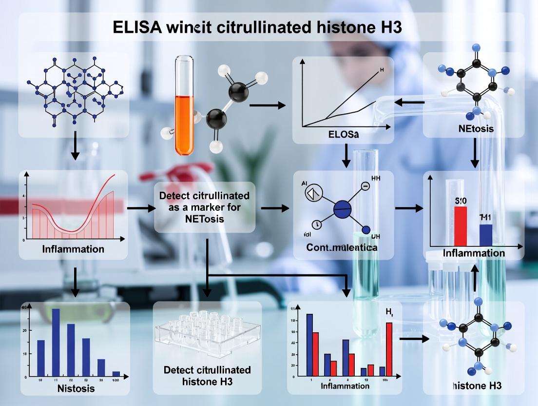
Quantifying NETosis: A Comprehensive Guide to ELISA-Based Detection of Citrullinated Histone H3 (H3Cit)
This article provides a detailed resource for researchers and drug development professionals on using Enzyme-Linked Immunosorbent Assay (ELISA) to detect citrullinated histone H3 (H3Cit), a key biomarker for Neutrophil Extracellular...
Quantifying NETosis: A Comprehensive Guide to ELISA-Based Detection of Citrullinated Histone H3 (H3Cit)
Abstract
This article provides a detailed resource for researchers and drug development professionals on using Enzyme-Linked Immunosorbent Assay (ELISA) to detect citrullinated histone H3 (H3Cit), a key biomarker for Neutrophil Extracellular Trap (NET) formation (NETosis). We cover the foundational biology of NETosis and the role of histone citrullination by PAD enzymes. A methodological deep-dive offers a step-by-step protocol for H3Cit ELISA, including sample preparation from blood, plasma, and tissues. The guide addresses common troubleshooting issues, optimization strategies for sensitivity and specificity, and critical validation steps. Finally, we compare H3Cit ELISA with alternative NETosis detection methods (e.g., microscopy, MPO-DNA complexes) and discuss its applications in inflammatory, autoimmune, and oncological research, providing a complete framework for integrating this assay into preclinical and clinical studies.
NETosis and Histone Citrullination: The Biological Basis for H3Cit as a Key Biomarker
NETosis is a unique form of programmed cell death distinct from apoptosis and necrosis, wherein neutrophils extrude decondensed chromatin structures decorated with antimicrobial granular proteins, known as Neutrophil Extracellular Traps (NETs). This process serves as a double-edged sword, providing a critical defense mechanism against pathogens while contributing to the pathogenesis of numerous inflammatory and autoimmune diseases when dysregulated. The detection of specific NETosis markers, such as citrullinated histone H3 (CitH3), via ELISA is a cornerstone of contemporary research in this field, bridging fundamental immunology with clinical diagnostics and therapeutic development.
Quantitative Data on NETosis in Disease Contexts
Table 1: Association of Circulating NETosis Markers (CitH3) with Disease Activity
| Disease Model / Condition | Reported CitH3 Level (Mean ± SD or Range) | Assay Used | Correlation with Clinical Score (r/p-value) | Key Reference (Year) |
|---|---|---|---|---|
| Severe COVID-19 | 4500 ± 1200 pg/mL | ELISA | r=0.78, p<0.001 | Zuo et al., 2020 |
| Rheumatoid Arthritis | 3200 ± 950 pg/mL | ELISA | r=0.65, p<0.01 | Dwivedi et al., 2019 |
| Systemic Lupus Erythematosus | 2800 ± 700 pg/mL | ELISA | r=0.71, p<0.001 | Thålin et al., 2017 |
| Sepsis | 5200 ± 1500 pg/mL | ELISA | r=0.82, p<0.001 | Park et al., 2021 |
| Healthy Controls | 450 ± 200 pg/mL | ELISA | N/A | Multiple |
Table 2: Pharmacological Modulation of NETosis In Vitro
| Compound/Inhibitor | Target Pathway/Enzyme | Concentration Tested | % Inhibition of CitH3 (vs. PMA control) | Key Reference |
|---|---|---|---|---|
| Cl-amidine | PAD4 (Pan-Inhibitor) | 10 µM | 85-95% | Li et al., 2010 |
| GSK484 | PAD4 (Specific) | 5 µM | 80-90% | Lewis et al., 2015 |
| DNase I | Degrades DNA backbone | 100 U/mL | Quantifies NET clearance | Hakkim et al., 2011 |
| Diphenyleneiodonium (DPI) | NADPH Oxidase (NOX) Inhibitor | 10 µM | 95-98% | Kirchner et al., 2012 |
| Dexamethasone | General Anti-inflammatory | 1 µM | 40-60% | Lapponi et al., 2013 |
Detailed Experimental Protocol: ELISA for Citrullinated Histone H3 (CitH3) in Human Plasma
Title: Protocol for Quantifying Circulating CitH3 via ELISA
I. Principle A sandwich ELISA specifically detects human CitH3 (typically at the R2+R8+R17 sites) using a capture antibody against a citrullinated peptide and a detection antibody against histone H3.
II. Reagents & Materials (The Scientist's Toolkit) Table 3: Essential Research Reagent Solutions for CitH3 ELISA
| Item | Function & Specification |
|---|---|
| Human CitH3 (R2+R8+R17) ELISA Kit (e.g., Cayman Chemical #501620) | Pre-optimized kit containing capture antibody-coated plate, detection antibodies, standards, and buffers. |
| Anti-Citrullinated Histone H3 (Clone 11D3) Antibody | Alternative monoclonal antibody for custom assay development. |
| Recombinant Human CitH3 Protein | Critical for generating standard curves and validating assay specificity. |
| PMA (Phorbol 12-myristate 13-acetate) | Potent inducer of NOX-dependent NETosis for positive controls (use at 25-50 nM). |
| Peripheral Blood Neutrophil Isolation Kit (e.g., Polymorphprep) | For generating in vitro NETosis samples. |
| Microplate Reader (450 nm filter) | For absorbance measurement of the colorimetric TMB reaction. |
| Plate Washer (or manual wash bottle) | Essential for removing unbound material between steps. |
| Plasma Collection Tubes (EDTA, with PAD inhibitor) | For clinical sampling; inhibits ex vivo NETosis during processing. |
III. Step-by-Step Procedure
- Sample Preparation: Collect blood into EDTA tubes supplemented with a PAD inhibitor (e.g., 10 mM Cl-amidine). Centrifuge at 2,000 x g for 15 minutes at 4°C. Aliquot plasma and store at -80°C. Avoid repeated freeze-thaw cycles.
- Standard Reconstitution: Reconstitute the provided CitH3 standard with the recommended assay buffer. Generate a 7-point standard curve via serial dilution (e.g., from 100 ng/mL to 0.78 ng/mL).
- Assay Setup: Add 100 µL of standard, plasma sample (diluted 1:2 in assay buffer), or blank to the antibody-coated wells. Incubate for 1 hour at room temperature (RT) on a plate shaker.
- Wash: Aspirate and wash each well 5 times with 300 µL of 1X wash buffer. Blot plate thoroughly on absorbent paper.
- Detection Antibody: Add 100 µL of the provided detection antibody (anti-histone H3 biotin conjugate). Incubate for 1 hour at RT on a shaker. Repeat wash step (Step 4).
- Streptavidin-Enzyme Conjugate: Add 100 µL of Streptavidin-HRP conjugate. Incubate for 30 minutes at RT on a shaker. Repeat wash step (Step 4).
- Substrate Development: Add 100 µL of TMB substrate solution. Incubate in the dark for 15-30 minutes at RT until color develops.
- Stop Reaction: Add 100 µL of stop solution (1M H2SO4 or equivalent). The color will change from blue to yellow.
- Measurement: Read absorbance immediately at 450 nm within 30 minutes.
IV. Data Analysis
- Subtract the average absorbance of the blank standard from all other readings.
- Generate a 4-parameter logistic (4PL) standard curve (Absorbance vs. Log[Conc.]).
- Interpolate sample concentrations from the curve, applying the appropriate dilution factor.
- Report values as pg/mL or ng/mL of CitH3 in plasma.
Pathway and Workflow Visualizations
Title: Major Signaling Pathway in NOX-Dependent NETosis
Title: CitH3 ELISA Experimental Workflow
Title: Research Context of CitH3 ELISA in NETosis Studies
The PAD Enzyme Family and Post-Translational Histone Modification
Within the context of ELISA detection of citrullinated histone H3 (CitH3) as a key marker for NETosis, understanding the Peptidyl Arginine Deiminase (PAD) enzyme family is fundamental. PADs catalyze the post-translational deimination of arginine residues to citrulline, a process central to the formation of Neutrophil Extracellular Traps (NETs). This application note details the role of PADs, protocols for studying their activity, and tools for detecting their histone modifications, specifically CitH3, in NETosis research and drug development.
The PAD Enzyme Family: Key Characteristics
PADs (PAD1-4 and PAD6) are calcium-dependent enzymes. PAD4 is the primary isoform involved in histone citrullination and NETosis due to its nuclear localization.
Table 1: The Human PAD Enzyme Family
| Isoform | Gene Name | Primary Tissue Expression | Key Substrates | Role in NETosis |
|---|---|---|---|---|
| PAD1 | PADI1 | Epidermis, Uterus | Keratin, Filaggrin | Not Direct |
| PAD2 | PADI2 | CNS, Muscle, Spleen | Myelin Basic Protein, Histones | Potential Contributor |
| PAD3 | PADI3 | Hair Follicles | Trichohyalin | Not Direct |
| PAD4 | PADI4 | Granulocytes, Immune Cells | Histone H3 (Arg2,8,17), Histone H4 | Primary Driver |
| PAD6 | PADI6 | Oocytes, Embryos | Unknown (Cytoplasmic) | Not Direct |
Table 2: Quantitative Data on PAD4-Mediated Histone H3 Citrullination in NETosis
| Parameter | Typical Experimental Value/Outcome | Detection Method |
|---|---|---|
| Key Citrullination Sites on H3 | R2 + R8 + R17 (Multi-site) | Mass Spectrometry, Site-Specific Antibodies |
| Calcium Requirement for PAD4 Activation | EC₅₀ ~ 5-10 µM | In vitro Activity Assay |
| Effect of PAD4 Inhibition (e.g., Cl-amidine) on NETosis | 70-90% Reduction in CitH3 Signal | ELISA, Immunofluorescence |
| Circulating CitH3 in Inflammatory Models (e.g., Sepsis) | 2- to 5-fold Increase vs. Control | Plasma/Sera ELISA |
Research Reagent Solutions Toolkit
Table 3: Essential Reagents for PAD & CitH3 NETosis Research
| Item | Function/Application | Example |
|---|---|---|
| PAD4 Inhibitors | Pharmacological blockade of citrullination for mechanistic studies/target validation. | Cl-amidine, GSK199, BB-Cl-amidine |
| Anti-Citrullinated Histone H3 Antibodies | Specific detection of citrullinated H3 for ELISA, WB, IF. | Clone 11D3 (CitH3 R2+R8+R17), Polyclonal Anti-CitH3 (various sites) |
| Recombinant Human PAD4 | In vitro citrullination assays, substrate specificity studies. | Commercial full-length, active enzyme. |
| NETosis Inducers | Positive controls for in vitro NETosis assays. | PMA (Phorbol Myristate Acetate), Ionomycin, Calcium Ionophore A23187 |
| Citrulline Detection Kit | Colorimetric/fluorimetric measurement of PAD enzyme activity. | Based on anti-citrulline antibody or chemical reaction (e.g., COLDER assay). |
| HDAC Inhibitors (e.g., TSA) | Enhance NETosis by increasing histone acetylation, a potential priming signal. | Used in combination stimulators. |
Protocols
Protocol 1:In VitroPAD4 Activity Assay Using a Histone Substrate
Purpose: To measure the enzymatic activity of recombinant or immunoprecipitated PAD4. Materials: Recombinant PAD4, purified core histones or recombinant histone H3, assay buffer (100 mM Tris-HCl pH 7.5, 10 mM CaCl₂, 5 mM DTT), PAD inhibitor (optional control), Citrulline Detection Kit. Procedure:
- Prepare reaction mix in a 50 µL volume: 1-2 µg histone substrate, 0.5-1 µg PAD4, 1x assay buffer.
- For inhibitor studies, pre-incubate PAD4 with inhibitor (e.g., 50 µM Cl-amidine) for 15 min on ice.
- Incubate reaction at 37°C for 60-90 minutes.
- Stop reaction with 5 µL of 0.5 M EDTA (final ~45 mM).
- Detect citrulline production per kit instructions (typically a colorimetric read at 450 nm).
- Normalize activity to a no-Ca²⁺ (EDTA added at start) control.
Protocol 2: Cell-Based NETosis Induction and CitH3 Detection via ELISA
Purpose: To quantify CitH3 release during NETosis from primary human neutrophils. Materials: Isolated human neutrophils, RPMI medium, NETosis inducer (e.g., 100 nM PMA), micrococcal nuclease, cell culture plates, CitH3 sandwich ELISA kit. Procedure:
- Neutrophil Isolation & Stimulation: Seed 2 x 10^5 neutrophils/well in a 96-well plate. Stimulate with PMA or vehicle for 3-4 hours at 37°C, 5% CO₂.
- NET Harvest: Post-incubation, add micrococcal nuclease (0.5 U/mL final), incubate 15 min at 37°C. Centrifuge plate (300 x g, 5 min) to pellet cells.
- Supernatant Collection: Carefully transfer supernatant (containing released NETs/CitH3) to a fresh tube. This is the sample for ELISA.
- CitH3 ELISA: Perform assay per manufacturer's protocol. a. Coat capture antibody (anti-CitH3). b. Block, then add samples and standards. c. Add detection antibody (biotinylated anti-H3 or anti-CitH3). d. Add streptavidin-HRP, then TMB substrate. e. Stop reaction and read absorbance at 450 nm.
- Analysis: Calculate CitH3 concentration from standard curve. Express as fold-change over unstimulated control.
Visualizations
Title: PAD4 Activation Drives CitH3 Formation and NETosis
Title: Workflow for Cell-Based NETosis and CitH3 ELISA Detection
Why Citrullinated Histone H3 (H3Cit) is a Specific Marker for NET Release.
1. Introduction Within the broader thesis on ELISA detection of Neutrophil Extracellular Trap (NET) markers, establishing specific and reliable biomarkers is paramount. NETosis, a distinct form of programmed cell death, results in the expulsion of decondensed chromatin decorated with granular and cytoplasmic proteins to trap pathogens. A critical biochemical event during NETosis is the peptidylarginine deiminase 4 (PAD4)-mediated conversion of arginine residues to citrulline on core histones, particularly histone H3. This citrullination drives chromatin decondensation, a prerequisite for NET release. Consequently, Citrullinated Histone H3 (H3Cit) is not merely present in NETs but is a functional driver of their formation, distinguishing it from passive leakage markers. Its detection, especially via ELISA, provides a specific readout for active, PAD4-dependent NETosis, crucial for research and drug development targeting dysregulated NET formation in inflammatory, thrombotic, and autoimmune diseases.
2. The Specificity of H3Cit for NETosis H3Cit’s specificity arises from its causal role in the NETotic pathway. Unlike markers like myeloperoxidase (MPO) or neutrophil elastase (NE), which are pre-formed in granules and can be released during other forms of cell death (e.g., necrosis, apoptosis), robust histone citrullination is tightly coupled to PAD4 activation during NETosis. The quantitative relationship between H3Cit levels and NET release has been consistently demonstrated.
Table 1: Key Evidence Establishing H3Cit as a Specific NETosis Marker
| Experimental Evidence | Quantitative Outcome | Implication for Specificity |
|---|---|---|
| PAD4 Inhibition/Knockout | H3Cit signal reduced by 85-95%; NET formation inhibited by ~80% (vs. controls). | H3Cit generation is PAD4-dependent and essential for NET release. |
| Time-Course Analysis | H3Cit modification peaks at 2-4 hours post-stimulation (e.g., with PMA 25-100 nM), preceding or coinciding with NET extrusion. | H3Cit is a mid-phase event in the active NETosis cascade, not a late necrotic byproduct. |
| Comparison with Other Death Pathways | Apoptotic stimuli (e.g., staurosporine) induce <5% of the H3Cit signal compared to NETotic stimuli. | Minimal citrullination occurs during apoptosis. |
| Co-localization Studies | >90% of extracellular DNA structures (NETs) are positive for H3Cit by immunofluorescence. | H3Cit is a consistent structural component of released NETs. |
| Correlation with NET Quantitation | Strong correlation (r² > 0.85) between H3Cit ELISA absorbance and independent NET quantitation (e.g., SYTOX Green fluorescence). | Soluble H3Cit levels reliably reflect the magnitude of NET release. |
3. Research Reagent Solutions: The H3Cit Detection Toolkit Table 2: Essential Reagents for H3Cit and NETosis Research
| Reagent / Material | Function & Rationale |
|---|---|
| PAD4-specific Inhibitors (e.g., GSK484, Cl-amidine) | To pharmacologically confirm the PAD4-dependence of observed H3Cit signal and NETosis. |
| NETosis Inducers (e.g., Phorbol Myristate Acetate (PMA), Ionomycin, Calcium Ionophore A23187) | Positive control stimuli to reliably trigger PAD4 activation and H3Cit formation. |
| H3Cit-Specific Antibodies (monoclonal, validated for ELISA) | Critical for specific capture/detection; must not cross-react with unmodified histone H3. |
| Pan-Histone H3 Antibodies | Useful as a normalization control for total histone content in some assay formats. |
| DNase I (RNase-free) | To digest NET matrices post-release for accurate quantification of H3Cit in supernatant or for cell-free DNA measurement. |
| SYTOX Green / Orange Nucleic Acid Stain | Impermeant dye for real-time, high-throughput quantification of extracellular DNA (NETs). |
| Neutrophil Isolation Kits (e.g., density gradient centrifugation) | To obtain high-purity primary human or murine neutrophils for in vitro studies. |
4. Detailed Experimental Protocols
Protocol 4.1: Induction of NETosis and Sample Preparation for H3Cit ELISA Objective: To generate cell culture supernatants and lysates containing H3Cit from activated neutrophils.
- Isolate human neutrophils from fresh peripheral blood using a density gradient centrifugation kit (e.g., Polymorphprep). Achieve purity >95%.
- Resuspend neutrophils in pre-warmed, serum-free RPMI 1640 medium at a density of 1 x 10⁶ cells/mL.
- Seed Cells: Add 500 µL of cell suspension (5 x 10⁵ cells) per well of a 24-well tissue culture plate.
- Stimulate NETosis:
- Test Condition: Add PMA to a final concentration of 25-100 nM. Mix gently.
- Negative Control: Add an equal volume of vehicle (e.g., DMSO, ≤0.1% final).
- Inhibition Control: Pre-incubate cells with 10 µM GSK484 (PAD4 inhibitor) for 30 min before adding PMA.
- Incubate plates at 37°C, 5% CO₂ for 4 hours.
- Sample Collection:
- For Supernatant H3Cit (Released): Gently collect the supernatant without disturbing the adherent NETs/cells. Centrifuge at 300 x g for 5 min to pellet any residual cells. Transfer the clear supernatant to a new tube. Add 1 U/mL DNase I and incubate for 15 min at 37°C to release NET-bound H3Cit. Aliquot and store at -80°C.
- For Total Cellular H3Cit (Cell-associated + Released): Lyse cells and NET structures directly in the well using 200 µL of complete ELISA lysis buffer (with protease inhibitors). Scrape well, transfer lysate, sonicate briefly (10 sec pulse), centrifuge at 10,000 x g for 10 min at 4°C. Collect supernatant. Aliquot and store at -80°C.
Protocol 4.2: Sandwich ELISA for Quantitative Detection of H3Cit Objective: To quantify H3Cit concentration in experimental samples.
- Coating: Dilute capture antibody (anti-H3Cit monoclonal) in carbonate-bicarbonate coating buffer (pH 9.6) to 2 µg/mL. Add 100 µL per well to a 96-well microplate. Seal and incubate overnight at 4°C.
- Wash & Block: Aspirate coating solution. Wash plate 3x with 300 µL PBS containing 0.05% Tween-20 (PBST). Block with 200 µL of 3% BSA in PBST for 2 hours at room temperature (RT). Wash 3x with PBST.
- Sample & Standard Incubation: Prepare serial dilutions of the recombinant H3Cit standard (e.g., 1000 pg/mL to 15.6 pg/mL) in assay diluent. Dilute test samples (from Protocol 4.1) 1:2 to 1:10 in assay diluent. Add 100 µL of standard or sample per well in duplicate. Include a blank (diluent only). Incubate for 2 hours at RT. Wash 5x with PBST.
- Detection Antibody Incubation: Add 100 µL per well of detection antibody (e.g., biotinylated anti-histone H3 antibody) at the manufacturer’s recommended dilution. Incubate for 1 hour at RT. Wash 5x with PBST.
- Streptavidin-Enzyme Conjugate: Add 100 µL per well of streptavidin-HRP conjugate (1:5000 dilution). Incubate for 30 min at RT, protected from light. Wash 5x with PBST.
- Substrate Development: Add 100 µL of TMB substrate solution per well. Incubate for 10-20 min at RT until blue color develops adequately.
- Stop & Read: Add 50 µL of 2N H₂SO₄ stop solution. Read absorbance immediately at 450 nm, with 570 nm or 620 nm as a reference wavelength.
- Analysis: Generate a 4-parameter logistic (4PL) standard curve. Interpolate sample concentrations, applying the appropriate dilution factor.
5. Visualizing the Pathway and Workflow
Diagram 1: H3Cit Generation in the PAD4-NETosis Pathway (76 chars)
Diagram 2: H3Cit ELISA Experimental Workflow (54 chars)
Application Notes
Citrullinated histone H3 (H3Cit), a specific marker of NETosis (Neutrophil Extracellular Trap formation), has emerged as a critical biomarker and pathogenic mediator across diverse disease states. Its detection via ELISA is central to both basic research and translational applications. This document outlines the clinical relevance of H3Cit and provides standardized protocols for its investigation within a thesis focusing on ELISA-based NETosis marker detection.
Table 1: H3Cit Association Across Disease Spectra
| Disease Category | Specific Condition | Key Association with H3Cit (Levels/Outcome) | Primary Sample Type in Studies |
|---|---|---|---|
| Sepsis & Critical Illness | Severe Sepsis/Septic Shock | ↑ Plasma H3Cit correlates with disease severity, organ dysfunction (SOFA score), and mortality. | Human Plasma/Serum |
| Autoimmunity | Rheumatoid Arthritis (RA) | ↑ Synovial fluid and serum H3Cit. Linked to disease activity, anti-CCP antibodies, and joint erosion. | Human Synovial Fluid, Serum, Murine Arthritic Joint Tissue |
| Autoimmunity | Systemic Lupus Erythematosus (SLE) | ↑ Serum H3Cit. Associated with lupus nephritis activity and type I interferon signature. | Human Serum, Renal Biopsies |
| Cancer | Deep Vein Thrombosis (DVT) in Cancer | ↑ Plasma H3Cit in cancer-associated DVT vs. DVT alone. Links NETosis to cancer pro-thrombotic state. | Human Plasma |
| Cancer | Metastasis (Preclinical) | Tumor-induced NETosis facilitates metastasis. H3Cit detection in pre-metastatic niches. | Murine Plasma, Metastatic Tissue Sections |
Detailed Experimental Protocols
Protocol 1: Quantitative Detection of Human H3Cit in Plasma by ELISA
Objective: To measure circulating H3Cit levels in human plasma samples from clinical cohorts (e.g., sepsis, autoimmune patients).
Materials (Research Reagent Solutions):
- Coating Antibody: Mouse anti-human H3Cit monoclonal antibody (clone 13D2 or equivalent). Function: Specifically captures H3Cit antigen.
- Detection Antibody: Rabbit anti-histone H3 (pan) polyclonal antibody. Function: Binds to captured histone H3 backbone.
- HRP-Conjugate: Anti-rabbit IgG, HRP-linked antibody. Function: Enzymatic tag for colorimetric detection.
- H3Cit Standard: Recombinant human citrullinated histone H3 protein. Function: Provides a standard curve for absolute quantification.
- DNase I, RNase A, Protease Inhibitors: Added to sample collection tubes. Function: Prevent degradation and artifactual NET release ex vivo.
- ELISA Substrate: TMB (3,3',5,5'-Tetramethylbenzidine). Function: HRP substrate for color development.
- Plate Reader: Spectrophotometer capable of reading at 450nm (reference 570nm or 620nm).
Procedure:
- Coating: Dilute capture antibody in carbonate-bicarbonate buffer (pH 9.6) to 2-4 µg/mL. Add 100 µL/well to a 96-well microplate. Incubate overnight at 4°C.
- Blocking: Wash plate 3x with PBS + 0.05% Tween-20 (PBST). Block with 200 µL/well of 5% BSA in PBS for 2 hours at room temperature (RT).
- Sample & Standard Incubation: Wash 3x. Dilute plasma samples (1:10 to 1:50 in assay diluent) and serially dilute H3Cit standard. Add 100 µL/well in duplicate. Incubate for 2 hours at RT.
- Detection Antibody: Wash 5x. Add 100 µL/well of detection antibody (diluted per manufacturer's recommendation). Incubate 1-2 hours at RT.
- HRP-Conjugate: Wash 5x. Add 100 µL/well of HRP-conjugated secondary antibody. Incubate 1 hour at RT.
- Detection: Wash 7x. Add 100 µL/well of TMB substrate. Incubate in the dark for 10-20 minutes.
- Stop & Read: Add 50 µL/well of 2N H₂SO₄ stop solution. Immediately read absorbance at 450nm.
Protocol 2: Immunofluorescence Co-staining for H3Cit and Neutrophil Markers in Tissue
Objective: To visualize NETosis in situ in tissue sections (e.g., rheumatoid synovium, lupus nephritis biopsies, metastatic tumors).
Procedure:
- Tissue Preparation: Deparaffinize and rehydrate formalin-fixed, paraffin-embedded (FFPE) tissue sections. Perform antigen retrieval using citrate buffer (pH 6.0) at 95-100°C for 20 minutes.
- Blocking: Block sections with 10% normal goat serum and 1% BSA in PBS for 1 hour at RT.
- Primary Antibodies: Incubate with primary antibody cocktail overnight at 4°C:
- Anti-H3Cit (Rabbit monoclonal, e.g., clone D2H4T): 1:100-1:200 dilution.
- Anti-Neutrophil Elastase (NE) (Mouse monoclonal) or Anti-MPO: 1:200 dilution. Function: Confirms neutrophil origin.
- Secondary Antibodies: Wash and incubate with species-specific fluorophore-conjugated secondary antibodies (e.g., Alexa Fluor 488 anti-rabbit, Alexa Fluor 594 anti-mouse) for 1 hour at RT in the dark.
- DNA Stain: Counterstain with DAPI (1 µg/mL) for 5 minutes.
- Mounting & Imaging: Mount with antifade medium. Image using a fluorescence microscope with appropriate filters. Colocalization of H3Cit, neutrophil marker, and diffuse DAPI signals indicates NET structures.
Visualizations
Title: NETosis Pathway Triggered by PAD4-Mediated H3 Citrullination
Title: Pathogenic Roles of H3Cit-NETs in Major Diseases
Title: ELISA Workflow for Quantifying H3Cit in Plasma
Research Reagent Solutions Table
| Reagent Category | Specific Example/Clone | Function in H3Cit/NETosis Research |
|---|---|---|
| Anti-H3Cit Antibodies | Mouse monoclonal (13D2), Rabbit monoclonal (D2H4T) | Specific immunocapture/detection for ELISA and IF; gold-standard for NET identification. |
| PAD4 Inhibitors | GSK484, Cl-amidine | Pharmacological tools to inhibit histone citrullination and NETosis in vitro/vivo. |
| Neutrophil Markers | Anti-Myeloperoxidase (MPO), Anti-Neutrophil Elastase (NE) | Confirm neutrophil origin in IF co-staining with H3Cit. |
| DNase I | Recombinant, grade I | Used to digest NETs in vitro to confirm DNA scaffold dependency; added to blood tubes to prevent ex vivo NETosis. |
| Recombinant H3Cit Protein | Human, full-length or N-terminal peptide | Essential standard for ELISA quantification and antibody validation. |
| NETosis Inducers | PMA (Phorbol Myristate Acetate), Ionomycin, LPS | Positive control stimuli for in vitro NETosis assays in human or murine neutrophils. |
Application Notes
Within research on neutrophil extracellular trap (NET) formation (NETosis), the detection of citrullinated histone H3 (H3Cit) by ELISA is a cornerstone technique. However, assay selection is critical and hinges on a fundamental biological distinction: specific residue citrullination versus total histone H3 citrullination.
Peptidylarginine deiminase 4 (PAD4) catalyzes the conversion of arginine to citrulline on histone H3. While multiple arginine residues (R2, R8, R17, R26) can be citrullinated, their timing and functional roles differ. Early in NETosis, PAD4 targets specific residues like H3R2, R8, and R17, which are crucial for chromatin decondensation. Assays targeting these individual modifications (e.g., H3R2Cit, H3R8Cit, H3R17Cit) serve as specific, early-stage markers of active PAD4-driven NETosis. In contrast, "pan-H3Cit" assays, which detect citrullination across multiple residues (often via antibodies against a generic H3Cit peptide), measure the total citrullination burden, reflecting cumulative NETosis activity.
Table 1: Comparative Analysis of H3Cit Assay Types
| Feature | H3R2/R8/R17-Specific Assays | Pan-H3Cit Assays |
|---|---|---|
| Target Epitope | A single, specific citrullinated residue (e.g., R2). | Multiple citrullinated residues across H3. |
| Biological Insight | Early, regulated PAD4 activity; residue-specific functions. | Total histone H3 citrullination load. |
| Temporal Sensitivity | Higher for detecting initiation of NETosis. | Integrates signal over time; may be better for late stages. |
| Specificity for NETosis | High when correlated with other NET markers. | Lower; can reflect other PAD4-mediated processes. |
| Typical Application | Mechanistic studies of NETosis pathways, early kinetics. | Biomarker studies in disease plasma/sera (e.g., RA, sepsis). |
| Potential Cross-Reactivity | Minimal between specific residues. | Higher potential for background in complex samples. |
Table 2: Illustrative Experimental Data from NETosis Induction (PMA, 100nM, 4 hrs)
| Sample Type | H3R8Cit (OD 450nm) | Pan-H3Cit (OD 450nm) | MPO-DNA Complex (ng/mL) |
|---|---|---|---|
| Unstimulated Neutrophils | 0.15 ± 0.03 | 0.22 ± 0.05 | 15 ± 4 |
| PMA-Stimulated Neutrophils | 1.45 ± 0.20 | 2.80 ± 0.30 | 320 ± 45 |
| PAD4 Inhibitor (YW3-56) + PMA | 0.25 ± 0.04 | 0.60 ± 0.10 | 40 ± 8 |
Experimental Protocols
Protocol 1: Cell-Based NETosis ELISA for H3R8Cit Detection Objective: Quantify early, residue-specific histone citrullination in stimulated human neutrophils.
- Neutrophil Isolation & Stimulation: Isolate human neutrophils from fresh blood using density gradient centrifugation. Seed 2.5 x 10^5 cells/well in a poly-L-lysine coated 96-well plate. Stimulate with 100 nM Phorbol 12-myristate 13-acetate (PMA) or vehicle in RPMI-1640 for 2-4 hours at 37°C, 5% CO2.
- Cell Fixation & Permeabilization: Gently remove medium. Fix cells with 4% paraformaldehyde for 15 min at RT. Permeabilize with 0.25% Triton X-100 in PBS for 10 min. Wash 3x with PBS.
- Blocking: Block with 3% BSA in PBS for 1 hour at RT.
- Primary Antibody Incubation: Incubate with anti-H3R8Cit monoclonal antibody (1:1000 in blocking buffer) overnight at 4°C. Wash 3x.
- Detection: Incubate with HRP-conjugated secondary antibody (1:2000) for 1 hour at RT. Wash 3x. Develop with TMB substrate for 10-15 min. Stop with 1M H2SO4 and read absorbance at 450nm.
- Normalization: Run parallel experiment for total histone H3 (ELISA or SYBR Green staining) to normalize H3R8Cit signal to histone content.
Protocol 2: Plasma Pan-H3Cit ELISA for Disease Biomarker Analysis Objective: Measure circulating H3Cit levels in patient plasma.
- Sample Preparation: Collect blood in citrate tubes. Centrifuge at 2000 x g for 15 min at 4°C. Aliquot plasma and store at -80°C. Avoid freeze-thaw cycles.
- Plate Coating: Coat high-binding 96-well plate with 2 µg/mL of capture anti-histone H3 antibody in carbonate coating buffer, overnight at 4°C.
- Blocking & Sample Incubation: Block with 5% BSA/PBS for 2 hours. Incubate 100 µL of diluted (1:10 in assay buffer) plasma sample or citrullinated H3 standard curve for 2 hours at RT. Wash 5x.
- Detection Antibody Incubation: Incubate with biotinylated pan-H3Cit detection antibody (1 µg/mL) for 1 hour at RT. Wash 5x.
- Signal Amplification & Development: Incubate with streptavidin-HRP (1:5000) for 45 min. Wash 5x. Develop with ultrasensitive TMB for 10 min. Stop and read at 450nm.
- Data Analysis: Quantify against the standard curve. Report as ng/mL of H3Cit equivalents.
Visualization
Title: Temporal Pathway of H3 Citrullination in NETosis
Title: Decision Flow for H3Cit Assay Selection
The Scientist's Toolkit: Research Reagent Solutions
| Reagent / Material | Function & Application Notes |
|---|---|
| Anti-H3R8Cit Monoclonal Antibody | Highly specific primary antibody for detecting early, residue-specific citrullination in cell-based ELISAs. |
| Biotinylated Pan-H3Cit Antibody | Detection antibody for sandwich ELISAs measuring total H3Cit in complex biological fluids like plasma. |
| Recombinant Citrullinated H3 Protein | Essential standard for generating quantitative calibration curves in both specific and pan-H3Cit assays. |
| PAD4 Inhibitor (e.g., GSK484, YW3-56) | Pharmacological tool to confirm PAD4-dependency of the citrullination signal and validate assay specificity. |
| Poly-L-Lysine Coated Plates | Enhances adherence of neutrophils during cell-based NETosis assays to prevent loss during washing steps. |
| Streptavidin-HRP Conjugate | High-sensitivity detection system for biotinylated antibodies in sandwich ELISA formats. |
| Cell Permeabilization Buffer (Triton X-100) | Allows intracellular antibody access to nuclear histones in fixed-cell ELISA protocols. |
| Cirrated Blood Collection Tubes | Preserves plasma samples by inhibiting coagulation, preventing platelet activation and histone release. |
Step-by-Step Protocol: Performing a Reliable H3Cit ELISA from Sample to Data
The detection of Citrullinated Histone H3 (H3Cit), a specific marker of Neutrophil Extracellular Trap (NET) formation, is a cornerstone in inflammation, autoimmune disease, and oncology research. Within the broader thesis on ELISA detection of NETosis markers, the choice between commercial kits and in-house developed assays presents a critical, practical crossroads for laboratories. This application note provides a structured evaluation, current data, and actionable protocols to inform this decision.
Quantitative Comparison: Commercial Kits vs. In-House Assays
Table 1: Comprehensive Practical Evaluation for H3Cit ELISA
| Evaluation Parameter | Commercial Kits | In-House Assays |
|---|---|---|
| Development Time | Minimal (0-1 week validation) | Extensive (3-6+ months for development/optimization) |
| Initial Cost (Setup) | Moderate ($500 - $2,500 per kit) | High ($2,000 - $10,000 for antibody procurement, plate coating, buffer optimization) |
| Cost per Sample (96-well plate) | High ($8 - $25 per sample) | Low ($1 - $5 per sample post-optimization) |
| Assay Reproducibility | High (CV typically <12%; lot-to-lot variation possible) | Variable (CV 5-20%; dependent on rigorous SOPs) |
| Sensitivity (Typical LOD) | 0.1 - 0.5 ng/mL (kit-dependent) | Can be optimized to 0.05 - 0.2 ng/mL |
| Specificity | Validated for specific H3Cit epitopes (e.g., H3R2+R8+R17) | Customizable to novel or multiplex epitopes |
| Flexibility & Customization | Low (fixed protocol, antibodies, buffers) | High (adjustable sample diluent, detection systems, multiplexing potential) |
| Technical Expertise Required | Low to Moderate | High (immunoassay development expertise critical) |
| Best Suited For | Diagnostic labs, standardized drug trials, labs with high throughput & low development bandwidth. | Research-focused labs investigating novel NETosis forms, requiring high flexibility, or running very large-scale studies where cost efficiency is paramount. |
Detailed Experimental Protocols
Protocol 3.1: Standardized Workflow for Evaluating a Commercial H3Cit ELISA Kit
Objective: To validate a commercial H3Cit ELISA kit for the detection of NETosis in stimulated human neutrophil supernatants.
Materials:
- Commercial Human H3Cit ELISA Kit (e.g., Cayman Chemical #501620, R&D Systems DY8137-05, or similar).
- Freshly isolated human neutrophils or relevant cell line (e.g., HL-60 differentiated).
- NETosis inducers: Phorbol 12-myristate 13-acetate (PMA, 25 nM), Calcium Ionophore A23187 (4 µM), or relevant disease-specific stimuli.
- Microplate reader capable of 450 nm measurement (with 540 nm or 570 nm correction).
Procedure:
- Neutrophil Isolation & Stimulation: Isolate neutrophils from healthy donor blood using density gradient centrifugation (e.g., Polymorphprep). Resuspend at 1x10^6 cells/mL in pre-warmed RPMI. Seed cells and stimulate with PMA or vehicle control for 3-4 hours at 37°C, 5% CO₂.
- Sample Preparation: Centrifuge culture plates at 300 x g for 5 min. Carefully collect cell-free supernatant. Store at -80°C if not used immediately.
- ELISA Execution: Follow the manufacturer's protocol precisely. Typically involves:
- Coating: Pre-coated plates provided.
- Standards & Samples: Add standards (reconstituted per kit) and undiluted/diluted samples in duplicate.
- Detection: Sequential incubations with detection antibody (often biotinylated anti-H3Cit), Streptavidin-HRP conjugate, and TMB substrate.
- Stop & Read: Add stop solution (acid) and read absorbance at 450 nm (reference 540-570 nm).
Protocol 3.2: Development of an In-House H3Cit Capture ELISA
Objective: To develop a customized, cost-effective sandwich ELISA for H3Cit detection using commercially available components.
Materials:
- Coating Antibody: Monoclonal anti-Histone H3 (clone H3-1E4) or similar pan-Histone H3 antibody.
- Detection Antibody: Rabbit polyclonal anti-Citrullinated Histone H3 (e.g., MilliporeSigma AB5103; recognizes H3Cit2,8,17).
- Standard: Recombinant human Histone H3.1 (citrullinated at R2, R8, R17) or nucleosomes from stimulated cells as a calibrant.
- Secondary Reagent: HRP-conjugated anti-rabbit IgG.
- Blocking Buffer: 1% BSA or 5% non-fat dry milk in PBS.
- Assay Diluent: PBS with 0.05% Tween-20 (PBST) + 1% BSA.
- TMB Substrate & Stop Solution.
Procedure:
- Plate Coating: Dilute capture antibody (1-5 µg/mL) in carbonate-bicarbonate coating buffer (pH 9.6). Add 100 µL/well to a 96-well microplate. Incubate overnight at 4°C.
- Blocking: Wash plate 3x with PBST. Block with 300 µL/well of blocking buffer for 1-2 hours at room temperature (RT). Wash 3x.
- Antigen Binding: Prepare a standard curve of recombinant H3Cit (0.1-50 ng/mL) in assay diluent. Add 100 µL of standards or samples (cell lysates/supernatants) per well in duplicate. Incubate 2 hours at RT or overnight at 4°C. Wash 5x.
- Detection Antibody Incubation: Add detection antibody (optimized dilution, typically 0.5-1 µg/mL in diluent), 100 µL/well. Incubate 1-2 hours at RT. Wash 5x.
- Secondary Antibody Incubation: Add HRP-conjugated anti-rabbit IgG (diluted ~1:2000 in diluent), 100 µL/well. Incubate 1 hour at RT. Wash 5-7x.
- Signal Development & Acquisition: Add TMB substrate (100 µL/well). Develop in the dark for 5-30 min. Stop reaction with 2M H₂SO₄. Read immediately at 450 nm (ref. 540 nm).
Visualized Workflows & Pathways
Title: H3Cit ELISA Experimental Workflow
Title: Key Signaling in NETosis Leading to H3Cit
The Scientist's Toolkit: Essential Research Reagents for H3Cit Detection
Table 2: Key Research Reagent Solutions for NETosis ELISA
| Reagent Category | Specific Example(s) | Function & Role in H3Cit Detection |
|---|---|---|
| Primary Capture Antibody | Mouse anti-Histone H3 (clone H3-1E4) | Binds total histone H3 framework, capturing both citrullinated and non-citrullinated forms for specific detection. |
| Primary Detection Antibody | Rabbit anti-Citrullinated Histone H3 (H3Cit2,8,17) | Specifically recognizes the citrullinated epitopes on histone H3, providing assay specificity for NETosis. |
| Positive Control / Standard | Recombinant Human Citrullinated Histone H3 Protein (H3.1 Cit R2/R8/R17) | Serves as a quantitative calibrator for generating a standard curve and determining H3Cit concentration in unknowns. |
| NETosis Inducer (Control) | Phorbol 12-Myristate 13-Acetate (PMA) | A potent PKC activator used as a positive control stimulus to induce robust NET formation and H3Cit generation in vitro. |
| Detection System | HRP-conjugated Anti-Rabbit IgG & TMB Substrate | Enzyme-conjugated secondary antibody amplifies the detection signal; TMB is the chromogenic substrate for colorimetric readout. |
| Critical Assay Buffer | PBS with 0.05% Tween-20 (PBST) & 1% BSA | Universal wash and diluent buffer; reduces non-specific binding, lowering background noise and improving signal-to-noise ratio. |
Within the broader thesis on ELISA detection of citrullinated histone H3 (CitH3) as a key marker of NETosis, the reliability of final quantitative data is fundamentally dependent on stringent pre-analytical protocols. Inflammatory mediators and proteases released during neutrophil extracellular trap (NET) formation can significantly alter analyte stability. This document provides detailed application notes and protocols for the critical initial stages of sample processing to ensure the integrity of CitH3 and other NETosis-related analytes.
Blood Collection for NETosis Marker Analysis
The choice of anticoagulant and collection technique is paramount for minimizing ex vivo NETosis and preserving the native CitH3 signal.
Protocol: Phlebotomy for Plasma Preparation
Objective: To collect whole blood suitable for subsequent plasma separation and CitH3 ELISA, while suppressing artifactual NET formation during draw and handling.
Materials:
- Tourniquet
- Sterile needles (21G recommended) and vacuum collection tubes
- Disinfectant (70% isopropanol)
- Timer
- Ice bucket or chilled rack (4°C)
Detailed Procedure:
- Anticoagulant Selection: Use pre-chilled citrate tubes (e.g., 3.2% sodium citrate). Avoid heparin, as it can interfere with some ELISA antibodies and may influence NETosis. EDTA is acceptable but citrate is preferred for neutrophil studies.
- Venipuncture: Apply tourniquet for minimal time (<1 minute). Perform clean venipuncture. Discard the first 1-2 mL of blood if using a standard needle to avoid tissue thromboplastin contamination.
- Tube Filling: Fill the citrate tube to the exact specified volume to ensure proper blood-to-anticoagulant ratio. Invert tube gently 5-8 times immediately after collection.
- Immediate Cooling: Place the filled tube immediately into a slurry of ice and water (0-4°C). Critical Step: This cooling step inhibits neutrophil activation and enzymatic degradation during transport.
- Processing Timeline: Process blood for plasma separation within 60 minutes of collection. Do not allow samples to sit at room temperature.
Quantitative Considerations for Blood Collection
Table 1: Impact of Pre-Analytical Variables on Plasma CitH3 Levels
| Variable | Condition | Observed Effect on Measured CitH3 | Recommendation |
|---|---|---|---|
| Anticoagulant | Sodium Citrate (3.2%) | Baseline reference | Preferred for NETosis studies |
| K2-EDTA | ± 15% variation vs. Citrate | Acceptable alternative | |
| Lithium Heparin | Significant assay interference & variable effects | Avoid | |
| Time to Processing | ≤ 1 hour on ice | Optimal recovery | Process within 60 min |
| 2 hours at RT | Increase of 30-50% (artifactual NETosis) | Strictly avoid | |
| Tube Fill Volume | Correct (100%) | Correct anticoagulant ratio | Follow manufacturer specs |
| Underfill (80%) | Possible clotting, altered results | Discard underfilled tubes | |
| Centrifugation Force | 2,000 x g | Standard platelet-poor plasma | Use consistent force |
| 500 x g | Platelet-rich plasma, higher variability | Not recommended |
Plasma and Serum Separation Protocol
The separation protocol aims to remove cells and platelets efficiently while preventing the release of analytes from cells during centrifugation.
Protocol: Preparation of Cell-Free Plasma for CitH3 ELISA
Objective: To obtain platelet-poor plasma with minimal contamination from platelet-derived histones or ex vivo activated neutrophils.
Materials:
- Refrigerated centrifuge capable of precise speed control
- Centrifuge tubes (if secondary transfer is needed)
- Adjustable-volume pipettes and sterile, low-protein-binding tips
- Cryovials for storage
- -80°C freezer
Detailed Procedure:
- Initial Spin: Keep blood tubes on ice until centrifugation. Centrifuge at 2,000 x g for 20 minutes at 4°C with the brake ON. Using the brake ensures rapid separation from cells.
- Plasma Extraction: Carefully remove the centrifuge tube. Without disturbing the buffy coat (white cell layer), use a pipette to aspirate the top plasma layer (~2/3 of the volume). Transfer to a clean, labeled polypropylene tube.
- Second Spin (Optional but Recommended for High Sensitivity): To ensure removal of residual platelets, perform a second centrifugation of the transferred plasma at 10,000 x g for 10 minutes at 4°C.
- Aliquoting: Pipette the cleared, platelet-poor plasma into pre-chilled, low-protein-binding cryovials. Avoid multiple freeze-thaw cycles by creating single-use aliquots (e.g., 50-100 µL).
- Storage: Immediately snap-freeze aliquots in liquid nitrogen or a dry-ice/ethanol bath, then transfer to a -80°C freezer for long-term storage. Analyze samples within 3 months for best results.
Cell Culture Supernatant Handling forIn VitroNETosis Studies
Supernatants from stimulated neutrophil cultures contain CitH3 and other NET components. Handling must inhibit protease activity.
Protocol: Harvesting Supernatant from NETosis Assays
Objective: To terminate NETosis reactions and stabilize CitH3 in cell culture supernatant for downstream ELISA.
Materials:
- Neutrophil culture plate (e.g., PMA or ionomycin-stimulated)
- Protease Inhibitor Cocktail (PIC), EDTA-free recommended
- Pre-chilled microcentrifuge tubes
- Refrigerated microcentrifuge
Detailed Procedure:
- Termination & Stabilization: At the assay endpoint, add 1x volume of PIC (according to manufacturer's instructions) directly to the culture well. Gently swirl to mix. Note: For time-course experiments, remove supernatant at each time point and immediately mix with PIC in a separate tube on ice.
- Cell Debris Removal: Carefully pipette the supernatant (now containing PIC) from the well, avoiding the adherent NETs and cell layer at the bottom. Transfer to a pre-chilled microcentrifuge tube.
- Clarification Spin: Centrifuge the collected supernatant at 4,000 x g for 5 minutes at 4°C to pellet any remaining cellular debris or large NET fragments.
- Harvest & Storage: Carefully pipette the clarified supernatant into fresh, pre-chilled tubes, avoiding the pellet. Aliquot immediately and store at -80°C. Perform ELISA within one week for optimal detection of labile markers.
The Scientist's Toolkit: Research Reagent Solutions
Table 2: Essential Materials for Pre-Analytical Processing of NETosis Samples
| Item | Function/Benefit | Example/Note |
|---|---|---|
| Sodium Citrate Vacutainers | Prevents coagulation; preferred anticoagulant for neutrophil studies. Minimizes ex vivo activation. | BD Vacutainer #363083 |
| Protease Inhibitor Cocktail (EDTA-free) | Stabilizes protein analytes like CitH3 by inhibiting serine proteases (e.g., Neutrophil Elastase) released during NETosis. | Roche cOmplete, EDTA-Free |
| Low-Protein-Binding Tubes & Tips | Minimizes adsorption of low-abundance proteins (like circulating CitH3) to plastic surfaces, improving recovery. | Eppendorf LoBind |
| Refrigerated Centrifuge | Maintains samples at 4°C during processing to slow enzymatic degradation and cellular metabolism. | Essential for all spins. |
| DNase I (for specific protocols) | Can be used to digest NET scaffolds post-supernatant collection to solubilize NET-bound proteins for complete analysis. | Add after PIC step if required. |
| PMA (Phorbol Myristate Acetate) | Standard pharmacological inducer of NETosis for positive control generation in cell culture supernatant experiments. | Commonly used at 25-100 nM. |
Visualizations
Diagram 1: Blood to Plasma Processing Workflow
Diagram 2: Key Influences on Pre-Analytical CitH3 Integrity
The reliable detection of citrullinated histone H3 (CitH3), a key marker of Neutrophil Extracellular Trap (NET) formation (NETosis), via ELISA is critical for research into inflammatory and autoimmune diseases (e.g., rheumatoid arthritis, sepsis). The accuracy of this detection is fundamentally limited by the efficacy of sample preparation. Complex biological matrices like solid tissues and synovial fluid present significant challenges due to their viscosity, cellular heterogeneity, and high protein/ protease content. This application note details optimized protocols for preparing these complex samples to ensure the accurate quantification of CitH3 and other NETosis markers, directly supporting robust and reproducible thesis research data.
Key Challenges & Solution Principles
| Challenge | Impact on CitH3 ELISA | Solution Principle |
|---|---|---|
| Incomplete Tissue Homogenization | Low analyte yield, high variability. | Mechanical disruption with optimized buffers. |
| High Viscosity (Synovial Fluid) | Pipetting errors, uneven analyte distribution. | Enzymatic (hyaluronidase) and/or dilution treatment. |
| Nuclease/Protease Activity | Degradation of histone targets. | Use of potent, broad-spectrum inhibitors. |
| High Abundant Proteins | Non-specific interference, matrix effects. | Clarification and targeted dilution. |
| Cellular Heterogeneity | Inconsistent NET marker concentration. | Standardized cell counting & lysis. |
Detailed Experimental Protocols
Protocol 1: Optimized Homogenization of Solid Tissue for CitH3 Extraction
Objective: To efficiently extract intact CitH3 from organ tissues (e.g., murine lung, liver) for ELISA. Materials: See Scientist's Toolkit. Procedure:
- Tissue Procurement: Immediately after sacrifice, snap-freeze tissue in liquid nitrogen. Store at -80°C.
- Pre-homogenization: Weigh ~30 mg of frozen tissue. Place in a pre-chilled tube with a 5mm stainless steel bead and 500 µL of Complete Homogenization Buffer.
- Mechanical Disruption: Homogenize using a tissue lyser at 30 Hz for 2 minutes. Keep samples on ice.
- Sonication: To shear chromatin and solubilize histones, sonicate the homogenate on ice using a microtip probe (3 pulses of 10 seconds at 30% amplitude, with 30-second rests on ice).
- Clarification: Centrifuge at 16,000 x g for 15 minutes at 4°C.
- Supernatant Collection: Carefully transfer the supernatant to a fresh tube.
- Total Protein Normalization: Determine protein concentration via BCA assay. Dilute all samples to a consistent concentration (e.g., 1 µg/µL) in homogenization buffer to standardize loading for ELISA.
- Assay: Aliquot and proceed with CitH3 ELISA. Store at -80°C.
Protocol 2: Preparation of Synovial Fluid for NETosis Marker Detection
Objective: To reduce viscosity and prepare synovial fluid for direct CitH3 and MPO- DNA complex ELISA. Materials: See Scientist's Toolkit. Procedure:
- Initial Handling: Thaw synovial fluid samples on ice.
- Viscosity Reduction: Add 1 U/mL of hyaluronidase (type IV-S) to the fluid. Incubate at 37°C for 15 minutes. Alternatively, for some ELISA kits, a 1:10 dilution in PBS may be sufficient.
- Cellular Debris Removal: Centrifuge at 2,000 x g for 10 minutes at 4°C to remove cells and large particles.
- Clarification: Transfer supernatant to a fresh tube and centrifuge at 16,000 x g for 20 minutes at 4°C to remove microvesicles and remaining debris.
- Aliquoting & Storage: Aliquot the clear supernatant to avoid freeze-thaw cycles. Store at -80°C.
- Assay: Proceed with ELISA. A dilution series (e.g., 1:2, 1:5, 1:10) is recommended to identify the optimal dilution that falls within the standard curve and minimizes matrix interference.
Data Presentation: Protocol Optimization Impact
Table 1: Effect of Homogenization Method on CitH3 Recovery from Murine Lung Tissue
| Homogenization Method | Mean [CitH3] (ng/mg tissue) | Coefficient of Variation (CV%) | Total Protein Yield (mg/mL) |
|---|---|---|---|
| Manual Grinding (Mortar & Pestle) | 1.5 | 25.4 | 3.2 |
| Rotor-Stator Homogenizer | 4.2 | 18.7 | 5.8 |
| Bead Mill Homogenizer (Optimized Protocol) | 6.8 | 9.3 | 7.1 |
| Ultrasonic Processor Only | 3.1 | 22.1 | 4.5 |
Table 2: Impact of Synovial Fluid Pretreatment on ELISA Performance
| Pretreatment Method | Apparent [CitH3] (ng/mL) | Intra-assay CV% | Spike Recovery (%) |
|---|---|---|---|
| No Treatment (Neat) | 15.2 | 28.5 | 62 |
| Dilution Only (1:10 in PBS) | 8.1 | 15.2 | 85 |
| Hyaluronidase Only | 12.7 | 12.8 | 102 |
| Hyaluronidase + Dilution (1:5) | 9.8 | 8.1 | 98 |
Visualization of Workflows
Diagram 1: Sample preparation workflows for tissue and synovial fluid.
The Scientist's Toolkit: Essential Reagents & Materials
Table 3: Research Reagent Solutions for NETosis Sample Prep
| Item | Function & Rationale | Example/Notes |
|---|---|---|
| Complete Homogenization Buffer | Lyses cells, inhibits proteases/deacetylases, stabilizes histones. | 50 mM Tris-HCl (pH 7.5), 150 mM NaCl, 1% NP-40, 0.5% Sodium Deoxycholate, 0.1% SDS, 1x Protease Inhibitor Cocktail, 5 mM Sodium Butyrate (deacetylase inhibitor). |
| High-Strength Inhibitor Cocktail | Prevents post-collection degradation of CitH3. | Commercially available broad-spectrum cocktails with serine, cysteine, metallo-protease, and nuclease inhibitors. |
| Hyaluronidase (Type IV-S) | Degrades hyaluronic acid, drastically reducing synovial fluid viscosity. | Use at 1-5 U/mL. Type IV-S has low protease activity. |
| Stainless Steel Beads (5mm) | Provides efficient, cold mechanical grinding for tissues in a bead mill. | Superior to ceramic or glass for tough fibrous tissues. |
| Nuclease-Free Tubes & Tips | Prevents exogenous nuclease contamination that degrades NET DNA complexes. | Critical for assays detecting MPO-DNA complexes. |
| Precision Tissue Lyser/Homogenizer | Ensures reproducible, high-yield disruption across multiple samples simultaneously. | Key for low CV% in tissue samples. |
| Ultrasonic Processor with Microtip | Shears chromatin to efficiently solubilize nucleosomal histones (CitH3). | Short pulses on ice are essential to prevent heating/ degradation. |
| Cryogenic Tubes | For immediate snap-freezing and stable -80°C storage of raw tissue/synovial fluid. | Preserves the in vivo NETosis signature at time of collection. |
Application Notes & Protocol for the Detection of Citrullinated Histone H3 in NETosis Research
Within a thesis investigating NETosis as a biomarker and therapeutic target in inflammatory and thrombotic diseases, the precise detection of citrullinated histone H3 (H3Cit) via ELISA is a foundational technique. This protocol details the critical steps for a sandwich ELISA optimized for H3Cit, enabling quantitative assessment of NET formation in experimental samples (e.g., stimulated neutrophil supernatants, plasma).
Table 1: Representative Standard Curve Parameters for H3Cit ELISA
| Parameter | Value/Range | Notes |
|---|---|---|
| Assay Range | 0.78 - 50 ng/mL | Typical dynamic range for commercial and custom assays. |
| Lower Limit of Detection (LLOD) | 0.2 - 0.5 ng/mL | Defined as mean + 3SD of zero standard. |
| Intra-assay Precision (CV) | < 8% | Variation within a single plate. |
| Inter-assay Precision (CV) | < 12% | Variation between different plates/runs. |
| Typical Sample Dilution | Plasma: 1:2 to 1:10 | Cell Culture Supernatant: Neat to 1:5 |
| Recovery Rate | 85 - 115% | Spike-and-recovery in sample matrix. |
Table 2: Key Experimental Controls
| Control Type | Purpose | Expected Result |
|---|---|---|
| Coating Blank | Background from capture antibody binding. | Low OD (< 0.1). |
| Sample Blank (Assay Diluent) | Background from detection system. | Low OD (< 0.15). |
| Positive Control | Recombinant H3Cit or known positive sample. | Signal within standard curve range. |
| Negative Control | Unstimulated neutrophil supernatant. | Signal near LLOD. |
| Specificity Control | Peptide competition with citrullinated vs. arginine peptide. | Signal inhibition only with citrullinated peptide. |
Detailed Experimental Protocol
I. Coating (Day 1)
Principle: Immobilize the capture antibody (anti-H3Cit monoclonal) on the polystyrene plate.
- Dilute Capture Antibody: Prepare coating buffer (0.1 M Carbonate-Bicarbonate, pH 9.6). Dilute anti-H3Cit antibody to 2-5 µg/mL in coating buffer.
- Coat Wells: Add 100 µL per well to a 96-well microplate. Seal plate and incubate overnight at 4°C.
II. Blocking (Day 2)
Principle: Saturate non-specific protein-binding sites to minimize background.
- Wash: Decant coating solution. Wash plate 3 times with 300 µL/well of Wash Buffer (0.05% Tween-20 in PBS, PBS-T). Blot dry on clean paper.
- Block: Add 200 µL per well of Blocking Buffer (3-5% BSA or 1% Casein in PBS). Incubate for 1-2 hours at room temperature (RT).
- Wash: Repeat wash step as above. Plate can now be used immediately or dried and stored sealed at 4°C for short-term.
III. Sample & Standard Incubation
Principle: Bind H3Cit antigen from samples to the immobilized capture antibody.
- Prepare Standards: Serially dilute recombinant H3Cit protein in Assay Diluent (1% BSA in PBS-T) to generate a 7-point standard curve (e.g., 50 to 0.78 ng/mL). Include a zero standard (Assay Diluent).
- Prepare Samples: Dilute test samples (plasma, serum, cell culture supernatant) in Assay Diluent.
- Incubate: Add 100 µL of standard or sample per well. Incubate for 2 hours at RT on a plate shaker.
- Wash: Wash plate 5 times thoroughly with Wash Buffer.
IV. Detection Antibody Incubation
Principle: Bind a detection antibody (e.g., biotinylated anti-histone H3 antibody) to captured H3Cit.
- Add Detection Antibody: Dilute detection antibody per manufacturer's recommendation in Assay Diluent. Add 100 µL per well.
- Incubate: Incubate for 1-2 hours at RT.
- Wash: Wash plate 5 times as before.
V. Signal Development & Readout
Principle: Amplify and quantify the detected antibody-antigen complex.
- Add Streptavidin-HRP: Dilute Streptavidin conjugated to Horseradish Peroxidase (HRP) in Assay Diluent. Add 100 µL per well. Incubate for 30-45 minutes at RT, protected from light.
- Wash: Wash plate 5 times.
- Add Substrate: Add 100 µL of TMB (3,3',5,5'-Tetramethylbenzidine) substrate per well. Incubate for 10-20 minutes at RT until blue color develops adequately.
- Stop Reaction: Add 50 µL of 2M H₂SO₄ stop solution per well. Color will change from blue to yellow.
- Read Absorbance: Measure absorbance at 450 nm (reference wavelength 570 or 620 nm) using a plate reader within 30 minutes.
Visualizations
The Scientist's Toolkit: Key Research Reagent Solutions
| Item | Function in H3Cit ELISA | Critical Considerations |
|---|---|---|
| Anti-H3Cit Monoclonal Antibody (Capture) | Specifically binds the citrullinated epitope on histone H3. | Clone specificity (e.g., clone 2D8 or 9C8); verify reactivity to target H3 citrullination sites (R2+R8+R17). |
| Biotinylated Anti-Histone H3 Antibody (Detection) | Binds conserved regions of histone H3, independent of citrullination. | Must not compete with capture antibody; confirms total histone capture. |
| Recombinant H3Cit Protein | Serves as the standard for quantitative curve generation. | Essential for assay standardization; purity and citrullination level must be certified. |
| High Protein-Binding ELISA Plates | Provides surface for passive adsorption of capture antibody. | Polystyrene, clear, flat-bottom plates ensure consistent coating efficiency. |
| TMB Substrate Solution | Chromogenic substrate for HRP, produces measurable color change. | Sensitivity and kinetics vary; use a high-sensitivity formulation for low-abundance targets. |
| Citrullinated Peptide (for Competition) | Validates assay specificity by inhibiting signal in a dose-dependent manner. | Control arginine-containing peptide should show no inhibition. |
| Neutrophil Isolation Kit | To obtain primary human or murine neutrophils for in vitro NETosis induction. | Purity (>95%) and viability are crucial for generating relevant biological samples. |
| PAD Enzyme Inhibitor (e.g., Cl-amidine) | Negative control in NETosis induction experiments. | Confirms that H3Cit signal is PAD-dependent. |
Within the broader thesis research on ELISA detection of citrullinated histone H3 (CitH3) as a NETosis marker, robust data analysis is paramount. Accurate quantification of CitH3 in plasma or cell culture supernatant samples via ELISA hinges on precise standard curve generation, interpolation of unknown sample values, and appropriate unit reporting (pg/mL or relative units). This protocol details the steps for analyzing data from typical sandwich ELISA experiments to quantify NETosis markers.
Standard Curve Generation Protocol
Principle: A series of known concentrations of the recombinant CitH3 antigen standard is assayed alongside samples. The measured optical density (OD) values are used to generate a mathematical model describing the concentration- response relationship.
Detailed Protocol:
- Standard Dilution Series: Prepare a 2-fold or 5-fold serial dilution of the recombinant CitH3 standard in the provided assay diluent, covering the range specified in the ELISA kit (e.g., 15.6 pg/mL to 1000 pg/mL). Include a zero standard (diluent alone).
- Assay Execution: Load standards and samples in duplicate onto the pre-coated ELISA plate. Follow the specific kit protocol for incubation with detection antibody, streptavidin-HRP, and TMB substrate, terminating the reaction with stop solution.
- Absorbance Measurement: Read the optical density (OD) at 450 nm, with a reference wavelength of 570 nm or 620 nm for correction. Subtract the reference OD from the 450 nm OD for all wells.
- Data Averaging: Calculate the mean absorbance for each standard and sample duplicate.
- Curve Fitting: Plot the mean corrected absorbance (y-axis) against the known standard concentration (x-axis) using graphing software (e.g., GraphPad Prism, SoftMax Pro). Common models include:
- Four-Parameter Logistic (4PL) Curve: Preferred for most sandwich ELISAs. It models the sigmoidal curve with lower asymptote, upper asymptote, slope factor, and inflection point (EC50).
- Log-Linear Regression: A simpler linear model applied to the central, linear portion of the sigmoidal curve after log10 transformation of concentrations.
- Quality Assessment: Ensure the curve's R² value is >0.99. The percent recovery for each standard point (calculated concentration/known concentration * 100%) should ideally be between 80-120%.
Interpolation & Quantification Protocol
Principle: The fitted standard curve equation is used to interpolate the concentration of CitH3 in unknown samples from their mean corrected OD values.
Detailed Protocol:
- Interpolation: Input the mean corrected OD value of each unknown sample into the standard curve equation (inverse prediction) to calculate its concentration.
- Dilution Factor Application: If samples were diluted prior to assay (often necessary for plasma/serum), multiply the interpolated concentration by the dilution factor to obtain the final concentration in the original sample.
- Unit Reporting:
- Absolute Quantification: Report concentrations in pg/mL or ng/mL based on the recombinant standard. This allows direct comparison across experiments.
- Relative Quantification: If a standard is unavailable, assign the control group sample an arbitrary unit (e.g., 1 U/mL or 100 RU) and express other samples relative to it. This is less common for CitH3 ELISAs.
- Validation: Include quality control (QC) samples with known concentrations in each plate. Their interpolated values should fall within the acceptable range (e.g., ±20% of expected value).
- Statistical Analysis: Perform appropriate statistical tests (e.g., t-test, ANOVA) on the final quantified concentrations (pg/mL) between experimental groups (e.g., stimulated vs. control, disease vs. healthy donor) to determine significance.
Example Quantitative Data from CitH3 ELISA Analysis
Table 1: Standard Curve Data for a Representative CitH3 ELISA Plate
| Standard Concentration (pg/mL) | Mean Corrected OD (450nm - 570nm) | Calculated Conc. (pg/mL) | % Recovery |
|---|---|---|---|
| 0 | 0.051 | N/A | N/A |
| 15.6 | 0.102 | 14.9 | 95.5 |
| 31.3 | 0.185 | 30.1 | 96.2 |
| 62.5 | 0.420 | 60.8 | 97.3 |
| 125 | 0.890 | 129.5 | 103.6 |
| 250 | 1.550 | 255.2 | 102.1 |
| 500 | 2.210 | 488.7 | 97.7 |
| 1000 | 2.598 | 1050.3 | 105.0 |
Curve Fit: 4PL, R² = 0.9993
Table 2: Quantification of CitH3 in NETosis Induction Experiment Samples
| Sample ID & Description | Mean Corrected OD | Interpolated Conc. (pg/mL) | Dilution Factor | Final Conc. (pg/mL) |
|---|---|---|---|---|
| PMA-Stimulated Neutrophils (1) | 1.856 | 307.4 | 5 | 1537.0 |
| PMA-Stimulated Neutrophils (2) | 1.902 | 317.2 | 5 | 1586.0 |
| Control Neutrophils (1) | 0.233 | 39.5 | 5 | 197.5 |
| Control Neutrophils (2) | 0.241 | 41.0 | 5 | 205.0 |
| Patient Plasma Sample A | 0.745 | 114.9 | 20 | 2298.0 |
| Healthy Donor Plasma B | 0.109 | 18.2 | 20 | 364.0 |
The Scientist's Toolkit: Key Research Reagent Solutions
Table 3: Essential Materials for CitH3 ELISA and Data Analysis
| Item | Function & Rationale |
|---|---|
| Commercial CitH3 Sandwich ELISA Kit | Provides pre-coated plates, matched antibody pair, recombinant CitH3 standard, and optimized buffers for specific, sensitive detection. Critical for standardization. |
| Recombinant CitH3 Protein Standard | Serves as the calibrator for generating the standard curve, enabling absolute quantification in pg/mL. Must be from the same source as the kit. |
| Microplate Reader with 450nm Filter | Instrument for measuring the colorimetric signal (OD) generated by the TMB substrate-HRP reaction. |
| Data Analysis Software (e.g., GraphPad Prism) | Used for curve fitting (4PL), interpolation, statistical analysis, and graph generation. Ensures accurate and reproducible quantification. |
| PBS or Assay Diluent | Used for serial dilution of standards and samples. Matrix should match to avoid interference. |
| Multichannel Pipettes & Sterile Reservoirs | Essential for precise and efficient reagent dispensing across the 96-well plate, minimizing well-to-well variability. |
Visualizing the Workflow and Pathway
Diagram 1: ELISA Data Analysis Workflow for NETosis Marker Quantification
Diagram 2: CitH3 as a NETosis Marker in Disease Research Context
Within the broader thesis on ELISA detection of citrullinated histone H3 as a key marker for NETosis (Neutrophil Extracellular Trap formation), this application note highlights a critical translational use. The PAD4 enzyme catalyzes the citrullination of histone H3, a committed step in NETosis implicated in autoimmune, inflammatory, and oncological diseases. Therefore, pharmacological inhibition of PAD4 presents a promising therapeutic strategy. Quantifying H3Cit via ELISA provides a direct, high-throughput functional readout of PAD4 activity, making it an indispensable tool for primary compound screening and lead optimization in drug discovery pipelines.
Table 1: Representative H3Cit ELISA Performance Metrics for Inhibitor Screening
| Parameter | Value / Specification | Relevance to Screening |
|---|---|---|
| Assay Type | Sandwich ELISA | Quantifies total H3Cit protein levels. |
| Dynamic Range | 0.78 - 50 ng/mL | Covers physiological and stimulated levels. |
| Lower Limit of Detection (LLOD) | 0.39 ng/mL | Sensitivity to detect low-level inhibition. |
| Intra-assay CV | < 8% | Ensures reproducibility within a plate. |
| Inter-assay CV | < 12% | Ensures consistency across screening runs. |
| Z'-Factor (Typical) | 0.5 - 0.7 | Sufficient for robust high-throughput screening. |
| Stimulus Used (Positive Control) | 100 nM PMA or 5 µM Ionomycin | Induces robust NETosis and H3Cit generation. |
| Readout | Colorimetric (450 nm) | Compatible with standard plate readers. |
Table 2: Example Data from a PAD4 Inhibitor Dose-Response Screen
| Test Compound | PAD4 IC₅₀ (Biochemical) | NETosis IC₅₀ (Cellular, H3Cit ELISA) | Max Inhibition at 10 µM (% of Control) | Cytotoxicity (CC₅₀) |
|---|---|---|---|---|
| GSK199 | 0.31 µM | 0.89 µM | 97% | >50 µM |
| BMS-P5 | 0.21 µM | 1.52 µM | 95% | >50 µM |
| Lead Candidate (XYZ-101) | 0.05 µM | 0.22 µM | 99% | >30 µM |
| DMSO Vehicle | N/A | N/A | 0% | N/A |
Experimental Protocols
Protocol 1: Primary Screening of PAD4 Inhibitors Using H3Cit ELISA
Objective: To identify hits that reduce H3Cit levels in stimulated human neutrophils.
Materials:
- Isolated human primary neutrophils.
- Test compounds in DMSO.
- 96-well cell culture-treated plates.
- NETosis inducer: Phorbol 12-myristate 13-acetate (PMA), 100 nM stock.
- H3Cit ELISA Kit (e.g., Cayman Chemical #501620, R&D Systems #EA-0037, or similar).
- Microplate reader capable of 450 nm measurement.
Method:
- Neutrophil Preparation: Isolate neutrophils from healthy donor blood using density gradient centrifugation. Resuspend in pre-warmed serum-free RPMI-1640 medium at 1x10⁶ cells/mL.
- Compound Treatment: In a 96-well plate, add 90 µL of cell suspension per well. Add 0.5 µL of test compound (or DMSO for controls) to achieve desired final concentration (e.g., 10 µM). Pre-incubate for 1 hour at 37°C, 5% CO₂.
- NETosis Induction: Add 10 µL of PMA (to a final concentration of 100 nM) or vehicle to appropriate wells. Incubate for 3-4 hours at 37°C, 5% CO₂.
- Cell Lysis & Sample Prep: Add 10 µL of 10X lysis buffer (supplied in ELISA kit) directly to each well. Gently mix and incubate on ice for 30 minutes. Centrifuge plate at 500 x g for 10 minutes to pellet debris.
- H3Cit ELISA: Transfer 50-100 µL of clear supernatant to the corresponding well of the pre-coated H3Cit ELISA plate. Perform assay strictly according to the manufacturer's instructions. Include H3Cit standards in duplicate.
- Data Analysis: Calculate H3Cit concentration in samples from the standard curve. Express inhibition as
% of PMA-stimulated control = (1 - [H3Cit]˅compound / [H3Cit]˅PMA) * 100.
Protocol 2: IC₅₀ Determination for Lead Compounds
Objective: To determine the potency of confirmed hits in inhibiting cellular H3Cit formation.
Method:
- Prepare a 10-point, 1:3 serial dilution of the lead compound in DMSO, spanning a range above and below the estimated IC₅₀ (e.g., 0.001 µM to 30 µM).
- Repeat steps 1-6 from Protocol 1, testing each compound dilution in triplicate.
- Plot
% Inhibitionvs.log₁₀[Compound]. Fit the data using a four-parameter logistic (4PL) nonlinear regression model to calculate the IC₅₀ value.
Diagrams
The Scientist's Toolkit
Table 3: Essential Research Reagent Solutions for H3Cit-Based PAD4i Screening
| Item | Function & Relevance | Example/Notes |
|---|---|---|
| Anti-H3Cit Coated Plate | The core of the sandwich ELISA; specifically captures citrullinated histone H3. | Plates pre-coated with monoclonal anti-H3Cit (e.g., clone 2D2). |
| Detection Antibody | A second, high-affinity anti-Histone H3 antibody detects total captured histone, quantifying the citrullinated fraction. | Often rabbit polyclonal anti-Histone H3, HRP-conjugated. |
| PAD4 Inhibitor Libraries | Diverse chemical compounds for primary screening. | Available from commercial libraries (e.g., Tocris, MedChemExpress) or proprietary collections. |
| NETosis Inducers | Positive control agents to trigger maximal H3Cit production. | PMA (protein kinase C activator), Ionomycin (calcium ionophore). |
| Human Primary Neutrophils | The physiologically relevant cell type for NETosis studies. | Isolated from donor blood via Ficoll-Percoll gradients or negative selection kits. |
| Cell Lysis Buffer | Releases intracellular H3Cit while preserving antigenicity. | Must be compatible with downstream ELISA; often a mild RIPA or kit-specific buffer. |
| H3Cit Standard | Quantifies the absolute concentration of H3Cit in samples. | Recombinant or derived citrullinated histone for generating the standard curve. |
| High-Throughput Microplate Reader | Enables rapid absorbance measurement for 96/384-well formats. | Essential for screening throughput and Z'-factor calculation. |
Solving Common H3Cit ELISA Problems: Optimization for Sensitivity, Specificity, and Reproducibility
Addressing High Background and Poor Signal-to-Noise Ratios
Within the context of research on the ELISA detection of citrullinated histone H3 (H3Cit), a key marker for Neutrophil Extracellular Trap (NET) formation (NETosis), achieving a high signal-to-noise (S/N) ratio is paramount. High background and poor S/N ratios are prevalent challenges that compromise assay sensitivity, specificity, and reproducibility. These issues can obscure the detection of low-abundance H3Cit in complex biological samples like serum or plasma, leading to false negatives or inflated quantitation. This application note details the systematic identification, troubleshooting, and resolution of factors contributing to high background in H3Cit ELISA, presenting optimized protocols and reagent solutions to enhance data reliability for research and drug development.
Non-Specific Binding (NSB)
NSB is the primary culprit for elevated background. It occurs when detection antibodies or enzymes bind indiscriminately to plate surfaces, capture antibodies, or sample components.
Mitigation Strategies:
- Enhanced Blocking: Use high-quality protein blockers (e.g., Casein, BSA) at optimized concentrations and durations.
- Sample Diluent Optimization: Include carrier proteins and mild detergents (e.g., Tween-20) in sample diluents to reduce matrix interference.
- Wash Stringency: Increase wash cycles and optimize wash buffer ionic strength and detergent concentration.
- Antibody Validation: Use affinity-purified, pre-adsorbed antibodies specifically validated for ELISA.
Endogenous Enzyme Activity
Samples such as hemolyzed serum may contain active peroxidases or phosphatases that directly react with the ELISA substrate.
Mitigation Strategies:
- Addition of Inhibitors: Include sodium azide (for HRP-based systems) or levamisole (for AP-based systems) in buffers.
- Sample Treatment: Implement centrifugation filters to remove particulates and potentially interfering molecules.
Cross-Reactivity
Antibodies may recognize epitopes similar to H3Cit on other proteins or non-citrullinated histone variants.
Mitigation Strategies:
- Antibody Characterization: Employ antibodies with documented specificity verified by peptide inhibition assays or Western blot.
- Competitive Assay Design: For complex matrices, a competitive ELISA format can sometimes offer superior specificity.
Substrate Degradation & Optimization
Chemical or microbial contamination of substrate buffers, or suboptimal incubation conditions, can cause premature chromogen conversion.
Mitigation Strategies:
- Fresh Substrate Preparation: Prepare substrate immediately before use and protect from light.
- Kinetic Read: Use kinetic rather than single endpoint measurements to identify linear signal ranges.
Quantitative Impact of Troubleshooting Steps
Table 1: Effect of Optimized Conditions on ELISA Performance Metrics for H3Cit Detection
| Parameter Optimized | Typical Baseline (O.D.) | Optimized Condition | Result Post-Optimization (O.D.) | Approx. S/N Ratio Improvement |
|---|---|---|---|---|
| Blocking Agent | 5% BSA, 1 hr | 3% Casein, 2 hrs | Background: 0.45 → 0.15 | 1.8x |
| Wash Buffer | PBS, 0.05% Tween-20 | 50mM Tris, 150mM NaCl, 0.1% Tween-20, pH 8.0 | Background: 0.20 → 0.08 | 1.4x |
| Sample Incubation | 2 hrs, RT | Overnight, 4°C | Signal (Low Std): 0.30 → 0.65 | 2.2x (for low conc.) |
| Detection Ab Dilution | 1:5000 (Vendor Rec.) | 1:15000 (User Titer) | Background: 0.25 → 0.10 | 2.0x |
| Substrate Incubation | 30 min, RT (Fixed) | Kinetic read (5-15 min linear range) | Background drift eliminated | N/A (More reliable data) |
Optimized Protocol for H3Cit Sandwich ELISA
Objective: To specifically detect citrullinated histone H3 in human plasma with minimal background.
Materials & Reagents:
- Coating Antibody: Mouse anti-H3Cit monoclonal (Clone 11D3, or equivalent).
- Capture Plate: High-binding, 96-well polystyrene plate.
- Blocking Buffer: 3% (w/v) Casein in PBS, pH 7.4.
- Wash Buffer (TBST): 50mM Tris, 150mM NaCl, 0.1% Tween-20, pH 8.0.
- Diluent for Samples/Ab: 1% BSA in TBST.
- Standards: Recombinant H3Cit peptide or nucleosome standard.
- Samples: Citrate or EDTA plasma, centrifuged at 20,000 g for 20 min at 4°C.
- Detection Antibody: Rabbit polyclonal anti-Histone H3 (pan) antibody, HRP-conjugated.
- Substrate: TMB (3,3’,5,5’-Tetramethylbenzidine).
- Stop Solution: 1M H₂SO₄.
- Microplate Reader: Capable of reading at 450 nm (reference 570 nm or 620 nm).
Detailed Protocol:
- Coating: Dilute capture antibody to 2 µg/mL in PBS. Add 100 µL/well. Seal plate and incubate overnight at 4°C.
- Washing: Aspirate liquid. Wash plate 3 times with ≥300 µL/well TBST. Blot thoroughly on absorbent paper.
- Blocking: Add 200 µL/well of 3% Casein blocking buffer. Incubate for 2 hours at room temperature (RT) on a plate shaker.
- Sample & Standard Incubation: Wash plate 3x (as step 2). Prepare standard curve dilutions and dilute samples (suggested 1:5-1:20) in sample diluent. Add 100 µL/well of standard or sample in duplicate. Incubate overnight at 4°C for maximal sensitivity and low background.
- Detection Antibody Incubation: Wash plate 5x. Dilute HRP-conjugated detection antibody in diluent per titer optimization (e.g., 1:15000). Add 100 µL/well. Incubate for 1.5 hours at RT on a shaker.
- Final Wash: Wash plate 5-7x thoroughly.
- Substrate Development: Add 100 µL/well of TMB substrate. Incubate in the dark for exactly 10-20 minutes (establish linear range kinetically) at RT.
- Stop & Read: Add 50 µL/well of 1M H₂SO₄ to stop reaction. Read absorbance at 450 nm within 30 minutes, subtracting reference wavelength.
The Scientist's Toolkit: Research Reagent Solutions
Table 2: Essential Reagents for High-Fidelity H3Cit ELISA
| Item | Recommended Solution / Product Example | Primary Function |
|---|---|---|
| Capture Antibody | Monoclonal anti-H3Cit (e.g., Clone 11D3, 13C2) | High-specificity binding to citrullinated epitope on H3. |
| Detection Antibody | HRP-conjugated anti-Histone H3 (pan) antibody | Binds to conserved regions of captured histone, enables detection. |
| Blocking Agent | Purified Casein or BSA, prepared in PBS. | Saturates non-specific protein-binding sites on plate and immune complexes. |
| Wash Buffer Additive | Tween-20 (Polysorbate 20) at 0.05-0.1% | Reduces NSB by disrupting hydrophobic interactions. |
| Plate Substrate | High-sensitivity, stabilized TMB (Single-Component) | Chromogenic substrate for HRP; produces measurable color change. |
| Plasma Sample Prep | Protease Inhibitor Cocktail (without EDTA), 0.22 µm spin filter | Inhibits NETosis post-collection and removes aggregates that cause NSB. |
| Standard | Recombinant H3Cit-containing nucleosomes or validated peptide | Provides accurate calibration curve for quantitation. |
Workflow and Pathway Diagrams
Diagram 1: ELISA Background Troubleshooting
Diagram 2: Optimized H3Cit ELISA Workflow
Optimizing Antibody Concentrations and Incubation Times for Your Sample Type
This application note is framed within a broader thesis research focusing on the ELISA detection of citrullinated histone H3 (CitH3), a key marker of NETosis (Neutrophil Extracellular Trap formation). Accurate quantification of this marker in complex biological samples (e.g., plasma, serum, cell culture supernatants, tissue lysates) is critical for studying inflammatory and thromboinflammatory diseases. A cornerstone of robust and reproducible ELISA data is the systematic optimization of primary and secondary antibody concentrations and their respective incubation times. This document provides a detailed protocol and data-driven guidance for this optimization process, tailored for CitH3 ELISA development.
The Scientist's Toolkit: Key Research Reagent Solutions
| Reagent/Material | Function in CitH3 ELISA Optimization |
|---|---|
| CitH3 Antigen Standard | Recombinant or synthetic peptide used to generate the standard curve. Essential for determining assay sensitivity and dynamic range. |
| Capture & Detection Antibodies | Matched antibody pair (typically monoclonal) specific for the citrullinated epitope on histone H3. The targets for concentration optimization. |
| High-Binding ELISA Plates | 96-well plates with surface treatment for optimal protein adsorption of the capture antibody. |
| Blocking Buffer (e.g., 5% BSA/PBS) | Prevents non-specific binding of proteins to wells, reducing background signal. |
| HRP-Conjugated Secondary Antibody | Enzyme-linked antibody that binds the detection antibody. Requires optimization of concentration and incubation time. |
| Chromogenic TMB Substrate | Colorimetric substrate for HRP. Reaction is stopped with acid before measurement. |
| Plate Reader (450 nm) | Instrument for measuring the optical density (OD) of the developed color in each well. |
| Microplate Washer (or Manual Washer) | Ensures consistent and thorough removal of unbound reagents between steps. |
| Sample Diluent (Assay Buffer) | Matrix-matched buffer for diluting samples and standards to minimize matrix effects. |
Protocol: Checkerboard Titration for Antibody Optimization
Objective
To determine the optimal combination of capture antibody coating concentration and detection antibody concentration that yields the highest signal-to-noise ratio (S/N) for CitH3 detection.
Materials
- Coating Buffer (0.1 M Carbonate-Bicarbonate, pH 9.6)
- PBS (Phosphate-Buffered Saline)
- Wash Buffer (PBS with 0.05% Tween 20, PBST)
- Blocking Buffer (5% w/v BSA in PBS)
- Antigen: CitH3 positive control/standard
- Capture Antibody (anti-CitH3 monoclonal)
- Detection Antibody (biotinylated or directly conjugated anti-CitH3 monoclonal)
- Streptavidin-HRP (if using biotinylated detection Ab) or HRP-conjugated secondary Ab
- TMB Substrate and Stop Solution (1M H2SO4 or 2M HCl)
Detailed Methodology
Day 1: Plate Coating
- Prepare a series of capture antibody dilutions in coating buffer (e.g., 0.5, 1, 2, 4 µg/mL).
- Add 100 µL of each dilution to separate rows of a 96-well plate according to a planned checkerboard layout. Include a row with coating buffer only as a blank.
- Seal plate and incubate overnight at 4°C.
Day 2: Blocking, Antigen Addition, and Detection
- Aspirate coating solution and wash plate 3x with 300 µL PBST per well.
- Add 300 µL of Blocking Buffer to each well. Incubate for 1-2 hours at room temperature (RT).
- Wash plate 3x with PBST.
- Add 100 µL of CitH3 antigen (at a mid-range concentration, e.g., 10 ng/mL) and blank (diluent) to designated columns. Incubate for 2 hours at RT.
- Wash plate 3x with PBST.
- Prepare a series of detection antibody dilutions in blocking buffer (e.g., 0.1, 0.2, 0.4, 0.8 µg/mL).
- Add 100 µL of each detection Ab dilution to separate columns according to the checkerboard layout.
- Incubate for 1 hour at RT. Wash plate 3x with PBST.
- If using a biotinylated detection Ab, add Streptavidin-HRP at recommended dilution for 30-45 min at RT. Wash plate 3x with PBST.
- Add 100 µL of TMB substrate. Develop in the dark for 5-20 minutes.
- Stop the reaction with 100 µL of stop solution.
- Read absorbance immediately at 450 nm.
Data Analysis
Calculate the average OD450 for each antibody combination. Subtract the background (blank well with no antigen). The optimal pair is the combination that gives the strongest specific signal for the antigen while maintaining a very low background (typically the lowest concentration that achieves this).
Optimization of Incubation Times
Protocol: Kinetic Assessment of Incubation Steps
Objective
To determine the minimal incubation time required for maximal specific signal for key assay steps: antigen binding and detection antibody binding.
Methodology
- Coat and block the plate using the optimized antibody concentration and standard times.
- Antigen Incubation Time: Add antigen to multiple wells. Remove sets of wells at different time points (e.g., 30, 60, 90, 120, 180 min), wash immediately, and proceed with the rest of the assay using optimized times for subsequent steps.
- Detection Antibody Incubation Time: After a fixed antigen incubation time, add the detection Ab. Remove sets of wells at different time points (e.g., 30, 60, 90, 120 min), wash, and complete the assay.
- Plot OD450 vs. incubation time for each variable. The optimal time is at the beginning of the signal plateau, balancing assay speed and sensitivity.
Table 1: Example Checkerboard Titration Results for a CitH3 ELISA
| Capture Ab (µg/mL) | Detection Ab (µg/mL) | Mean OD450 (Antigen) | Mean OD450 (Blank) | Signal-to-Noise (S/N) |
|---|---|---|---|---|
| 0.5 | 0.1 | 0.45 | 0.05 | 9.0 |
| 0.5 | 0.2 | 0.82 | 0.06 | 13.7 |
| 0.5 | 0.4 | 1.10 | 0.08 | 13.8 |
| 1.0 | 0.1 | 0.68 | 0.05 | 13.6 |
| 1.0 | 0.2 | 1.25 | 0.06 | 20.8 |
| 1.0 | 0.4 | 1.45 | 0.10 | 14.5 |
| 2.0 | 0.1 | 0.75 | 0.07 | 10.7 |
| 2.0 | 0.2 | 1.30 | 0.12 | 10.8 |
| 2.0 | 0.4 | 1.50 | 0.20 | 7.5 |
Optimal Combination: Capture Ab at 1.0 µg/mL, Detection Ab at 0.2 µg/mL.
Table 2: Incubation Time Optimization Results
| Step | Incubation Time (min) | Mean OD450 | % of Max Signal |
|---|---|---|---|
| Antigen Binding | 30 | 0.85 | 65% |
| 60 | 1.15 | 88% | |
| 90 | 1.25 | 96% | |
| 120 | 1.30 | 100% | |
| Detection Ab Binding | 30 | 1.05 | 81% |
| 60 | 1.25 | 96% | |
| 90 | 1.30 | 100% | |
| 120 | 1.30 | 100% |
Optimal Time: Antigen = 90-120 min; Detection Ab = 60-90 min.
Visualizing the Workflow and Pathway
Title: ELISA Optimization Workflow Steps
Title: CitH3 as a NETosis Marker for ELISA
Within the broader thesis investigating ELISA detection of citrullinated histone H3 (H3Cit) as a key NETosis marker in inflammatory and thrombotic diseases, accurate quantification in biological matrices is paramount. Plasma and serum are complex, presenting significant matrix effects that can interfere with antibody binding, leading to inaccurate H3Cit concentration readings. This application note details the use of spike-and-recovery experiments to validate ELISA performance in these matrices, ensuring data reliability for researchers and drug development professionals assessing NETosis inhibition therapies.
Understanding Matrix Effects in H3Cit ELISA
Matrix effects arise from components in plasma/serum (e.g., lipids, heterophilic antibodies, complement, other proteins) that non-specifically interfere with the antigen-antibody interaction. For H3Cit, this can result in:
- Signal Suppression: Underestimation of true H3Cit levels, critical for detecting baseline NETosis.
- Signal Enhancement: Overestimation, potentially leading to false-positive efficacy signals in drug trials. A spike-and-recovery experiment directly measures these effects by adding a known quantity of purified analyte into the matrix and calculating the percentage recovered by the assay.
Table 1: Representative Spike-and-Recovery Data for a Commercial H3Cit ELISA in Various Matrices
| Matrix Type | Endogenous H3Cit (Background) | Spike Concentration (ng/mL) | Measured Concentration (Mean ± SD) | % Recovery (Ideal: 100%) | Interpretation |
|---|---|---|---|---|---|
| Assay Diluent (Standard Curve) | 0.0 ng/mL | 10.0 | 9.8 ± 0.5 | 98% | Assay performs optimally in ideal buffer. |
| Normal Human Plasma (Citrate) | 2.1 ng/mL | 10.0 | 10.9 ± 0.8 | 88%* | Acceptable recovery; indicates mild matrix suppression. |
| Normal Human Serum | 1.8 ng/mL | 10.0 | 14.5 ± 1.2 | 127%* | Unacceptable enhancement; serum requires validated dilution. |
| Disease-State Plasma (Sepsis) | 15.5 ng/mL | 10.0 | 22.7 ± 1.5 | 72%* | Significant suppression; highlights need for matrix-matched calibration. |
| Acceptance Criteria | — | — | — | 80-120% | Typical validation benchmark for ligand-binding assays. |
*Recovery % calculated as: [(Measured - Endogenous) / Spike] * 100.
Detailed Experimental Protocol
Protocol: Spike-and-Recovery Validation for H3Cit ELISA
I. Principle: A known amount of recombinant or purified H3Cit protein is spiked into the test matrix and a parallel sample of assay diluent. The recovery of the spike from the matrix relative to the diluent is calculated to quantify interference.
II. Key Research Reagent Solutions & Materials
| Item | Function/Explanation |
|---|---|
| Validated H3Cit ELISA Kit | Provides pre-coated plates, detection antibodies, and optimized buffers. Critical for consistency. |
| Recombinant H3Cit Protein | Purified antigen for spiking. Must be identical or highly similar to the analyte detected by the ELISA. |
| Test Matrices | Pooled or individual donor samples of plasma (EDTA, citrate, heparin) and serum. Aliquoted and stored at -80°C. |
| Assay Diluent/Calibrator Diluent | The kit's buffer; serves as the interference-free reference matrix. |
| Matrix-Matched Calibrators | ELISA standards prepared in a validated, "clean" matrix (e.g., stripped plasma) to correct for background. |
| Blocking Agent (e.g., Heterophilic Block) | Added to samples to mitigate interference from heterophilic antibodies, common in serum. |
| Precision Pipettes & Calibrated Liquid Handler | Essential for accurate low-volume spiking, especially for creating serial dilutions. |
III. Procedure
- Sample Preparation:
- Prepare a high-concentration stock of recombinant H3Cit in a compatible buffer.
- Prepare the "spike" solution at 5x the desired final concentration in the sample. Final spike levels should bracket the expected physiological range (e.g., 5, 20, 50 ng/mL for H3Cit).
- For each test matrix (e.g., Normal Plasma), prepare two sets of tubes:
- Unspiked Matrix Control: 80 µL matrix + 20 µL assay diluent.
- Spiked Matrix Sample: 80 µL matrix + 20 µL spike solution.
- Prepare the reference set in Assay Diluent:
- Unspiked Diluent Control: 80 µL diluent + 20 µL assay diluent.
- Spiked Diluent Control: 80 µL diluent + 20 µL spike solution.
- Mix all samples thoroughly. Incubate at room temperature for 30-60 minutes to allow interaction with matrix components.
ELISA Execution:
- Run the prepared samples, controls, and the kit's standard curve on the same plate in duplicate according to the manufacturer's protocol.
- Include appropriate QC samples.
Data Analysis:
- Generate the standard curve from the kit's calibrators.
- Interpolate the concentration of all samples from the curve.
- Calculate % Recovery for each matrix and spike level:
% Recovery = [(Spiked Matrix Conc. - Unspiked Matrix Conc.) / (Spiked Diluent Conc. - Unspiked Diluent Conc.)] x 100 - Average recoveries across spike levels and replicates.
IV. Interpretation & Mitigation Strategies
- Recovery within 80-120%: ELISA is valid for use with that specific matrix without modification.
- Recovery outside 80-120%: Implement mitigation:
- Sample Dilution: Dilute samples with assay diluent. Re-test to find a dilution where recovery falls within acceptance criteria.
- Use of Calibrators in Modified Matrix: Prepare the ELISA standard curve in a validated, analyte-stripped version of the problematic matrix (e.g., immunoaffinity-depleted plasma).
- Addition of Blocking Reagents: Incorporate commercial heterophilic blocking reagents or additional protein (e.g., IgG, casein) to the diluent to minimize nonspecific binding.
Visualization: Workflow and Mitigation Pathways
Diagram 1: Spike-and-Recovery Validation & Mitigation Workflow
Within the broader thesis on ELISA detection of citrullinated histone H3 (CitH3) as a NETosis marker, a critical challenge is assay specificity. Anti-CitH3 antibodies, central to detection, may cross-react with other citrullinated proteins (e.g., filaggrin, vimentin) or non-H3 citrullinated histones present in serum or tissue samples. This cross-reactivity generates false-positive signals, compromising data integrity in both basic NETosis research and drug development pipelines targeting NET formation. These Application Notes detail protocols and validation strategies to identify and mitigate such cross-reactivity.
Quantitative Cross-Reactivity Assessment Data
To evaluate potential cross-reactivity, a panel of common citrullinated antigens was tested against a commercial anti-CitH3 (clone 11D3) ELISA. The results are summarized below.
Table 1: Cross-Reactivity Profile of a Commercial Anti-CitH3 Antibody (Clone 11D3)
| Antigen Coated on Plate | Mean Absorbance (450 nm) | % Cross-Reactivity* |
|---|---|---|
| Citrullinated H3 (R2+R8+R17) | 2.850 ± 0.120 | 100% (Reference) |
| Citrullinated H2A | 0.480 ± 0.045 | 16.8% |
| Citrullinated H4 | 0.210 ± 0.032 | 7.4% |
| Citrullinated Filaggrin (CCP1) | 1.150 ± 0.098 | 40.4% |
| Citrullinated Vimentin (Mutated) | 0.320 ± 0.028 | 11.2% |
| Native (Unmodified) Histone H3 | 0.095 ± 0.012 | 3.3% |
| Assay Buffer (Blank) | 0.062 ± 0.008 | -- |
*% Cross-Reactivity = (Mean Absorbance of Test Antigen / Mean Absorbance of Target CitH3) x 100.
Experimental Protocols
Protocol 1: Screening Antibody Specificity by Peptide Inhibition ELISA
Objective: To confirm that the ELISA signal is specifically due to recognition of the CitH3 epitope and not other citrullinated motifs. Materials: CitH3 ELISA kit, biotinylated candidate interfering peptides (CitH3 target peptide, citrullinated filaggrin peptide, citrullinated H2A peptide, arginine-control peptides), streptavidin-HRP, standard ELISA equipment. Procedure:
- Prepare the capture antibody-coated plate as per the standard CitH3 ELISA protocol.
- Pre-incubate the detector antibody (anti-CitH3) with a 10-fold molar excess of each biotinylated peptide (or buffer control) for 1 hour at room temperature with gentle agitation.
- Add the pre-incubated mixture to the plate wells. Simultaneously, add known concentrations of purified CitH3 protein to generate a standard curve.
- Proceed with the standard ELISA protocol for washing, incubation with streptavidin-HRP (if needed), substrate addition, and stop solution.
- Analysis: Compare the standard curve slopes and signal intensity in the presence of different peptides. Significant inhibition (>70%) only by the target CitH3 peptide indicates high specificity. Inhibition by off-target citrullinated peptides reveals cross-reactive epitopes.
Protocol 2: Parallel Testing with Citrullinated Whole Protein Arrays
Objective: To empirically identify cross-reactivity against full-length, post-translationally modified proteins. Materials: Commercial or custom citrullinated protein array/membrane, anti-CitH3 primary antibody, matched HRP-conjugated secondary antibody, chemiluminescent substrate, imaging system. Procedure:
- Block the protein array membrane with 5% BSA in TBST for 1 hour.
- Incubate the membrane with the anti-CitH3 antibody (at the same concentration used in ELISA) overnight at 4°C on a shaker.
- Wash the membrane 3x for 10 minutes each with TBST.
- Incubate with the appropriate HRP-conjugated secondary antibody for 1 hour at RT.
- Wash again as in step 3.
- Develop using a chemiluminescent substrate and image. Spots corresponding to non-H3 citrullinated proteins (e.g., H2A, H4, filaggrin) that show signal indicate antibody cross-reactivity at the whole-protein level.
Visualizations
Diagram Title: Sources of Cross-Reactivity in CitH3 ELISA
Diagram Title: Workflow for Validating Anti-CitH3 Antibody Specificity
The Scientist's Toolkit: Research Reagent Solutions
Table 2: Essential Reagents for Cross-Reactivity Investigation
| Reagent / Material | Function & Rationale |
|---|---|
| Anti-CitH3 Monoclonal Antibody (Clone 11D3) | Primary detection tool; clone selection is critical as different clones recognize distinct citrulline epitopes (e.g., R2+R8+R17 vs. R3+R26). |
| Citrullinated Peptide Panel (Biotinylated) | Synthetic peptides (e.g., from H3, H2A, H4, filaggrin) used in inhibition assays to map antibody epitope and quantify competitive cross-reactivity. |
| Citrullinated Whole-Protein Array | Contains immobilized, in vitro citrullinated full-length proteins. Provides a systems-level view of antibody cross-reactivity beyond linear epitopes. |
| Peptidylarginine Deiminase (PAD) Enzyme | Used to generate in-house citrullinated protein controls (e.g., from recombinant histones) to confirm antibody activity against physiologically relevant conformations. |
| High-Purity Native & Citrullinated Histones | Essential for creating standard curves and as blocking agents (e.g., native H3) to absorb antibodies targeting non-citrullinated histone regions. |
| Citrullinated Fetal Calf Serum (FCS) | Pre-absorption of primary antibody with citrullinated FCS can remove antibodies that react with common citrullinated serum proteins, reducing background. |
Within the context of ELISA-based detection of citrullinated histone H3 (CitH3) as a NETosis marker, sample integrity is paramount. Circulating nucleosomes and CitH3 are susceptible to ex vivo degradation by serum/plasma proteases, and repetitive freeze-thaw cycles can exacerbate this degradation, leading to analyte loss and irreproducible results. These Application Notes detail protocols and data for assessing and mitigating these pre-analytical variables to ensure robust quantification of NETosis markers.
Table 1: Effect of Repeated Freeze-Thaw Cycles on Measured CitH3 Concentration in Human Plasma
| Freeze-Thaw Cycles | Mean CitH3 (ng/mL) ± SD | Percent Recovery vs. Baseline |
|---|---|---|
| 1 (Baseline) | 15.2 ± 1.1 | 100% |
| 2 | 13.8 ± 1.4 | 90.8% |
| 3 | 11.5 ± 1.8 | 75.7% |
| 4 | 8.9 ± 2.2 | 58.6% |
Note: Samples frozen at -80°C and thawed on ice. Degradation accelerates after 2 cycles.
Table 2: Efficacy of Protease Inhibition Cocktails on CitH3 Stability at 4°C
| Protease Inhibitor Condition | CitH3 after 24h at 4°C (% of T0) | Key Proteases Targeted |
|---|---|---|
| None (Control) | 62.3% ± 7.1 | Serine, Metallo, Cathepsins |
| AEBSF + EDTA | 85.4% ± 4.3 | Serine proteases, Metalloproteases |
| Commercial Broad-Spectrum Cocktail* | 94.7% ± 2.8 | Serine, Cysteine, Metallo, Calpain, Proteasome |
| 10 μM DFP (PMSF analog) | 88.1% ± 3.9 | Serine proteases (irreversible) |
e.g., containing Aprotinin, Bestatin, E-64, Leupeptin, EDTA.
Detailed Experimental Protocols
Protocol 1: Assessing Freeze-Thaw Stability of NETosis Markers
Objective: To determine the maximum tolerable number of freeze-thaw cycles for plasma/serum samples intended for CitH3 ELISA.
Materials:
- Freshly collected human plasma (EDTA or Citrate).
- Aliquot tubes (low protein binding).
- -80°C freezer.
- Ice bath.
- Microcentrifuge.
- Validated CitH3 (or MPO-DNA) ELISA kit.
Procedure:
- Sample Preparation: Centrifuge fresh blood at 2000 x g for 10 min at 4°C. Collect plasma into a fresh tube.
- Baseline Aliquot: Immediately aliquot a portion for "Cycle 0" analysis. Process for ELISA or snap-freeze in liquid N₂ before storage at -80°C.
- Freeze-Thaw Cycling: Aliquot remaining plasma into multiple identical tubes. Freeze completely at -80°C for ≥12 hours.
- Thawing: Remove one set of aliquots and thaw on ice (approx. 60 min) or in a refrigerated bath (4°C). Avoid room temperature thawing.
- Analysis: Centrifuge thawed samples at 10,000 x g for 5 min at 4°C to remove precipitates. Analyze immediately by ELISA.
- Re-freezing: Return the thawed, analyzed aliquots to -80°C for the next cycle.
- Repetition: Repeat steps 4-6 for subsequent cycles (2, 3, 4, etc.). Always compare to the original "Cycle 0" or "Cycle 1" baseline.
Protocol 2: Implementing Protease Inhibition for Optimal Pre-Analytical Stability
Objective: To prepare plasma/serum samples with added protease inhibitors to preserve CitH3 epitopes during processing and storage.
Materials:
- Blood collection tubes.
- Prepared Protease Inhibitor Cocktail (PIC) stock solutions or commercial tablets/vials.
- ͏0.5 M EDTA, pH 8.0.
- Benchtop centrifuge.
Procedure:
- Inhibitor Preparation: Prepare a 100X Broad-Spectrum PIC in DMSO or recommended buffer. A typical formulation includes:
- 2 mM AEBSF (Serine protease inhibitor)
- 1 mM EDTA (Metalloprotease inhibitor)
- 130 μM Bestatin (Aminopeptidase inhibitor)
- 14 μM E-64 (Cysteine protease inhibitor)
- 1 μM Leupeptin (Serine/Cysteine protease inhibitor) Alternatively, use a commercial tablet dissolved per manufacturer's instructions.
- Blood Collection & Processing: a. Draw blood into anticoagulant tubes (e.g., EDTA). b. Immediately add 10 μL of 100X PIC per 1 mL of whole blood. Invert gently to mix. c. Centrifuge at 2000 x g for 10-15 min at 4°C. d. Carefully collect the plasma supernatant, avoiding the buffy coat.
- Aliquot and Store: Aliquot plasma into small, single-use volumes to avoid repeat freeze-thaws. Snap-freeze in liquid nitrogen and store at -80°C.
- ELISA Analysis: Thaw samples on ice, centrifuge, and run ELISA. Include a non-inhibitor control set from the same donor to quantify stabilization efficacy.
Visualizations
Diagram 1: Pre-analytical factors affecting NETosis marker stability
Diagram 2: Mechanism of protease inhibition for CitH3 stability
The Scientist's Toolkit: Key Reagent Solutions
Table 3: Essential Reagents for NETosis Sample Stabilization & Detection
| Item / Reagent | Primary Function in NETosis Research | Key Considerations |
|---|---|---|
| EDTA or Citrate Blood Collection Tubes | Anticoagulation; inhibits Ca²⁺-dependent protease activity and NETosis ex vivo. | Prefer over heparin for downstream ELISA; heparin can interfere. |
| Broad-Spectrum Protease Inhibitor Cocktail (PIC) | Potently inhibits serine, cysteine, metallo-, and calpain proteases to preserve protein epitopes. | Add immediately post-blood draw. Use DMSO-stable cocktails for versatility. |
| AEBSF (Serine Protease Inhibitor) | Irreversible, less toxic alternative to PMSF. Crucial for inhibiting neutrophil-derived proteases. | Prepare fresh in aqueous solution. Stable for ~1 week at 4°C. |
| EDTA (0.5 M, pH 8.0) | Chelates divalent cations (Mg²⁺, Ca²⁺), inhibiting metalloproteases and nucleases. | Often included in PIC. Also present in anticoagulant tubes. |
| Single-Use, Low-Protein-Bind Tubes | For aliquoting plasma/serum to minimize analyte adhesion and freeze-thaw requirements. | Polypropylene or specific low-bind materials are essential. |
| Validated CitH3 (or MPO-DNA) ELISA Kit | Quantification of specific NETosis markers. | Ensure the kit has been validated for use with human plasma/serum. Check epitope recognition specificity. |
| Liquid Nitrogen / -80°C Freezer | Rapid snap-freezing and long-term stable storage to halt all enzymatic activity. | Snap-freezing is superior to slow freezing. Maintain consistent -80°C. |
Troubleshooting a Flat Standard Curve or Out-of-Range Samples
Within the context of ELISA detection of citrullinated histone H3 (CitH3) as a NETosis marker for inflammatory and thrombo-inflammatory disease research, obtaining a robust standard curve is paramount. A flat or non-ideal curve, or samples falling outside the assay range, compromise data integrity, leading to unreliable quantification of NET formation.
Table 1: Common Causes and Diagnostic Checks for Suboptimal ELISA Performance
| Issue Symptom | Potential Root Cause | Diagnostic Experiment | Expected Outcome if Cause is Confirmed |
|---|---|---|---|
| Flat standard curve (low OD) | Antibody concentration too high (prozone effect) | Run a checkerboard titration of capture and detection antibodies. | OD decreases at very high antibody concentrations. |
| Flat standard curve (high OD) | Standard degradation or improper reconstitution | Run a fresh aliquot of standard alongside the old. | Fresh standard yields a normal sigmoidal curve. |
| High background across all wells | Insufficient plate washing or contaminated wash buffer | Compare background in wells with vs. without detection antibody. | High background persists even without detection Ab. |
| Samples consistently above ULOQ | Unexpectedly high CitH3 concentration in sample (e.g., severe sepsis) | Re-run samples at higher dilution (e.g., 1:10, 1:50). | Sample ODs fall within the linear range of the curve. |
| Poor replicate agreement (high CV%) | Inconsistent pipetting or plate sealing during incubations | Audit pipette calibration and technique; ensure proper sealing. | CV% improves to <15% between replicates. |
Detailed Protocol: Checkerboard Titration for Antibody Optimization
Purpose: To identify the optimal pair of concentrations for capture and detection antibodies that yields the highest signal-to-noise ratio for CitH3 detection.
Reagents:
- Coating Buffer (0.1 M Carbonate-Bicarbonate, pH 9.6)
- Capture Antibody (Anti-CitH3, e.g., clone 11D3)
- Recombinant CitH3 Standard
- Detection Antibody (Biotinylated Anti-Histone H3)
- Streptavidin-HRP
- Wash Buffer (PBS + 0.05% Tween-20)
- Blocking Buffer (PBS + 1% BSA or 5% non-fat dry milk)
- TMB Substrate Solution
- Stop Solution (1M H₂SO₄ or 2M HCl)
Procedure:
- Coating: Prepare a series of capture antibody dilutions in coating buffer (e.g., 0.5, 1, 2, 4 µg/mL). Add 100 µL per well to a 96-well plate. Seal and incubate overnight at 4°C.
- Washing: Aspirate and wash plate 3x with wash buffer (300 µL/well).
- Blocking: Add 200 µL blocking buffer per well. Incubate for 1-2 hours at room temperature (RT). Wash 3x.
- Antigen Addition: Add 100 µL of a fixed, mid-range concentration of CitH3 standard (e.g., 5 ng/mL) and blank (diluent) to duplicate wells for each capture Ab concentration.
- Incubation: Incubate 2 hours at RT. Wash 3x.
- Detection: Prepare a series of detection antibody dilutions (e.g., 0.25, 0.5, 1, 2 µg/mL). Add 100 µL of each dilution to the antigen/blank wells across all capture Ab concentrations.
- Incubation: Incubate 1-2 hours at RT. Wash 5x.
- Enzyme Conjugate: Add Streptavidin-HRP at manufacturer's recommended dilution (e.g., 1:5000). Incubate 30-45 min at RT, protected from light. Wash 5x.
- Substrate & Stop: Add TMB substrate (100 µL/well). Incubate 5-15 min. Stop with equal volume of stop solution.
- Analysis: Read absorbance at 450 nm (ref. 570/620 nm). Plot signal (antigen blank) vs. antibody concentrations. Select the pair giving the highest specific signal with lowest background.
Diagram: CitH3 ELISA Workflow & Troubleshooting Points
The Scientist's Toolkit: Key Reagents for CitH3 ELISA
| Reagent / Material | Function & Importance in CitH3 NETosis ELISA |
|---|---|
| High-Binding 96-Well Plate | Ensures efficient adsorption of the capture antibody. Critical for assay sensitivity and consistency. |
| CitH3-Specific Monoclonal Antibody (e.g., clone 11D3) | Capture antibody that specifically recognizes the citrullinated epitope on H3 (R2+R8+R17). Specificity is non-negotiable for NETosis detection. |
| Biotinylated Pan-Histone H3 Antibody | Detection antibody that binds to the histone backbone, confirming the captured protein is histone H3. |
| Recombinant Citrullinated Histone H3 Protein | Essential for generating the standard curve. Must be properly aliquoted and stored at -80°C to prevent degradation. |
| Streptavidin-Horseradish Peroxidase (SA-HRP) | High-affinity conjugate that amplifies the detection signal. Lot-to-lot consistency is key. |
| TMB (3,3',5,5'-Tetramethylbenzidine) Substrate | HRP chromogenic substrate. Sensitive to light and contamination; use clear, single-use aliquots. |
| Plate Washer (or Manual Wash Manifold) | Consistent and thorough washing is the single most important step to reduce background and improve precision. |
| Precision Multichannel & Single-Channel Pipettes | Accurate liquid handling is critical for sample and standard serial dilution, and reagent addition. Regular calibration is required. |
Validating Your H3Cit ELISA Data and Comparing NETosis Detection Methodologies
Reliable detection of citrullinated histone H3 (H3Cit), a key marker of Neutrophil Extracellular Trap (NET) formation, is critical for research into inflammatory, thrombotic, and autoimmune diseases. This protocol details the essential validation of an H3Cit-specific ELISA within the context of a NETosis thesis, ensuring data robustness for preclinical drug development.
Validation Parameters: Definitions & Protocols
Precision (Repeatability & Reproducibility)
Precision measures the assay's random error, expressed as the coefficient of variation (%CV) of repeated measurements.
Protocol: Intra- and Inter-Assay Precision
- Prepare three quality control (QC) samples: Low (near LOQ), Mid (mid-range), and High (near upper limit of quantification - ULOQ) concentrations of recombinant H3Cit in appropriate matrix (e.g., cell lysate or plasma surrogate).
- Intra-Assay: On one plate, run each QC sample in 10 replicates within the same assay.
- Inter-Assay: Run each QC sample in triplicate across 5 separate assays performed on different days by different analysts.
- Calculate mean, standard deviation (SD), and %CV for each level.
Table 1: Precision Data for H3Cit ELISA
| Parameter | QC Level | Mean Concentration (ng/mL) | SD | %CV | Acceptance Criteria (Typical) |
|---|---|---|---|---|---|
| Intra-Assay (n=10) | Low (1.5 ng/mL) | 1.52 | 0.12 | 7.9% | ≤15% |
| Mid (25 ng/mL) | 24.8 | 1.4 | 5.6% | ≤12% | |
| High (75 ng/mL) | 77.1 | 4.0 | 5.2% | ≤12% | |
| Inter-Assay (n=5 runs) | Low (1.5 ng/mL) | 1.58 | 0.21 | 13.3% | ≤20% |
| Mid (25 ng/mL) | 25.3 | 2.8 | 11.1% | ≤15% | |
| High (75 ng/mL) | 76.4 | 6.2 | 8.1% | ≤15% |
Accuracy (Recovery)
Accuracy determines the systematic error by measuring the closeness of the measured value to the true value, often assessed via spike-and-recovery.
Protocol: Spike-and-Recovery
- Use a matrix expected for samples (e.g., normal human plasma depleted of native H3Cit).
- Spike known concentrations of purified H3Cit standard into the matrix at Low, Mid, and High levels (e.g., 2, 20, 60 ng/mL). Prepare unspiked matrix and standard in buffer as controls.
- Analyze all samples in triplicate.
- Calculate % Recovery:
(Measured [Spiked] – Measured [Unspiked]) / Theoretical Spike Concentration * 100.
Table 2: Accuracy (Recovery) Data
| Spike Level (ng/mL) | Measured Concentration (ng/mL) | % Recovery | Acceptance Range |
|---|---|---|---|
| 2.0 | 1.86 ± 0.18 | 93.0% | 80-120% |
| 20.0 | 18.9 ± 1.3 | 94.5% | 85-115% |
| 60.0 | 63.6 ± 3.8 | 106.0% | 85-115% |
Limit of Detection (LOD) & Limit of Quantification (LOQ)
LOD is the lowest detectable analyte level. LOQ is the lowest concentration quantifiable with acceptable precision and accuracy.
Protocol: LOD and LOQ Determination
- Run at least 16 replicates of the "zero" analyte sample (assay diluent or blank matrix).
- Run 6-10 replicates of very low concentration standards near the expected LOD/LOQ.
- LOD Calculation: Mean of blank + 3*(SD of blank).
- LOQ Calculation: The lowest standard concentration that yields an inter-assay CV ≤20% and a recovery of 80-120%. Confirm with at least 6 replicates across multiple runs.
Table 3: LOD and LOQ Summary
| Parameter | Value (ng/mL) | Method of Determination |
|---|---|---|
| Limit of Detection (LOD) | 0.45 | Mean Blank (0.12) + 3*SD (0.11) |
| Limit of Quantification (LOQ) | 1.5 | Lowest calibrator with CV=18.2%, Recovery=93% |
Dynamic Range (Linearity)
The range from LOQ to ULOQ where the response is linear, and precision/accuracy criteria are met.
Protocol: Linear Range Assessment
- Prepare a dilution series of the H3Cit standard from above the expected ULOQ to below the LOQ.
- Run duplicates in one assay. Plot measured concentration vs. expected concentration.
- Perform linear regression. The range where back-calculated values meet accuracy (80-120%) and precision (CV<20%) criteria defines the dynamic range.
Table 4: Dynamic Range Linearity
| Expected (ng/mL) | Measured (ng/mL) | % of Expected | CV% |
|---|---|---|---|
| 100.0 | 105.2 | 105.2 | 4.1 |
| 50.0 | 48.9 | 97.8 | 5.5 |
| 25.0 | 24.1 | 96.4 | 7.0 |
| 10.0 | 9.7 | 97.0 | 8.9 |
| 5.0 | 4.9 | 98.0 | 12.3 |
| 1.5 (LOQ) | 1.52 | 101.3 | 18.2 |
| 0.5 | 0.61 | 122.0 | 25.1 |
Reported Dynamic Range: 1.5 – 100 ng/mL (r² = 0.998 for linear fit).
Application: Validated Protocol for H3Cit Measurement in NETosis Induction
Validated ELISA Workflow for PMA-Stimulated Neutrophils
Diagram Title: Validated H3Cit ELISA Workflow for NETosis Samples
The Scientist's Toolkit: Key Reagent Solutions
Table 5: Essential Reagents for H3Cit NETosis Research
| Item | Function & Specification |
|---|---|
| High-Binding ELISA Plates | 96-well plates for optimal antibody immobilization. |
| Recombinant H3Cit Protein | Primary standard for calibration curve generation. |
| Anti-H3Cit Monoclonal Antibody | Capture antibody; specificity for citrullinated epitope is critical. |
| Validated Detection Antibody | Biotinylated or HRP-conjugated antibody; often anti-histone H3. |
| Streptavidin-HRP (if needed) | Amplification conjugate for biotinylated detection antibodies. |
| TMB Substrate | Chromogenic substrate for HRP, producing measurable color. |
| Cell Lysis Buffer (RIPA + PIC) | Extracts H3Cit from neutrophils; must include protease inhibitors. |
| NETosis Inducers (PMA, Iono/Ca2+) | Positive controls for assay validation (e.g., 25 nM PMA). |
| Precision QC Samples | Low, Mid, High H3Cit pools in matrix for run acceptance. |
| Matrix (e.g., H3Cit-depleted plasma) | For preparing standards/spikes to mimic sample background. |
Application Notes
Within the broader thesis investigating ELISA-based detection of citrullinated histone H3 (H3Cit) as a circulating NETosis marker, validation against morphological gold standards is paramount. This document outlines the rationale and protocol for correlating quantitative H3Cit ELISA data from cell culture supernatants or patient plasma with qualitative, cell-based NET imaging via immunofluorescence (IF) co-staining for Neutrophil Elastase (NE) and Myeloperoxidase (MPO).
A core challenge in NET research is the disconnect between soluble biomarkers and cellular events. ELISA offers high-throughput, quantifiable data on NET release but cannot confirm the presence of intact NET structures or differentiate specific cell death modalities. IF microscopy visually identifies decondensed chromatin co-localized with granular proteins, confirming NETosis. Correlating these methods strengthens data interpretation, allowing researchers to assert that elevated H3Cit levels measured by ELISA originate from bona fide NET structures rather than other forms of cell death or histone release.
Key Correlation Insights:
- A strong positive correlation between supernatant H3Cit (ELISA) and the percentage of NET-releasing neutrophils (IF) validates the ELISA for specific NETosis induction models (e.g., PMA, ionomycin).
- Discrepancies, such as high H3Cit ELISA signals with low NET counts, may indicate:
- The presence of H3Cit in other forms (e.g., microparticles, cell-free DNA complexes).
- Background citrullination in non-NETosis contexts.
- NET degradation/lysis releasing H3Cit without intact structures.
- Standardizing neutrophil isolation and stimulation protocols between ELISA and IF samples is critical for direct comparison.
Table 1: Representative Correlation Data from PMA-Stimulated Human Neutrophils
| Stimulus (Duration) | H3Cit in Supernatant (ELISA, ng/mL) | % NETotic Cells (IF, NE/MPO/DNA+) | Correlation Coefficient (Pearson r) | P-value |
|---|---|---|---|---|
| Unstimulated (4h) | 1.2 ± 0.3 | 2.1 ± 0.8 | 0.92 | <0.0001 |
| PMA (100nM, 4h) | 45.8 ± 12.7 | 68.5 ± 9.2 | 0.89 | <0.0001 |
| Ionomycin (5µM, 4h) | 32.4 ± 8.5 | 52.3 ± 10.4 | 0.87 | <0.0001 |
| LPS (1µg/mL, 4h) | 8.5 ± 2.1 | 15.7 ± 4.3 | 0.85 | <0.001 |
Table 2: Comparison of NET Detection Methodologies
| Method | Detected Output | Advantage | Limitation | Primary Use |
|---|---|---|---|---|
| H3Cit ELISA | Soluble, citrullinated histone H3 | Quantitative, high-throughput, scalable | No morphological confirmation; measures release only. | Screening drug inhibitors; biomarker studies. |
| IF Microscopy (NE/MPO/DNA) | Morphological NET structures (decondensed chromatin co-localized with NE/MPO) | Gold-standard for visual confirmation; specific. | Qualitative/semi-quantitative; low-throughput; subjective. | Mechanism validation; secondary confirmation. |
Experimental Protocols
Protocol 1: Neutrophil Isolation, Stimulation, and Parallel Sample Preparation for ELISA & IF
Objective: Generate matched samples for H3Cit ELISA and IF microscopy from the same donor/experiment.
- Neutrophil Isolation: Isolate human neutrophils from fresh peripheral blood using density gradient centrifugation (e.g., Polymorphprep) followed by dextran sedimentation and RBC lysis. Resuspend in pre-warmed RPMI-1640 (no phenol red, serum-free).
- Cell Plating & Stimulation:
- For ELISA: Seed neutrophils in a 24-well plate at 5x10^5 cells/well in 500 µL.
- For IF: Seed neutrophils on poly-L-lysine-coated coverslips in a 24-well plate at 2x10^5 cells/well in 500 µL.
- Stimulate both sets in parallel with desired agonists (e.g., PMA 100 nM, Ionomycin 5 µM) or vehicle control. Incubate at 37°C, 5% CO₂ for 3-4 hours.
- Sample Harvest:
- ELISA Sample: Carefully collect the supernatant from ELISA-designated wells without disturbing the pellet. Centrifuge at 300 x g for 5 min to remove any cells. Transfer the clarified supernatant to a new tube and store at -80°C until H3Cit ELISA analysis.
- IF Sample: Proceed directly to fixation of cells on coverslips (Protocol 2).
Protocol 2: Immunofluorescence Staining for NETs (NE/MPO/DNA)
Objective: Visualize and quantify NETotic cells based on NE/MPO co-localization with decondensed chromatin.
- Fixation: Aspirate medium from wells containing coverslips. Fix cells with 4% paraformaldehyde in PBS for 15 min at RT. Wash 3x with PBS.
- Permeabilization & Blocking: Permeabilize with 0.5% Triton X-100 in PBS for 10 min. Wash 3x with PBS. Block with 5% BSA in PBS for 1 hour at RT.
- Primary Antibody Incubation: Prepare primary antibody cocktail in 1% BSA/PBS: mouse anti-Neutrophil Elastase (1:500) and rabbit anti-Myeloperoxidase (1:500). Apply 100 µL droplet on parafilm, invert coverslip onto droplet. Incubate overnight at 4°C in a humid chamber.
- Secondary Antibody & DNA Stain: Wash coverslips 3x with PBS. Apply secondary antibody cocktail: Alexa Fluor 488-conjugated anti-mouse and Alexa Fluor 647-conjugated anti-rabbit (1:1000) with Hoechst 33342 (1 µg/mL) in 1% BSA/PBS for 1 hour at RT in the dark. Wash 3x with PBS.
- Mounting: Mount coverslips on slides using anti-fade mounting medium. Seal with nail polish.
- Imaging & Analysis: Image using a 60x or 63x oil immersion objective on a fluorescence or confocal microscope. Acquire z-stacks for 3D NET visualization. Score at least 5 random fields per condition (≥200 cells total). A NETotic cell is defined by diffuse, cloud-like decondensed chromatin (Hoechst) co-localizing with both NE (green) and MPO (far-red) signals. Calculate % NETotic cells = (Number of NETotic Cells / Total Neutrophils) x 100.
Protocol 3: H3Cit ELISA on Cell Culture Supernatants
Objective: Quantify released citrullinated histone H3 (H3Cit) from stimulated neutrophils.
- Assay Principle: Use a commercial Human H3Cit ELISA Kit (e.g., Cayman Chemical, Cell Biolabs). This is typically a sandwich ELISA using a capture antibody against histone H3 and a detection antibody specific for citrulline residues.
- Procedure:
- Thaw clarified supernatants on ice.
- Add 100 µL of standard or sample to appropriate wells of the pre-coated plate. Incubate 1-2 hours at RT.
- Wash plate 4x with provided wash buffer.
- Add 100 µL of detection antibody. Incubate 1 hour at RT. Wash.
- Add 100 µL of HRP-conjugated secondary antibody (if needed). Incubate 1 hour at RT. Wash.
- Add 100 µL of TMB substrate. Incubate 15-30 min in the dark.
- Add 100 µL of stop solution. Read absorbance immediately at 450 nm.
- Analysis: Generate a standard curve from known H3Cit concentrations. Interpolate sample concentrations, applying any necessary dilution factor.
Pathway and Workflow Diagrams
Title: Workflow for Correlating ELISA and IF NET Detection
Title: PAD4 Pathway Links H3Cit to NET Structure
The Scientist's Toolkit: Research Reagent Solutions
Table 3: Essential Materials for Correlated NETosis Studies
| Item & Example Product | Function in NET Research |
|---|---|
| Neutrophil Isolation Kit (e.g., EasySep Direct Human Neutrophil Isolation Kit) | Rapid, negative selection isolation of high-purity neutrophils from whole blood with minimal activation. |
| H3Cit ELISA Kit (e.g., Cayman Chemical Citrullinated Histone H3 (Clone 11D3) ELISA Kit) | Quantitative detection of soluble H3Cit in supernatants, plasma, or serum. The key translational assay. |
| Anti-Neutrophil Elastase Antibody (Clone, e.g., EPR23244-103) | Primary antibody for IF; specific marker for azurophilic granules, localizing to NET filaments. |
| Anti-Myeloperoxidase Antibody (Polyclonal) | Primary antibody for IF; specific marker for primary granules, confirms neutrophil origin of structures. |
| Nuclear Stain (e.g., Hoechst 33342, Sytox Green) | DNA dye for visualizing chromatin morphology. Distinguishes condensed nuclei vs. decondensed NETs. |
| Fluorophore-Conjugated Secondary Antibodies (e.g., Alexa Fluor 488, 647) | Enable multiplex IF detection of NE and MPO with minimal cross-talk. |
| NETosis Inducers (PMA, Ionomycin, nigericin) | Positive control agonists to induce robust, reproducible NET formation for assay validation. |
| PAD4 Inhibitor (e.g., GSK484, Cl-amidine) | Critical control to demonstrate that H3Cit signal and NET morphology are PAD4-dependent. |
| Poly-L-Lysine Coated Coverslips | Ensures neutrophil adhesion during stimulation and washing steps for consistent IF imaging. |
| Anti-Fade Mounting Medium (e.g., ProLong Diamond) | Preserves fluorescence signal during microscopy and allows for long-term slide storage. |
How H3Cit ELISA Compares to Other Soluble NET Markers (MPO-DNA Complexes, Cell-Free DNA)
Within the broader thesis investigating ELISA-based detection of citrullinated histone H3 (H3Cit) as a specific marker for NETosis, it is critical to benchmark its performance against other established soluble NET biomarkers. This application note provides a comparative analysis of H3Cit, myeloperoxidase-DNA (MPO-DNA) complexes, and cell-free DNA (cfDNA), detailing their respective strengths, limitations, and experimental protocols for quantitative assessment in biological fluids.
Comparative Marker Characteristics and Data
The following table summarizes the key attributes and quantitative performance metrics of the three primary soluble NET markers.
Table 1: Comparative Analysis of Soluble NETosis Markers
| Feature | Citrullinated Histone H3 (H3Cit) | MPO-DNA Complexes | Cell-Free DNA (cfDNA) |
|---|---|---|---|
| Specificity for NETosis | High. Direct product of PAD4 enzyme activity during NETosis. | Moderate. Integral component of NETs but can be released from other cell death pathways. | Low. Ubiquitous marker of cellular turnover, apoptosis, and necrosis. |
| Assay Format | Sandwich ELISA (commercial kits available). | Capture ELISA (anti-MPO capture, anti-DNA detection). | Fluorometric assays (e.g., PicoGreen, Sybr Gold) or PCR. |
| Sample Types | Plasma, serum, synovial fluid, cell culture supernatant. | Plasma, serum. | Plasma, serum, other bodily fluids. |
| Typical Basal Levels in Healthy Plasma | Very low to undetectable. | 20-50 OD450nm units* (assay-dependent). | 5-50 ng/mL (highly variable). |
| Fold-Increase in Disease (e.g., SLE, Sepsis) | 5- to 20-fold increases reported. | 2- to 5-fold increases common. | 2- to 10-fold increases, but broad reference range. |
| Key Advantage | High mechanistic link to NETotic process. | Measures a stable NET component. | Simple, rapid, and inexpensive quantification. |
| Key Limitation | Potential instability; requires protease/PAD inhibition. | Can be influenced by autoantibodies. | Lacks specificity for NETosis. |
| Correlation with Disease Activity | Strong in RA, vasculitis, some cancers. | Moderate in SLE, APS, sepsis. | Weak to moderate, non-specific. |
*OD450nm: Optical Density at 450 nm.
Detailed Experimental Protocols
Protocol 1: Quantitative Detection of H3Cit by ELISA
Principle: A two-site sandwich ELISA using a capture antibody specific to the citrullinated epitope on histone H3 and a detection antibody against histone H3.
Materials:
- Pre-coated anti-H3Cit microplate.
- Standards (recombinant citrullinated histone H3).
- Sample diluent (with protease and PAD inhibitor).
- Detection antibody (biotinylated anti-histone H3).
- Streptavidin-HRP conjugate.
- TMB substrate solution.
- Stop solution (1M H2SO4).
- Plate washer and microplate reader.
Procedure:
- Sample Preparation: Collect blood into EDTA tubes supplemented with 10 µM PPACK (protease inhibitor) and 10 mM Sodium Fluoride (PAD inhibitor). Centrifuge at 2,000 x g for 15 min at 4°C. Aliquot plasma and store at -80°C. Avoid repeated freeze-thaw cycles.
- Assay: Thaw samples on ice. Dilute samples 1:2 in provided diluent.
- Add 100 µL of standard or sample to appropriate wells. Incubate for 2 hours at room temperature (RT) on a plate shaker.
- Aspirate and wash wells 4 times with 300 µL wash buffer.
- Add 100 µL of detection antibody. Incubate for 1 hour at RT.
- Repeat wash step (4x).
- Add 100 µL of Streptavidin-HRP. Incubate for 45 minutes at RT, protected from light.
- Repeat wash step (4x).
- Add 100 µL of TMB substrate. Incubate for 15-30 minutes at RT until color develops.
- Stop reaction with 100 µL stop solution. Read absorbance at 450 nm within 30 minutes.
- Analysis: Generate a standard curve using 4-parameter logistic regression. Report concentrations in ng/mL.
Protocol 2: Detection of MPO-DNA Complexes by Capture ELISA
Principle: DNA is captured via pre-coated anti-DNA antibodies; associated MPO is detected with an anti-MPO antibody.
Materials:
- Anti-DNA coated microplate (e.g., from In Vitro SSB ELISA kit).
- Standards (purified neutrophil NET supernatant).
- PBS-T (0.05% Tween-20).
- Blocking buffer (1% BSA in PBS).
- Detection antibody (HRP-conjugated anti-human MPO).
- TMB substrate and stop solution.
- Plate washer and reader.
Procedure:
- Sample Prep: Platelet-poor plasma is essential. Centrifuge blood at 2,000 x g for 15 min, then collect and re-centrifuge supernatant at 10,000 x g for 10 min. Store at -80°C.
- Block plate with 200 µL blocking buffer for 1 hour at RT.
- Wash plate 3x with PBS-T.
- Add 100 µL of sample or standard (diluted 1:2 in blocking buffer). Incubate overnight at 4°C.
- Wash plate 5x with PBS-T.
- Add 100 µL of HRP-anti-MPO antibody (1:1000 dilution in blocking buffer). Incubate for 2 hours at RT.
- Wash plate 5x with PBS-T.
- Develop with TMB as in Protocol 1 (steps 9-10).
- Analysis: Express results as arbitrary units (AU) relative to a pooled standard or as OD450nm.
Protocol 3: Quantification of Cell-Free DNA by Fluorometric Assay
Principle: Fluorescent dyes that bind double-stranded DNA are used for quantification.
Materials:
- Quant-iT PicoGreen dsDNA reagent.
- TE buffer (10 mM Tris-HCl, 1 mM EDTA, pH 7.5).
- Lambda DNA standard.
- Black 96-well microplate.
- Fluorescence microplate reader (excitation ~480 nm, emission ~520 nm).
Procedure:
- Sample Prep: Obtain plasma as in Protocol 2, Step 1. For cell culture, centrifuge at 300 x g for 5 min to pellet cells.
- Prepare a standard curve of Lambda DNA in TE buffer (0-1000 ng/mL range).
- Dilute plasma samples 1:10 in TE buffer.
- In a black plate, mix 50 µL of standard or sample with 50 µL of PicoGreen working solution (1:200 dilution in TE).
- Incubate for 5 minutes at RT, protected from light.
- Measure fluorescence.
- Analysis: Calculate cfDNA concentration from the standard curve. Report in ng/mL.
Visualizing NETosis Biomarker Context and Workflow
Diagram Title: NETosis Pathway and Resulting Soluble Biomarkers
Diagram Title: Workflow for Comparative Analysis of NET Biomarkers
The Scientist's Toolkit: Key Research Reagent Solutions
Table 2: Essential Reagents and Materials for NET Marker Studies
| Item | Function & Importance |
|---|---|
| PPACK (D-Phe-Pro-Arg chloromethyl ketone) | Irreversible thrombin inhibitor. Critical for blood collection to prevent platelet activation and plasma clotting, which can confound NET marker levels. |
| Sodium Fluoride (NaF) | Pan-PAD enzyme inhibitor. Added to blood collection tubes to prevent ex vivo citrullination of histones after draw, preserving the in vivo H3Cit signal. |
| Anti-H3Cit Monoclonal Antibody (Clone 11D3 or similar) | The cornerstone of specific H3Cit detection. Used for capture in ELISA; clone specificity (e.g., for H3R2+R8+R17Cit) is crucial. |
| Anti-DNA Coated Microplates | Essential for the MPO-DNA complex capture ELISA. Pre-coated plates (e.g., from In Vitro SSB ELISA) provide consistency and save time. |
| Quant-iT PicoGreen dsDNA Assay Kit | Gold-standard fluorescent reagent for sensitive, specific quantification of double-stranded cfDNA. Linear range from 1 pg/mL to 1 µg/mL. |
| Recombinant Citrullinated Histone H3 Protein | Required for generating a standard curve in H3Cit ELISA. Validates assay performance and allows absolute quantification. |
| HRP-conjugated Anti-Myeloperoxidase (MPO) Antibody | Detection antibody in MPO-DNA ELISA. Must be validated for lack of cross-reactivity in the ELISA format. |
| Neutrophil NETosis Inducer (e.g., PMA, Ionomycin) | Positive control for assay development. Used to generate NET-rich supernatant for use as a standard or control in all three assay types. |
Within the context of a thesis investigating citrullinated histone H3 (CitH3) as a key biomarker for NETosis (Neutrophil Extracellular Trap formation), selecting the appropriate detection method is critical. This application note provides a comparative analysis and detailed protocols to guide researchers in choosing Enzyme-Linked Immunosorbent Assay (ELISA) over flow cytometry or microscopy for NETosis studies.
Comparative Analysis of Detection Methods for CitH3
The choice of assay depends on the research question, sample type, required throughput, and data granularity. The following table summarizes key parameters.
Table 1: Method Comparison for CitH3/NETosis Detection
| Parameter | ELISA | Flow Cytometry | Microscopy (Immunofluorescence) |
|---|---|---|---|
| Primary Output | Quantitative, concentration (e.g., ng/mL) | Semi-quantitative, cell count & fluorescence intensity | Qualitative/Semi-quantitative, spatial localization |
| Throughput | High (96/384-well plates) | Medium | Low |
| Sample Type | Lysates, plasma, serum, supernatant | Single-cell suspensions | Adherent cells, fixed tissue |
| Spatial Info | No | No | Yes (subcellular localization) |
| Multiplexing | Low (single analyte per well) | High (multiple markers per cell) | Medium (2-4 colors typical) |
| Key Strength | Superior quantification for soluble/ released CitH3; High sensitivity for low-abundance targets. | Phenotyping of CitH3+ cell populations; Analysis of rare events. | Visual confirmation of NET structures; Co-localization studies. |
| Key Limitation | No single-cell data; Requires cell lysis. | Complex data analysis; Cannot confirm NET morphology. | Low throughput; Subjective quantification. |
| Ideal Use Case | Screening drug compounds for NETosis inhibition in preclinical models; Longitudinal studies requiring precise serum CitH3 levels. | Identifying % of neutrophils undergoing NETosis in heterogeneous blood samples. | Mechanistic studies to visualize NET fibers and confirm CitH3 citrullination within chromatin. |
Decision Framework: Choose ELISA when the primary goal is to obtain absolute, quantitative data on CitH3 levels across many samples (e.g., from drug-treated animals or patient cohorts). It is the optimal tool for measuring released or total cellular CitH3 as a soluble biomarker. Choose flow cytometry or microscopy when single-cell resolution or visual confirmation of NET structures is paramount.
Detailed Protocol: Sandwich ELISA for Quantifying CitH3 in Cell Culture Supernatants and Lysates
This protocol is optimized for detecting CitH3 released during NETosis or present in total cell lysates.
I. Reagents and Materials (The Scientist's Toolkit)
Table 2: Essential Research Reagent Solutions
| Item | Function & Specification |
|---|---|
| Capture Antibody | Mouse anti-Citrullinated Histone H3 (Clone 11D3 or similar). Binds specifically to CitH3 antigen. |
| Detection Antibody | Rabbit anti-Histone H3 (pan) antibody. Recognizes total H3, confirming chromatin origin. |
| HRP-Conjugated Secondary Antibody | Goat anti-rabbit IgG-HRP. Enables colorimetric detection. |
| Recombinant CitH3 Protein | Critical for generating a standard curve (0-100 ng/mL). |
| NETosis Inducer | Phorbol 12-myristate 13-acetate (PMA), 100 nM final concentration. Positive control. |
| NETosis Inhibitor | Cl-amidine (PAD inhibitor), 50 µM final concentration. Negative control. |
| Coating Buffer | 0.1 M Carbonate-Bicarbonate buffer, pH 9.6. For immobilizing capture antibody. |
| Blocking Buffer | 5% Bovine Serum Albumin (BSA) in PBS. Reduces non-specific binding. |
| TMB Substrate | 3,3',5,5'-Tetramethylbenzidine. HRP substrate for color development. |
| Stop Solution | 2 N Sulfuric Acid (H₂SO₄). Halts enzymatic reaction. |
| Cell Lysis Buffer | RIPA buffer supplemented with protease inhibitors and PAD inhibitors (e.g., DFMJ). |
II. Step-by-Step Workflow
Day 1: Coating
- Dilute the capture antibody to 2-5 µg/mL in coating buffer.
- Add 100 µL per well to a 96-well microplate. Seal and incubate overnight at 4°C.
Day 2: Blocking & Sample Incubation
- Aspirate wells and wash 3x with 300 µL PBS-T (PBS with 0.05% Tween-20).
- Add 200 µL of blocking buffer per well. Incubate for 2 hours at room temperature (RT).
- Prepare standards (0-100 ng/mL recombinant CitH3) and samples (cell culture supernatants centrifuged at 300 x g to remove debris, or total cell lysates normalized for total protein).
- Wash plate 3x with PBS-T.
- Add 100 µL of standards and samples to appropriate wells. Include assay controls (untreated, PMA-induced, PMA+Cl-amidine inhibited). Incubate for 2 hours at RT.
Antigen Detection
- Wash plate 5x with PBS-T.
- Add 100 µL of detection antibody (diluted in blocking buffer) per well. Incubate for 1-2 hours at RT.
- Wash plate 5x with PBS-T.
- Add 100 µL of HRP-conjugated secondary antibody (diluted in blocking buffer) per well. Incubate for 1 hour at RT in the dark.
Signal Development & Analysis
- Wash plate 7x with PBS-T.
- Add 100 µL of TMB substrate per well. Incubate for 15-30 minutes at RT in the dark until blue color develops.
- Stop the reaction by adding 50 µL of stop solution per well (color changes to yellow).
- Immediately read absorbance at 450 nm on a plate reader.
- Generate a 4-parameter logistic (4PL) standard curve and interpolate sample concentrations.
Diagram 1: CitH3 ELISA Experimental Workflow
Diagram 2: NETosis Signaling & CitH3 Release Context
Within the broader thesis investigating ELISA-based detection of citrullinated histone H3 (H3Cit) as a definitive marker for NETosis, the selection of an optimal commercial assay is critical. Variability in antibody specificity, citrullination site target (e.g., H3R2+R8+R17, H3R8), and kit components can significantly impact research reproducibility and data interpretation in studies of autoimmune diseases, sepsis, and cancer. This document provides a detailed feature comparison and standardized protocols for benchmarking leading commercial H3Cit ELISA kits, enabling researchers to make informed selections and generate reliable, comparable data.
Comparative Kit Analysis
The following table summarizes key specifications and quantitative performance metrics for four leading commercial H3Cit ELISA kits, as compiled from current manufacturer datasheets and recent peer-reviewed evaluations.
Table 1: Feature and Performance Comparison of Commercial H3Cit ELISA Kits
| Feature / Metric | Kit A (Site Multi) | Kit B (Site R8) | Kit C (Site R2+R8+R17) | Kit D (Site R8+R17) |
|---|---|---|---|---|
| Manufacturer | Company Alpha | Company Beta | Company Gamma | Company Delta |
| Target Citrullination Site | H3R2+R8+R17 | H3R8 | H3R2+R8+R17 | H3R8+R17 |
| Capture Antibody | Monoclonal anti-H3Cit | Monoclonal anti-H3Cit | Rabbit polyclonal anti-H3 | Monoclonal anti-H3Cit |
| Detection Antibody | HRP-anti-Histone H3 | HRP-anti-Histone H3 | HRP-anti-H3Cit (monoclonal) | Biotin-anti-Histone H3 |
| Assay Format | Sandwich ELISA | Sandwich ELISA | Indirect Capture ELISA | Sandwich ELISA |
| Sample Type Validated | Cell Lysate, Serum | Cell Lysate | Cell Lysate, Plasma | Cell Lysate, Tissue |
| Assay Time | 4 hours | 3.5 hours | 4.5 hours | Overnight + 4 hours |
| Dynamic Range (per ml) | 0.5 - 100 ng | 0.2 - 50 ng | 1 - 200 ng | 0.15 - 75 ng |
| Sensitivity (LOD) | 0.2 ng/ml | 0.1 ng/ml | 0.5 ng/ml | 0.08 ng/ml |
| Intra-Assay CV (%) | < 8% | < 10% | < 12% | < 7% |
| Inter-Assay CV (%) | < 12% | < 15% | < 15% | < 10% |
| Key Distinguishing Feature | Broad site detection | High specificity | Total H3 normalization | Highest sensitivity |
Core Benchmarking Protocol
This protocol outlines a standardized experiment to critically evaluate kit performance using a controlled NETosis model.
Protocol 2.1: Parallel Kit Benchmarking Using PMA-Stimulated Neutrophils
Objective: To compare the sensitivity, dynamic range, and reproducibility of different H3Cit ELISA kits using a standardized NETosis inducer.
Research Reagent Solutions:
- Primary Cells/Line: Human peripheral blood neutrophils isolated via density gradient centrifugation.
- NETosis Inducer: Phorbol 12-myristate 13-acetate (PMA), 25 nM.
- Inhibition Control: NADPH oxidase inhibitor (DPI, 10 µM).
- Lysis Buffer: Kit-specific or standardized RIPA buffer with protease inhibitors.
- Quantification Standard: Recombinant citrullinated histone H3 protein (where available).
- Detection Instrument: Microplate reader capable of measuring absorbance at 450 nm (with 570/620 nm correction).
Methodology:
- Neutrophil Isolation & Stimulation: Isolate neutrophils from healthy donor blood using a Polymorphprep gradient. Resuspend at 1x10⁶ cells/ml in RPMI. Aliquot cells into three groups:
- Group 1 (Unstimulated): Media only.
- Group 2 (PMA-Stimulated): Treat with 25 nM PMA for 4 hours at 37°C, 5% CO₂.
- Group 3 (Inhibited Control): Pre-treat with 10 µM DPI for 1 hour, then add 25 nM PMA for 4 hours.
- Sample Preparation: After incubation, gently pellet cells. Lyse the pellet in 100 µl of ice-cold lysis buffer per 1x10⁶ cells. Centrifuge at 12,000xg for 10 minutes at 4°C. Transfer supernatant to a new tube. Determine total protein concentration via BCA assay.
- Parallel ELISA Execution: Perform each commercial ELISA kit strictly according to its manufacturer's protocol. Use the same set of prepared samples (unstimulated, PMA-stimulated, DPI+PMA) across all kits. Load samples based on equal total protein (e.g., 10 µg/well) and/or equal cell equivalents. Include each kit's full standard curve in duplicate.
- Data Normalization & Analysis: Calculate H3Cit concentration from standard curves. Normalize data to both total protein input and, if possible, to total histone H3 (measured via a separate pan-Histone H3 ELISA). Calculate fold-change relative to unstimulated controls. Compare absolute signal strength, background (unstimulated), and signal-to-noise ratio (PMA/Unstimulated) across kits.
Critical Validation Protocol
To assess kit specificity, a confirmatory experiment using a deimination inhibition assay is required.
Protocol 3.1: Specificity Verification via PAD Enzyme Inhibition
Objective: To confirm that ELISA signals are specific to PAD-mediated citrullination and not due to assay cross-reactivity.
Methodology:
- Generate samples using a cell-free system: Treat recombinant histone H3 protein with active human PAD4 enzyme in the presence of calcium.
- Create parallel reaction sets with the PAD inhibitor Cl-amidine (5 mM) or a vehicle control.
- After the citrullination reaction, measure H3Cit levels in the PAD4-treated, inhibitor-treated, and untreated histone samples using each kit.
- Specific kits will show high signal in the PAD4-treated sample and a >90% reduction in signal in the inhibitor-treated sample, confirming detection specificity.
Visual Summaries
Title: H3Cit ELISA Kit Benchmarking Experimental Workflow
Title: Specificity Test via PAD Inhibition
Integrating H3Cit Data with Other Biomarkers for a Comprehensive NETosis Profile
Application Notes
The quantification of citrullinated histone H3 (H3Cit) via ELISA has become a cornerstone in Neutrophil Extracellular Trap (NET) formation research. However, relying on a single biomarker can be reductive. A multifaceted NETosis profile, integrating H3Cit with complementary biomarkers, is essential for distinguishing between vital and suicidal NETosis, assessing NET burden in vivo, and evaluating the efficacy of therapeutic interventions targeting NETs. This integrated approach is critical for research in sepsis, autoimmune diseases (e.g., SLE, RA), oncology, and thrombo-inflammatory conditions.
Table 1: Core Biomarkers for a Comprehensive NETosis Profile
| Biomarker Category | Specific Marker(s) | Biological Significance | Detection Method (Example) | Correlation with H3Cit |
|---|---|---|---|---|
| Histone Citrullination | H3Cit (R2+R8+R17) | Direct marker of PAD4 activity; central to chromatin decondensation. | Sandwich ELISA, IHC, WB | Primary & essential marker. |
| Neutrophil Enzymes | Myeloperoxidase (MPO) | Component of NET backbone; released during NETosis. | ELISA, Activity Assay, IHC | Confirms neutrophil origin; correlates with NET structures. |
| Neutrophil Enzymes | Neutrophil Elastase (NE) | Serine protease that degrades histones to facilitate NET release. | ELISA, Activity Assay, IHC | Often precedes H3Cit in some NETosis pathways. |
| NET Scaffold | Cell-Free DNA (cfDNA) / Nucleosomes | Measures the extracellular DNA meshwork. | Fluorescent dyes (Sytox Green), Picogreen Assay | Quantifies NET bulk; requires specificity controls. |
| Neutrophil Activation | Calprotectin (S100A8/A9) | Abundant cytosolic protein released during NETosis; pro-inflammatory. | ELISA | Indicates neutrophil activation and lytic death. |
| In Vivo Marker | DNA-MPO Complexes | Specific complexes of NET DNA and MPO. | Capture ELISA (anti-DNA & anti-MPO) | Highly specific for NETs in circulation; complements soluble H3Cit. |
Integrated Experimental Protocol: Serum/Plasma Analysis for NET Burden
- Objective: To concurrently quantify H3Cit, MPO, and DNA-MPO complexes from a single patient plasma sample set.
- Sample Preparation: Collect blood into citrate or EDTA tubes. Process plasma within 1 hour (double spin at 2,500 x g, 15 min, 4°C). Aliquot and store at -80°C. Avoid freeze-thaw cycles.
- Workflow:
- DNA-MPO Complex ELISA: Use a commercial or custom sandwich ELISA. Coat plate with anti-MPO antibody. Incubate with diluted plasma. Detect with anti-DNA antibody (e.g., HRP-conjugated).
- H3Cit ELISA: Use a validated commercial ELISA (e.g., detection of H3Cit R2+R8+R17). Use a separate plate or a sequential assay format.
- MPO ELISA: Use a standard commercial MPO Quantification ELISA on the same sample dilutions.
- Data Integration: Normalize all values to total protein or use absolute concentrations. Present as a multi-parameter panel.
Table 2: Key Research Reagent Solutions
| Reagent / Material | Function & Importance in NETosis Profiling |
|---|---|
| H3Cit (R2+R8+R17) Specific Antibodies | Critical for specific detection of the key PAD4-mediated citrullination epitopes on histone H3. |
| Citrullinated Histone H3 Standard | Essential for generating a standard curve in ELISA for absolute quantification of H3Cit. |
| PAD4 Inhibitor (e.g., GSK484) | Used in control experiments to confirm PAD4-dependent NETosis and H3Cit generation. |
| DNase I | Used to digest NET structures in control wells to confirm specificity of DNA-based assays (cfDNA, DNA-MPO). |
| Sytox Green / Orange | Cell-impermeant nucleic acid stains for visualizing and quantifying extracellular NET DNA in live-cell imaging. |
| Anti-Myeloperoxidase Antibody | For detection of MPO in ELISA, IHC, or as part of a DNA-MPO complex capture assay. |
| Recombinant Human Calprotectin | Standard for quantifying this inflammatory biomarker often co-released with NETs. |
| Cell-Free DNA Isolation Kit | For clean extraction of cfDNA from plasma/serum prior to quantification, improving accuracy. |
Protocol: In Vitro NETosis Induction & Multi-Parameter Assessment
- Neutrophil Isolation: Isolate human neutrophils from healthy donor blood using density gradient centrifugation (e.g., Polymorphprep).
- NETosis Induction:
- Positive Control (PMA): Stimulate cells with 25 nM Phorbol 12-myristate 13-acetate for 3-4 hours.
- Alternative Inducers: Use calcium ionophore A23187 (2-5 µM) for PAD4-dependent NETosis, or nigericin for ATP-mediated NETosis.
- Inhibitor Control: Pre-treat with 10 µM GSK484 (PAD4 inhibitor) for 1 hour prior to PMA stimulation.
- Multi-Parameter Readout:
- Microscopy (Qualitative): Fix cells and immunostain for H3Cit (red) and MPO (green), counterstain DNA with DAPI (blue). Co-localization indicates NETosis.
- Quantitative ELISA (Supernatant): Collect supernatant. Run separate ELISAs for H3Cit and MPO.
- cfDNA Quantification (Supernatant): Use a fluorescent DNA-binding dye (e.g., Picogreen) to measure released DNA.
PAD4-Dependent NETosis Signaling Pathway
Multi-Biomarker Serum Profiling Workflow
In Vitro Multi-Parameter NETosis Assay
Conclusion
The detection of citrullinated histone H3 via ELISA has emerged as a robust, quantitative, and scalable method for assessing NETosis in both fundamental research and translational applications. This guide underscores that a deep understanding of the underlying biology (Intent 1) is paramount for meaningful interpretation of H3Cit data. A meticulously optimized and executed protocol (Intent 2 & 3) is critical for generating reliable and reproducible results, requiring careful attention to sample handling and assay validation. Finally, researchers must contextualize H3Cit ELISA data within the broader methodological landscape (Intent 4), recognizing it as a powerful tool for measuring a specific molecular event within the complex NETotic process. Future directions involve standardizing assays across laboratories, developing high-throughput platforms for clinical screening, and correlating circulating H3Cit levels with disease activity and therapeutic outcomes, thereby solidifying its role as a key biomarker in inflammatory and thrombo-inflammatory diseases.