Silicon Meets Cells
How Neural Progenitor Cells Are Revolutionizing Brain-Computer Interfaces
Article Navigation
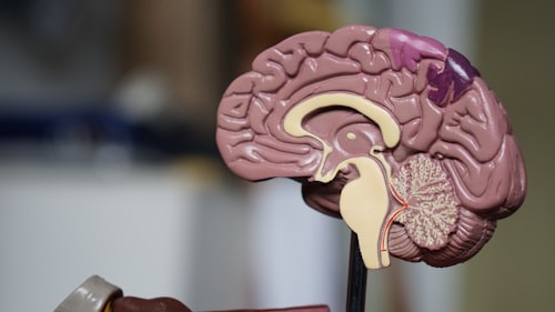
Introduction: Neural Interfaces and the Biological Challenge
Imagine a future where paralyzed individuals can control robotic limbs with their thoughts, where blind people can see through artificial vision systems, or where neurological disorders like Parkinson's disease are treated with precise neural stimulation. This isn't just science fiction—it's the promising potential of neural interface technology.
"At the heart of this technology are silicon-based neural probes: tiny devices that can record from and stimulate neurons in the brain. But there's a problem: our brains aren't exactly welcoming to these artificial intruders."
When scientists implant these silicon probes into brain tissue, the brain mounts a defensive response that ultimately leads to the device being walled off by scar tissue. This biological response significantly diminishes the functionality of these devices over time, presenting a major hurdle to developing reliable long-term neural interfaces.
What Are Neural Progenitor Cells? The Brain's Natural Repair System
Before we dive into how these cells can help neural probes, let's understand what neural progenitor cells are. NPCs are multipotent cells found in various regions of the brain that have the remarkable ability to differentiate into the main cell types of the nervous system: neurons, astrocytes, and oligodendrocytes.
Think of them as the brain's built-in repair crew—they're naturally involved in processes like learning, memory, and tissue repair.
What makes NPCs particularly interesting for neural interfaces is their ability to release neurotrophic factors (natural substances that promote neuron survival and function) and their potential to reduce inflammatory responses 1 .
Researchers have discovered ways to generate these cells from various sources, including adult brain tissue and—more recently—from induced pluripotent stem cells (iPSCs) 7 . This breakthrough allows scientists to create patient-specific NPCs without ethical concerns, opening the door to personalized neural interface treatments.
The Silicon Frontier: Why We Need Neural Probes
Silicon-based neural probes are among the most advanced tools for interfacing with the brain. Their manufacturing leverages decades of expertise from the semiconductor industry, allowing for incredibly precise arrays of recording sites that can monitor the activity of hundreds of neurons simultaneously.
- High-density electrode arrays
- Precise manufacturing
- Simultaneous recording/stimulation
- Unprecedented neural insights
- Foreign body response
- Glial scar formation
- Diminished signal quality
- Neuronal death around implant
Despite their technological sophistication, these probes face a fundamental biological challenge: the foreign body response. When implanted, the brain recognizes the probe as a foreign object and triggers an immune response. Microglia (the brain's immune cells) and astrocytes (star-shaped support cells) migrate to the implantation site, eventually forming a protective glial scar that insulates the probe from the very neurons it's trying to monitor 1 .
The Innovative Approach: Seeding Probes With Neural Progenitor Cells
The exciting solution that researchers have developed involves creating a biological interface between the silicon probe and brain tissue by pre-seeding the devices with NPCs before implantation.
Traditional Approach
Fighting against biological response
Paradigm Shift
From fighting to collaborating with biology
New Approach
Guiding biological response beneficially
This approach represents a paradigm shift in neural interface design. Instead of viewing the implant as a purely artificial device, researchers are now creating biohybrid interfaces that combine the best of both technological and biological worlds.
How NPCs Enhance Integration
- 1 Providing a more biocompatible surface
- 2 Releasing neurotrophic factors
- 3 Differentiating into connecting neurons
- 4 Modulating inflammatory response 1
Inside a Groundbreaking Experiment: Step-by-Step
Let's take a closer look at one of the pioneering studies that demonstrated the potential of this approach 1 :
Methodology: Building a Biological Interface
Surface Modification
Silicon probes were coated with laminin using silane chemistry to create covalent bonds between the silicon surface and the protein molecules.
Cell Seeding
Neural progenitor cells derived from adult murine subependyma were genetically engineered to express green fluorescent protein (GFP).
In Vitro Testing
The cells were grown on both modified and unmodified silicon surfaces for 14 days with evaluations of adhesion, growth, and differentiation.
Shear Force Testing
Controlled shear forces were applied to test cell attachment strength under implantation-like conditions.
In Vivo Implantation
The pre-seeded probes were implanted into the cortex of mice, with evaluations after 1 and 7 days.
Results and Analysis: Promising Findings
The results of this experiment were encouraging across multiple dimensions:
Enhanced Cell Adhesion
Maintained Differentiation
In Vivo Survival
Reduced Glial Response
Summary of Results
- The laminin modification resulted in significantly improved NPC attachment
- NPCs on modified surfaces showed differentiation potential similar to standard cultures
- GFP-labeled NPCs remained viable after implantation
- Preliminary observations suggested reduced astrocytic reaction 1
The Scientist's Toolkit: Research Reagent Solutions
Creating these biohybrid neural interfaces requires specialized materials and reagents. Here are some of the key components researchers use:
| Reagent/Material | Function | Example Use Case |
|---|---|---|
| Laminin | Extracellular matrix protein that promotes cell adhesion | Coating silicon probes to improve NPC attachment |
| Silane coupling agents | Create covalent bonds between silicon and organic molecules | Immobilizing laminin on silicon surfaces |
| Neural progenitor cells | Biological interface component | Seeding on probes before implantation |
| GFP labeling | Cell tracking and visualization | Monitoring NPC survival and location after implantation |
| Doxycycline-inducible systems | Controlled gene expression | Regulating differentiation of seeded NPCs 2 |
| Conductive polymers (e.g., polypyrrole) | Create electroactive surfaces | Enhancing electrical communication with tissue 4 |
Implications and Future Directions: Toward Next-Generation Neural Interfaces
The approach of seeding neural probes with NPCs represents just the beginning of what might be possible with biohybrid interfaces. Researchers are already exploring ways to enhance this technology further:
Recent advances in controlling NPC differentiation could lead to more specialized interfaces. Scientists have developed methods to promote oligodendrocyte differentiation from NPCs using inducible expression of the Olig2 transcription factor 2 .
Combining NPC-seeded probes with conductive polymer scaffolds might further improve integration. Studies show electrical stimulation can modulate NPC behavior and enhance neurotrophic factor production 4 .
The move toward xeno-free and GMP-compatible NPC generation methods is crucial for clinical applications. Protocols to generate NPCs under completely defined conditions are being developed 7 .
Potential Clinical Applications
| Application | Potential Benefit | Current Status |
|---|---|---|
| Parkinson's disease | Restore neural circuitry with improved recording/stimulation | Preclinical research |
| Paralysis | Enable stable control of prosthetic limbs | Preclinical research |
| Stroke recovery | Promote tissue repair and functional recovery | Early clinical trials 3 |
| Epilepsy management | Improved detection and intervention for seizures | Preclinical research |
| Chronic pain | More targeted neuromodulation therapies | Preclinical research |
Advanced Monitoring Techniques
Novel imaging approaches, such as the 18F-SynVesT-1 PET imaging technique mentioned in recent research 3 , could allow clinicians to non-invasively monitor synapse formation and integration of transplanted cells in patients receiving neural implants.
Conclusion: Bridging the Biological-Digital Divide
The integration of neural progenitor cells with silicon-based neural probes represents an exciting convergence of biology and technology. By learning to work with the brain's natural repair mechanisms rather than against them, scientists are developing neural interfaces that could maintain their functionality for years rather than months.
The Future of Neural Interfaces
This approach highlights a broader shift in medical technology: from fighting biology to collaborating with it. As research progresses, we move closer to a future where seamless integration between human tissue and advanced electronics enables revolutionary treatments for neurological conditions that currently have limited therapeutic options.
The path forward will require continued collaboration between materials scientists, electrical engineers, cell biologists, and clinicians. But the potential reward—transforming how we treat neurological disorders and potentially even enhancing human capabilities—makes this one of the most exciting frontiers in modern science.
Collaborative Science
Multidisciplinary approaches driving innovation
Ethical Considerations
As these technologies develop, important ethical considerations will need to be addressed regarding the appropriate use of brain-computer interface technologies. Nevertheless, the careful and thoughtful development of biohybrid neural interfaces offers tremendous hope for millions of people affected by neurological conditions worldwide.