Systemic Inflammation Biomarkers in Disease Prognostication: A Comprehensive Analysis of NLR, PLR, and LMR Across Clinical Contexts
This comprehensive review synthesizes current evidence on neutrophil-to-lymphocyte ratio (NLR), platelet-to-lymphocyte ratio (PLR), and lymphocyte-to-monocyte ratio (LMR) as prognostic biomarkers across inflammatory diseases and cancer immunotherapy.
Systemic Inflammation Biomarkers in Disease Prognostication: A Comprehensive Analysis of NLR, PLR, and LMR Across Clinical Contexts
Abstract
This comprehensive review synthesizes current evidence on neutrophil-to-lymphocyte ratio (NLR), platelet-to-lymphocyte ratio (PLR), and lymphocyte-to-monocyte ratio (LMR) as prognostic biomarkers across inflammatory diseases and cancer immunotherapy. Drawing from recent meta-analyses and clinical studies, we examine the foundational biology underlying these hematological indices, standardized methodological approaches for their application, strategies to address measurement variability, and comparative performance validation across diverse clinical contexts including inflammatory bowel disease, non-alcoholic fatty liver disease, gastric cancer, and melanoma. The analysis demonstrates that elevated NLR and PLR consistently correlate with poorer survival outcomes and increased disease activity, while higher LMR generally indicates improved prognosis. These readily accessible biomarkers offer significant potential for enhancing risk stratification, treatment monitoring, and clinical decision-making in both inflammatory conditions and oncology.
The Biology of Systemic Inflammation: Understanding NLR, PLR, and LMR as Pathophysiological Indicators
The systemic immune status of an individual provides crucial insights into their health, particularly in the context of chronic diseases like cancer, autoimmune disorders, and inflammatory conditions. Peripheral blood cell ratios—specifically the Neutrophil-to-Lymphocyte Ratio (NLR), Platelet-to-Lymphocyte Ratio (PLR), and Lymphocyte-to-Monocyte Ratio (LMR)—have emerged as accessible, cost-effective, and reproducible biomarkers that reflect the underlying balance between pro-inflammatory and anti-inflammatory pathways in the body. These hematological indices, derived from routine complete blood count (CBC) parameters, offer a window into the host's immune response and have demonstrated significant prognostic value across diverse medical conditions. Their calculation integrates multiple immune cell populations, providing a more comprehensive assessment of systemic inflammation than individual cell counts alone.
The biological rationale for these ratios lies in their representation of competing immune processes. Neutrophils drive pro-inflammatory responses and can promote tumor progression and tissue damage, while lymphocytes mediate anti-tumor and anti-inflammatory responses. Platelets contribute to inflammatory processes and thrombosis, and monocytes differentiate into tumor-associated macrophages that facilitate immune suppression. Therefore, elevated NLR and PLR typically indicate a pro-inflammatory, immunosuppressive state, while a higher LMR reflects robust immune surveillance. The integration of these markers provides clinicians and researchers with valuable tools for prognostic stratification, treatment monitoring, and clinical decision-making across oncology, gastroenterology, and other medical specialties.
Comparative Analysis of NLR, PLR, and LMR Across Conditions
Prognostic Utility in Malignant Conditions
Table 1: Prognostic Value of Blood Cell Ratios in Oncology
| Cancer Type | NLR Impact (Cut-off) | PLR Impact (Cut-off) | LMR Impact (Cut-off) | Outcomes Measured | Citation |
|---|---|---|---|---|---|
| Non-Small Cell Lung Cancer | HR: 9.923 (≥3.57) | HR: 9.978 (≥216.00) | Not assessed | OS, PFS | [1] |
| Melanoma (ICI Treatment) | HR: 2.21 OS, 1.80 PFS | HR: 2.15 OS, 1.67 PFS | HR: 0.36 OS, 0.56 PFS | OS, PFS | [2] |
| Early-Stage NSCLC | Significant (102.7 vs 109.4 months) | Significant (104.1 vs 110.1 months) | Significant (101.0 vs 110.3 months) | OS, DFS | [3] |
| Osteosarcoma | HR: 1.88 OS, 1.67 DFS | Not significant | Not significant | OS, DFS | [4] |
| Lip Cancer | HR: 5.885 (>2.134) | Not independent predictor | Not independent predictor | OS | [5] |
The consistent pattern across oncologic studies demonstrates that elevated NLR and PLR are associated with poorer survival outcomes, while higher LMR typically correlates with improved prognosis. In NSCLC, elevated NLR (≥3.57), PLR (≥216.00) were independently associated with worse survival outcomes with hazard ratios approaching 10, indicating substantial prognostic impact [1]. The combination of these inflammatory markers further enhanced prognostic discrimination, with area under the curve (AUC) values reaching 0.906 for overall survival prediction, significantly outperforming individual markers [1].
In melanoma patients receiving immune checkpoint inhibitors, elevated NLR and PLR were associated with significantly poorer overall survival (HR=2.21 and HR=2.15, respectively) and progression-free survival [2]. Conversely, an elevated LMR was associated with improved survival outcomes (HR=0.36 for OS), highlighting its protective role [2]. This inverse relationship pattern for LMR is consistent across multiple cancer types, reflecting its representation of effective immune surveillance.
Diagnostic Accuracy in Inflammatory and Immune Conditions
Table 2: Inflammatory Biomarkers in Non-Malignant Conditions
| Condition | NLR Performance | PLR Performance | LMR Performance | Clinical Utility | Citation |
|---|---|---|---|---|---|
| Inflammatory Bowel Disease | WMD=1.50 (active vs remission) | WMD=69.02 (active vs remission) | WMD=-1.14 (active vs remission) | Disease activity monitoring | [6] |
| Indeterminate Thyroid Nodules | AUC=0.685 (cut-off=2.202) | Not significant | Not significant | Malignancy prediction | [7] |
| COVID-19 Serology | Not assessed | Not assessed | Not assessed | Infection detection | [8] |
In non-malignant conditions, these inflammatory markers demonstrate distinct patterns. For inflammatory bowel disease (IBD), NLR and PLR were significantly higher in active disease compared to remission (WMD=1.50 and WMD=69.02, respectively), while LMR was significantly lower (WMD=-1.14) [6]. This pattern reinforces the concept that NLR and PLR reflect inflammatory activity, while LMR represents regulatory capacity.
For thyroid nodules with indeterminate cytology, NLR demonstrated prognostic capability for predicting malignancy with an AUC of 0.685 at a cut-off of 2.202 [7]. This application highlights the potential role of inflammatory biomarkers in preoperative risk stratification beyond traditional oncologic applications.
Methodological Framework for Biomarker Assessment
Standardized Experimental Protocols
The investigation of hematological ratios requires standardized methodologies to ensure reproducible and comparable results across studies. The following protocols represent consolidated approaches from multiple research investigations:
Blood Collection and Processing Protocol:
- Sample Collection: Fasting venous blood (3-5 mL) is collected in EDTA tubes within 15 days prior to treatment initiation or intervention [1] [3]. A 12-hour overnight fast is recommended for standardization [1].
- Laboratory Analysis: Complete blood count with differential is performed using automated hematology analyzers (e.g., Sysmex XN-3000, Mindray BC-6800, or Beckman Coulter UniCel DxH 800) [3]. Quality control procedures follow manufacturer specifications and institutional standards.
- Parameter Calculation:
- Statistical Analysis: Receiver operating characteristic (ROC) curves determine optimal cut-off values for each ratio based on clinical outcomes [1] [5]. Kaplan-Meier survival analysis and Cox proportional hazards models assess prognostic significance, with multivariate analysis adjusting for potential confounders [1] [3].
Quality Assurance Considerations:
- Exclusion of patients with active infection, hematologic disorders, autoimmune diseases, or recent blood transfusions that could affect inflammatory markers [1] [3]
- Timing consistency for blood collection relative to diagnosis or treatment initiation
- Analysis of fresh blood samples within 2-4 hours of collection to prevent cellular degradation
- Use of internal controls and participation in external quality assurance schemes [9]
Advanced Immune Function Assessments
Beyond basic ratio calculations, advanced immune function assessments provide deeper insights into mechanistic pathways:
Lymphocyte Phenotyping: Flow cytometric analysis of CD4+ and CD8+ T-lymphocytes provides detailed immunophenotyping. A decrease in CD4+ and a CD4+:CD8+ ratio of less than 1.5 correlates with immune impairment and increased susceptibility to infection [10].
Lymphocyte Stimulation Assays: Functional assessments include in vitro stimulation with plant mitogens (e.g., phytohemagglutinin), recall antigens, or allogeneic cells to measure lymphocyte blastogenesis and proliferation. Measurable indicators include [3H]thymidine incorporation into DNA, expression of cell-surface activation antigens (CD25), cytokine release, and shedding of soluble IL-2 receptors [10].
Circulating Cytokine and Soluble Receptor Assays: Immunoassays and bioassays measure circulating cytokines (e.g., IL-1β, TNF-α, IFN-γ, IL-10) and soluble receptors, though methodological considerations include the effects of cytokine-binding proteins, antagonists, and assay sensitivity limitations [10].
Visualizing Systemic Immune Signaling Pathways
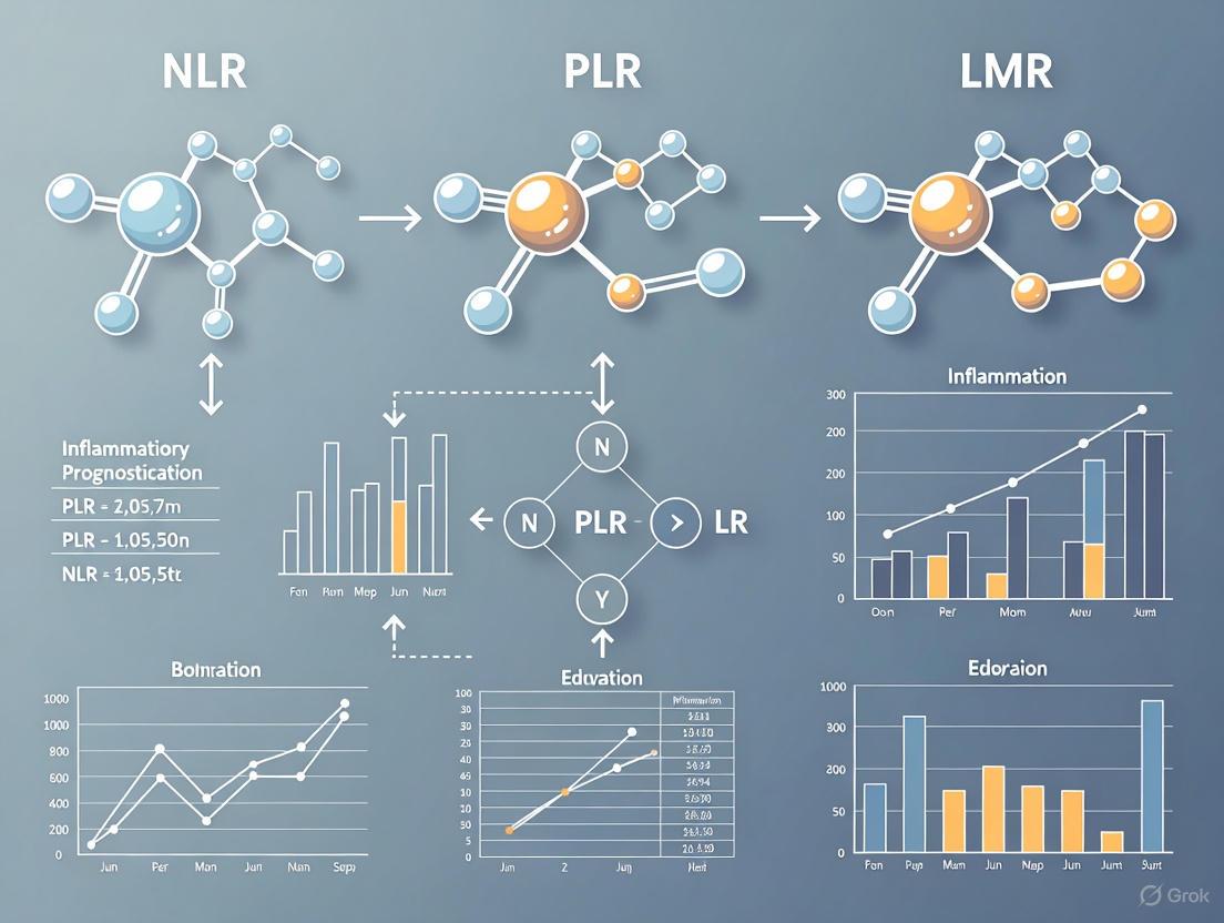
Systemic Immune Signaling Pathway
This diagram illustrates the pathophysiological mechanisms linking inflammatory stimuli to altered blood cell ratios and subsequent systemic immunosuppression. The process begins with various inflammatory triggers (cancer, infection, or autoimmune conditions) activating bone marrow hematopoiesis, resulting in increased release and activation of neutrophils. These activated neutrophils secrete pro-inflammatory cytokines including IL-6 and TNF-α, which simultaneously induce lymphocyte suppression and apoptosis while promoting platelet activation and monocyte differentiation into M2 macrophages. These cellular changes directly manifest in the peripheral blood as elevated NLR (increased neutrophils/decreased lymphocytes), elevated PLR (increased platelets/decreased lymphocytes), and decreased LMR (decreased lymphocytes/increased monocytes). Collectively, these altered ratios reflect a state of systemic immunosuppression that facilitates disease progression across various pathological conditions [1] [2] [3].
Essential Research Reagent Solutions
Table 3: Essential Research Reagents for Biomarker Investigation
| Reagent/Category | Specification | Research Function | Representative Example |
|---|---|---|---|
| Blood Collection | EDTA tubes | Pre-analytical sample preservation for CBC with differential | K2EDTA Vacutainer tubes (BD) [3] |
| Hematology Analyzer | Automated CBC with 5-part differential | Absolute cell counts for neutrophil, lymphocyte, platelet, monocyte quantification | Sysmex XN-3000, Mindray BC-6800 [3] |
| Flow Cytometry | CD4+, CD8+ monoclonal antibodies | Lymphocyte subpopulation phenotyping for immune status assessment | CD4-FITC/CD8-PE antibodies [10] |
| Cytokine Assays | ELISA kits for IL-6, TNF-α, IL-10 | Quantification of pro-inflammatory and anti-inflammatory cytokines | High-sensitivity ELISA kits [10] |
| Statistical Software | SPSS, STATA, R | ROC analysis, survival analysis, multivariate regression | SPSS v26.0 (IBM) [1] [5] |
The research reagents and instruments listed in Table 3 represent the essential infrastructure for conducting rigorous investigations into blood cell ratios and systemic immune status. Automated hematology analyzers with 5-part differential capabilities provide the fundamental cellular quantification necessary for ratio calculations, with strict quality control procedures ensuring analytical precision [3]. Flow cytometry reagents enable deeper immunophenotyping beyond standard complete blood count parameters, particularly for assessing T-cell subsets (CD4+, CD8+) and their ratios, which provide additional insights into immune competence [10].
Enzyme-linked immunosorbent assay (ELISA) kits for cytokine quantification allow researchers to correlate cellular ratios with soluble inflammatory mediators, establishing mechanistic links between cellular patterns and inflammatory pathways [10]. Specialized statistical software packages are indispensable for determining optimal cut-off values through ROC analysis, conducting survival analyses, and performing multivariate adjustments for potential confounding factors [1] [5]. Together, these research tools enable comprehensive assessment of systemic immune status through multiple complementary methodologies.
Blood cell ratios—NLR, PLR, and LMR—provide valuable insights into systemic immune status by integrating information from multiple cellular components of the innate and adaptive immune systems. The consistent prognostic performance of these biomarkers across diverse conditions, including various cancers, inflammatory diseases, and infection responses, underscores their fundamental role in reflecting the balance between pro-inflammatory and anti-inflammatory pathways. Their accessibility, cost-effectiveness, and reproducibility make them particularly valuable for both clinical practice and research applications.
The combination of these ratios often enhances prognostic discrimination beyond individual markers, as demonstrated by the substantial improvement in AUC values when NLR, PLR, and SII are combined for predicting overall survival in NSCLC patients [1]. Furthermore, the inverse relationship patterns observed across conditions—where elevated NLR and PLR typically indicate poorer outcomes while elevated LMR suggests better prognosis—reinforce the biological plausibility of these markers as representations of competing immune processes. As research in this field advances, standardization of methodological approaches and cut-off values will further enhance the comparability and clinical utility of these promising biomarkers.
Chronic systemic inflammation is a fundamental component of the pathophysiology of numerous diseases, from cancer and cardiovascular conditions to autoimmune disorders and severe infections. The complex interplay between the innate and adaptive immune systems creates a biochemical signature that can be measured through peripheral blood parameters. Among these, the Neutrophil-to-Lymphocyte Ratio (NLR) has emerged as a particularly compelling biomarker that integrates two crucial arms of the immune response: neutrophils as mediators of innate immunity and lymphocytes as effectors of adaptive immunity [11]. Calculated as a simple ratio between absolute neutrophil and lymphocyte counts from routine complete blood tests, NLR provides a window into the body's inflammatory status and stress response that is both economically accessible and routinely obtainable in clinical settings worldwide.
The clinical significance of NLR stems from its ability to mirror the delicate homeostasis between pro-inflammatory and anti-inflammatory pathways. Under conditions of physiological stress, whether from acute infection, malignancy, or cardiovascular events, the body typically mounts a neutrophilic response while simultaneously suppressing lymphocyte counts through increased cortisol and catecholamine release [11]. This dynamic shift creates an elevated NLR that has demonstrated prognostic value across an astonishingly broad spectrum of pathologies. The biomarker's strength lies in its synthesis of two complementary biological narratives: neutrophil elevation represents the acute phase of inflammatory response, while lymphopenia reflects physiological stress and impaired immune surveillance [12] [11].
In comparative inflammometry research, NLR is frequently evaluated alongside other ratio-based biomarkers, particularly the Platelet-to-Lymphocyte Ratio (PLR) and Lymphocyte-to-Monocyte Ratio (LMR), which provide additional dimensions of inflammatory and immune status. While PLR integrates thrombotic and inflammatory pathways, and LMR reflects monocyte-driven chronic inflammation and lymphocytic immune competence, NLR remains the most extensively validated ratio across conditions and populations [13] [14]. This review employs a comparative framework to objectively examine the experimental evidence supporting NLR's performance characteristics against these alternative inflammatory ratios, with particular attention to methodological standardization, prognostic accuracy, and clinical applicability across diverse disease states and patient populations.
Experimental Protocols and Methodological Standards
Standardized Protocol for NLR Determination
The measurement of NLR follows a straightforward protocol that can be implemented in virtually any clinical or research setting with access to basic hematological analysis capabilities. The standard methodology involves the collection of peripheral venous blood samples in EDTA tubes to prevent coagulation, with analysis typically performed within 2-4 hours of collection to ensure cellular integrity [3]. Automated hematology analyzers (such as Sysmex, Mindray, or Beckman Coulter systems) provide the absolute neutrophil and lymphocyte counts through impedance technology and flow cytometry principles. The NLR is then calculated by dividing the absolute neutrophil count by the absolute lymphocyte count, with no unit designation as it represents a pure ratio [3].
Critical methodological considerations include the timing of blood collection relative to disease onset or therapeutic interventions, as NLR demonstrates dynamic fluctuations in response to clinical status. For prognostic studies, baseline NLR is typically measured before initiation of treatment or at initial diagnosis [12] [15]. Additionally, researchers must account for potential confounders including age (NLR naturally increases with advanced age), exogenous steroid administration, hematological disorders, acute physiological stress, and certain medications that can affect white cell subpopulations [11]. The stability of NLR measurements under proper storage conditions and the minimal intra-individual diurnal variation further enhance its reliability as a biomarker when standardized protocols are followed.
Comparative Methodologies for PLR and LMR Determination
The PLR is calculated by dividing the absolute platelet count by the absolute lymphocyte count, while LMR is derived by dividing the absolute lymphocyte count by the absolute monocyte count [13] [14]. All three ratios utilize components of the complete blood count with differential, but they reflect distinct physiological pathways. The analytical protocols share common pre-analytical requirements regarding blood collection and processing, though the interpretation of each ratio emphasizes different aspects of the inflammatory cascade: NLR primarily reflects acute inflammation and stress, PLR integrates thrombotic and inflammatory pathways, and LMR emphasizes adaptive immune function against monocyte-driven chronic inflammation [13] [14] [16].
Table 1: Standardized Experimental Protocol for Inflammatory Ratio Biomarkers
| Protocol Step | NLR-Specific Considerations | PLR-Specific Considerations | LMR-Specific Considerations |
|---|---|---|---|
| Sample Collection | EDTA venous blood; fasting not required | EDTA venous blood; fasting not required | EDTA venous blood; fasting not required |
| Time to Analysis | Within 2-4 hours | Within 2-4 hours (platelets more sensitive to time) | Within 2-4 hours |
| Required Parameters | Absolute neutrophil count, Absolute lymphocyte count | Platelet count, Absolute lymphocyte count | Absolute lymphocyte count, Absolute monocyte count |
| Calculation Formula | Neutrophils ÷ Lymphocytes | Platelets ÷ Lymphocytes | Lymphocytes ÷ Monocytes |
| Primary Biological Reflection | Innate vs adaptive immune balance | Thrombotic-inflammatory interplay | Immune competence vs monocyte-driven inflammation |
| Key Confounders | Steroids, acute stress, infection | Thrombocytopenia, splenectomy | Chronic infections, hematological disorders |
Comparative Performance Across Disease States
Prognostic Performance in Oncological Conditions
The prognostic value of inflammatory ratios has been most extensively validated in oncology, where systemic inflammation plays a crucial role in tumor progression, metastasis, and response to therapy. A comprehensive retrospective analysis of individual patient data from five Phase III clinical trials across multiple cancer types demonstrated that elevated baseline NLR was significantly associated with worse overall survival (OS) and progression-free survival (PFS) [12]. In Cox multivariate analyses, NLR remained an independent predictor of OS with a hazard ratio (HR) of 1.508 (95% CI: 1.390–1.636, p<0.001), outperforming both isolated neutrophil count (N1 HR: 1.390) and lymphocyte count (L1 HR: 0.801) [12]. The superior prognostic performance of NLR compared to its individual components highlights the clinical value of evaluating the balance between these immune compartments rather than absolute counts alone.
In early-stage non-small cell lung cancer (NSCLC), a multicenter study of 2,159 surgical patients found that elevated preoperative NLR was associated with significantly shorter overall survival (102.7 vs. 109.4 months, p=0.040) [3]. The comparative analysis in the same cohort revealed that high PLR was also a poor prognostic factor for both OS (104.1 vs. 110.1 months, p=0.017) and disease-free survival (102.5 vs. 108.7 months, p=0.021), while low LMR was associated with worse OS (101 vs. 110.3 months, p<0.001) and DFS (100.2 vs. 108.6 months, p=0.020) [3]. This large-scale investigation demonstrates that while all three inflammatory ratios provide prognostic information, their effect sizes and significance levels vary, with NLR and LMR showing particularly robust associations with survival outcomes.
In the emerging field of immunotherapy, NLR has shown particular promise as a predictive biomarker. A multicentric study of 135 patients with recurrent/metastatic head and neck squamous cell carcinoma (HNSCC) treated with immune checkpoint inhibitors found that patients with baseline NLR ≤4 had significantly superior outcomes across multiple endpoints [15]. The median overall survival was 37.4 months for low-NLR patients compared to 23.1 months for high-NLR patients (p=0.002), while progression-free survival was 20 months versus 6.5 months, respectively (p=0.013) [15]. The objective response rate was similarly stratified by NLR (20% for NLR≤4 vs. 12.5% for NLR>4), suggesting that NLR may help identify patients most likely to benefit from immunotherapeutic approaches [15].
For gastric cancer patients receiving immunotherapy, a systematic review and meta-analysis revealed that LMR demonstrated particularly strong prognostic performance, with high pre-treatment LMR associated with improved progression-free survival (HR=0.58; 95% CI: 0.47–0.71, p<0.00001) and overall survival (HR=0.51, 95% CI: 0.33–0.79; p=0.003) [17]. The post-treatment LMR dynamics also showed prognostic significance for PFS (HR=0.48; 95% CI: 0.29–0.79; p=0.004), suggesting potential utility in monitoring treatment response [17].
Table 2: Comparative Performance of Inflammatory Ratios in Oncology
| Cancer Type | NLR Performance | PLR Performance | LMR Performance | Study Details |
|---|---|---|---|---|
| Multiple Cancers | OS HR: 1.508 (1.390-1.636) p<0.001 | Not assessed | Not assessed | 5 Phase III trials retrospective analysis [12] |
| Early-Stage NSCLC | OS: 102.7 vs 109.4 months (p=0.040) | OS: 104.1 vs 110.1 months (p=0.017) DFS: 102.5 vs 108.7 months (p=0.021) | OS: 101 vs 110.3 months (p<0.001) DFS: 100.2 vs 108.6 months (p=0.020) | 2,159 patients, multicenter [3] |
| HNSCC (Immunotherapy) | OS: 23.1 vs 37.4 months (p=0.002) PFS: 6.5 vs 20 months (p=0.013) ORR: 12.5% vs 20% | Not assessed | Not assessed | 135 patients, NLR cut-off=4 [15] |
| Gastric Cancer (Immunotherapy) | Not assessed | Not assessed | PFS HR: 0.58 (0.47-0.71) p<0.00001 OS HR: 0.51 (0.33-0.79) p=0.003 | 815 patients, meta-analysis [17] |
| Breast Cancer (Neoadjuvant Therapy) | Not assessed | Not assessed | Higher in pCR patients (p<0.05) Predictive of treatment response | 70 patients vs 48 controls [16] |
Cardiovascular and Metabolic Disease Applications
In cardiovascular disease, NLR has demonstrated prognostic value for mortality and adverse events, reflecting the fundamental role of inflammation in atherosclerosis and thrombosis. A comprehensive analysis of hypertensive individuals from the NHANES database (n=15,483) revealed distinctive prognostic patterns for PLR, which exhibited a U-shaped relationship with all-cause mortality and a linear association with cardiovascular mortality [18]. Those in the highest PLR quartile had significantly elevated risks of all-cause mortality (HR=1.16, 95% CI: 1.05–1.29, p=0.004) and cardiovascular mortality (HR=1.47, 95% CI: 1.20–1.80, p<0.001) after multivariate adjustment [18]. The study identified a PLR threshold of 118.83 as indicative of adverse prognosis for all-cause mortality, providing a potential clinical decision point for risk stratification [18].
The pathophysiological basis for inflammatory ratios in cardiovascular disease stems from the integral role of immune cells in atherosclerosis progression and plaque instability. Neutrophils contribute to plaque vulnerability through the release of proteolytic enzymes and neutrophil extracellular traps (NETs), while lymphocytes exhibit atheroprotective effects through immunoregulatory functions [19]. This balance is captured by NLR, which has been shown to improve prognostic classification beyond traditional risk scores like Framingham [19]. The association between elevated NLR and poor outcomes in heart failure, coronary artery disease, and acute coronary syndromes underscores the clinical relevance of this inflammatory biomarker across the cardiovascular spectrum.
Inflammatory and Infectious Disease Applications
Inflammatory bowel disease (IBD) represents another condition where ratio-based biomarkers have shown significant utility. A meta-analysis of 23 cohort studies involving 3,550 IBD patients and 1,010 healthy controls found that both NLR and PLR were significantly elevated in IBD patients compared to healthy populations (NLR WMD=1.57, 95% CI: 1.14–2.01, p<0.001; PLR WMD=60.66, 95% CI: 51.68–69.64, p<0.001) [13]. Furthermore, these ratios effectively discriminated between active and remission disease stages, with significant differences observed for NLR (WMD=1.50, 95% CI: 1.23–1.78, p<0.001), PLR (WMD=69.02, 95% CI: 39.66–98.39, p<0.001), and LMR (WMD=-1.14, 95% CI: -1.43–-0.86, p<0.001) [13]. The diagnostic accuracy for predicting clinical activity was favorable across markers, with a pooled AUC of 0.72 (95% CI: 0.69–0.75, p<0.001) [13].
In infectious diseases, particularly sepsis and pneumonia, NLR has emerged as an early marker of physiological stress that can precede other laboratory parameters. In sepsis, NLR values correlate with disease severity, with one prospective observational study reporting NLR values of 9.53±2.31 in septic ICU patients, correlating with SOFA scores (R=0.65) and presepsin levels (R=0.56) [11]. The same study found significantly higher NLR in patients with septic shock (10.31±2.32), suggesting potential utility in stratification algorithms [11]. For community-acquired pneumonia, NLR has demonstrated strong predictive value for short- and long-term mortality, need for ICU admission, and re-hospitalization, in some cases outperforming traditional pneumonia severity scores [11].
Pathophysiological Framework and Signaling Pathways
The biological plausibility underlying NLR as a biomarker stems from its representation of competing immunological pathways. Neutrophils mediate the innate immune response through phagocytosis, release of reactive oxygen species, granular proteins, cytokines, and formation of neutrophil extracellular traps (NETs) [12] [11]. In cancer biology, neutrophils contribute to multiple stages of tumor progression including carcinogenesis (through ROS-induced DNA damage), immunosuppression (via arginase-1 release inhibiting T-cell function), and metastasis (through angiogenesis promotion and reactivation of dormant cells) [12].
Conversely, lymphocytes represent adaptive immunity and are crucial for effective antitumor response and immune surveillance. Lymphopenia reflects impaired immune competence and has been associated with poor prognosis across multiple cancers [12]. The NLR thus captures the balance between pro-tumor inflammatory forces and anti-tumor immune defense, providing a quantitative measure of the host's immune status in relation to disease burden.
The following diagram illustrates the key pathophysiological pathways reflected by inflammatory ratio biomarkers:
Immune Signaling Pathways Captured by Inflammatory Ratios
The diagram illustrates how different physiological stressors disrupt immune homeostasis, leading to characteristic cellular responses that are quantified by inflammatory ratios. NLR captures the balance between innate neutrophilic inflammation and adaptive lymphocytic immunity, representing acute phase response and physiological stress. PLR integrates thrombotic (platelet) and immune (lymphocyte) pathways, reflecting the interplay between coagulation and inflammation. LMR emphasizes the relationship between adaptive immune competence (lymphocytes) and chronic inflammation (monocytes), particularly relevant in cancer immunology.
Research Reagent Solutions and Methodological Toolkit
The experimental determination of inflammatory ratios relies on standardized hematological analytical systems and reagents. The following table outlines essential research materials and their applications in inflammometry studies:
Table 3: Essential Research Reagents and Methodological Toolkit
| Reagent/Instrument Category | Specific Examples | Research Application | Technical Considerations |
|---|---|---|---|
| Blood Collection Systems | EDTA vacuum tubes, tourniquets, venipuncture kits | Standardized sample acquisition for complete blood count with differential | EDTA preferred over heparin for morphology; time to processing critical for platelet integrity |
| Hematology Analyzers | Sysmex XN-3000, Mindray BC-6800, Beckman Coulter UniCel DxH 800 | Automated determination of absolute neutrophil, lymphocyte, monocyte, and platelet counts | Different platforms may show slight variability; consistency within studies essential |
| Quality Control Materials | Commercial whole blood controls at normal and abnormal levels, proficiency testing programs | Ensuring analytical precision and accuracy across measurement runs | Should include three levels of controls covering normal, low, and high ranges |
| Data Analysis Tools | Statistical software (R, SPSS, STATA), sample size calculation tools (G*Power) | Power analysis, cutoff determination, survival analysis | ROC analysis often used for optimal cutoff determination; Cox regression for survival outcomes |
Comparative Limitations and Standardization Challenges
Despite the compelling evidence supporting NLR's prognostic utility, several methodological challenges require consideration in comparative inflammometry research. The determination of optimal cut-off values remains a significant hurdle, with reported thresholds varying substantially across studies. For NLR, proposed cut-offs range from 2.5 to 5.0 across different conditions and populations, while PLR cut-offs show even wider variation from approximately 120 to 180 [12] [18] [15]. This heterogeneity stems from multiple factors including differences in patient demographics, disease stages, laboratory methodologies, and statistical approaches for cutoff determination.
Biological and clinical confounders present additional challenges in the interpretation of inflammatory ratios. NLR is influenced by age (typically higher in elderly populations), sex (generally higher in males), exogenous steroid administration, hematological disorders, and acute physiological stress [11]. The normal reference range for NLR in healthy adult populations is generally considered to be between 0.78 and 3.53, though population-specific standards continue to be refined [11]. The Rotterdam Study, a large population-based prospective cohort, reported a mean NLR of 1.76 in the general population, with 2.5% and 97.5% limits of 0.83 and 3.92, respectively [11].
For PLR, reference values also demonstrate population variability, with studies reporting means ranging from approximately 120 in European populations to 132 in South Korean cohorts [14]. The dynamic nature of these ratios in response to clinical status necessitates careful consideration of timing in relation to disease course and therapeutic interventions. While single measurements provide valuable prognostic information, serial measurements may offer additional insights into treatment response and disease trajectory.
In the comparative landscape of inflammatory biomarkers, NLR has established itself as a robust, accessible, and clinically informative parameter that reflects fundamental immune balance between innate and adaptive responses. The extensive validation across diverse disease states—from oncology and cardiology to infectious and inflammatory conditions—supports its utility as a prognostic tool and potential predictive biomarker. When evaluated against PLR and LMR, NLR demonstrates particular strength in acute inflammatory conditions and scenarios of physiological stress, while LMR may offer advantages in chronic inflammatory states and specific immunotherapy contexts.
The integration of inflammatory ratios into clinical decision-making requires ongoing standardization efforts, particularly regarding optimal cutoff determination and accounting for population-specific variations. Future research directions should include prospective validation of dynamic ratio monitoring during treatment courses, exploration of composite models integrating multiple inflammatory parameters, and investigation of the molecular mechanisms underlying the consistent prognostic performance of these hematological biomarkers across such diverse pathological states.
For researchers and drug development professionals, NLR represents a practical biomarker that can be immediately implemented in clinical trials for patient stratification and outcome assessment. Its low cost, universal availability, and strong prognostic performance position it as an valuable tool in the era of personalized medicine, particularly when interpreted within the broader context of clinical presentation and complementary biomarkers.
The Platelet-to-Lymphocyte Ratio (PLR) has emerged as a significant biomarker in the landscape of inflammatory prognostication research. As an integrative measure derived from routine complete blood counts, PLR reflects the delicate balance between two fundamental biological systems: the pro-thrombotic, inflammatory functions of platelets and the regulatory, adaptive capabilities of lymphocytes [20]. Within the context of comparative studies involving the Neutrophil-to-Lymphocyte Ratio (NLR) and Lymphocyte-to-Monocyte Ratio (LMR), PLR offers unique insights into the interplay between hemostasis and immune regulation across various pathological states, particularly in thrombosis, cancer progression, and cardiovascular diseases. The value of PLR lies in its ability to bridge these interconnected pathways, providing clinicians and researchers with a cost-effective, readily accessible tool for risk stratification and prognostic assessment [21]. This review systematically compares the prognostic performance, methodological standardization, and clinical applications of PLR against NLR and LMR, with a specific focus on thrombotic and immune regulatory mechanisms.
Comparative Performance of Inflammatory Ratios
Table 1: Prognostic Performance of Inflammatory Ratios Across Pathologies
| Condition | Biomarker | Cut-off Value | Outcome Association | Hazard Ratio (HR) / Correlation | Source |
|---|---|---|---|---|---|
| Colon Cancer | PLR | ≈150 | Positive correlation with tumor size & stage | r=0.428 (p<0.001) for tumor size | [22] |
| NLR | ≈3 | No significant correlation with tumor aggressiveness | Not significant | [22] | |
| Melanoma (ICI-treated) | PLR | Variable | Poorer OS & PFS | OS: HR=2.15; PFS: HR=1.67 | [2] |
| NLR | Variable | Poorer OS & PFS | OS: HR=2.21; PFS: HR=1.80 | [2] | |
| LMR | Variable | Improved OS & PFS | OS: HR=0.36; PFS: HR=0.56 | [2] | |
| Gastric Cancer (ICI-treated) | PLR | Variable | Poorer OS & PFS | OS: HR=1.57; PFS: HR=1.52 | [23] |
| NLR | Variable | Poorer OS & PFS | OS: HR=2.01; PFS: HR=1.59 | [23] | |
| LMR | Variable | Improved OS & PFS | OS: HR=0.62; PFS: HR=0.69 | [23] | |
| Lip Cancer | PLR | >146.5 | Increased mortality | Significant (univariate) | [5] |
| NLR | >2.13 | Independent predictor of OS | HR=5.885 (multivariate) | [5] | |
| LMR | ≤4.00 | Increased mortality | Significant (univariate) | [5] | |
| Hypertension | PLR | 118.8 (threshold) | U-shaped (all-cause); Linear (CVD mortality | All-cause: Q4 HR=1.16; CVD: Q4 HR=1.47 | [18] |
| Contrast-Induced Nephropathy | PLR | 143 | CIN development | p=0.006 | [24] |
| NLR | 3.3 | CIN development | p<0.001 | [24] | |
| LMR | 2.8 | CIN development | p=0.016 | [24] |
The comparative analysis of inflammatory ratios reveals distinct prognostic strengths across various clinical contexts. In oncological applications, PLR demonstrates particular utility in assessing tumor aggressiveness, as evidenced by its significant correlation with tumor size (r=0.428, p<0.001) and stage in colon cancer, whereas NLR showed no significant association in the same cohort [22]. In immunotherapy-treated malignancies, NLR consistently demonstrates strong prognostic value for overall survival (OS) and progression-free survival (PFS), with hazard ratios often exceeding those of PLR [2] [23]. The differential performance highlights how each biomarker reflects distinct aspects of the immune response: PLR better reflects tumor burden and platelet-mediated processes, while NLR is more associated with systemic inflammation, and LMR indicates immune competence and nutritional status [22] [25].
Table 2: Biological Rationale and Clinical Context of Inflammatory Ratios
| Biomarker | Biological Rationale | Primary Clinical Utility | Strengths | Limitations |
|---|---|---|---|---|
| PLR | Reflects platelet-mediated inflammation & thrombotic activity + lymphocyte-mediated immune regulation | Assessing tumor aggressiveness, cardiovascular risk, thrombotic states | Integrates coagulation & immunity; strong in tumor burden assessment | Affected by non-inflammatory thrombocytosis; limited in lymphopenic states |
| NLR | Balances innate inflammatory response (neutrophils) vs. adaptive immune regulation (lymphocytes) | Predicting systemic inflammation, infection severity, overall survival | Strong prognostic value across multiple cancers; technically robust | Less specific to thrombotic processes; confounded by many inflammatory conditions |
| LMR | Represents immune competence (lymphocytes) vs. inflammatory monocyte activity | Nutritional status, immune competence, response to immunotherapy | Strong positive prognostic marker; reflects host immune status | Limited value in isolated hematologic disorders; less studied in thrombosis |
PLR in Thrombosis and Immune Regulation: Mechanisms
The PLR serves as a integrated measure of two interconnected physiological pathways: platelet-mediated thrombosis and inflammation, and lymphocyte-mediated immune regulation. Platelets contribute to inflammatory and thrombotic processes through multiple mechanisms: they release proinflammatory agents and microparticles, express P-selectin which facilitates interactions with leukocytes and lymphocytes, and form conjugates with neutrophils that intensify inflammatory responses [21]. Through P-selectin mediated interactions with T-lymphocytes, platelets can reduce lymphocyte proliferation and modulate cytokine production, decreasing proinflammatory cytokines like TNF-α and IL-17 while increasing anti-inflammatory IL-10 [21]. This direct crosstalk establishes the pathophysiological basis for PLR as a biomarker bridging thrombotic and immune pathways.
In cancer contexts, platelets facilitate tumor growth and metastasis through multiple mechanisms: releasing pro-angiogenic factors, forming microthrombi that protect circulating tumor cells, and promoting epithelial-mesenchymal transition [22]. Simultaneously, cancer-induced lymphocytopenia reflects impaired cell-mediated immunity, reducing tumor surveillance and enabling immune evasion [22] [2]. The PLR thus captures this dual dysregulation - increased thrombotic activity and diminished immune surveillance - making it particularly valuable in cancer prognostication, especially for assessing tumor aggressiveness [22].
Figure 1: PLR in Thrombosis and Immune Regulation Pathways. This diagram illustrates the interconnected biological pathways reflected by the Platelet-to-Lymphocyte Ratio, showing how platelet activity and lymphocyte regulation converge to influence disease processes.
Methodological Standards and Experimental Protocols
Blood Collection and Processing Protocols
Standardized protocols for PLR measurement begin with proper blood collection. Venous blood samples should be collected in ethylenediaminetetraacetic acid (EDTA) tubes and processed within 30-120 minutes of collection to prevent platelet activation or lymphocyte degradation [22] [24]. Complete blood count analysis should be performed using automated hematology analyzers (e.g., Beckman Coulter analyzers), with manual smear review recommended for abnormal results [18] [24]. For research consistency, blood samples should ideally be drawn after an overnight fast and at consistent times of day to minimize diurnal variation effects [22].
Calculation and Cut-off Standards
PLR is calculated by dividing the absolute platelet count (×10³/μL) by the absolute lymphocyte count (×10³/μL). While study-specific optimal cut-offs should be determined via receiver operating characteristic curve analysis, commonly used thresholds in research include approximately 150 for PLR and 3 for NLR [22]. Recent large-scale studies have identified specific thresholds; for instance, in hypertension, a PLR of 118.83 was identified as prognostic for all-cause mortality [18], while in lip cancer, optimal thresholds were PLR >146.5 and NLR >2.13 [5]. For renal cell carcinoma, established cut-offs include NLR >3.05 and PLR >154.97 [25].
Table 3: Standardized Experimental Protocol for Inflammatory Ratio Analysis
| Step | Parameter | Standard Protocol | Quality Control Measures |
|---|---|---|---|
| 1. Patient Preparation | Fasting status | Overnight fast recommended | Document non-fasting status if applicable |
| Time of collection | Morning collection preferred | Record exact collection time | |
| 2. Blood Collection | Tube type | EDTA vacuum tubes | Check for proper filling and mixing |
| Processing time | Within 30-120 minutes | Document processing delays | |
| 3. Laboratory Analysis | Analyzer type | Automated hematology analyzer | Daily calibration verification |
| Manual review | For abnormal results or flags | Document review findings | |
| 4. Data Collection | Parameters | Absolute platelet, lymphocyte, neutrophil counts | Verify automated vs. manual counts |
| Calculation | PLR: platelets/lymphocytes; NLR: neutrophils/lymphocytes | Independent double-calculation | |
| 5. Statistical Analysis | Cut-off determination | ROC curve analysis | Report AUC with confidence intervals |
| Outcome analysis | Cox proportional hazards for survival | Multivariate adjustment for confounders |
Figure 2: Experimental Workflow for Inflammatory Biomarker Research. This diagram outlines the standardized methodology for conducting prognostic studies on PLR, NLR, and LMR, from patient selection through statistical analysis.
Essential Research Toolkit
Table 4: Research Reagent Solutions for PLR Studies
| Category | Essential Materials | Specifications & Functions | Representative Examples |
|---|---|---|---|
| Blood Collection | EDTA Vacuum Tubes | Prevents coagulation; preserves cell morphology | K2EDTA or K3EDTA tubes (lavender top) |
| Sterile Phlebotomy Needles | Standardized blood draw | 21-23 gauge safety-winged needles | |
| Laboratory Analysis | Automated Hematology Analyzer | Provides complete blood count with differential | Beckman Coulter analyzers, Sysmex systems |
| Quality Control Materials | Ensures analyzer precision and accuracy | Commercial quality control whole blood | |
| Staining Reagents | For manual differential verification | Wright-Giemsa stain, microscopic slides | |
| Data Analysis | Statistical Software | For ROC, survival, and multivariate analysis | SPSS, R, SAS, JASP |
| Database Management | Secure data storage and retrieval | REDCap, Microsoft SQL Server | |
| Specialized Assays | Flow Cytometry Panels | Immune cell subset characterization | CD45, CD3, CD4, CD8, CD19 antibodies |
| Cytokine Assays | Validation of inflammatory status | ELISA for IL-6, TNF-α, CRP |
Discussion and Future Directions
The comparative analysis of PLR, NLR, and LMR within inflammatory prognostication research reveals a complex landscape where each biomarker offers distinct advantages depending on clinical context and pathological processes. PLR demonstrates particular strength in conditions where thrombotic mechanisms intersect with immune dysregulation, such as in cancer progression [22] and cardiovascular diseases [18]. NLR consistently emerges as a robust marker of systemic inflammation across diverse conditions, while LMR appears particularly valuable in assessing nutritional status and immune competence in cancer patients [25].
The integration of these inflammatory ratios with novel biomarkers represents the future of inflammatory prognostication. Promising directions include combining PLR with circulating tumor DNA for monitoring immunotherapy response [26], integrating multiple ratios into comprehensive scoring systems like the Memorial Sloan Kettering Prognostic Score [25], and exploring hemorheological parameters such as blood viscosity that may complement cellular ratios [26]. Additionally, standardized serial monitoring of these ratios during treatment could provide dynamic assessment of therapeutic response and disease progression, particularly for immunotherapies where traditional response metrics often lag behind clinical outcomes [2] [23].
For researchers and drug development professionals, these inflammatory ratios offer practical advantages: they are cost-effective, readily obtainable from standard blood tests, and can be implemented across diverse healthcare settings without specialized equipment [20] [26]. However, methodological standardization remains crucial, as variations in blood processing, analyzer platforms, and statistical approaches can significantly impact results and limit comparability across studies [22] [21]. Future prospective studies with uniform protocols and adequately powered sample sizes will be essential to establish definitive cut-off values and implement these biomarkers in clinical decision-making algorithms.
The prognostic assessment of inflammatory status is a cornerstone of research across oncology, cardiology, and immunology. Among the most investigated hematologic biomarkers are the Neutrophil-to-Lymphocyte Ratio (NLR), Platelet-to-Lymphocyte Ratio (PLR), and Lymphocyte-to-Monocyte Ratio (LMR). These systemic immune-inflammatory biomarkers, derived from routine complete blood counts (CBC), provide crucial insights into the balance between pro-inflammatory and anti-inflammatory pathways, serving as accessible, cost-effective prognostic tools [27] [28]. While NLR and PLR have historically received significant attention, emerging evidence positions LMR as a particularly sensitive indicator of immunocompetence, reflecting the dynamic interplay between adaptive immune activation (lymphocytes) and innate immune mobilization (monocytes) [29] [30].
This comparative analysis objectively evaluates the performance of LMR against NLR and PLR across diverse clinical contexts, supported by experimental data and standardized methodologies. The lymphocyte-to-monocyte ratio is calculated by dividing the absolute lymphocyte count by the absolute monocyte count from peripheral blood samples [31] [30]. Its prognostic strength stems from its representation of two critical immune axes: lymphocytopenia indicates impaired adaptive immunity and weakened anti-tumor or anti-inflammatory responses, while monocytosis reflects increased monocyte recruitment and differentiation into tumor-associated macrophages (TAMs) that promote disease progression through growth factors, proteolytic enzymes, and immunosuppressive cytokines [29] [30]. Consequently, a decreased LMR signifies a compromised immune state, associated with poorer outcomes across numerous conditions.
Comparative Performance Data Across Disease States
Extensive research has quantified the prognostic value of LMR, NLR, and PLR across various diseases. The table below summarizes key comparative data from recent studies, highlighting their association with clinical outcomes.
Table 1: Comparative Performance of Inflammatory Biomarkers Across Diseases
| Disease Context | Biomarker | Cut-off Value | Association with Outcomes | Study Details |
|---|---|---|---|---|
| Various Cancers (Meta-analysis) | LMR | Variable (1.0-4.0) | Low LMR associated with shorter Overall Survival (HR: 0.59 for solid tumors, 0.44 for hematological tumors) [29]. | 56 studies, 20,248 patients [29]. |
| Non-Alcoholic Fatty Liver Disease (NAFLD) | LMR | Continuous | Significant positive association with NAFLD risk (OR=1.39) [27]. | |
| NLR | Continuous | Significant positive association with NAFLD risk (OR=1.25) [27]. | ||
| PLR | Nonlinear (ln(PLR)=4.64 | Inverted U-shaped relationship; risk increases until threshold, then decreases [27]. | 10,821 adults from NHANES [27]. | |
| Erectile Dysfunction (ED) | LMR | 3.50 | L-shaped association; odds of ED decrease with increasing LMR up to 3.50 (OR=0.67), plateauing beyond [31]. | 2,965 participants from NHANES [31]. |
| Deep Neck Infections (DNI) | LMR | Pre- vs. Post-Treatment | Significantly increases with successful treatment (e.g., from 1.98 to 2.90 in males) [28]. | |
| NLR | Pre- vs. Post-Treatment | Significantly decreases with successful treatment (e.g., from 7.60 to 4.23 in males) [28]. | ||
| PLR | Pre- vs. Post-Treatment | Significantly decreases with successful treatment [28]. | 965 patients; pre/post treatment analysis [28]. | |
| Contrast-Induced Nephropathy (CIN) | LMR | 2.52 | Independent predictor of CIN; LMR < 2.52 predicts development with 66.3% sensitivity, 55.8% specificity [32]. | 873 patients with ACS [32]. |
| Cervical Cancer Risk | LMR | 4.49 | Predicts higher-grade lesions; cutoff <4.49 shows 82.6% sensitivity, 50.0% specificity for invasive carcinoma [30]. | 374 patients undergoing LEEP [30]. |
The data consistently demonstrate LMR's robust prognostic capability. In oncology, a meta-analysis of over 20,000 patients established that a low LMR is a significant predictor of reduced overall survival, with its impact notably pronounced in hematological malignancies [29]. In non-alcoholic fatty liver disease (NAFLD), LMR shows a stronger positive association with disease risk (OR=1.39) compared to NLR (OR=1.25), while PLR exhibits a more complex, non-linear relationship [27]. Furthermore, LMR demonstrates dynamic responsiveness to clinical status, as evidenced by its significant increase following effective treatment for deep neck infections, paralleling improvements in NLR and PLR [28].
Experimental Protocols and Methodologies
Core Laboratory Protocol for Biomarker Calculation
The calculation of LMR, NLR, and PLR relies on standardized complete blood count (CBC) analysis, making the protocol highly accessible and reproducible.
Table 2: Essential Research Reagent Solutions and Materials
| Item | Function/Description | Example Methodology |
|---|---|---|
| EDTA Blood Collection Tubes | Prevents coagulation and preserves cellular integrity for accurate CBC. | Venous blood drawn into tubes and mixed thoroughly [31]. |
| Automated Hematology Analyzer | Precisely counts and differentiates blood cells. | Beckman Coulter analyzers or Siemens Advia 2120i systems are commonly used [31] [33]. |
| Quality Control Reagents | Ensures analytical precision and accuracy of the analyzer. | Used according to manufacturer and laboratory standards [33]. |
Step-by-Step Workflow:
- Blood Sample Collection: Collect a venous blood sample from participants following a standardized protocol, using EDTA tubes as the anticoagulant [31] [30].
- Sample Processing: Analyze the blood sample using an automated hematology analyzer within a specified time frame (e.g., within 30 minutes to 2 hours of collection) to ensure cell count stability [32] [30].
- Data Extraction: Record the absolute counts for lymphocytes, monocytes, neutrophils, and platelets from the CBC report.
- Biomarker Calculation:
Statistical Analysis Framework for Prognostication
Robust statistical analysis is critical for validating the prognostic value of these biomarkers. The standard approach includes:
- Cut-off Determination: Many studies determine the clinically significant cut-off value for LMR using Receiver Operating Characteristic (ROC) curve analysis, selecting the value that optimizes sensitivity and specificity for the outcome of interest [32] [30].
- Association Analysis: The association between the biomarker (either as a continuous variable or categorized by the cut-off) and clinical outcomes (e.g., survival, disease presence) is typically assessed using multivariate logistic regression (for odds ratios) or Cox proportional hazards regression (for hazard ratios), adjusting for relevant confounders like age, sex, and comorbidities [31] [27].
- Non-Linear Relationship Testing: Studies increasingly use restricted cubic spline regression models or segmented regression to identify potential non-linear relationships, such as the L-shaped curve found between LMR and erectile dysfunction [31] [27].
Diagram 1: LMR pathophysiological rationale.
Discussion: LMR as a Pivotal Immunocompetence Indicator
Within the comparative framework of inflammatory prognostication, LMR emerges with distinct advantages. Its biological rationale is compelling: it directly reflects the critical balance between the adaptive immune system's cytotoxic capacity (lymphocytes) and the pro-tumor, pro-inflammatory potential of the innate immune system (monocytes) [29] [30]. This is mechanistically clearer than NLR, which primarily indicates a general stress and inflammatory state, or PLR, which incorporates platelet activity that can be influenced by non-inflammatory conditions.
The experimental data reveals LMR's consistent predictive power across a remarkably broad spectrum of conditions, from solid and hematological cancers [29] to cardiovascular complications [32], metabolic liver disease [27], and even non-cancerous inflammatory states like deep neck infections [28]. Furthermore, the identification of non-linear relationships, such as the L-shaped association with erectile dysfunction, underscores the biomarker's complexity and suggests the existence of threshold effects beyond which further immune modulation may not yield additional benefit [31].
Diagram 2: Biomarker analysis workflow.
For researchers and drug development professionals, LMR represents a readily deployable tool for patient stratification, monitoring treatment response, and understanding the immune context of disease. Its calculation from routine CBC data makes it exceptionally cost-effective for large-scale studies. Future research should focus on standardizing disease-specific cut-off values and further elucidating the molecular pathways linking lymphocyte-monocyte balance to clinical outcomes, thereby solidifying its role in the era of precision medicine.
Systemic inflammatory markers derived from routine complete blood counts—specifically the neutrophil-to-lymphocyte ratio (NLR), platelet-to-lymphocyte ratio (PLR), and lymphocyte-to-monocyte ratio (LMR)—have emerged as pivotal, cost-effective tools for prognostication in a diverse range of diseases [34]. These biomarkers reflect the underlying balance between pro-inflammatory and anti-inflammatory immune components, offering insights into the host's immune status and systemic inflammatory response. Their role in predicting disease severity, treatment response, and overall survival is increasingly recognized across oncology, gastroenterology, cardiology, and metabolic diseases [7] [35] [6]. This guide provides a comparative analysis of NLR, PLR, and LMR, detailing their cellular mechanisms, clinical performance data, and associated experimental protocols.
Comparative Performance of NLR, PLR, and LMR Across Disease States
The prognostic utility of NLR, PLR, and LMR varies significantly across different pathological conditions. The table below summarizes key performance metrics from recent clinical studies.
Table 1: Comparative Performance of Inflammatory Biomarkers Across Diseases
| Disease Context | Biomarker | Performance and Clinical Association | Key Quantitative Findings |
|---|---|---|---|
| Indeterminate Thyroid Nodules (Thyr 3B) [7] | NLR | Prognosticates malignancy | AUC: 0.685; Optimal Cut-off: 2.202 |
| PLR, LMR | Not significant predictors of malignancy | ||
| Gastric Cancer (with Immunotherapy) [23] | High NLR | Poorer Overall Survival (OS) & Progression-Free Survival (PFS) | HR for OS: 2.01 (95% CI: 1.72-2.34) |
| High PLR | Poorer OS & PFS | HR for OS: 1.57 (95% CI: 1.25-1.96) | |
| High LMR | Improved OS & PFS | HR for OS: 0.62 (95% CI: 0.47-0.81) | |
| Inflammatory Bowel Disease (IBD) [6] | NLR, PLR | Significantly higher in active disease vs. remission | NLR WMD: 1.50; PLR WMD: 69.02 |
| LMR | Significantly lower in active disease | LMR WMD: -1.14 | |
| Non-Alcoholic Fatty Liver Disease (NAFLD) [35] | NLR, LMR | Linear positive association with NAFLD risk | NLR OR: 1.25; LMR OR: 1.39 |
| PLR | Inverted U-shaped relationship with risk | ||
| Pancreatic Cancer (Resected) [36] | High NLR | Worse Median Overall Survival | 13 vs. 32.4 months (HR: 2.43) |
| High PLR | Weak correlation with residual tumour post-chemo | Correlation coefficient: 0.21 | |
| Preeclampsia-Acute Kidney Injury (PE-AKI) [37] | NLR, MLR, PLR | Positive linear association with PE-AKI risk | Highest OR for MLR: 6.02 (95% CI: 4.68-7.73) |
| Stroke (All-Cause Mortality) [38] | NLR | Independent predictor of mortality | HR: 1.09 (95% CI: 1.06-1.12) |
Cellular Mechanisms and Pathogenic Links
The biological significance of these ratios lies in their representation of specific immune cell populations and their complex interactions within the disease microenvironment.
Neutrophil-to-Lymphocyte Ratio (NLR): A high NLR signifies a predominance of pro-inflammatory neutrophils over lymphocytes, which are crucial for adaptive anti-tumor or anti-pathogen immunity [23] [36]. Neutrophils promote tumor progression and tissue damage by releasing reactive oxygen species (ROS) and facilitating DNA damage [34] [36]. They also secrete cytokines and chemokines (e.g., CXCR2 ligands) that enhance cancer cell migration and invasion [36]. Concurrently, a low lymphocyte count indicates an impaired adaptive immune response, allowing for disease progression. This imbalance is a robust marker of a pro-tumor and pro-inflammatory state.
Platelet-to-Lymphocyte Ratio (PLR): An elevated PLR reflects increased platelet counts and/or decreased lymphocytes. Platelets contribute to inflammation and thrombosis by releasing various growth factors and pro-inflammatory mediators [37]. They can also facilitate tumor cell proliferation and metastasis by protecting circulating tumor cells from immune attacks and promoting their extravasation [23]. Similar to NLR, a low lymphocyte count in this ratio underscores immune suppression.
Lymphocyte-to-Monocyte Ratio (LMR): A high LMR is generally associated with better outcomes, indicating a robust lymphocyte-mediated anti-tumor response and a relative decrease in pro-tumor monocytes/macrophages [6] [23]. Monocytes can differentiate into tumor-associated macrophages (TAMs) in tissues, which often adopt an M2 phenotype that promotes tissue repair, angiogenesis, and tumor growth while suppressing effective T-cell responses [23]. Therefore, a low LMR signifies an immunosuppressive microenvironment.
Diagram: Inflammatory Marker Pathways in Disease Pathogenesis
Experimental Protocols for Biomarker Analysis
The measurement of NLR, PLR, and LMR is standardized and relies on common laboratory procedures.
Sample Collection and Hematological Analysis
- Blood Sample Collection: Venous blood is collected from participants into vacuum tubes containing the anticoagulant ethylenediaminetetraacetic acid (EDTA) to preserve cellular integrity [37].
- Cell Counting and Differentiation: A complete blood count (CBC) with white blood cell (WBC) differential is performed using an automated hematology analyzer (e.g., SYSMEX-XN9000) [37]. This instrument uses principles of flow cytometry and impedance to accurately quantify the absolute counts of neutrophils, lymphocytes, monocytes, and platelets.
- Calculation of Ratios:
Statistical Analysis and Validation
- ROC Curve Analysis: Used to determine the predictive accuracy of each biomarker for a specific clinical outcome (e.g., malignancy, disease activity) and to identify optimal cut-off values that maximize both sensitivity and specificity [7].
- Regression Models: Logistic or Cox proportional hazards regression are employed to assess the independent association between the inflammatory markers and outcomes, while adjusting for potential confounders such as age, gender, and body mass index [7] [37] [38].
- Survival Analysis: Kaplan-Meier curves and log-rank tests are used to compare survival outcomes (e.g., Overall Survival, Progression-Free Survival) between patient groups with high and low biomarker values [23] [36].
Diagram: Experimental Workflow for Biomarker Research
The Scientist's Toolkit: Essential Research Reagents and Materials
The following table lists key materials and reagents required for conducting research on systemic inflammatory biomarkers.
Table 2: Essential Research Reagents and Materials
| Item Name | Function/Application | Specific Examples / Assay Details |
|---|---|---|
| EDTA Blood Collection Tubes | Prevents coagulation and preserves cellular morphology for accurate full blood count analysis. | K2E or K3E EDTA tubes [37]. |
| Automated Hematology Analyzer | Provides precise and high-throughput quantification of blood cells, including neutrophils, lymphocytes, monocytes, and platelets. | SYSMEX-XN9000 series [37]. |
| Quality Control Materials | Ensures the accuracy and precision of the hematology analyzer results. | Commercial controls traceable to international standards [37]. |
| Statistical Analysis Software | For performing complex statistical analyses, including ROC curves, regression models, and survival analysis. | SPSS, R, Stata, Python (with scikit-survival library) [36] [38]. |
NLR, PLR, and LMR serve as accessible and powerful windows into the systemic inflammatory state, with demonstrated prognostic value across a spectrum of chronic diseases. The collective evidence indicates that NLR is often the most consistently powerful prognostic marker, particularly in oncological contexts, showing a strong correlation with survival outcomes [23] [36]. PLR provides valuable supplementary information, often related to thromboinflammatory pathways [37] [23]. In contrast, LMR generally serves as an inverse marker of disease activity, where lower values indicate a more immunosuppressive state [6] [23].
The choice of biomarker and its interpretive cut-off value is highly disease-specific, necessitating rigorous clinical validation. Their integration into clinical practice and research protocols offers a promising strategy for improving patient risk stratification and guiding therapeutic decisions in a cost-effective manner. Future prospective studies are essential to further standardize their application and fully elucidate their role in personalized medicine.
Standardized Measurement and Clinical Implementation of Inflammatory Ratios
In the evolving landscape of medical research, inflammatory prognostication has emerged as a critical field for optimizing patient outcomes across diverse clinical conditions. The comparative study of neutrophil-to-lymphocyte ratio (NLR), platelet-to-lymphocyte ratio (PLR), and lymphocyte-to-monocyte ratio (LMR) represents a paradigm shift in how clinicians approach prognosis and treatment stratification. These hematological biomarkers, derived from routine complete blood count (CBC) parameters, offer a non-invasive, cost-effective, and readily accessible window into the patient's systemic inflammatory status and immune response [2] [3].
The clinical significance of these markers stems from their ability to reflect the delicate balance between different components of the immune system. NLR encapsulates the interplay between innate immunity (represented by neutrophils) and adaptive immunity (represented by lymphocytes). PLR reflects platelet activation and their interaction with lymphoid cells, while LMR indicates the balance between adaptive immunity and monocyte-mediated inflammatory responses [5]. The optimization of cut-off values for these ratios across various conditions enables healthcare providers to transform routine blood parameters into powerful prognostic tools, facilitating risk stratification and personalized treatment approaches without additional financial burden on healthcare systems.
Comparative Analysis of Optimal Cut-off Values Across Conditions
Established Thresholds for Cancer Prognostication
Table 1: Optimal Cut-off Values for NLR, PLR, and LMR in Oncology
| Cancer Type | NLR Cut-off | PLR Cut-off | LMR Cut-off | Prognostic Significance | Study Details |
|---|---|---|---|---|---|
| Melanoma (with ICIs) | - | - | - | High NLR/PLR & low LMR → poorer OS/PFS [2] | 22 studies, 3,235 patients [2] |
| Early-stage NSCLC | Not specified | Not specified | Not specified | High NLR/PLR & low LMR → worse OS/DFS [3] | 2,159 patients, multicenter [3] |
| Lip Cancer | >2.134 | >146.528 | ≤4.000 | NLR independent predictor of OS (HR=5.885) [5] | 122 patients [5] |
| Colorectal Cancer | 2.0 | 134.6 | 5.8 | NLR superior prognostic indicator [39] | 1,744 patients [39] |
| Muscle-Invasive Bladder Cancer | ≥2.15 | ≥110.15 | <4.97 | All predicted overall survival [40] | 100 patients [40] |
| Locally Advanced Rectal Cancer | >1.2 (predicts pCR) | - | >0.18 (MLR, predicts DFS) | NLR predicted pathological complete response [41] | 808 patients, multicentric [41] |
The established cut-off values demonstrate significant variability across cancer types, reflecting the unique tumor microenvironment and host immune response in each malignancy. In melanoma patients receiving immune checkpoint inhibitors, elevated NLR and PLR were consistently associated with poorer overall survival (OS) and progression-free survival (PFS), while increased LMR correlated with improved outcomes [2]. The derived NLR (dNLR) demonstrated particularly strong prognostic value in this population, with a hazard ratio of 2.34 for OS [2].
For early-stage non-small cell lung cancer (NSCLC), a multicenter study of 2,159 patients revealed that high NLR and PLR, along with low LMR, were associated with worse overall and disease-free survival, though these markers did not retain independent significance in multivariate analysis [3]. In lip cancer, NLR emerged as a particularly powerful independent prognostic factor, with patients exceeding the cut-off of 2.134 experiencing a nearly 6-fold increased risk of mortality [5].
The extensive colorectal cancer study highlighted NLR's superiority over other inflammatory markers, establishing a cut-off of 2.0 as the most reliable predictor of survival outcomes [39]. This large-scale analysis demonstrated that NLR provided enhanced prognostic discrimination when combined with traditional TNM staging systems.
Inflammatory Conditions and Other Applications
Table 2: Optimal Cut-off Values for Non-Malignant Conditions
| Condition | NLR Cut-off | PLR Cut-off | LMR Cut-off | Clinical Application | Study Details |
|---|---|---|---|---|---|
| COVID-19 (Intubation) | Day 1: >5.06Day 4: >6.40 | Day 1: >262.2Day 4: >217.3 | - | Predicts need for mechanical ventilation [42] | 393 patients, accounting for immunosuppression [42] |
| COVID-19 (Mortality) | Day 1: >4.82Day 4: >6.41 | Day 1: >229Day 4: >205.4 | - | Predicts probability of death [42] | Same cohort as above [42] |
| Ulcerative Colitis (Diagnosis) | 2.26 | 179.8 | - | Differentiates UC from healthy controls [43] | 48 patients, 96 controls [43] |
| Ulcerative Colitis (Severe Inflammation) | 3.44 | 175.9 | - | Identifies severe endoscopic inflammation [43] | Compared to fecal calprotectin [43] |
Beyond oncology, inflammatory ratios demonstrate significant utility in infectious and inflammatory conditions. In COVID-19, NLR and PLR values obtained early during hospitalization strongly predicted disease progression, with dynamic monitoring offering enhanced prognostic capability [42]. The study notably established different optimal thresholds for intubation and mortality outcomes, and these markers maintained predictive value regardless of the patient's immunosuppression status.
For ulcerative colitis, these ratios served both diagnostic and severity-assessment functions. NLR particularly distinguished patients from healthy controls with high specificity (90.6%), while both NLR and PLR correlated with endoscopic disease severity, performing comparably to more established markers like fecal calprotectin [43].
Methodological Framework for Cut-off Determination
Standardized Experimental Protocols
The determination of optimal cut-off values for inflammatory ratios follows rigorous methodological frameworks across studies. The predominant approach involves retrospective analysis of patient cohorts with clearly defined endpoints, typically overall survival (OS), disease-free survival (DFS), or specific clinical outcomes like intubation in COVID-19 patients [3] [42].
The standard laboratory protocol begins with venous blood collection in EDTA tubes performed during routine clinical assessment, typically within 15 days before treatment initiation for oncology studies or at hospital admission for acute conditions [3] [42]. Hematological analysis is then conducted using automated analyzers such as Sysmex XN-3000, Mindray BC-6800, or Beckman Coulter UniCel DxH 800 systems [3]. Absolute counts of neutrophils, lymphocytes, platelets, and monocytes are extracted from complete blood count results, and ratios are calculated using standardized formulas:
- NLR = Neutrophil count / Lymphocyte count
- PLR = Platelet count / Lymphocyte count
- LMR = Lymphocyte count / Monocyte count [3] [40]
Statistical determination of optimal cut-offs primarily utilizes receiver operating characteristic (ROC) curve analysis, with thresholds selected to maximize the area under the curve (AUC) or according to Youden's index [5] [40]. Some large-scale studies employ alternative statistical approaches like Harrell's concordance index (c-index) to optimize discrimination between outcome groups [39]. Multi-institutional collaboration strengthens these findings, as evidenced by studies incorporating 9 centers for rectal cancer [41] and NSCLC research [3].
Prognostic Validation Methodologies
Validation of the prognostic significance of established cut-offs typically employs survival analysis techniques, primarily Kaplan-Meier curves with log-rank tests for univariate assessment, followed by multivariate Cox proportional hazards models to determine independent prognostic value [5] [39] [40]. This methodological rigor ensures that identified cut-offs provide genuine clinical insight beyond traditional staging systems.
The following diagram illustrates the standard research workflow for establishing and validating optimal cut-off values:
Biological Mechanisms and Signaling Pathways
The prognostic significance of inflammatory ratios stems from their ability to quantify the systemic inflammatory response and immune homeostasis in various disease states. NLR effectively represents the balance between pro-inflammatory, tumor-promoting neutrophils and anti-tumor, cytotoxic lymphocytes [5]. Elevated neutrophils facilitate tumor progression through the release of pro-inflammatory cytokines, vascular endothelial growth factor (VEGF), and matrix metalloproteinases, while lymphocytes play a crucial role in cancer immunosurveillance and eradication [40].
PLR reflects the interplay between coagulation and inflammation pathways. Platelets contribute to tumor metastasis by protecting circulating tumor cells from immune elimination and promoting angiogenesis, while lymphocytes inhibit tumor progression. Thus, elevated PLR indicates a pro-thrombotic, immunosuppressive state [40]. LMR represents the balance between adaptive immunity (lymphocytes) and monocyte-driven inflammation, with monocytes differentiating into tumor-associated macrophages that promote tumor invasion and metastasis [5] [40].
The following diagram illustrates the biological significance of these inflammatory ratios in the tumor microenvironment:
Essential Research Reagents and Methodologies
Table 3: Research Reagent Solutions for Inflammatory Ratio Studies
| Reagent/Equipment | Function | Specification | Application Context |
|---|---|---|---|
| EDTA Blood Collection Tubes | Prevents coagulation and preserves blood cell morphology | K2E or K3EDTA 1.5-2.0 mg/mL | Standard for complete blood count analysis [3] |
| Automated Hematology Analyzer | Quantifies absolute blood cell counts | Sysmex XN-3000, Mindray BC-6800, or Beckman Coulter UniCel DxH 800 | Provides neutrophil, lymphocyte, platelet, and monocyte values [3] |
| Statistical Software | Cut-off determination and survival analysis | SPSS, R, STATA, MedCalc | ROC analysis, Kaplan-Meier curves, Cox regression [5] [39] |
| Electronic Health Record System | Patient data aggregation and outcome tracking | Institutional EHR with structured data fields | Retrospective cohort identification and follow-up data [3] |
The research infrastructure required for inflammatory ratio studies emphasizes standardization and quality control throughout the analytical process. Blood collection must follow standardized phlebotomy procedures to avoid cellular activation or degradation, with analysis ideally performed within 2-4 hours of collection [3]. Automated hematology analyzers provide the necessary precision for absolute cell counts, though consistency in instrumentation within studies is crucial to minimize inter-assay variability.
Statistical packages capable of advanced survival analysis and ROC curve analysis are indispensable, with studies utilizing specialized software like MedCalc Statistical Software [40] and G*Power for sample size calculations [3]. The multicenter approach adopted by several major studies in this field requires particularly rigorous standardization protocols across participating institutions to ensure data harmonization [3] [41].
The establishment of condition-specific optimal cut-off values for NLR, PLR, and LMR represents a significant advancement in inflammatory prognostication research. The consistent demonstration of their prognostic utility across diverse malignancies and inflammatory conditions underscores the fundamental role of systemic inflammation in disease progression. These ratios provide clinicians with accessible, cost-effective tools for risk stratification and treatment personalization.
Future research directions should focus on prospective validation of the identified cut-offs in larger, multi-institutional cohorts, standardization of measurement timing and methodology, and integration of these hematological markers with other biomarkers and genomic data for enhanced prognostic precision [2] [3]. The emerging field of dynamic monitoring of these ratios during treatment courses represents another promising avenue, potentially enabling real-time assessment of treatment response and disease evolution [42].
As the field progresses, the incorporation of inflammatory ratios into clinical decision-support systems and prognostic nomograms will further enhance their utility in personalized medicine, ultimately improving patient outcomes across a spectrum of diseases through optimized risk assessment and treatment stratification.
In inflammatory prognostication research, biomarkers like the Neutrophil-to-Lymphocyte Ratio (NLR), Platelet-to-Lymphocyte Ratio (PLR), and Lymphocyte-to-Monocyte Ratio (LMR) have gained prominence as cost-effective, accessible indicators of systemic inflammation. Their utility spans a spectrum of conditions, including inflammatory bowel disease [13], renal cell carcinoma [25], IgA nephropathy [44], and pulmonary hypertension [45]. However, the reliability of these and other molecular biomarkers is profoundly influenced by pre-analytical variables—the procedures surrounding sample collection, processing, timing, and storage. Variations in these factors can alter analyte stability, leading to degraded sample quality and ultimately compromising the accuracy and reproducibility of research data. This guide objectively compares the effects of key pre-analytical considerations, providing supporting experimental data to inform robust laboratory protocols.
Comparative Performance of Inflammatory Ratios
The NLR, PLR, and LMR are derived from routine complete blood count (CBC) data, but their clinical utility and stability under pre-analytical stress can differ. The table below summarizes their comparative performance across different diseases, which can be influenced by underlying sample integrity.
Table 1: Comparative Prognostic Utility of Inflammatory Ratios in Disease
| Disease Context | Most Prognostic Marker(s) | Performance Notes | Key Supporting Data |
|---|---|---|---|
| Inflammatory Bowel Disease (IBD) | NLR & PLR | Significantly higher in active IBD vs. healthy controls and remission states. LMR was less reliable [13]. | Active vs. Remission: NLR WMD=1.50; PLR WMD=69.02; LMR WMD=-1.14 [13]. |
| IgA Nephropathy (IgAN) | NMR & NLR | Neutrophil-to-Monocyte Ratio (NMR) was an independent risk factor. NLR had the highest AUROC (0.622) [44]. | NMR emerged as an independent predictor after multivariate adjustment, unlike NLR and SII [44]. |
| Renal Cell Carcinoma (RCC) | NLR & PNI | Prognostic Nutritional Index (PNI) was an independent prognostic factor, suggesting nutritional parameters may be highly influential [25]. | NLR (AUC=0.720) and PNI (AUC=0.683) were significant in univariate analysis for survival [25]. |
| Pulmonary Hypertension | NLR, SII, NPAR | In PAH, SII and NPAR predicted mortality. In CTEPH, NLR was a strong predictor [45]. | SII associated with in-hospital mortality in PAH (OR=1.001); NLR predicted mortality in CTEPH (OR=1.289) [45]. |
The Critical Variable of Time: Delayed Sample Processing
The time interval between blood collection and processing is a major pre-analytical factor. Delays can lead to cellular degradation, metabolic activity, and gene expression changes, which directly impact the accuracy of downstream analyses, including cellular counts for NLR/PLR/LMR and molecular assays.
Experimental Evidence on Processing Delays
- Impact on mRNA Biomarkers: A study investigating transcriptome profiles of peripheral white blood cells found that storage of whole blood at 4°C for ≥8 hours prior to processing caused significant changes in gene expression. While samples stored for 3-6 hours showed no significant differences, those stored for 24 hours had 515 differentially expressed genes and a lower RNA Integrity Number (RIN), indicating RNA degradation [46].
- Altered Transcript Detection: Research using next-generation sequencing demonstrated that delayed processing (cold storage at 4°C for 24-48 hours) led to a ≥40% reduction in the number of detectable transcripts. Crucially, specific mRNA biomarkers for diseases like coronary artery disease (e.g., CXCR1) were only detected in delayed-process samples, not in those processed immediately, highlighting the potential for false positives/negatives [47].
- Feasibility in Multicenter Trials: The PREOPANC-2 trial established that with a robust logistics protocol, it is feasible to process certain blood tubes (CellSave, EDTA, Tempus) within a 72-hour window after blood draw for central storage. However, serum samples required separation within 4 hours at local centers before shipping, underscoring the analyte-specific nature of timing requirements [48].
Table 2: Impact of Processing Delay on Sample Quality
| Delay Duration | Storage Temp | Analyte Type | Key Observed Effects | Source |
|---|---|---|---|---|
| ≥8 hours | 4°C | mRNA (Transcriptome) | Significant alteration in transcriptome profiles; 4 differentially expressed genes at 8h, 515 at 24h [46]. | [46] |
| 24-48 hours | 4°C | mRNA (NGS) | ≥40% reduction in detectable contigs/transcripts; altered detection of specific disease biomarkers [47]. | [47] |
| 24 hours | 4°C | Total RNA | Statistically significant decrease in RNA Integrity Number (RIN) [46]. | [46] |
| 72 hours (Max) | Room Temp | Plasma (ctDNA) | Established as acceptable maximum delay with proper logistics for specific tube types in a clinical trial [48]. | [48] |
Recommended Protocol for Time-Sensitive Processing
Diagram 1: Blood Sample Processing Workflow. The red arrow indicates the optimal processing pathway for preserving sample integrity, while the yellow path shows the maximum acceptable delay for certain analytes based on experimental data [46] [48].
Long-Term Storage: Temperature and Stability
Long-term storage conditions are critical for preserving samples in biobanks for future research. The temperature and duration of storage can significantly affect the stability of proteins, nucleic acids, and metabolites.
Experimental Data on Long-Term Storage
- Stability at -80°C: A study measuring Alzheimer's disease biomarkers (Aβ40, Aβ42, Total Tau, NfL) in serum and plasma found that samples stored at -80°C for up to 20 years yielded measurable concentrations within expected ranges. However, a slight increase in variability was noted in samples stored for 14 or more years, indicating that even at -80°C, very long storage may have minor effects [49].
- -80°C vs. -20°C for Metabolomics/Proteomics: A comparative analysis of serum samples stored at -80°C versus -20°C for a median of 4.2 years identified 15 analytes (out of 193) that were clearly susceptible to degradation at -20°C. The study also identified the serum glutamate/glutamine ratio >0.20 as a biomarker indicative of sub-optimal storage at -20°C [50]. Conversely, 120 analytes were found to be unaffected, providing a catalog of stable biomarkers [50].
- Pre-analytical Variations by Assay: The impact of pre-analytical variations can depend on the assay method used. For instance, delayed processing caused a decrease in CCR2 expression in microarray data but led to its detection in NGS data, emphasizing that storage recommendations might be technology-specific [47].
Table 3: Effects of Long-Term Storage Conditions on Blood Analytes
| Storage Condition | Analyte Category | Key Findings on Stability | Source |
|---|---|---|---|
| -80°C for 20 years | Neurodegenerative Biomarkers (Aβ40, Aβ42, TTau, NfL) | Concentrations within expected ranges; small increase in variability after 14+ years [49]. | [49] |
| -20°C vs. -80°C (4.2 years) | Metabolites & Proteins | 120 analytes unaffected; 15 clearly susceptible to -20°C storage. Glutamate/glutamine ratio >0.20 indicates -20°C storage [50]. | [50] |
| Multiple Freeze-Thaw Cycles | General Best Practice | To be avoided for all sample types; aliquoting into single-use vials is recommended [50] [48]. | [50] [48] |
The Scientist's Toolkit: Essential Research Reagent Solutions
Successful biomarker research requires careful selection of collection tubes and reagents, each designed to stabilize specific analytes.
Table 4: Essential Materials for Blood Sample Collection and Processing
| Reagent Solution | Primary Function | Key Considerations |
|---|---|---|
| EDTA Tubes | Anticoagulant for plasma collection; used for CBC (NLR/PLR/LMR) and cell-free DNA analysis. | Prevents coagulation by chelating calcium. Standard for hematological parameters. Process within 4-6h [48]. |
| Serum Separator Tubes | Collection of serum for proteomic and metabolomic studies. | Contains a gel separator and clot activator. Requires 30-45 min clotting time before centrifugation [49] [48]. |
| CellSave Tubes / EDTA Tubes (for PBMCs) | Preservation of circulating tumor cells (CTCs) and isolation of Peripheral Blood Mononuclear Cells (PBMCs). | CellSave contains a preservative. For PBMC isolation via Ficoll-Paque, processing within 4-72h is reported [48]. |
| Tempus Tubes | Stabilization of RNA in whole blood for transcriptomic studies. | Contains an RNA-stabilizing reagent. Critical for preserving accurate gene expression profiles and preventing delays [46] [48]. |
| TRIzol/RLT Reagent | Lysis and stabilization of RNA/DNA/proteins from isolated cells (e.g., PBMCs). | Effective for preserving high-quality RNA (RIN > 8) suitable for sequencing when processed promptly [46]. |
| Ficoll-Paque | Density gradient medium for isolation of PBMCs from whole blood. | Essential for obtaining a pure lymphocyte/monocyte population for functional studies or specific molecular analyses [48]. |
Integrated Workflow and Concluding Recommendations
Diagram 2: Impact of Pre-analytical Variables on Data Quality. The diagram summarizes how key pre-analytical decisions can lead to either reliable data (green) or compromised results (red), based on experimental evidence [47] [49] [46].
The consistency of inflammatory prognostication research using NLR, PLR, and LMR is fundamentally dependent on rigorous pre-analytical practices. Key recommendations based on the presented experimental data include:
- Minimize Processing Delays: Process blood samples for plasma/serum separation and nucleic acid stabilization within 6 hours of collection, with immediate processing being ideal [46].
- Standardize Long-Term Storage: Store samples at -80°C for long-term preservation, as -20°C is suboptimal for many proteins and metabolites [50].
- Avoid Freeze-Thaw Cycles: Aliquot samples prior to initial freezing to avoid repeated freeze-thaw cycles, which degrade most analytes [50] [48].
- Match Tube to Analyte: Select blood collection tubes based on the target analyte (e.g., EDTA for cellular ratios, Tempus for RNA, Serum tubes for proteomics) [48].
Adherence to these standardized protocols, informed by empirical data, is essential for generating reliable, reproducible biomarker data that can accurately inform clinical prognostication and therapeutic development.
In the evolving landscape of clinical prognostication, systemic inflammatory biomarkers have emerged as critical tools for risk stratification across diverse medical specialties. The neutrophil-to-lymphocyte ratio (NLR), platelet-to-lymphocyte ratio (PLR), and lymphocyte-to-monocyte ratio (LMR) represent readily calculable hematological indices that provide valuable insights into the balance between pro-inflammatory and anti-inflammatory pathways. These markers serve as proxies for the systemic inflammatory response, which plays a fundamental role in disease progression, particularly in oncology, cardiology, and critical care medicine. Their calculation derives from routine complete blood count (CBC) parameters, offering a cost-effective and universally accessible means of assessing inflammatory status without requiring advanced laboratory techniques. This guide provides a comprehensive comparison of the calculation methodologies, reference ranges, and experimental applications of NLR, PLR, and LMR within inflammatory prognostication research, equipping investigators with standardized protocols for their implementation in clinical studies.
Core Definitions and Calculation Formulas
The NLR, PLR, and LMR are derived from differential white blood cell counts and platelet measurements obtained through standard complete blood count analysis. Each ratio reflects a distinct aspect of the systemic inflammatory response and immune status.
Neutrophil-to-Lymphocyte Ratio (NLR): Calculated by dividing the absolute neutrophil count by the absolute lymphocyte count. Neutrophils represent the innate immune system's first line of defense and drive inflammatory processes, while lymphocytes mediate adaptive immunity. The NLR thus quantifies the balance between these competing immune pathways, with elevation indicating a predominance of pro-inflammatory activity [51] [52].
Platelet-to-Lymphocyte Ratio (PLR): Determined by dividing the absolute platelet count by the absolute lymphocyte count. Platelets contribute to inflammation through cytokine release and interactions with inflammatory cells, while lymphocytes reflect immunocompetence. The PLR thus integrates thrombotic and inflammatory pathways with immune response status [51] [22].
Lymphocyte-to-Monocyte Ratio (LMR): Derived by dividing the absolute lymphocyte count by the absolute monocyte count. Monocytes differentiate into tissue macrophages that promote inflammation and immunosuppression in pathological states. The LMR therefore represents the balance between adaptive immunity (lymphocytes) and innate inflammatory response (monocytes) [51] [53].
The formulas for calculating these ratios are mathematically straightforward but require precise laboratory measurements of the complete blood count components as detailed in Table 1.
Table 1: Calculation Formulas and Components for Inflammatory Ratios
| Ratio | Calculation Formula | Numerator Component | Denominator Component |
|---|---|---|---|
| NLR | Absolute Neutrophil Count / Absolute Lymphocyte Count | Neutrophils (mature, bands) | Lymphocytes (T-cells, B-cells, NK cells) |
| PLR | Platelet Count / Absolute Lymphocyte Count | Platelets (thrombocytes) | Lymphocytes (T-cells, B-cells, NK cells) |
| LMR | Absolute Lymphocyte Count / Absolute Monocyte Count | Lymphocytes (T-cells, B-cells, NK cells) | Monocytes (circulating precursors) |
Reference Values from Healthy Populations
Establishing reference ranges from healthy populations is essential for contextualizing pathological deviations. A large-scale study conducted in South Korea provides robust reference data derived from 12,160 healthy individuals, offering age- and sex-specific values for these inflammatory ratios as shown in Table 2 [51].
Table 2: Reference Values for Inflammatory Ratios in Healthy Populations
| Ratio | Overall Population Mean (SD) | Male Mean (SD) | Female Mean (SD) | Notes |
|---|---|---|---|---|
| NLR | 1.65 (0.79) | 1.63 (0.76) | 1.66 (0.82) | Sex differences become more pronounced with age due to hormonal influences on hematopoiesis |
| PLR | 132.40 (43.68) | - | - | Values generally higher in women than men |
| LMR | 5.31 (1.68) | - | - | Values generally higher in women than men |
Several factors influence these baseline values and must be considered in research design:
Ethnic Variations: The Korean study population demonstrated lower mean NLR values (1.65) compared to those reported in Western populations, where average NLR exceeds 2.0 in some cohorts [51]. This highlights the necessity of population-specific reference ranges.
Age and Sex Dynamics: The NLR exhibits distinct patterns according to age and sex, particularly in women during menopausal transition. Estrogen influences neutrophil recruitment and apoptosis, resulting in decreased neutrophil counts and consequent NLR reduction in postmenopausal women [51].
Physiological Influences: Strenuous exercise, particularly high-intensity interval training, can transiently elevate NLR. Pregnancy also affects these ratios, with NLR peaking during the second trimester [54].
Disease-Specific Cutoff Values and Prognostic Utility
In clinical research contexts, disease-specific cutoff values for NLR, PLR, and LMR demonstrate significant prognostic utility across various medical specialties, particularly in oncology, cardiology, and neurology as summarized in Table 3.
Table 3: Clinically Validated Cutoff Values Across Medical Specialties
| Medical Context | NLR Cutoff | PLR Cutoff | LMR Cutoff | Prognostic Significance | Citation |
|---|---|---|---|---|---|
| Heart Failure | 3.56 | - | - | Independent predictor of all-cause long-term mortality | [52] |
| Renal Cell Carcinoma | 3.05 | 154.97 | - | Predictive of overall survival in univariate analysis | [25] |
| Colon Cancer | ~3.0 | ~150.0 | - | PLR correlated with tumor size and stage; NLR more associated with systemic inflammation | [22] |
| Oral Squamous Cell Carcinoma | - | - | Varies | LMR with other indices (ALI, PNI) predict surgical outcomes | [53] |
| Ischemic Stroke with Delirium | Quartile-based | 154.97 (Q2) | Quartile-based | Highest NLR quartile: OR 2.08 for delirium | [55] |
The application of these ratios extends beyond oncology to neurological and psychiatric conditions:
Delirium in Ischemic Stroke: Research utilizing the MIMIC-IV database demonstrated that ischemic stroke patients in the highest NLR quartile had a 2.08-fold increased odds of delirium compared to those in the lowest quartile, establishing NLR as an independent predictor of this neuropsychiatric complication [55].
Depression: Investigations through NHANES data have revealed non-linear relationships between NLR, PLR, and major depression, suggesting complex interactions between inflammatory pathways and mood disorders [56].
The comparative prognostic strength of these indices varies by pathological context. In heart failure, NLR emerges as the most robust predictor, independently correlating with long-term mortality, whereas in colon cancer, PLR demonstrates superior correlation with tumor burden characteristics such as size and stage [52] [22].
Standardized Experimental Protocols
Implementing standardized methodologies for blood collection, processing, and analysis is paramount to ensuring the reliability and reproducibility of NLR, PLR, and LMR measurements in research settings.
Blood Collection and Hematological Analysis
Specimen Collection: Venous blood samples should be collected in EDTA-containing tubes following standard phlebotomy procedures. Fasting samples are preferred to minimize postprandial inflammation variability, though this requirement may be protocol-specific [22].
Time Considerations: For surgical studies, baseline samples should be obtained preoperatively (day of surgery or one day prior). In critical care settings, initial samples are typically drawn within 24 hours of ICU admission [25] [55].
Analytical Methodology: Automated hematology analyzers (e.g., Sysmex XN-series) provide differential counts. Laboratories should implement regular quality control using calibrators (e.g., XN-CAL) and participate in external quality assurance programs [51].
Calculation and Statistical Analysis
Data Extraction: Absolute counts for neutrophils, lymphocytes, monocytes, and platelets should be obtained from complete blood count reports. Manual calculations or automated scripts can derive the ratios.
Threshold Determination: Receiver operating characteristic (ROC) curve analysis against clinical endpoints (e.g., mortality, progression) identifies optimal cohort-specific cutoffs. Alternatively, established literature-based thresholds (Table 3) may be applied [25] [52].
Statistical Approaches: Researchers typically employ Kaplan-Meier survival analysis with log-rank tests for univariate assessment, followed by multivariate Cox proportional hazards regression to adjust for clinical confounders such as age, stage, and comorbidities [25] [52] [53].
Signaling Pathways and Physiological Relationships
The biological significance of NLR, PLR, and LMR stems from their reflection of underlying inflammatory pathophysiology. The following diagram illustrates the interconnected pathways and cellular relationships that these ratios represent:
Systemic Inflammation Pathways Captured by Hematological Ratios
This diagram illustrates how systemic inflammation simultaneously activates pro-inflammatory cellular elements (neutrophils, platelets, monocytes) while suppressing adaptive immune components (lymphocytes). The resulting hematological ratios thus provide composite measures of this pathophysiological balance.
Essential Research Reagents and Materials
Implementation of NLR, PLR, and LMR measurement in research requires specific laboratory materials and analytical tools as detailed in Table 4.
Table 4: Essential Research Materials for Inflammatory Ratio Studies
| Category | Specific Items | Research Application | Technical Notes |
|---|---|---|---|
| Blood Collection | EDTA vacuum tubes, venipuncture equipment, specimen transport containers | Standardized biological specimen acquisition | Maintain cold chain for processing within 2-4 hours |
| Laboratory Analysis | Automated hematology analyzer (e.g., Sysmex XN-9000), calibrators (XN-CAL), quality control materials | Precise differential cell counting | Implement internal QC every 8 hours; participate in external QA programs |
| Data Management | Electronic health record access, statistical software (SPSS, R, JASP), database management tools | Data extraction, calculation, and statistical analysis | Ensure data anonymization for retrospective studies |
| Calculation Tools | MDCalc NLR calculator, custom spreadsheet templates, programming scripts (Python/R) | Efficient ratio computation | Verify unit consistency before calculation |
| Reference Materials | Population-specific reference ranges, clinical cutoff values, standardized protocols | Results interpretation and contextualization | Account for ethnic, age, and sex variations |
NLR, PLR, and LMR represent computationally simple yet biologically sophisticated biomarkers that integrate multiple dimensions of the systemic inflammatory response. Their standardized calculation from routine complete blood count parameters enables widespread research application across medical disciplines. This comparative guide establishes that while these ratios share common methodological foundations, their prognostic utility varies significantly according to clinical context, with NLR demonstrating particular strength in cardiology, PLR in oncological tumor burden assessment, and LMR in immunonutritional evaluation. Future research directions should prioritize the validation of ethnicity-specific reference ranges, standardization of pre-analytical variables, and exploration of dynamic ratio changes in response to therapeutic interventions. The implementation of standardized protocols outlined in this guide will enhance the reproducibility and clinical translatability of future inflammatory prognostication research.
The advent of immune checkpoint inhibitors (ICIs) has fundamentally transformed the landscape of cancer treatment, enabling durable responses across multiple cancer types. However, a significant challenge persists: only 20-30% of patients achieve lasting benefits from these powerful therapies, while others face potential toxicity without clinical improvement [57] [58]. This pressing clinical problem has accelerated the search for reliable, accessible predictive biomarkers to guide treatment decisions. Within this context, easily derived inflammatory biomarkers from routine blood tests—specifically the neutrophil-to-lymphocyte ratio (NLR), platelet-to-lymphocyte ratio (PLR), and lymphocyte-to-monocyte ratio (LMR)—have emerged as potentially valuable prognostic tools. These markers serve as quantifiable reflections of the host's systemic inflammatory response, which plays a fundamental role in shaping the tumor microenvironment and influencing response to immunotherapy [59] [60]. This guide provides a comparative analysis of these biomarkers, evaluates their performance against emerging alternatives, and details the experimental methodologies essential for their investigation in ICI response prediction.
Comparative Performance of NLR, PLR, and LMR Across Cancers
Extensive clinical research has investigated the prognostic value of NLR, PLR, and LMR across various malignancies. The table below summarizes key performance data from recent studies, highlighting their association with survival outcomes in patients receiving immunotherapy.
Table 1: Prognostic Performance of Inflammatory Biomarkers Across Cancer Types
| Cancer Type | Biomarker | Cut-off Value | Outcome Measure | Hazard Ratio (HR) | 95% CI | P-value | Study Details |
|---|---|---|---|---|---|---|---|
| Multiple Myeloma | NLR | - | OS | 2.06 | 1.72 - 2.47 | - | Meta-analysis of 27 studies [59] |
| Multiple Myeloma | NLR | - | PFS | 1.70 | 1.32 - 2.19 | - | Meta-analysis of 27 studies [59] |
| Multiple Myeloma | LMR | - | OS | 0.58 | - | - | Meta-analysis, low LMR = poor outcome [59] |
| Lip Cancer | NLR | >2.134 | OS | 5.885 | 2.131 - 16.256 | <0.001 | Multivariate analysis, n=122 [5] |
| Early-Stage NSCLC | NLR | - | OS | - | - | 0.040 | High NLR = worse OS, n=2,159 [3] |
| Early-Stage NSCLC | LMR | - | OS | - | - | <0.001 | Low LMR = worse OS, n=2,159 [3] |
| Early-Stage NSCLC | PLR | - | OS | - | - | 0.017 | High PLR = worse OS, n=2,159 [3] |
| Hilar Cholangiocarcinoma | LMR | 4.02 | OS | - | - | - | Independent prognostic factor [61] |
| Intrahepatic Cholangiocarcinoma | LMR | 3.62 | OS | 2.082 | 1.218 - 3.558 | 0.007 | Training cohort, n=123 [60] |
Interpretation of Comparative Data
- NLR consistently demonstrates strong prognostic value across diverse cancers. Elevated NLR is significantly associated with poorer overall survival (OS) and progression-free survival (PFS) in multiple myeloma and other solid tumors, suggesting it is a robust indicator of an unfavorable immune contexture [59] [3].
- LMR shows particular promise as an independent prognostic factor. A lower LMR is reliably linked to worse survival outcomes in several cancers, including intrahepatic and hilar cholangiocarcinoma, where it was identified as the only independent inflammation-based predictor in multivariate analysis [60] [61].
- PLR, while informative, generally shows less consistent predictive power compared to NLR and LMR. In the multiple myeloma meta-analysis, PLR showed no significant association with prognosis, and its performance varies considerably across cancer types [59] [3].
Experimental Protocols for Biomarker Evaluation
Standardized Methodology for Blood-Based Inflammatory Biomarkers
The investigation of NLR, PLR, and LMR follows a structured experimental workflow to ensure reproducibility and clinical relevance.
Table 2: Key Research Reagent Solutions and Materials
| Item | Specification/Function | Experimental Role |
|---|---|---|
| EDTA Blood Collection Tubes | Prevents coagulation and preserves cellular morphology. | Standardized sample collection for complete blood count (CBC). |
| Automated Hematology Analyzer | e.g., Sysmex XN-3000, Mindray BC-6800. | Provides absolute counts of neutrophils, lymphocytes, platelets, and monocytes. |
| Immunohistochemistry Kits | For detecting CD3+, CD4+, CD8+ T-cells. | Validates correlation between peripheral blood markers and tumor immune contexture. |
| Statistical Analysis Software | SPSS, R. | Cut-off determination, survival analysis, and multivariate regression. |
Workflow Diagram: Experimental Protocol for Biomarker Analysis
Step-by-Step Protocol:
- Patient Selection and Blood Collection: Define inclusion/exclusion criteria, ensuring patients have not received recent treatments that alter blood counts (e.g., steroids, antibiotics). Collect peripheral blood samples in EDTA tubes within 1-2 weeks before initiating ICI therapy [60] [61].
- Complete Blood Count (CBC) Analysis: Process samples using an automated hematology analyzer to obtain absolute counts for neutrophils, lymphocytes, platelets, and monocytes.
- Biomarker Calculation:
- NLR = Absolute Neutrophil Count / Absolute Lymphocyte Count
- PLR = Absolute Platelet Count / Absolute Lymphocyte Count
- LMR = Absolute Lymphocyte Count / Absolute Monocyte Count
- Cut-off Value Determination: Use receiver operating characteristic (ROC) curve analysis against a primary clinical endpoint (e.g., 5-year overall survival) to identify the optimal cut-off value that maximizes sensitivity and specificity for each biomarker and cancer type [5]. Alternatively, use web-based tools like "Cut-off Finder" [61].
- Patient Stratification and Survival Analysis: Stratify patients into "high" and "low" ratio groups based on the determined cut-off. Compare overall survival (OS) and progression-free survival (PFS) between groups using Kaplan-Meier curves and the log-rank test.
- Multivariate Analysis: Perform Cox proportional hazards regression to determine if the inflammatory biomarker is an independent prognostic factor when adjusted for clinicopathological variables such as TNM stage, age, and sex.
- Correlation with Tumor Microenvironment (TME): In a subset of patients with available tissue, perform immunohistochemical staining for tumor-infiltrating lymphocytes (TILs). Use antibodies against CD3+, CD4+, and CD8+ to quantify T-cell infiltration and correlate these densities with peripheral blood ratios [61].
Beyond Traditional Ratios: The Rise of Integrated Prediction Models
While NLR, PLR, and LMR provide valuable insights, the field is rapidly evolving toward more sophisticated, multi-feature prediction models. The limitations of single biomarkers—including variable performance and moderate predictive accuracy—have spurred the development of artificial intelligence (AI)-driven tools.
Table 3: Comparison of Traditional Biomarkers and Advanced AI Models
| Feature | NLR/PLR/LMR | SCORPIO AI Model | Traditional Biomarkers (PD-L1, TMB) |
|---|---|---|---|
| Data Source | Routine CBC | Routine blood tests + clinical data [62] | Tumor tissue (IHC, genomic sequencing) [63] |
| Predictive Power (AUC/C-index) | Variable, often moderate | 0.763 (median AUC for OS) [62] | Limited (PD-L1 predictive in only 28.9% of FDA approvals) [57] |
| Cost & Accessibility | Low, highly accessible | Low (uses existing data) [64] | High (requires specialized equipment/expertise) [62] |
| Key Strength | Simple, rapid, inexpensive | Integrates multiple variables, superior performance [62] [64] | FDA-approved, biologically validated |
| Primary Limitation | Single-dimensional view | "Black box" interpretability, requires validation [57] | Tissue heterogeneity, lack of standardization [57] [63] |
The SCORPIO Model: A New Paradigm
The SCORPIO machine learning system represents a significant leap forward. Developed using data from 9,745 ICI-treated patients across 21 cancer types, it utilizes routine blood tests and clinical characteristics without needing complex genomic sequencing [62] [64].
- Performance: SCORPIO demonstrated a median time-dependent area under the curve (AUC(t)) of 0.763 for predicting overall survival at 6-30 months, significantly outperforming tumor mutational burden (TMB), which had an AUC of ~0.52 [62].
- Clinical Utility: This model successfully predicts both survival and clinical benefit (tumor response or prolonged stability), offering a more nuanced and accurate tool for patient stratification than single biomarkers like NLR or PD-L1 [62].
Conceptual Diagram: Evolution of ICI Response Prediction
The comparative analysis presented in this guide underscores a clear trajectory in the field of cancer immunotherapy prognostication. Simple inflammatory ratios like NLR and LMR provide accessible and prognostically significant information that can enhance risk stratification in clinical practice today. However, their limitations necessitate a more integrated approach. The future lies in AI-driven models like SCORPIO, which leverage the power of multiple routine data points to achieve superior predictive performance. For researchers and drug development professionals, this evolution implies a dual focus: continuing to refine our understanding of the biological significance of systemic inflammation markers while actively contributing to the development, validation, and clinical implementation of multi-dimensional predictive algorithms. The ultimate goal is a future where sophisticated, accessible, and accurate tools seamlessly guide immunotherapy decisions for every patient.
The management of chronic inflammatory diseases increasingly relies on objective, reproducible, and non-invasive biomarkers to guide treatment decisions. Among these, hematologic ratios derived from routine complete blood count (CBC) and differential measurements—specifically the neutrophil-to-lymphocyte ratio (NLR), platelet-to-lymphocyte ratio (PLR), and lymphocyte-to-monocyte ratio (LMR)—have emerged as promising tools for assessing disease activity and severity. These biomarkers integrate information from multiple immune pathways into accessible indices that reflect systemic inflammatory status. Their calculation requires only standard laboratory data, making them particularly attractive for both clinical practice and research settings where cost and accessibility are concerns. This review synthesizes current evidence on the application of NLR, PLR, and LMR in two distinct chronic inflammatory conditions: inflammatory bowel disease (IBD), including Crohn's disease (CD) and ulcerative colitis (UC), and non-alcoholic fatty liver disease (NAFLD), with a focus on their comparative performance characteristics and clinical utility.
Performance Comparison in Inflammatory Bowel Disease
In IBD, the correlation between NLR, PLR, LMR and disease activity has been extensively studied. Recent meta-analyses provide robust quantitative estimates of their performance characteristics.
Table 1: Performance of Hematologic Ratios in Inflammatory Bowel Disease (IBD)
| Biomarker | Active vs. Remission IBD | IBD vs. Healthy Controls | Disease Severity Discrimination | Key Clinical Associations |
|---|---|---|---|---|
| NLR | SMD = 1.01, 95% CI: 0.73-1.29, P < 0.001 [65] | WMD = 1.57, 95% CI: 1.14-2.01, P < 0.001 [66] | OR = 1.18, 95% CI: 1.04-1.34, P = 0.001 [65] | Predicts relapse (OR=1.35) and steroid responsiveness (SMD=0.50) [65] |
| PLR | SMD = 0.60, 95% CI: 0.46-0.74, P < 0.001 [65] | WMD = 60.66, 95% CI: 51.68-69.64, P < 0.001 [66] | SMD = 1.08, 95% CI: 0.60-1.55, P < 0.001 [65] | Associated with endoscopic response in IBD patients [65] |
| LMR | SMD = -1.14, 95% CI: -1.43 to -0.86, P < 0.001 [66] | Not consistently significant | Associated with both severity and activity of IBD [65] | Reduced LMR correlates with increased disease activity [66] |
The diagnostic accuracy of these markers in predicting clinical activity of IBD is relatively good, with a pooled area under the curve (AUC) of 0.72 (95% CI: 0.69-0.75, P<0.001) according to one meta-analysis [66]. NLR and PLR show particular promise as effective biomarkers for assessing IBD activity, providing valuable insights for treatment decisions [66]. The 2025 ACG clinical guideline for Crohn's disease, while emphasizing a treat-to-target approach with fecal calprotectin and CRP, acknowledges the value of hematologic indices as ancillary measures of inflammatory burden [67].
Performance Comparison in Non-Alcoholic Fatty Liver Disease
In NAFLD, research on hematologic ratios is more recent and shows somewhat different patterns compared to IBD.
Table 2: Performance of Hematologic Ratios in Non-Alcoholic Fatty Liver Disease (NAFLD)
| Biomarker | NAFLD vs. Healthy Controls | Performance Characteristics | Clinical Utility | Notes |
|---|---|---|---|---|
| NLR | SMD = 0.43, 95% CI: 0.28-0.58, P < 0.001 [68] | Moderate diagnostic accuracy | Potential for early detection and risk stratification | Consistent elevation across multiple studies |
| PLR | SMD = -0.29, 95% CI: -0.41 to -0.17, P < 0.001 [68] | Lower in NAFLD patients vs. controls | Limited diagnostic utility in NAFLD | Inverse relationship compared to IBD |
| LMR | No significant difference (SMD = 0.08, 95% CI: -0.00 to 0.17, P = 0.051) [68] | Poor discrimination | Limited value in NAFLD diagnosis | Not recommended as independent marker |
A large cross-sectional study of 10,821 adults from NHANES found significant positive associations between NLR (OR=1.25, 95% CI: 1.05-1.49, P=0.015) and NAFLD risk after full adjustment for confounders [35]. Interestingly, this study identified a nonlinear relationship with PLR, demonstrating an inverted "U"-shaped association with NAFLD risk, with an inflection point at ln(PLR)=4.64 [35].
Novel derivatives of these ratios, such as the neutrophil percentage-to-albumin ratio (NPAR) and neutrophil-to-albumin ratio (NAR), have also been investigated in NAFLD. A 2025 meta-analysis found both NPAR (SMD=0.28, 95% CI: 0.22-0.35, P<0.01) and NAR (SMD=0.69, 95% CI: 0.44-0.93, P<0.01) were significantly elevated in NAFLD patients compared to healthy individuals [69]. NPAR demonstrated a pooled sensitivity of 69.5% and specificity of 63.1% (AUC=76.05%) for NAFLD diagnosis [69].
Experimental Protocols and Methodologies
Standardized Calculation Methods
The hematologic ratios discussed require precise calculation from complete blood count (CBC) with differential:
- NLR Calculation: Absolute neutrophil count (cells/μL) divided by absolute lymphocyte count (cells/μL)
- PLR Calculation: Absolute platelet count (cells/μL) divided by absolute lymphocyte count (cells/μL)
- LMR Calculation: Absolute lymphocyte count (cells/μL) divided by absolute monocyte count (cells/μL)
All parameters are typically obtained from the same blood sample collected in EDTA tubes and analyzed through automated hematology analyzers within 2-4 hours of collection to ensure accuracy [35].
Research Protocol for Biomarker Validation Studies
Typical high-quality studies investigating these ratios follow standardized protocols:
- Patient Selection: Clearly defined diagnostic criteria for IBD (clinical, endoscopic, histologic) or NAFLD (imaging or histologic with exclusion of other liver diseases and significant alcohol consumption)
- Control Groups: Healthy controls matched for age, sex, and key demographic variables
- Disease Stratification: Active vs. remission states defined by standardized clinical indices (Mayo score for UC, CDAI for CD, USFLI >30 for NAFLD)
- Blood Collection: Venipuncture following standard phlebotomy procedures after appropriate fasting
- Laboratory Analysis: Automated complete blood count with differential using standardized calibrations
- Statistical Analysis: Appropriate comparative statistics (t-tests, Mann-Whitney U), calculation of effect sizes, receiver operating characteristic (ROC) analysis for cutoff determination, and multivariate regression to adjust for potential confounders
Most recent meta-analyses required studies to have NOS scores ≥7 indicating high methodological quality [66] [68].
Pathway Diagrams and Mechanistic Insights
Biomarker Pathogenesis Pathway
This diagram illustrates the pathophysiological basis for hematologic ratios as biomarkers. Chronic inflammation drives immune cell activation, altering differential counts. NLR elevation reflects increased neutrophils and decreased lymphocytes, indicating heightened innate immunity and suppressed adaptive immunity. PLR elevation signals platelet activation and lymphopenia. LMR reduction indicates monocyte expansion with lymphocytopenia [66] [35] [65].
Experimental Workflow Diagram
The Scientist's Toolkit: Essential Research Reagents and Materials
Table 3: Essential Research Materials for Hematologic Ratio Studies
| Category | Specific Items | Research Function | Notes |
|---|---|---|---|
| Sample Collection | EDTA vacuum tubes, tourniquets, sterile needles, venipuncture supplies | Standardized blood collection for complete blood count | EDTA prevents coagulation while preserving cell morphology |
| Laboratory Equipment | Automated hematology analyzer (Sysmex, Beckman Coulter, Abbott systems), calibrators, quality control materials | Precise quantification of blood cell subsets | Required for absolute counts, not just percentages |
| Data Collection Tools | Electronic case report forms, clinical data management system | Standardized capture of patient demographics, clinical indices, and disease characteristics | Critical for multivariate adjustment |
| Statistical Software | R, STATA, SPSS, MedCalc | Statistical analysis including ROC analysis, regression models, meta-analysis | Specialized packages needed for diagnostic test evaluation |
| Reference Materials | Clinical practice guidelines, disease activity indices (Mayo, CDAI, USFLI) | Standardized patient stratification and outcome assessment | Enables cross-study comparisons |
Comparative Analysis and Clinical Implications
The evidence reveals distinct patterns of biomarker performance across IBD and NAFLD. In IBD, all three ratios demonstrate significant correlations with disease activity, with NLR showing the most consistent and robust effect sizes. The elevated NLR and PLR in active IBD reflect the interplay between innate immunity (neutrophils, platelets) and impaired adaptive immune regulation (lymphopenia). The diagnostic accuracy of NLR (AUC 0.72) approaches that of more established biomarkers like fecal calprotectin, though direct comparative studies are limited [66] [70].
In NAFLD, the inflammatory signature differs, with NLR showing more modest elevation and PLR demonstrating an inverse relationship compared to IBD. This likely reflects the distinct pathogenesis of NAFLD, where metabolic dysfunction rather than classic immune activation drives disease progression. The relatively weaker performance of these ratios in NAFLD may also relate to the different inflammatory milieu, characterized by adipose tissue inflammation and insulin resistance rather than the mucosal inflammation seen in IBD [68] [35].
For researchers designing studies involving these biomarkers, NLR appears to be the most consistently useful across both conditions, while PLR and LMR show disease-specific patterns. The inverse relationship of PLR with NAFLD highlights the importance of disease context in interpreting these ratios. When incorporating these biomarkers into research protocols, investigators should consider establishing population-specific reference ranges and cutoff values, as these may vary across ethnic groups and geographic regions [68] [35].
Future Research Directions
Several knowledge gaps persist in the application of hematologic ratios for chronic inflammatory diseases. First, standardized cutoff values for disease activity and treatment response are needed, potentially through large prospective consortium studies. Second, the integration of these ratios with other biomarkers (e.g., fecal calprotectin in IBD, CK-18 in NAFLD) in multiparameter models may enhance diagnostic and prognostic accuracy. Third, longitudinal studies examining ratio trajectories in relation to treatment response and disease flares would strengthen their utility in monitoring applications. Finally, more research is needed on the molecular mechanisms underlying the differential performance of these ratios across disease states, which could reveal novel insights into disease pathogenesis [66] [71].
The evolving landscape of disease definitions, particularly the transition from NAFLD to MASLD (metabolic dysfunction-associated steatotic liver disease), may also impact future research on inflammatory biomarkers in liver disease [72]. Similarly, the increasing emphasis on treat-to-target strategies in IBD [70] creates opportunities for hematologic ratios to serve as accessible monitoring tools between more invasive endoscopic assessments.
In conclusion, NLR, PLR, and LMR represent readily accessible biomarkers that reflect underlying inflammatory processes in chronic diseases. Their performance characteristics differ substantially between IBD and NAFLD, highlighting the importance of disease-specific validation and application. For researchers and drug development professionals, these ratios offer cost-effective tools for patient stratification and treatment response assessment, particularly in resource-limited settings or large-scale epidemiological studies.
Addressing Variability and Enhancing Predictive Accuracy of Inflammatory Biomarkers
The neutrophil-to-lymphocyte ratio (NLR), platelet-to-lymphocyte ratio (PLR), and lymphocyte-to-monocyte ratio (LMR) have emerged as cost-effective systemic inflammatory biomarkers with significant prognostic value in oncology and chronic inflammatory diseases [13]. These hematological indices, derived from routine complete blood count (CBC) parameters, reflect the complex interplay between systemic inflammation, immune response, and disease progression [3]. However, their widespread clinical adoption has been hampered by substantial variability in performance characteristics across different disease contexts and patient populations. This comparative guide objectively evaluates the performance of NLR, PLR, and LMR across multiple clinical domains, examining the physiological, technical, and clinical confounders that contribute to their variable prognostic utility. By synthesizing current evidence from rigorous clinical studies and meta-analyses, this analysis provides researchers and drug development professionals with a framework for selecting, standardizing, and interpreting these inflammatory biomarkers in research and clinical practice.
Comparative Performance Across Disease Contexts
Performance in Oncologic Applications
In non-small cell lung cancer (NSCLC), a retrospective study of 183 patients demonstrated that elevated pretreatment levels of NLR (≥3.57), PLR (≥216.00), and systemic immune-inflammation index (SII) (≥969.50) were independently associated with worse overall survival (OS) and progression-free survival (PFS) [1]. The combination of all three biomarkers significantly enhanced prognostic accuracy compared to individual markers alone, achieving an area under the curve (AUC) of 0.906 for OS prediction [1].
Table 1: Prognostic Performance of Inflammatory Biomarkers in Non-Small Cell Lung Cancer
| Biomarker | Cut-off Value | AUC for OS | Specificity | Sensitivity | HR for OS |
|---|---|---|---|---|---|
| NLR | ≥3.57 | 0.714 | 97.08% | 41.30% | 9.923 |
| PLR | ≥216.00 | 0.808 | 85.40% | 63.04% | 9.978 |
| SII | ≥969.50 | 0.752 | 63.50% | 78.26% | 4.913 |
| Combined Model | - | 0.906 | 81.02% | 84.78% | - |
In early-stage NSCLC, a multicenter study of 2,159 surgical patients found that high NLR and PLR, along with low LMR, were associated with worse OS, though these markers lost statistical significance in multivariate analysis [3]. This suggests that in early-stage disease, traditional prognostic factors may outweigh inflammatory biomarkers.
For breast cancer patients receiving neoadjuvant chemotherapy (NACT), a meta-analysis of 24 studies (n=7,557) demonstrated that elevated PLR was significantly associated with reduced pathological complete response (pCR) rates (HR=1.51), shorter OS (HR=1.64), and decreased disease-free survival (DFS) (HR=2.29) [73]. In triple-negative breast cancer (TNBC), PLR showed significant association with DFS but not with OS or PFS in the overall analysis [74].
In lip cancer, a comprehensive evaluation of seven inflammatory biomarkers identified NLR as an independent prognostic factor for OS (HR=5.885, 95% CI: 2.131-16.256), while other markers including PLR and LMR showed predictive trends but did not reach statistical significance in multivariate analysis [5].
Performance in Inflammatory Bowel Disease
A meta-analysis of 23 cohort studies involving 3,550 IBD patients and 1,010 healthy controls demonstrated significant differences in NLR, PLR, and LMR between active and remission stages of IBD [13] [6]. NLR and PLR were significantly elevated in IBD patients compared to healthy populations, while LMR was significantly reduced during active disease phases.
Table 2: Inflammatory Biomarker Performance in Inflammatory Bowel Disease
| Biomarker | Active vs. Remission WMD | Moderate vs. Severe WMD | IBD vs. Healthy WMD | Diagnostic AUC |
|---|---|---|---|---|
| NLR | 1.50 (95% CI: 1.23-1.78) | -1.41 (95% CI: -2.13- -0.69) | 1.57 (95% CI: 1.14-2.01) | 0.72 (95% CI: 0.69-0.75) |
| PLR | 69.02 (95% CI: 39.66-98.39) | -112.03 (95% CI: -143.87- -80.19) | 60.66 (95% CI: 51.68-69.64) | - |
| LMR | -1.14 (95% CI: -1.43- -0.86) | - | - | - |
The meta-analysis concluded that NLR and PLR serve as effective biomarkers for assessing IBD activity, while LMR may not be a reliable independent marker due to conflicting or non-significant results across studies [13].
Performance in Thyroid Cancer Diagnostics
In the assessment of indeterminate thyroid nodules (Thyr 3A and 3B), NLR demonstrated prognostic capability for malignancy specifically in the Thyr 3B subgroup, with an AUC of 0.685 and an optimal cutoff of 2.202 [7]. Neither PLR nor LMR showed significant predictive value for thyroid malignancy in this context, highlighting the tissue-specific variability in biomarker performance.
Experimental Protocols and Methodologies
Blood Sample Collection and Processing
Standardized protocols across studies required venous blood collection in EDTA tubes after a 12-hour overnight fast [1]. Blood samples were typically obtained within 15 days before treatment initiation or surgical intervention to minimize acute inflammatory confounders [3]. Complete blood count analysis was performed using automated hematology analyzers such as Sysmex XN-3000, Mindray BC-6800, or Beckman Coulter UniCel DxH 800 systems [3].
Biomarker Calculation Methods
The inflammatory ratios were calculated using absolute cell counts from CBC analysis:
- NLR = Absolute Neutrophil Count (ANC) ÷ Absolute Lymphocyte Count (LY) [1]
- PLR = Absolute Platelet Count (PLT) ÷ Absolute Lymphocyte Count (LY) [1]
- LMR = Absolute Lymphocyte Count (LY) ÷ Absolute Monocyte Count [75]
- SII = (Platelet Count × Neutrophil Count) ÷ Lymphocyte Count [1]
- PIV = (Platelet Count × Neutrophil Count × Monocyte Count) ÷ Lymphocyte Count [3]
Statistical Analysis Framework
Studies consistently employed receiver operating characteristic (ROC) curve analysis to determine optimal cut-off values for each biomarker, maximizing the Youden index or achieving balanced sensitivity and specificity [1] [5]. Survival analyses utilized Kaplan-Meier curves with log-rank tests for univariate assessment and Cox proportional hazards models for multivariate adjustment [1] [3]. Diagnostic performance was evaluated through area under the curve (AUC) calculations with 95% confidence intervals [75].
Biomarker Research Workflow: Standardized Process from Sample to Interpretation
Physiological and Pre-analytical Confounders
Multiple physiological factors significantly influence inflammatory biomarker levels and contribute to measurement variability:
- Circadian rhythms: Neutrophil and lymphocyte counts exhibit diurnal variation, necessitating standardized collection times [1]
- Acute infections: Recent infections within one month of testing can profoundly elevate NLR and PLR, requiring careful patient screening [3]
- Comorbid conditions: Autoimmune diseases, hematological disorders, and chronic inflammatory conditions exclude patients from most studies to minimize confounding [3] [5]
- Medications: Immunosuppressive therapies, corticosteroids, and recent blood transfusions significantly alter cell counts and ratios [3]
Technical and Analytical Variability
Substantial methodological differences across studies introduce significant variability in reference ranges and performance characteristics:
- Instrumentation variability: Different automated hematology analyzers (Sysmex, Mindray, Beckman Coulter) may yield slightly different absolute cell counts [3]
- Sample processing delays: Extended time between blood collection and analysis can affect cell stability and differential counts
- Calculation methodologies: Some studies use absolute counts while others utilize percentage differentials, creating inconsistency
Clinical and Disease-Specific Confounders
Disease stage, histology, and patient demographics significantly impact biomarker performance:
- Cancer stage: In early-stage NSCLC, inflammatory biomarkers showed weaker prognostic value compared to advanced stages [3]
- Molecular subtypes: In breast cancer, PLR prognostic value varies significantly between triple-negative, HER2-positive, and hormone receptor-positive subtypes [74] [73]
- Geographic and ethnic variations: Subgroup analyses revealed differential performance of PLR in Asian versus Caucasian populations [74]
Research Reagent Solutions and Essential Materials
Table 3: Essential Research Materials for Inflammatory Biomarker Studies
| Item | Specification | Research Function |
|---|---|---|
| EDTA Blood Collection Tubes | K2EDTA or K3EDTA, 3-5 mL | Anticoagulant preservation for complete blood count analysis |
| Automated Hematology Analyzer | Sysmex XN-3000, Mindray BC-6800, or Beckman Coulter UniCel DxH 800 | Absolute cell count determination for neutrophil, lymphocyte, platelet, and monocyte populations |
| Statistical Analysis Software | SPSS (v20.0+), R (v4.3.2+), Review Manager (v5.4) | ROC analysis, survival curves, multivariate regression, and meta-analysis |
| Laboratory Information System | Electronic medical record integration | Clinical data extraction and correlation with outcomes |
| Quality Control Materials | Commercial whole blood controls at normal and abnormal levels | Instrument calibration and result verification |
Major Variability Sources: Key Confounders Affecting Biomarker Reliability
The comparative analysis of NLR, PLR, and LMR reveals a complex landscape of prognostic utility characterized by significant disease-specific and context-dependent performance. NLR demonstrates the most consistent prognostic value across multiple cancer types, including NSCLC, lip cancer, and thyroid malignancy assessment. PLR shows particular strength in predicting treatment response in breast cancer patients undergoing neoadjuvant therapy, while LMR exhibits more variable performance with limitations as a standalone prognostic indicator. The substantial variability introduced by physiological, technical, and clinical confounders underscores the necessity for rigorous standardization of pre-analytical conditions, analytical methodologies, and statistical approaches. For researchers and drug development professionals, these findings highlight the importance of context-specific biomarker selection and the potential advantage of multi-marker panels over single-parameter approaches. Future studies should prioritize prospective validation of optimal cut-off values across diverse populations and the development of standardized reporting frameworks to enhance comparability across research initiatives.
Inflammatory prognostication research has increasingly recognized the value of combining novel hematologic ratios with established biomarkers to create a more comprehensive assessment of disease activity. The neutrophil-to-lymphocyte ratio (NLR), platelet-to-lymphocyte ratio (PLR), and lymphocyte-to-monocyte ratio (LMR) have emerged as accessible, cost-effective inflammatory markers that reflect systemic immune responses. When integrated with traditional biomarkers like C-reactive protein (CRP) and fecal calprotectin (FCP), as well as imaging modalities, these ratios provide a multi-dimensional view of inflammation that enhances clinical decision-making. This integration is particularly valuable in inflammatory bowel disease (IBD), where management requires accurate assessment of both systemic and localized intestinal inflammation [76] [13].
The drive toward biomarker integration stems from the recognition that each marker provides distinct yet complementary information. CRP serves as a rapid indicator of systemic inflammation, while FCP offers specificity for intestinal inflammation. Hematologic ratios like NLR and PLR provide insight into the systemic immune response and cellular inflammatory milieu. Imaging modalities add anatomical context and assessment of transmural disease. Together, these tools facilitate a precision medicine approach to inflammatory conditions, potentially allowing for better disease monitoring, treatment response assessment, and prognostication [76] [13].
Comparative Performance Profiles of Inflammatory Biomarkers
Technical and Performance Characteristics
Table 1: Comparative characteristics of key inflammatory biomarkers
| Biomarker | Biological Source | Primary Clinical Utility | Key Performance Limitations | Approximate Cost |
|---|---|---|---|---|
| NLR/PLR/LMR | Peripheral blood | Assess systemic inflammatory response | Limited specificity for intestinal inflammation | Low |
| CRP | Serum (liver) | Measure systemic inflammation | Poor correlation with mild mucosal inflammation | Low |
| Fecal Calprotectin | Stool (neutrophils) | Detect intestinal inflammation | Patient acceptance, sample processing requirements | Moderate |
| Endoscopy | Direct visualization | Gold standard for mucosal assessment | Invasive, costly, risk of complications | High |
The performance characteristics of inflammatory biomarkers vary significantly, creating both challenges and opportunities for their integrated use. NLR, PLR, and LMR are derived from complete blood count parameters, making them inexpensive and readily available in most clinical settings. A 2025 meta-analysis confirmed that NLR and PLR were significantly elevated in IBD patients compared to healthy populations, with NLR showing a weighted mean difference (WMD) of 1.57 and PLR showing a WMD of 60.66 [13].
CRP, an acute-phase protein synthesized by the liver in response to interleukin-6 stimulation, serves as a sensitive marker of systemic inflammation but demonstrates variable performance in different IBD subtypes. It shows stronger correlation with disease activity in Crohn's disease than in ulcerative colitis, and is more reflective of moderate-to-severe disease than mild inflammation [76]. Fecal calprotectin, representing neutrophil migration to the gastrointestinal tract, provides superior specificity for intestinal inflammation but faces limitations related to patient acceptance and variable assay performance [76] [77].
Diagnostic and Prognostic Performance Data
Table 2: Quantitative performance of inflammatory biomarkers in disease activity assessment
| Biomarker | Sensitivity for Active IBD | Specificity for Active IBD | Correlation with Endoscopic Activity | AUC for Disease Activity |
|---|---|---|---|---|
| NLR | Moderate | Moderate | Spearman's rho: ~0.4-0.6 | 0.72 (pooled) |
| PLR | Moderate | Moderate | Spearman's rho: ~0.4-0.6 | Data limited |
| LMR | Moderate | Moderate | Inverse correlation | Data limited |
| CRP | 50-60% (UC) | Moderate | Spearman's rho: ~0.5-0.7 | 0.65-0.75 |
| Fecal Calprotectin | 82-95% | 72-85% | Spearman's rho: ~0.7-0.9 | 0.84-0.95 |
Recent research has provided quantitative data on the performance of these biomarkers both individually and in combination. A 2024 practical guide on IBD biomarkers highlighted that FCP demonstrates superior sensitivity for intestinal inflammation compared to CRP, particularly in ulcerative colitis [76]. This was supported by a 2025 study which reported FCP sensitivity of 95% and specificity of 85% at a cut-off value of 200 mg/kg, with an area under the curve (AUC) of 0.95 for diagnosing IBD activity [77].
The diagnostic accuracy of hematologic ratios was established in a 2025 meta-analysis, which found a pooled AUC of 0.72 for NLR, PLR, and LMR in predicting IBD clinical activity [13]. The same analysis demonstrated that NLR and PLR were significantly higher during active disease versus remission phases (NLR WMD=1.50; PLR WMD=69.02), while LMR was significantly lower during active disease (WMD=-1.14) [13].
Experimental Protocols for Biomarker Assessment
Standardized Methodology for Biomarker Quantification
Blood Collection and Hematologic Parameter Analysis: Peripheral blood samples should be collected in EDTA tubes within 15 days of clinical or endoscopic assessment. Automated hematology analyzers (e.g., Sysmex XN-3000, Mindray BC-6800, or Beckman Coulter UniCel DxH 800) are used for complete blood count analysis. NLR is calculated by dividing absolute neutrophil count by absolute lymphocyte count; PLR by dividing absolute platelet count by absolute lymphocyte count; and LMR by dividing absolute lymphocyte count by absolute monocyte count [3] [78].
CRP Measurement: Serum CRP is typically measured using nephelometric techniques (e.g., C-Reactive Protein Reagent, IMMAGE Immunochemistry Systems). Results are available within hours, with levels >5 mg/L generally considered abnormal, though specific cut-offs for disease activity may vary [76] [77].
Fecal Calprotectin Assessment: Stool samples are collected in pre-weighed containers and mixed with extraction buffer (typically phosphate-buffered saline in a 1:9 ratio). After homogenization and centrifugation, the supernatant is analyzed using enzyme-linked immunosorbent assay (ELISA) kits (e.g., My Biosource Co., Ltd., catalog number MBS7606803) or fluorescence enzyme immunoassay on platforms like the Phadia 250 immunoanalyzer [79] [77].
Integrated Assessment Protocols
Disease Activity Monitoring Protocol: For comprehensive IBD assessment, collect simultaneous blood and stool samples within 24 hours of endoscopic evaluation. Process samples within 2 hours of collection or store at -20°C for batch analysis. Calculate NLR, PLR, and LMR from complete blood count data, quantify CRP levels, and measure FCP concentration. Correlate biomarker levels with endoscopic scores such as the Mayo Endoscopic Subscore (MES) or Ulcerative Colitis Endoscopic Index of Severity (UCEIS) [76] [79].
Treatment Response Assessment: Obtain baseline biomarker measurements before initiating therapy, then repeat at 4-8 week intervals during induction and 12-16 week intervals during maintenance. A composite response may be defined as reduction in CRP >50%, FCP >50%, and normalization of NLR/PLR, which has been associated with improved long-term outcomes in some studies [76].
Biomarker Integration Framework and Clinical Applications
Conceptual Framework for Multi-Biomarker Integration
The relationship between different biomarker classes and their clinical applications can be visualized through an integrated framework that accounts for their complementary roles in inflammatory assessment.
This conceptual framework illustrates how integrated biomarker assessment informs clinical decision-making in inflammatory conditions. Systemic inflammation markers (NLR, PLR, LMR, CRP) and localized intestinal inflammation markers (fecal calprotectin, imaging) provide complementary data streams that collectively enable a precision medicine approach to disease management [76] [13].
Synergistic Applications in Clinical Practice
The integration of hematologic ratios with established biomarkers creates synergistic relationships that enhance clinical utility. NLR and PLR provide valuable prognostic information that complements the disease activity assessment provided by CRP and FCP. A 2025 meta-analysis demonstrated that elevated NLR and PLR were associated with increased disease activity and severity in both ulcerative colitis and Crohn's disease, while elevated LMR was linked to reduced disease activity [13].
This integrated approach is particularly valuable in specific clinical scenarios. In acute severe ulcerative colitis, the ECCO guidelines recommend using CRP >30 mg/L in combination with clinical symptoms (bloody diarrhea >6/day) to identify patients requiring intensive treatment [76]. Meanwhile, research suggests that asymptomatic patients with elevated inflammatory markers (particularly CRP and NLR) have a seven-fold higher risk of worse disease trajectories, highlighting the prognostic value of combined biomarker assessment [76].
The combination of biomarkers may also help overcome individual limitations. While CRP demonstrates poor sensitivity for isolated small bowel Crohn's disease, the addition of NLR and PLR may improve detection of systemic inflammatory response in these patients [76] [13]. Similarly, in patients with normal CRP but active symptoms, elevated FCP and abnormal hematologic ratios may prompt further investigation that would otherwise be delayed.
The Researcher's Toolkit: Essential Reagents and Materials
Table 3: Essential research reagents and materials for inflammatory biomarker studies
| Category | Specific Products/Assays | Primary Application | Technical Notes |
|---|---|---|---|
| Blood Collection | EDTA tubes (e.g., K2E K3E EDTA) | CBC and differential analysis | Maintain sample stability <2 hours at room temperature |
| Automated Hematology Analyzers | Sysmex XN-3000, Mindray BC-6800, Beckman Coulter UniCel DxH 800 | Absolute neutrophil, lymphocyte, monocyte, and platelet counts | Standardize calculation methods for NLR, PLR, LMR |
| CRP Assays | Nephelometric assays (e.g., C-Reactive Protein Reagent, IMMAGE) | Quantitative CRP measurement | Results typically available within hours |
| Fecal Calprotectin Kits | ELISA kits (e.g., My Biosource MBS7606803), Phadia Calprotectin 2 | Quantitative fecal calprotectin measurement | Requires sample extraction with PBS buffer |
| Automated Immunoassay Platforms | Phadia 250 immunoanalyzer | High-sensitivity calprotectin measurement | Uses fluorescence enzyme immunoassay principles |
| Endoscopic Scoring Instruments | Mayo Endoscopic Subscore (MES), UCEIS, CDEIS | Gold standard reference for validation | Critical for correlative studies |
This toolkit represents essential materials and platforms required for comprehensive inflammatory biomarker research. Standardization across these elements is critical for generating comparable data across studies and institutions. Particular attention should be paid to consistent calculation methods for hematologic ratios, standardized extraction protocols for fecal biomarkers, and uniform endoscopic scoring to ensure research quality and reproducibility [79] [3] [77].
The integration of NLR, PLR, and LMR with established biomarkers like CRP and fecal calprotectin represents a significant advancement in inflammatory prognostication research. This multi-modal approach leverages the unique strengths of each biomarker while mitigating their individual limitations, creating a more comprehensive assessment of inflammatory activity. The resulting composite inflammatory profile enables improved disease monitoring, treatment response assessment, and prognostic stratification across various inflammatory conditions, particularly inflammatory bowel disease.
Future research directions should focus on validating standardized cut-off values for hematologic ratios across different patient populations, establishing optimal testing intervals for integrated biomarker monitoring, and developing algorithmic approaches to biomarker interpretation that can guide therapeutic decisions. As precision medicine continues to evolve in gastroenterology and other fields of inflammation research, the strategic combination of accessible hematologic ratios with specific protein biomarkers and imaging findings will likely play an increasingly important role in optimizing patient outcomes.
In the evolving landscape of clinical research, the prognostic utility of peripheral blood inflammatory biomarkers has gained significant prominence. Among these, the neutrophil-to-lymphocyte ratio (NLR), platelet-to-lymphocyte ratio (PLR), and lymphocyte-to-monocyte ratio (LMR) have emerged as cost-effective, accessible, and reproducible indicators of systemic inflammatory responses across diverse pathological conditions. These biomarkers reflect the delicate balance between pro-inflammatory and immunomodulatory pathways, offering valuable insights into disease progression, treatment response, and survival outcomes. The temporal dynamics of these ratios—how they fluctuate in response to disease activity and therapeutic interventions—provide a critical window into understanding patient trajectories.
This comparative guide objectively evaluates the performance characteristics of NLR, PLR, and LMR across multiple clinical contexts, with a specific focus on their prognostic utility in malignant and inflammatory conditions. The analysis synthesizes current evidence from rigorous clinical studies, detailing experimental methodologies, performance metrics, and practical applications for researchers and drug development professionals. By examining serial measurements and trend analyses, this guide aims to establish a framework for interpreting the dynamic nature of these inflammatory biomarkers in both research and clinical settings.
Experimental Protocols and Methodologies
Standardized Measurement Protocols
The quantification of NLR, PLR, and LMR follows consistent methodological principles across studies, ensuring comparability of results. The fundamental protocol begins with peripheral venous blood collection, typically drawn into EDTA-anticoagulated tubes to preserve cellular integrity. Most studies specify that baseline samples should be obtained prior to any therapeutic intervention—whether surgical, radiological, or pharmacological—to establish a true baseline inflammatory state uncontaminated by treatment effects [80] [5]. For serial measurement studies, subsequent samples are collected at standardized timepoints following specific clinical events (e.g., post-treatment, at suspected recurrence, or at regular intervals during monitoring).
Laboratory processing involves automated complete blood count (CBC) analysis using standardized hematology analyzers. The DxH800 analyzer (Beckman Coulter) is commonly referenced in the literature for this purpose [81]. The critical parameters extracted from the CBC include absolute neutrophil count, absolute lymphocyte count, absolute monocyte count, and platelet count. The calculation of ratios follows these standardized formulas:
- NLR = Neutrophil count (10⁹/L) / Lymphocyte count (10⁹/L)
- PLR = Platelet count (10⁹/L) / Lymphocyte count (10⁹/L)
- LMR = Lymphocyte count (10⁹/L) / Monocyte count (10⁹/L) [80] [81] [24]
For studies evaluating dynamic changes, the rate of change between sequential measurements is often calculated using the formula: (Follow-up value - Baseline value) / Time interval. This quantifies the velocity of inflammatory progression or resolution.
Statistical Analysis Framework
The analytical approach for determining prognostic value follows a consistent statistical framework across studies. Receiver operating characteristic (ROC) curve analysis is routinely employed to identify optimal cutoff values that maximize sensitivity and specificity for predicting clinical outcomes [80] [5] [81]. These cutoff values stratify patients into high-risk and low-risk groups for subsequent survival analyses.
Survival analysis typically utilizes the Kaplan-Meier method with log-rank tests to compare survival distributions between groups stratified by biomarker thresholds [80] [25]. The prognostic independence of these biomarkers is evaluated through multivariate Cox regression models, adjusting for established clinical covariates such as tumor stage, grade, age, and other disease-specific factors [80] [25] [82]. Results are expressed as hazard ratios (HR) with 95% confidence intervals (CI), quantifying the magnitude of association between biomarker levels and clinical outcomes.
For meta-analyses, weighted mean differences (WMD) with 95% CI are calculated to compare biomarker levels between patient groups, using random-effects models to account for between-study heterogeneity [13]. Diagnostic accuracy is summarized using the area under the curve (AUC) from pooled ROC analyses.
Comparative Performance Across Clinical Contexts
Oncological Applications
In lip cancer, a comprehensive retrospective study of 122 patients established optimal cutoff values for inflammatory biomarkers and evaluated their prognostic significance for survival outcomes. The NLR emerged as the most powerful independent predictor of overall survival in multivariate analysis (HR=5.885, 95% CI: 2.131-16.256, P<0.001), outperforming other biomarkers in this specific cancer type [80] [5]. The established optimal cutoff values were: NLR >2.134, PLR >146.528, and LMR ≤4.000, with all three ratios significantly associated with mortality in univariate analysis [80].
In hepatocellular carcinoma (HCC) patients with cirrhosis undergoing transarterial chemoembolization (TACE), a retrospective analysis of 216 patients demonstrated that elevated PIV, PLR, NLR, and NPR, along with decreased LMR, were significantly associated with poor prognosis [81]. Multivariate analysis identified PIV and NPR as the strongest independent predictors of poor prognosis, with PIV showing the greatest predictive accuracy (AUC=0.803) [81].
For renal cell carcinoma (RCC), a study of 120 patients found that NLR, PLR, and PNI all significantly impacted survival in univariate analysis [25]. However, in multivariate analysis, PNI emerged as a more influential independent prognostic factor than inflammatory markers, suggesting that nutritional parameters may have greater prognostic value than inflammatory markers in RCC [25].
Table 1: Prognostic Performance of Inflammatory Biomarkers in Oncology
| Cancer Type | NLR Cutoff | PLR Cutoff | LMR Cutoff | Strongest Predictor | HR (95% CI) | AUC |
|---|---|---|---|---|---|---|
| Lip Cancer [80] [5] | >2.134 | >146.528 | ≤4.000 | NLR | 5.885 (2.131-16.256) | - |
| Hepatocellular Carcinoma [81] | - | - | - | PIV | - | 0.803 |
| Renal Cell Carcinoma [25] | >3.05 | >154.97 | - | PNI | - | - |
Inflammatory and Cardiovascular Conditions
In inflammatory bowel disease (IBD), a meta-analysis of 23 cohort studies involving 3,550 patients demonstrated that NLR and PLR were significantly higher in IBD patients compared to healthy populations (NLR WMD=1.57, 95% CI: 1.14-2.01, P<0.001; PLR WMD=60.66, 95% CI: 51.68-69.64, P<0.001) [13]. These markers also significantly differentiated between active and remission disease stages, with NLR and PLR being higher in active disease, while LMR was significantly lower (WMD=-1.14, 95% CI: -1.43--0.86, P<0.001) [13]. The diagnostic accuracy for predicting clinical activity was favorable (AUC=0.72, 95% CI: 0.69-0.75, P<0.001) [13].
In acute coronary syndrome (ACS), a retrospective study of 814 patients evaluated the predictive value of these ratios for contrast-induced nephropathy (CIN) [24]. Patients who developed CIN had significantly higher NLR (3.3 vs. 2.6, p<0.001) and PLR (143 vs. 117, p=0.006), and lower LMR (2.8 vs. 3.3, p=0.016) compared to those without CIN [24]. ROC analysis showed that NLR had the highest specificity (84%) among the hematologic indices for predicting CIN development [24].
Table 2: Inflammatory Biomarkers in Non-Malignant Conditions
| Condition | Study Focus | NLR Findings | PLR Findings | LMR Findings | Key Conclusion |
|---|---|---|---|---|---|
| Inflammatory Bowel Disease [13] | Disease Activity | Higher in active vs remission (WMD=1.50) | Higher in active vs remission (WMD=69.02) | Lower in active vs remission (WMD=-1.14) | NLR and PLR effective for assessing IBD activity |
| Acute Coronary Syndrome [24] | Contrast-Induced Nephropathy | Higher in CIN patients (3.3 vs 2.6) | Higher in CIN patients (143 vs 117) | Lower in CIN patients (2.8 vs 3.3) | Hematologic indices useful for CIN risk stratification |
| Cancer Survivorship [82] | All-Cause Mortality | Positive association with ACM (HR=1.10) | - | Negative association with ACM (HR=0.91) | NLR and LMR independent predictors of prognosis |
Visualization of Experimental Workflows and Biomarker Dynamics
Biomarker Calculation and Analysis Workflow
Temporal Dynamics and Prognostic Relationships
The Scientist's Toolkit: Essential Research Reagents and Materials
Table 3: Essential Research Materials for Inflammatory Biomarker Studies
| Item | Specification | Research Function |
|---|---|---|
| Blood Collection Tubes | EDTA-anticoagulated (purple top) | Preserves cellular morphology for accurate complete blood count |
| Automated Hematology Analyzer | DxH800 (Beckman Coulter) or equivalent | Provides precise differential counts of leukocyte subsets and platelets |
| Statistical Software | SPSS, R, or Python with survival analysis packages | EnROC curve analysis, survival modeling, and multivariate regression |
| Laboratory Information System | - | Tracks serial measurements and links to clinical outcome data |
| Quality Control Materials | Commercial hematology controls at normal & abnormal levels | Ensures analytical precision and accuracy across measurements |
| Clinical Data Repository | HIPAA-compliant database | Links biomarker data with patient demographics, treatment, and outcomes |
The comprehensive analysis of NLR, PLR, and LMR across multiple studies reveals distinct patterns of prognostic performance contextualized by specific disease states. The NLR consistently demonstrates robust prognostic value across the broadest spectrum of conditions, emerging as an independent predictor in oncological, inflammatory, and cardiovascular contexts. Its strength lies in reflecting both the pro-inflammatory drive (through neutrophils) and immune competence (through lymphocytes), providing a balanced perspective on the host response. The PLR offers particular utility in conditions where platelet activation and thrombosis contribute to pathophysiology, such as in cardiovascular diseases and certain cancers. The LMR appears most valuable in contexts where monocyte-driven inflammation and lymphocyte exhaustion play central roles in disease progression.
For researchers and drug development professionals, these inflammatory biomarkers offer practical advantages for stratifying patient risk, monitoring treatment response, and identifying potential candidates for immunomodulatory therapies. Their calculation from routine complete blood counts makes them economically efficient for both retrospective studies and prospective trial designs. When implementing these biomarkers in research protocols, attention to standardized measurement timing, consistent laboratory methods, and disease-specific cutoff values is essential for generating reliable, reproducible data. The temporal dynamics of these ratios—their trajectory over time—often provide more valuable prognostic information than single measurements, highlighting the importance of serial assessment in both clinical practice and research design.
The prognostic value of the neutrophil-to-lymphocyte ratio (NLR), platelet-to-lymphocyte ratio (PLR), and lymphocyte-to-monocyte ratio (LMR) is well-established in inflammatory prognostication research. However, their interpretation is highly context-dependent, varying significantly across patient populations due to factors such as age, underlying comorbidities, and concomitant medications. A comparative analysis reveals that these biomarkers demonstrate unique prognostic cut-offs and clinical utilities in different diseases, underscoring the critical need for population-specific application in both research and clinical decision-making.
Comparative Performance of NLR, PLR, and LMR Across Populations
The table below summarizes the prognostic performance and optimal cut-off values of NLR, PLR, and LMR across various patient populations, illustrating significant population-specific variations.
| Patient Population | NLR Prognosis & Cut-off | PLR Prognosis & Cut-off | LMR Prognosis & Cut-off | Key Associated Comorbidities/Medications | Primary Research Source |
|---|---|---|---|---|---|
| Early-Stage NSCLC (Surgery) [3] | Worse OS (≥ cut-off); Mean OS: 102.7 vs 109.4 mos [3] | Worse OS & DFS (≥ cut-off); Mean OS: 104.1 vs 110.1 mos [3] | Worse OS & DFS (≤ cut-off); Mean OS: 101.0 vs 110.3 mos [3] | Excluded: Neoadjuvant/adjuvant therapy, steroids, immunosuppressants, renal/hepatic insufficiency [3] | Multicenter Retrospective Cohort (n=2,159) [3] |
| Melanoma (Immunotherapy) [2] | Poorer OS & PFS (High); HR for OS: 2.21 [2] | Poorer OS & PFS (High); HR for OS: 2.15 [2] | Improved OS & PFS (High); HR for OS: 0.36 [2] | Patients on PD-1, PD-L1, CTLA-4 inhibitors; irAEs affect inflammatory status [2] | Meta-Analysis (22 studies, n=3,235) [2] |
| Lip Cancer (Surgery/Radiotherapy) [5] | Independent predictor of worse OS; Cut-off: >2.134 [5] | Associated with mortality; Cut-off: >146.528 [5] | Associated with mortality; Cut-off: ≤4.000 [5] | Excluded: Pre-existing active infection, autoimmune disorders [5] | Retrospective Cohort (n=122) [5] |
| Inflammatory Bowel Disease [13] | Higher in active vs. remission disease; WMD=1.50 [13] | Higher in active vs. remission disease; WMD=69.02 [13] | Lower in active vs. remission disease; WMD=-1.14 [13] | Confounding by non-inflammatory GI symptoms in remission [13] | Meta-Analysis (23 studies, n=3,550) [13] |
| Indeterminate Thyroid Nodules [7] | Predictor of malignancy in Thyr 3B nodules; Cut-off: >2.202 [7] | Not significant in predicting malignancy [7] | Not significant in predicting malignancy [7] | Studied in context of hypertension, diabetes, smoking [7] | Retrospective Diagnostic Study (n=353) [7] |
| Preeclampsia w/ Kidney Injury [37] | Positive association with AKI risk; OR=3.93 [37] | Positive association with AKI risk [37] | MLR (not LMR) showed the strongest association with AKI risk [37] | Excluded: chronic kidney disease, chronic hypertension with PE, immune diseases [37] | Retrospective Observational (n=4,071) [37] |
| Stroke (All-Cause Mortality) [38] | Independent predictor of mortality; Optimal cut-off varies [38] | Not a major independent predictor in multivariate/machine learning models [38] | Not a major independent predictor in multivariate/machine learning models [38] | Comorbidities like hypertension, diabetes, malignant neoplasms influence mortality [38] | Prospective Cohort (UK Biobank, n=7,220) [38] |
| Non-Alcoholic Fatty Liver Disease [27] | Linear association with increased risk; OR=1.25 [27] | Nonlinear, inverted U-shaped relationship with risk [27] | Linear association with increased risk; OR=1.39 [27] | Closely linked with metabolic syndrome, obesity, insulin resistance, T2DM [27] | Cross-Sectional (NHANES, n=10,821) [27] |
Detailed Experimental Protocols and Methodologies
The comparative data presented rely on rigorous experimental protocols. The following details are representative of the high-quality studies cited.
Protocol 1: Multicenter Retrospective Cohort Study in NSCLC
This protocol exemplifies large-scale surgical oncology research [3].
- Patient Selection and Eligibility: The study analyzed data from 2,159 patients across nine centers who underwent R0 lung resection for stage I-IIA NSCLC between 2010 and 2022. Key inclusion criteria were accessible electronic files, preoperative staging with PET/CT or chest CT, normal preoperative complete blood count (CBC) and respiratory function tests, and at least 12 months of follow-up. Critical exclusion criteria were designed to control for confounders: active infection within the last month, other malignancies within 5 years, hematologic/rheumatologic/autoimmune/chronic inflammatory diseases, receipt of neoadjuvant/adjuvant therapy or corticosteroids/immunosuppressants, and severe renal/hepatic insufficiency [3].
- Blood Sample Analysis and Biomarker Calculation: Peripheral venous blood samples were collected in EDTA tubes within 15 days before surgery. CBC analyses were performed using standardized automated hematology analyzers (e.g., Sysmex XN-3000, Mindray BC-6800). The inflammatory indices were calculated as follows:
- NLR = Absolute Neutrophil Count / Absolute Lymphocyte Count
- PLR = Absolute Platelet Count / Absolute Lymphocyte Count
- LMR = Absolute Lymphocyte Count / Absolute Monocyte Count [3]
- Statistical Analysis for Prognostication: Overall survival (OS) and disease-free survival (DFS) were the primary outcomes. The mean OS was compared between groups with high vs. low marker values using statistical methods like the log-rank test. A formal sample size calculation was performed a priori using G*Power software to ensure adequate statistical power (≥95%) for detecting clinically significant effects [3].
Protocol 2: Systematic Review and Meta-Analysis in Immunotherapy
This protocol is standard for synthesizing evidence from multiple cohort studies, particularly in fast-evolving fields like immuno-oncology [2].
- Literature Search and Study Selection: A comprehensive search was conducted in PubMed, Embase, Web of Science, and Cochrane databases up to July 2024. Search terms included MeSH words and keywords for "Melanoma," "Immune Checkpoint Inhibitors," "NLR," "PLR," and "LMR." The study was registered in PROSPERO (CRD42024573406) [2].
- Inclusion and Exclusion Criteria: The analysis included cohort studies that involved melanoma patients treated with ICIs (e.g., anti-PD-1, anti-CTLA-4) and reported the prognostic value of NLR, PLR, or LMR for overall survival (OS) or progression-free survival (PFS). Studies were excluded if they lacked sufficient data to extract Hazard Ratios (HRs) and 95% Confidence Intervals (CIs) [2].
- Data Extraction and Statistical Synthesis: Two researchers independently extracted data. The prognostic value was estimated by pooling HRs using appropriate statistical models. Heterogeneity was assessed using the I² statistic. A high level of NLR, PLR, and a low level of LMR were evaluated for their association with poorer OS and PFS [2].
Signaling Pathways and Systemic Inflammation Workflow
The following diagram illustrates the core pathophysiological concept connecting systemic inflammation, as measured by NLR, PLR, and LMR, to disease progression across various conditions. This overarching mechanism is contextualized by population-specific factors.
The Scientist's Toolkit: Essential Research Reagent Solutions
The following table details key materials and reagents essential for conducting research on NLR, PLR, and LMR, based on the methodologies reported in the cited literature.
| Item Name | Function/Application | Specific Examples from Research |
|---|---|---|
| EDTA Blood Collection Tubes | Anticoagulation and preservation of whole blood for CBC analysis. | Standard for pre-operative blood draws in the NSCLC cohort study [3]. |
| Automated Hematology Analyzer | Precise quantification of absolute neutrophil, lymphocyte, monocyte, and platelet counts. | Sysmex XN-3000, Mindray BC-6800, Beckman Coulter UniCel DxH 800 [3]. |
| Standardized Biochemical Analyzers | Measurement of serum parameters (creatinine, albumin) to assess comorbidities and nutritional status. | Used for measuring albumin for PNI calculation and creatinine for renal function exclusion criteria [5] [37]. |
| Immunoassay Systems | Quantification of specific proteins (e.g., PD-L1, angiogenic factors) in immunotherapy or specialized diagnostic studies. | SYSMEX HISCL-5000 system for serum parameters in preeclampsia studies [37]. |
| Statistical Analysis Software | For robust survival analysis, calculation of hazard ratios, ROC analysis to determine cut-offs, and meta-analysis. | IBM SPSS (v26.0) for ROC and survival analysis in lip cancer study [5]; R or Stata for meta-analyses [13] [2]. |
In the evolving landscape of clinical prognostication, complete blood count (CBC)-derived inflammatory biomarkers have emerged as powerful, accessible, and cost-effective tools for risk stratification across diverse pathological conditions. The neutrophil-to-lymphocyte ratio (NLR), platelet-to-lymphocyte ratio (PLR), and lymphocyte-to-monocyte ratio (LMR) reflect the complex interplay between systemic inflammation, immune response, and disease progression. These indices provide a more nuanced understanding of patient-specific immune status compared to single-parameter measurements, integrating information from both innate and adaptive immune pathways [83].
The physiological basis for these ratios lies in the fundamental processes of immunothrombosis and thromboinflammation, where immune cells (neutrophils, lymphocytes, monocytes) and platelets interact within the vascular environment. Neutrophils serve as first responders, releasing neutrophil extracellular traps (NETs) and inflammatory mediators, while lymphocytes modulate adaptive immune responses. Platelets act as "sentinels of vascular integrity," promoting thrombus formation and immune cell recruitment, and monocytes further amplify inflammatory cascades. The NLR, PLR, and LMR quantitatively capture these cellular interactions, providing integrated measures of systemic inflammatory status that have demonstrated significant prognostic value in conditions ranging from severe traumatic brain injury and sepsis to cancer and venous thromboembolism [83].
Multivariate modeling approaches that combine these inflammatory ratios with clinical parameters offer enhanced prognostic capability, enabling more accurate risk stratification and personalized treatment strategies. This comparative guide examines the experimental evidence, methodological considerations, and clinical applications of NLR, PLR, and LMR across different disease contexts, providing researchers with a comprehensive framework for developing and validating multivariate prognostic models.
Comparative Prognostic Performance Across Disease States
Severe Traumatic Brain Injury (sTBI)
In severe traumatic brain injury, inflammatory responses play a crucial role in secondary brain injury and functional outcomes. A 2025 retrospective study of 118 sTBI patients investigated the dynamic changes in inflammatory indexes at 1, 3, and 7 days after admission, with Glasgow Outcome Scale (GOS) at 3 months as the primary endpoint [84].
The study revealed that NLR measured at 3 days post-admission emerged as the most robust independent prognostic factor for clinical outcomes, with higher absolute values significantly associated with unfavorable outcomes. Specifically, patients in the unfavorable outcome group demonstrated significantly elevated white blood cell (WBC) and neutrophil counts at 1, 3, and 7 days, along with higher monocyte counts at 1 day and NLR at 3 days compared to those with favorable outcomes. Receiver operating characteristic (ROC) curve analysis and multivariate logistic regression confirmed NLR at 3 days as an independent prognostic factor for GOS in sTBI patients, suggesting this specific temporal measurement may reflect critical neuroinflammatory processes during the peak secondary injury phase [84].
Table 1: Prognostic Performance of Inflammatory Biomarkers in Severe Traumatic Brain Injury
| Biomarker | Measurement Timing | Association with Outcomes | Statistical Significance | Prognostic Value |
|---|---|---|---|---|
| NLR | Day 3 post-admission | Higher in unfavorable outcome group | p < 0.05 (ROC and multivariate analysis) | Independent prognostic factor |
| WBC | Days 1, 3, 7 | Higher in unfavorable outcome group | Significant (specific p-value not provided) | Associated with outcomes |
| Neutrophil | Days 1, 3, 7 | Higher in unfavorable outcome group | Significant (specific p-value not provided) | Associated with outcomes |
| Monocyte | Day 1 post-admission | Higher in unfavorable outcome group | Significant (specific p-value not provided) | Associated with outcomes |
Sepsis and Urosepsis
In sepsis and urosepsis, NLR and PLR have demonstrated significant diagnostic and prognostic value, with specific cutoff values established for risk stratification. A 2025 prospective observational study of 223 patients with urosepsis evaluated the predictive power of NLR and PLR for septic shock development and in-hospital mortality, following Sepsis-3 criteria [85].
The study established that an NLR threshold ≥ 13 at admission served as a strong independent predictor for both septic shock (adjusted Odds Ratio [OR] 2.10, 95% Confidence Interval [CI] 1.25–3.54) and in-hospital mortality (adjusted OR 2.45, 95% CI 1.40–4.28). While PLR provided moderate prognostic value, NLR demonstrated superior predictive power, performing comparably to established clinical scores like SOFA (Sequential Organ Failure Assessment) and NEWS (National Early Warning Score). The overall mortality rate was 19.3%, with significantly higher mortality in the septic shock group (39.1%) compared to the urosepsis group (11.3%) [85].
Supporting evidence comes from a 2021 retrospective study of 251 septic patients outside the intensive care unit, which identified an NLR cutoff of 7.97 for sepsis diagnosis (sensitivity 64.26%, specificity 80.16%, AUC 0.74) and 9.05 for predicting 90-day mortality (sensitivity 69.57%, specificity 61.44%, AUC 0.66) [86]. The prognostic performance significantly improved when NLR was combined with PLR, clinical scores (SOFA, qSOFA, SIRS), and other biomarkers (CRP, PCT, MR-proADM), highlighting the value of multivariate approaches in sepsis prognostication [86] [87].
Table 2: Established Cutoff Values for NLR and PLR in Sepsis and Urosepsis
| Condition | Biomarker | Cutoff Value | Predictive For | Adjusted OR (95% CI) | Sensitivity/Specificity |
|---|---|---|---|---|---|
| Urosepsis | NLR | ≥ 13 | Septic shock | 2.10 (1.25–3.54) | Not specified |
| Urosepsis | NLR | ≥ 13 | In-hospital mortality | 2.45 (1.40–4.28) | Not specified |
| Sepsis (general) | NLR | 7.97 | Sepsis diagnosis | Not applicable | 64.26%/80.16% |
| Sepsis (general) | NLR | 9.05 | 90-day mortality | Not applicable | 69.57%/61.44% |
Non-Small Cell Lung Cancer (NSCLC)
In oncology, systemic inflammation markers have shown significant prognostic value for survival outcomes. A 2025 multicenter retrospective study of 2,159 patients with early-stage (I-IIA) NSCLC evaluated the prognostic significance of preoperative NLR, LMR, PLR, and pan-immune inflammation value (PIV) following surgical resection [3].
The results demonstrated that elevated NLR (≥ defined cutoff) was associated with significantly worse overall survival (OS) (102.7 vs. 109.4 months, p = 0.040). Similarly, low LMR correlated with poorer OS (101 vs. 110.3 months, p < 0.001) and worse disease-free survival (DFS) (100.2 vs. 108.6 months, p = 0.020). High PLR predicted inferior OS (104.1 vs. 110.1 months, p = 0.017) and DFS (102.5 vs. 108.7 months, p = 0.021), while high PIV was associated with worse DFS (101.2 vs. 109.8 months, p = 0.003) [3].
The study also revealed clinically significant correlations between inflammatory markers and postoperative outcomes. Patients with high PIV had prolonged chest tube duration (6.9 vs. 6.7 days, p = 0.049) and longer hospital stays (8.6 vs. 8.2 days, p < 0.001). Complication rates were significantly higher in patients with low LMR (33.8% vs. 29.4%, p = 0.028) and high PLR (38.1% vs. 33.1%, p = 0.016). However, in multivariate analysis, none of the inflammatory markers retained independent prognostic significance, suggesting they should be integrated with other clinical and pathological factors in comprehensive prognostic models for NSCLC [3].
COVID-19 and Venous Thromboembolism
The prognostic utility of inflammatory ratios extends to infectious diseases like COVID-19 and vascular conditions such as venous thromboembolism (VTE). A study of 93 COVID-19 patients identified NLR as the most significant hematological predictor of poor clinical outcomes, exhibiting the largest area under the ROC curve (AUC 0.841) with 88% sensitivity and 63.6% specificity. Multivariate COX regression confirmed elevated NLR as an independent predictor of poor outcomes (hazard ratio 2.46, 95% CI 1.98–4.57), along with advanced age (HR 2.52, 95% CI 1.65–4.83) [88].
In venous thromboembolism, CBC-derived immuno-inflammatory indices have gained attention as biomarkers reflecting pro-thrombotic states driven by immunothrombosis and thromboinflammation. The systemic immune-inflammation index (SII), calculated as NLR multiplied by platelet count, has demonstrated predictive value for thromboembolic events, along with NLR and PLR. These indices capture the complex interactions between neutrophils, platelets, and the endothelium that promote thrombus formation, particularly through the formation of platelet-neutrophil aggregates (PNAs), which were significantly elevated in DVT patients compared to non-DVT controls (OR = 3.60 for DVT occurrence with PNA cutoff >7.4%) [83].
Methodological Framework for Multivariate Modeling
Experimental Protocols and Data Collection
Standardized methodologies are essential for valid comparison and interpretation of inflammatory biomarkers across studies. The following experimental protocols are derived from the cited literature:
Blood Sample Collection and Processing: Across studies, venous blood samples are typically collected in EDTA tubes prior to treatment initiation or surgical intervention. In the sTBI study, samples were obtained at 1, 3, and 7 days after hospital admission to capture dynamic inflammatory changes [84]. For elective conditions like early-stage NSCLC, preoperative samples within 15 days before surgery were utilized [3]. In sepsis studies, baseline samples were collected at emergency department admission before antibiotic administration [85].
Laboratory Analysis: Complete blood count (CBC) analysis is performed using automated hematology analyzers such as Sysmex XN-3000, Mindray BC-6800, or Beckman Coulter UniCel DxH 800 systems [3]. Absolute counts of neutrophils, lymphocytes, platelets, and monocytes are derived from the CBC differential. In sepsis research, additional inflammatory biomarkers including C-reactive protein (CRP), procalcitonin (PCT), and mid-regional pro-adrenomedullin (MR-proADM) are often measured alongside CBC parameters to enable comprehensive biomarker comparison [86].
Calculation of Inflammatory Ratios:
- NLR = Absolute neutrophil count / Absolute lymphocyte count [84] [3]
- PLR = Absolute platelet count / Absolute lymphocyte count [84] [3]
- LMR = Absolute lymphocyte count / Absolute monocyte count [84] [3]
- PIV = (Absolute neutrophil count × Absolute platelet count × Absolute monocyte count) / Absolute lymphocyte count [3]
- SII = NLR × Platelet count [83]
Clinical Data Collection: Comprehensive clinical data should include demographic information, comorbidities, disease severity scores (GCS for TBI, SOFA/qSOFA for sepsis, TNM staging for cancer), treatment modalities, and outcome measures (mortality, functional outcomes, survival data). Standardized follow-up periods (e.g., 3-month GOS for sTBI, 90-day mortality for sepsis, 5-year survival for cancer) ensure consistent endpoint assessment [84] [86] [3].
Diagram 1: Experimental workflow for developing multivariate prognostic models
Statistical Analysis and Model Development
Robust statistical methodology is essential for validating the prognostic value of inflammatory ratios and developing multivariate models:
Univariate Analysis: Initial assessment typically involves comparing biomarker values between outcome groups using appropriate statistical tests (Student's t-test, Mann-Whitney U test, or Chi-square test for categorical variables). In the NSCLC study, mean overall survival was compared between high and low ratio groups using log-rank tests [3].
Cutoff Determination: Receiver operating characteristic (ROC) curve analysis identifies optimal cutoff values that maximize sensitivity and specificity for predicting outcomes. The Youden index is commonly employed for cutoff selection. Studies should report area under the curve (AUC) values with confidence intervals and statistical significance [86] [85].
Multivariate Analysis: Cox proportional hazards regression or logistic regression models assess independent prognostic value after adjusting for relevant clinical covariates (age, disease severity, comorbidities). Results are reported as hazard ratios (HR) or odds ratios (OR) with 95% confidence intervals [3] [85] [88].
Model Validation: Internal validation through bootstrapping or cross-validation techniques assesses model robustness. For clinical application, external validation in independent cohorts is essential to demonstrate generalizability.
Model Performance Metrics: Comprehensive model evaluation should include discrimination measures (AUC, C-statistic), calibration (Hosmer-Lemeshow test), and overall model fit (likelihood ratio test) [86] [85].
Pathway Integration and Biological Mechanisms
The prognostic value of inflammatory ratios stems from their ability to quantitatively reflect underlying biological pathways connecting inflammation, immune response, and disease progression:
Immunothrombosis and Thromboinflammation: NLR, PLR, and LMR capture cellular interactions central to immunothrombosis - the process where immune cells with procoagulant activity create intravascular scaffolds in response to pathogens or tissue damage. Neutrophils release neutrophil extracellular traps (NETs) that promote platelet activation and thrombus formation. Platelets, in turn, express pattern recognition receptors that detect damage-associated molecular patterns (DAMPs), further amplifying inflammatory cascades relevant in sepsis, VTE, and cancer-associated thrombosis [83].
Cellular Dynamics in Disease Progression: Elevated NLR reflects two complementary processes: increased neutrophil activation (innate immune response) and relative lymphopenia (immune exhaustion or apoptosis). In cancer, this ratio indicates an immunosuppressive tumor microenvironment that facilitates progression and metastasis. In sepsis, extreme NLR elevation signifies overwhelming inflammation and compensatory anti-inflammatory response syndrome (CARS) with lymphocyte depletion [86] [3] [85].
Platelet-Lymphocyte Interactions: PLR integrates information about both thrombotic activity (platelet count) and immune competence (lymphocyte count). Elevated PLR indicates enhanced platelet turnover and activation alongside impaired adaptive immunity, a combination particularly prognostic in solid tumors and severe infections [86] [3].
Monocyte-Mediated Inflammation: LMR reflects the balance between adaptive immunity (lymphocytes) and monocyte-mediated inflammation. Decreased LMR indicates monocyte activation and differentiation into pro-inflammatory macrophages, with simultaneous lymphocyte reduction. This pattern is associated with poor outcomes across multiple conditions, including cancer and cardiovascular disease [84] [3].
Diagram 2: Biological pathways linking inflammatory ratios to clinical outcomes
The Scientist's Toolkit: Essential Research Reagents and Solutions
Table 3: Essential Research Materials for Inflammatory Biomarker Studies
| Category | Specific Product/Platform | Research Application | Key Features |
|---|---|---|---|
| Blood Collection | EDTA Vacutainer Tubes | Sample collection for complete blood count | Prevents coagulation while preserving cellular morphology |
| Hematology Analyzers | Sysmex XN-3000, Mindray BC-6800, Beckman Coulter UniCel DxH 800 | Automated complete blood count with differential | Provides absolute counts of neutrophils, lymphocytes, platelets, monocytes |
| Inflammatory Biomarkers | CRP, Procalcitonin, MR-proADM assays | Additional inflammatory biomarker measurement | Complementary biomarkers for multivariate model development |
| Data Management Software | Veeva, Medidata, Oracle InForm | Electronic data capture and management | Streamlines clinical data collection, integration, and quality control |
| Statistical Analysis | MedCalc, R, Python with scikit-learn | Statistical analysis and model development | ROC analysis, multivariate regression, machine learning algorithms |
| Multivariate Data Analysis | SIMCA | Multivariate data analysis and predictive modeling | PCA, PLS, OPLS algorithms for complex biomarker data |
Comparative Analysis and Integration Strategies
Relative Strengths and Clinical Applications
Each inflammatory ratio provides distinct information with varying prognostic strength across clinical contexts:
NLR has demonstrated the most consistent prognostic value across multiple conditions, with strong evidence in sepsis (cutoff 7.97-13), sTBI (day 3 measurement), and NSCLC. Its strength lies in capturing both acute inflammatory response (neutrophils) and immune competence (lymphocytes), making it particularly valuable in conditions where this balance determines outcomes [84] [86] [3].
PLR shows particular utility in oncology contexts and as a complementary marker to NLR in sepsis. Its value derives from integrating information about thrombotic tendency (platelets) and immune status (lymphocytes), making it especially relevant in conditions where thrombosis and inflammation intersect, such as cancer and VTE [83] [3].
LMR provides unique prognostic information, particularly in oncology, where it reflects monocyte-driven inflammation and tumor microenvironment characteristics. The consistent association between low LMR and poor outcomes across cancer types suggests its value in assessing tumor-associated immunosuppression [3].
Multivariate Integration Approaches
Developing comprehensive prognostic scores requires strategic integration of inflammatory ratios with clinical parameters:
Temporal Considerations: The optimal timing for ratio measurement varies by condition. In sTBI, NLR at 3 days post-injury provided superior prognostic value compared to earlier (day 1) or later (day 7) measurements, reflecting the dynamic nature of neuroinflammatory responses [84]. In elective surgical settings like NSCLC resection, preoperative baseline values are most appropriate [3].
Condition-Specific Combinations: Different ratio combinations maximize prognostic value across conditions:
- Sepsis: NLR + PLR + clinical scores (SOFA/qSOFA) [86] [85]
- sTBI: Serial NLR measurements (especially day 3) + GCS [84]
- Early-stage NSCLC: NLR + LMR + PLR + PIV [3]
- VTE: NLR + PLR + SII [83]
Composite Scores: The pan-immune inflammation value (PIV), which integrates all four hematological parameters (neutrophils, platelets, monocytes, lymphocytes), represents a promising approach for more comprehensive immune status assessment. In NSCLC, PIV demonstrated significant association with disease-free survival, postoperative complications, and hospital stay, suggesting potential advantages over simpler ratios [3].
The comparative analysis of NLR, PLR, and LMR across multiple disease states demonstrates their significant value as accessible, cost-effective prognostic biomarkers. NLR has emerged as the most consistently powerful single ratio across conditions, while PLR and LMR provide complementary information that enhances prognostic accuracy in specific contexts. The development of multivariate models that integrate these inflammatory ratios with clinical parameters, disease-specific factors, and additional biomarkers represents the most promising approach for comprehensive prognostication. Future research should focus on validating optimal cutoff values across diverse populations, standardizing measurement protocols, and developing integrated scoring systems that can be readily implemented in clinical practice to guide personalized treatment strategies and improve patient outcomes across the spectrum of inflammatory diseases.
Comparative Performance and Clinical Validation Across Disease States
Table 1: Pooled Prognostic Value of Inflammatory Biomarkers in Gastrointestinal Cancers
| Biomarker | Pooled HR for OS (95% CI) | Pooled HR for PFS/DFS (95% CI) | Number of Studies/Patients | Cancer Types | Evidence Strength |
|---|---|---|---|---|---|
| NLR (High) | 2.21 (1.62, 3.02) [2] | 1.80 (1.40, 2.30) [2] | 22 studies / 3,235 patients [2] | Melanoma (on ICIs) [2] | Strong |
| PLR (High) | 2.15 (1.66, 2.80) [2] | 1.67 (1.31, 2.12) [2] | 22 studies / 3,235 patients [2] | Melanoma (on ICIs) [2] | Strong |
| LMR (High) | 0.36 (0.19, 0.70) [2] | 0.56 (0.40, 0.79) [2] | 22 studies / 3,235 patients [2] | Melanoma (on ICIs) [2] | Strong |
| NPS (High) | Significant association with worse OS [89] | Significant association with poor DFS [89] | 28 studies / 10,874 patients [89] | Various GI Cancers [89] | Moderate to Strong |
| HALP (High) | 1.76 (1.57, 1.98) [90] | 1.84 (1.31, 2.59) for DFS [90] | 30 articles / 9,389 patients [90] | Digestive System Cancers [90] | Strong |
Abbreviations: CI: Confidence Interval; HR: Hazard Ratio; OS: Overall Survival; PFS: Progression-Free Survival; DFS: Disease-Free Survival; ICIs: Immune Checkpoint Inhibitors. Note: An HR > 1 for NLR/PLR indicates worse survival with a high value; an HR < 1 for LMR and HALP indicates improved survival with a high value.
This guide provides a systematic comparison of the prognostic value of key inflammatory biomarkers—the Neutrophil-to-Lymphocyte Ratio (NLR), Platelet-to-Lymphocyte Ratio (PLR), and Lymphocyte-to-Monocyte Ratio (LMR)—in gastrointestinal (GI) cancers. The data, synthesized from recent meta-analyses, demonstrates that these easily accessible blood-based markers hold significant value for risk stratification and prognosis. The evidence confirms that elevated NLR and PLR are consistently associated with poorer survival outcomes, while a high LMR is a robust indicator of improved survival. This comparative analysis equips researchers and clinicians with the quantitative evidence needed to evaluate and apply these biomarkers in prognostication research and clinical trial design.
Detailed Methodological Protocols of Cited Meta-Analyses
The robustness of the pooled data presented in this guide relies on the rigorous methodologies employed by the constituent meta-analyses. The following protocols detail the systematic approaches used to generate the evidence.
Protocol 1: General Framework for Inflammatory Marker Meta-Analysis This protocol outlines the standard methodology used in meta-analyses like those investigating NLR, PLR, and LMR [2] [91].
- Research Registration: The protocol is prospectively registered in an international repository such as PROSPERO [2] [91].
- Systematic Search Strategy:
- Databases: Comprehensive searches are conducted in major electronic databases including PubMed, Embase, Web of Science, and Cochrane Library [2].
- Search Terms: MeSH terms and keywords related to the target cancer (e.g., "gastrointestinal neoplasms"), the biomarkers (e.g., "neutrophil-to-lymphocyte ratio," "NLR," "PLR," "LMR"), and outcomes (e.g., "prognosis," "overall survival," "survival outcome") are used to construct the search string [2] [91].
- Time Frame: Searches are typically up to the most recent date before publication (e.g., July 2024) [2].
- Study Selection & Eligibility Criteria:
- Inclusion: Studies must be cohort studies involving patients with the cancer of interest, assess the pre-treatment biomarker, and report survival outcomes (e.g., Overall Survival (OS), Progression-Free Survival (PFS)) as Hazard Ratios (HRs) with 95% Confidence Intervals (CIs) or provide Kaplan-Meier curves for estimation [2] [91].
- Exclusion: Non-clinical studies, conference abstracts, reviews, and studies with insufficient data are excluded [91].
- Data Extraction & Quality Assessment:
- Data Items: Author, publication year, sample size, patient characteristics, biomarker cut-off values, HRs for OS and PFS, and follow-up period are extracted [2] [91].
- Quality Assessment: The Newcastle-Ottawa Scale (NOS) is used to evaluate the quality of included cohort studies, judging them on selection, comparability, and outcome [2].
- Statistical Synthesis:
- Pooled Effect Size: Pooled HRs and 95% CIs are calculated. An HR > 1 for NLR/PLR indicates worse survival with high values; an HR < 1 for LMR indicates better survival with high values [2].
- Effect Model: A random-effects model is generally used to account for anticipated heterogeneity between studies [2] [90].
- Heterogeneity: Assessed using Cochran's Q test and the I² statistic. I² > 50% indicates substantial heterogeneity [90].
- Publication Bias: Evaluated using funnel plots and Egger's test [90].
Protocol 2: Specific Protocol for Melanoma and Immune Checkpoint Inhibitors This protocol details the methods from the meta-analysis of NLR, PLR, and LMR in melanoma patients treated with ICIs, which provides the core comparative data for this guide [2].
- Objective: To explore the association between NLR, PLR, LMR, dNLR, ANC, and prognostic factors in melanoma patients treated with ICIs.
- Search & Selection: As per Protocol 1, with a final inclusion of 22 cohort studies involving 3,235 melanoma patients [2].
- Analysis: Pooled HRs for OS and PFS were calculated for each biomarker (NLR, PLR, LMR) to directly compare their prognostic strength in a homogeneous clinical context [2].
Signaling Pathways and Experimental Workflows
Inflammatory Biomarkers in Cancer Prognostication
Meta-Analysis Workflow for Prognostic Biomarkers
The Scientist's Toolkit: Research Reagent Solutions
Table 2: Essential Materials and Reagents for Inflammatory Prognostication Research
| Item / Solution | Function / Application | Experimental Context |
|---|---|---|
| Complete Blood Count (CBC) Analyzer | Provides absolute counts of neutrophils, lymphocytes, platelets, and monocytes from patient blood samples. The foundational source data for calculating NLR, PLR, and LMR. | Standard clinical equipment used in all included studies to obtain the raw hematological parameters [2] [92]. |
| Clinical Data Warehouse | A centralized database containing de-identified patient records, including laboratory results, cancer diagnoses, staging, treatment history, and survival outcomes. | Essential for retrospective cohort studies, enabling the linkage of biomarker values with long-term prognostic data [93] [92]. |
| Statistical Software (e.g., STATA, R) | Used to perform statistical analyses, including Kaplan-Meier survival analysis, Cox proportional hazards regression for calculating HRs, and random-effects models for meta-analysis. | Critical for both primary study analysis and the meta-analytical synthesis of results [2] [90]. |
| PROBAST Tool | The "Prediction model Risk Of Bias Assessment Tool." A structured tool to evaluate the methodological quality and risk of bias in studies developing or validating prognostic prediction models. | Used in systematic reviews to assess the quality of included prognostic model studies [93] [94]. |
| Newcastle-Ottawa Scale (NOS) | A quality assessment tool for non-randomized studies, specifically cohort and case-control studies, used in meta-analyses. | Applied to judge the quality of each included cohort study based on selection, comparability, and outcome [90] [95]. |
Inflammatory Bowel Disease (IBD), encompassing Ulcerative Colitis (UC) and Crohn's Disease (CD), is characterized by chronic, relapsing inflammation of the gastrointestinal tract. A cornerstone of effective clinical and research management is accurately differentiating the active from the remission state of the disease. This distinction is critical for therapeutic decision-making, assessing treatment efficacy, and predicting long-term outcomes. While endoscopy remains the diagnostic gold standard, its invasiveness, cost, and limited practicality for frequent monitoring have driven the search for reliable, non-invasive biomarkers [66] [6].
Within this context, the neutrophil-to-lymphocyte ratio (NLR), platelet-to-lymphocyte ratio (PLR), and lymphocyte-to-monocyte ratio (LMR) have emerged as promising, cost-effective serum inflammatory markers derived from routine complete blood counts [13]. This guide provides a comparative analysis of the performance of NLR, PLR, and LMR in discriminating between active and remission states in UC and CD, framed within a broader thesis on their role in inflammatory prognostication research.
Comparative Performance of NLR, PLR, and LMR
Extensive research has quantified the ability of NLR, PLR, and LMR to distinguish disease states in IBD. The following sections and tables summarize the key comparative data.
Meta-Analysis Findings on Disease Activity and Severity
A 2025 meta-analysis of 23 cohort studies involving 3,550 IBD patients and 1,010 healthy controls provides high-quality evidence for the utility of these markers. The results demonstrate significant differences in marker levels between various disease states [66] [13].
Table 1: Meta-Analysis Results for NLR, PLR, and LMR in IBD (2025)
| Comparison | Marker | Weighted Mean Difference (WMD) / Standardized Mean Difference (SMD) | 95% Confidence Interval | P-value |
|---|---|---|---|---|
| IBD vs. Healthy | NLR | WMD = 1.57 | [1.14, 2.01] | <0.001 |
| PLR | WMD = 60.66 | [51.68, 69.64] | <0.001 | |
| Active vs. Remission | NLR | WMD = 1.50 | [1.23, 1.78] | <0.001 |
| PLR | WMD = 69.02 | [39.66, 98.39] | <0.001 | |
| LMR | WMD = -1.14 | [-1.43, -0.86] | <0.001 | |
| Moderate vs. Severe IBD | NLR | WMD = -1.41 | [-2.13, -0.69] | <0.001 |
| PLR | WMD = -112.03 | [-143.87, -80.19] | <0.001 |
Another 2025 meta-analysis of 35 studies (5,870 patients) corroborated these findings, showing NLR (SMD=1.01), PLR (SMD=0.60), and other ratios are potentially linked to disease activity in IBD patients [65].
Diagnostic Accuracy and Proposed Cut-off Values
Individual studies have further defined the diagnostic performance of these markers by establishing optimal cut-off values for differentiating disease states, particularly in UC.
Table 2: Diagnostic Cut-off Values for Differentiating UC States
| Clinical Task | Marker | Optimal Cut-off | Sensitivity | Specificity | Area Under Curve (AUC) | Citation |
|---|---|---|---|---|---|---|
| Diagnosing UC vs. Healthy | NLR | 2.26 | 54.2% | 90.6% | 0.774 | [96] |
| PLR | 179.8 | 35.4% | 90.6% | 0.654 | [96] | |
| Differentiating Severe from Mild-Moderate Endoscopic Activity | NLR | 3.44 | 63.6% | 81.1% | 0.714 | [96] |
| PLR | 175.9 | 90.9% | 78.4% | 0.897 | [96] | |
| LMR | - | - | - | - | - |
The overall diagnostic accuracy for predicting clinical activity in IBD was found to be favorable, with a pooled AUC of 0.72 [66]. Notably, for differentiating severe from mild-moderate endoscopic activity in UC, PLR demonstrated superior performance with an AUC of 0.897, outperforming both NLR and fecal calprotectin in this specific study [96].
Experimental Protocols and Methodologies
The robust data presented above are derived from standardized experimental and analytical workflows. The following section details the key methodological approaches used in the cited studies.
Laboratory Measurement and Calculation
The fundamental process for obtaining NLR, PLR, and LMR is straightforward and reproducible, leveraging routine clinical laboratory data.
Clinical Study Design and Analysis
The protocols for clinical validation studies, particularly systematic reviews and meta-analyses, follow rigorous, pre-defined steps to ensure reliability and minimize bias.
- Protocol Registration: The study protocol is prospectively registered in international databases like PROSPERO (e.g., CRD42024608118) [66].
- Systematic Literature Search: Comprehensive searches are performed across multiple electronic databases (e.g., PubMed, Embase, Web of Science, Cochrane). Search strategies use controlled vocabulary and keywords related to "IBD," "NLR," "PLR," and "LMR" without language or study-type restrictions, with searches updated through 2024-2025 [66] [13].
- Study Selection and Data Extraction: Two researchers independently screen titles/abstracts and full texts against pre-specified inclusion/exclusion criteria (e.g., clinically confirmed IBD, reporting of marker values in different disease states). Data on study characteristics, patient populations, and outcomes are extracted in duplicate [13] [65].
- Quality Assessment and Data Synthesis: The quality of included studies is assessed using tools like the Newcastle-Ottawa Scale (NOS). Pooled effect estimates, such as Weighted Mean Difference (WMD) or Standardized Mean Difference (SMD) with 95% confidence intervals, are calculated using random-effects or fixed-effect models based on heterogeneity (I² statistic) [65]. Diagnostic accuracy metrics like AUC are also pooled.
The Scientist's Toolkit: Research Reagent Solutions
The investigation of hematologic ratios in IBD relies on a suite of essential materials and reagents. The following table details key components of the research toolkit.
Table 3: Essential Research Materials and Reagents
| Item | Function/Description | Application in NLR/PLR/LMR Research |
|---|---|---|
| EDTA Blood Collection Tubes | Prevents coagulation by chelating calcium, preserving cellular morphology for accurate full blood count analysis. | Standard sample collection for complete blood count (CBC) with differential. |
| Automated Hematology Analyzer | Instrument that uses principles of flow cytometry and impedance to rapidly quantify and differentiate blood cells. | Provides the absolute counts for neutrophils, lymphocytes, monocytes, and platelets required to calculate ratios. |
| Clinical Data Warehouse | A centralized database that aggregates anonymized patient data from electronic health records (EHR). | Source for retrieving historical CBC results, clinical disease activity indices (e.g., Mayo score, CDAI), and endoscopic reports for correlative analysis. |
| Statistical Analysis Software (e.g., R, STATA) | Software packages used for advanced statistical computing and graphics. | Essential for performing meta-analyses, generating receiver operating characteristic (ROC) curves, calculating AUC, and determining optimal cut-off values. |
Integrated Pathway of Biomarker Application in IBD
The journey of these biomarkers from a simple blood draw to clinical and research application involves a multi-step process that integrates laboratory science, data analysis, and clinical correlation. The following diagram synthesizes the experimental workflow with the resultant clinical discriminative performance of the biomarkers, illustrating their integrated pathway in IBD prognostication.
The comparative analysis demonstrates that NLR and PLR are robust, reproducible biomarkers for discriminating active from remission states in both UC and CD. Their elevation is consistently associated with increased disease activity and severity. NLR shows broad utility across different contexts, while PLR may offer exceptional performance in identifying severe endoscopic disease in UC. In contrast, while LMR is significantly different between states, current evidence suggests it is a less reliable independent marker.
For researchers and drug development professionals, these ratios offer a cost-effective, accessible, and non-invasive tool for patient stratification in clinical trials, monitoring treatment response, and advancing the understanding of inflammatory prognostication in IBD. Their integration into standardized research protocols, as outlined in this guide, can enhance the quality and efficiency of IBD research.
Non-alcoholic fatty liver disease (NAFLD) represents a significant global health challenge, affecting approximately 25% of the global population and emerging as a leading cause of chronic liver disease worldwide [68] [27]. This hepatic disorder is characterized by abnormal fat accumulation in liver cells without significant alcohol consumption and is closely linked to metabolic syndrome components including insulin resistance, obesity, and dyslipidemia [68]. The spectrum of NAFLD ranges from simple steatosis to non-alcoholic steatohepatitis (NASH), which can progress to advanced fibrosis, cirrhosis, and hepatocellular carcinoma [27].
In recent years, systemic immune-inflammatory biomarkers—specifically the neutrophil-to-lymphocyte ratio (NLR), platelet-to-lymphocyte ratio (PLR), and lymphocyte-to-monocyte ratio (LMR)—have garnered substantial research interest for their potential role in NAFLD diagnosis and prognostication [68] [35]. These readily calculable biomarkers, derived from routine complete blood count tests, reflect the complex interplay between inflammatory pathways and immune responses in NAFLD pathogenesis [35] [27]. This comparative analysis synthesizes current evidence on the diagnostic and prognostic utilities of NLR, PLR, and LMR within the broader context of inflammatory prognostication research, providing researchers and drug development professionals with objective performance assessments of these biomarker alternatives.
Pathophysiological Framework of Inflammation in NAFLD
The development and progression of NAFLD involve a complex interplay between metabolic dysfunction and chronic inflammation. Immune-mediated inflammation has been identified as a key driver in both the onset and advancement of NAFLD [68]. During NAFLD progression, particularly in the transition to NASH, significant changes occur in the composition of hepatic immune cells, accompanied by disruptive interactions between immune cells and parenchymal cells [35] [27].
Multiple immune cell types contribute to disease pathogenesis, with their activation and recruitment correlating with the severity of hepatic steatosis, fibrosis, inflammation, and cellular injury [27]. The systemic inflammatory response in NAFLD involves coordinated activation of various leukocyte populations. Neutrophils represent the initial innate immune responders, releasing pro-inflammatory cytokines and proteases that promote hepatocyte injury. Lymphocytes reflect adaptive immune regulation, with specific subsets influencing disease progression through both protective and pathogenic mechanisms. Monocytes differentiate into tissue macrophages that drive fibrotic responses, while platelets contribute through thromboinflammatory pathways that amplify hepatic damage [35] [37].
This pathophysiological understanding provides the rationale for investigating ratios of circulating immune cells as biomarkers that capture the net inflammatory state in NAFLD patients.
Comparative Performance of Inflammatory Ratios in NAFLD
Quantitative Synthesis of Diagnostic Performance
Table 1: Diagnostic Performance of Inflammatory Ratios in NAFLD Based on Meta-Analysis
| Biomarker | Studies (n) | Total Participants | Standardized Mean Difference | 95% Confidence Interval | P-value | Diagnostic Utility |
|---|---|---|---|---|---|---|
| NLR | 20 | 67,192 | 0.43 | 0.28-0.58 | <0.001 | Significant |
| PLR | 20 | 67,192 | -0.29 | -0.41 to -0.17 | <0.001 | Significant |
| LMR | 20 | 67,192 | 0.08 | -0.00 to 0.17 | 0.051 | Not Significant |
Data sourced from a comprehensive meta-analysis of 20 studies including 25,252 NAFLD patients and 41,940 controls [68].
Table 2: Association Between Inflammatory Ratios and NAFLD Risk Based on NHANES Study
| Biomarker | Odds Ratio | 95% Confidence Interval | P-value | Relationship with NAFLD |
|---|---|---|---|---|
| lnSII | 1.46 | 1.27-1.69 | <0.001 | Positive, linear |
| NLR | 1.25 | 1.05-1.49 | 0.015 | Positive, linear |
| LMR | 1.39 | 1.14-1.69 | 0.002 | Positive, linear |
| lnPLR (≤4.64) | 1.55 | 1.05-2.31 | <0.05 | Positive, nonlinear |
| lnPLR (>4.64) | 0.60 | 0.44-0.81 | <0.05 | Negative, nonlinear |
Data from a cross-sectional analysis of 10,821 adults from six NHANES cycles (2007-2018) [35] [27].
Comparative Analysis of Biomarker Performance
The neutrophil-to-lymphocyte ratio (NLR) demonstrates the most consistent diagnostic performance across studies. The significant elevation in NAFLD patients (SMD = 0.43, p < 0.001) reflects a predominance of innate inflammatory responses relative to adaptive immune regulation [68]. NLR's diagnostic value stems from its ability to capture the balance between neutrophil-driven inflammatory damage and lymphocyte-mediated immune regulation in the hepatic microenvironment.
The platelet-to-lymphocyte ratio (PLR) shows a more complex association pattern with NAFLD risk. The meta-analysis revealed significantly lower PLR levels in NAFLD patients (SMD = -0.29, p < 0.001) [68], while the large NHANES study identified a nonlinear relationship characterized by an inverted U-shaped curve with a threshold effect at ln(PLR) = 4.64 [35] [27]. This discrepancy highlights the potential influence of population characteristics and disease stage on PLR dynamics, possibly reflecting varying contributions of platelet activation and consumption across the NAFLD spectrum.
The lymphocyte-to-monocyte ratio (LMR) presents conflicting evidence across studies. The meta-analysis found no significant difference between NAFLD patients and controls (SMD = 0.08, p = 0.051) [68], suggesting limited diagnostic utility. However, the NHANES analysis reported a significant positive association with NAFLD risk (OR = 1.39, 95% CI: 1.14-1.69, p = 0.002) [35] [27]. This contradiction may reflect differences in study populations, diagnostic criteria for NAFLD, or adjustment for confounding variables.
Experimental Protocols and Methodologies
Standardized Measurement Protocols
Table 3: Essential Research Reagent Solutions and Methodologies
| Research Component | Specifications | Function/Application |
|---|---|---|
| Blood Collection | EDTA-anticoagulated venous blood | Preservation of cellular integrity for complete blood count |
| Cell Enumeration | Automated hematology analyzer (e.g., SYSMEX-XN9000) | Quantification of neutrophil, lymphocyte, monocyte, and platelet counts |
| NAFLD Diagnosis | Ultrasound, MRI-PDFF, H-MRS, or liver biopsy | Reference standard for patient stratification |
| Biomarker Calculation | NLR = neutrophil count/lymphocyte count | Assessment of innate-adaptive immune balance |
| PLR = platelet count/lymphocyte count | Evaluation of thromboinflammatory pathways | |
| LMR = lymphocyte count/monocyte count | Measurement of immune regulation potential |
Methodologies for inflammatory ratio assessment require standardized protocols to ensure reproducibility across studies [35] [37]. Blood samples should be collected in EDTA-containing tubes to maintain cellular integrity, with complete blood counts performed using validated automated hematology analyzers following manufacturer specifications and established quality control procedures [37].
The NHANES study implemented rigorous methodology, calculating inflammatory ratios from complete blood counts performed according to standardized Laboratory Procedures Manual protocols [35] [27]. NAFLD was defined using the US Fatty Liver Index (USFLI) score exceeding 30, excluding other causes of liver disease including significant alcohol consumption and viral hepatitis [35].
Statistical Analysis Framework
Comprehensive meta-analyses employed random-effects models to calculate standardized mean differences with 95% confidence intervals, accounting for heterogeneity across studies [68]. Heterogeneity was assessed using Cochran's Q statistic with a significance threshold of p < 0.10, while publication bias was evaluated through funnel plot inspection and Egger's test [68].
Large-scale epidemiological studies like the NHANES analysis utilized survey-weighted logistic regression to examine associations between inflammatory biomarkers and NAFLD risk, with restricted cubic spline regression models characterizing nonlinear relationships and threshold effects [35] [27]. Multivariable adjustments incorporated demographic, metabolic, and lifestyle factors including age, sex, ethnicity, socioeconomic status, body mass index, diabetes, hypertension, hyperlipidemia, and smoking status [35].
Comparative Visualization of Research Workflows
Diagram 1: Comprehensive Research Workflow for Inflammatory Ratio Studies in NAFLD
Clinical and Research Implications
Applications in Drug Development and Clinical Trials
Inflammatory ratios offer practical advantages for clinical trial design and patient stratification in NAFLD drug development. Their derivation from routine complete blood counts makes them economically efficient biomarkers that can be implemented without additional specialized testing [35] [27]. This accessibility facilitates their incorporation into large-scale clinical trials as secondary endpoints or stratification markers.
The distinct pathophysiological correlates of each ratio provide opportunities for targeted application in specific research contexts. NLR demonstrates particular utility for studies focusing on inflammatory pathways and innate immune activation, potentially serving as a pharmacodynamic marker for anti-inflammatory interventions. PLR's complex, nonlinear association with NAFLD risk suggests value in trials targeting platelet-mediated inflammatory processes or thromboinflammation. While evidence for LMR is less consistent, it may provide insights into immunological reprogramming in response to therapeutic interventions.
Integration with Multi-Omics Approaches
The future application of inflammatory ratios in NAFLD research lies in their integration with multi-omics platforms. Combining these readily accessible cellular ratios with genomic, transcriptomic, proteomic, and metabolomic data could yield comprehensive biomarker panels that enhance prognostic accuracy and pathophysiological insight.
Such integrated approaches could help resolve current contradictions in the literature, particularly regarding LMR's association with NAFLD risk, by identifying patient subgroups with distinct inflammatory endotypes. Furthermore, serial measurement of these ratios throughout therapeutic interventions could provide dynamic insights into treatment-induced immunological changes and help identify responders earlier in the treatment course.
This comparative analysis demonstrates that inflammatory ratios—particularly NLR and PLR—offer valuable insights into NAFLD-related inflammatory dysregulation, with distinct performance characteristics and potential applications. NLR emerges as the most consistently associated with NAFLD across studies, showing significant elevation in patients compared to controls. PLR demonstrates a more complex, nonlinear relationship with NAFLD risk, while evidence for LMR remains conflicting.
These biomarkers provide researchers and drug development professionals with accessible, cost-effective tools for patient stratification and inflammatory monitoring in NAFLD studies. Their calculation from routine complete blood counts facilitates implementation across diverse healthcare settings and study designs. However, inconsistencies in the literature highlight the need for standardized measurement protocols, uniform cutoff values, and consideration of population-specific factors in their application.
Future research directions should focus on validating optimal cutoff values across diverse populations, delineating longitudinal trajectory patterns associated with disease progression, and integrating these cellular ratios with multi-omics approaches to develop comprehensive biomarker panels. Such advances will strengthen the utility of inflammatory ratios in both clinical management and therapeutic development for NAFLD.
In the evolving landscape of medical prognostication, there is a growing emphasis on discovering biomarkers that are not only predictive but also cost-effective and readily available. The neutrophil-to-lymphocyte ratio (NLR), platelet-to-lymphocyte ratio (PLR), and lymphocyte-to-monocyte ratio (LMR) have emerged as promising inflammatory markers derived from routine complete blood count tests. These biomarkers integrate the balance between different immune cell populations, offering insights into the systemic inflammatory response, which is a critical driver of disease progression in conditions ranging from cancer to chronic inflammatory disorders. This review conducts a rigorous head-to-head analysis of NLR, PLR, and LMR to evaluate their comparative predictive power across various clinical contexts, providing evidence-based guidance for researchers and clinicians in prognostic stratification and therapeutic decision-making.
Methodology of Comparative Analysis
Literature Search and Study Selection
The comparative analysis of NLR, PLR, and LMR predictive value was conducted through systematic literature reviews adhering to PRISMA (Preferred Reporting Items for Systematic Reviews and Meta-Analyses) guidelines. Researchers performed comprehensive searches across major databases including PubMed, Embase, Web of Science, and Cochrane Library, with search timelines extending through 2025 to incorporate the most recent evidence [6] [97] [13]. The search strategy employed Medical Subject Headings (MeSH) terms and keywords related to each biomarker and various disease conditions, ensuring extensive literature coverage.
Data Extraction and Statistical Synthesis
For the quantitative synthesis, researchers extracted hazard ratios (HRs), odds ratios (ORs), and weighted mean differences (WMDs) with corresponding 95% confidence intervals (CIs) from included studies. Meta-analyses were performed using random-effects models to account for between-study heterogeneity. The predictive performance of each biomarker was assessed through receiver operating characteristic (ROC) curve analysis, with the area under the curve (AUC) values serving as key metrics for comparative accuracy [6] [1]. Heterogeneity was quantified using I² statistics, and subgroup analyses were conducted to explore potential sources of variation. Study quality was evaluated using the Newcastle-Ottawa Scale (NOS) for cohort studies, with scores ≥7 indicating high methodological quality [97] [98].
Performance Comparison Across Disease States
Inflammatory Bowel Disease
In inflammatory bowel disease (IBD), including ulcerative colitis and Crohn's disease, NLR and PLR demonstrate significant utility in distinguishing disease activity states. A meta-analysis of 23 cohort studies involving 3,550 IBD patients and 1,010 healthy controls revealed that both NLR and PLR were significantly elevated in IBD patients compared to healthy populations (NLR WMD=1.57, 95% CI: 1.14-2.01, p<0.001; PLR WMD=60.66, 95% CI: 51.68-69.64, p<0.001) [6] [13].
When comparing active versus remission stages of IBD, both NLR and PLR showed significant differences (NLR WMD=1.50, 95% CI: 1.23-1.78, p<0.001; PLR WMD=69.02, 95% CI: 39.66-98.39, p<0.001). Similarly, LMR demonstrated significant differences between active and remission stages (WMD=-1.14, 95% CI: -1.43--0.86, p<0.001), though with an inverse relationship [6]. For distinguishing moderate from severe IBD, both NLR and PLR showed significant differences (NLR WMD=-1.41, 95% CI: -2.13--0.69, p<0.001; PLR WMD=-112.03, 95% CI: -143.87--80.19, p<0.001) [13]. The diagnostic accuracy for predicting clinical activity in IBD was favorable across markers, with a pooled AUC of 0.72 (95% CI: 0.69-0.75, p<0.001) [6].
Table 1: Performance of Inflammatory Markers in Inflammatory Bowel Disease
| Comparison | Marker | Effect Size (WMD) | 95% CI | P-value |
|---|---|---|---|---|
| IBD vs. Healthy | NLR | 1.57 | 1.14 - 2.01 | <0.001 |
| PLR | 60.66 | 51.68 - 69.64 | <0.001 | |
| Active vs. Remission | NLR | 1.50 | 1.23 - 1.78 | <0.001 |
| PLR | 69.02 | 39.66 - 98.39 | <0.001 | |
| LMR | -1.14 | -1.43 - -0.86 | <0.001 | |
| Moderate vs. Severe | NLR | -1.41 | -2.13 - -0.69 | <0.001 |
| PLR | -112.03 | -143.87 - -80.19 | <0.001 |
Cancer Prognostication
Non-Small Cell Lung Cancer (NSCLC)
In NSCLC, inflammatory markers demonstrate significant prognostic value for survival outcomes. A retrospective study of 183 NSCLC patients evaluated NLR, PLR, and the systemic immune-inflammation index (SII) for predicting 3-year overall survival (OS) and progression-free survival (PFS) [1]. Non-survivors exhibited significantly elevated levels of all three markers compared to survivors (p<0.001). ROC analysis demonstrated moderate predictive accuracy for individual biomarkers, with AUC values of 0.714 for NLR, 0.808 for PLR, and 0.752 for SII in predicting OS. Notably, a combination model substantially enhanced prognostic discrimination (AUC=0.906 for OS; AUC=0.812 for PFS) [1].
Multivariate analysis identified high NLR (≥3.57, OR=9.923), PLR (≥216.00, OR=9.978), and SII (≥969.50, OR=4.913) as independent factors associated with worse survival outcomes (p<0.05) [1]. In early-stage NSCLC, a multicenter study of 2,159 patients found that high NLR was associated with shorter mean OS (102.7 vs. 109.4 months, p=0.040), while low LMR was associated with worse OS (101 vs. 110.3 months, p<0.001) and worse disease-free survival (DFS) (100.2 vs. 108.6 months, p=0.020) [3]. High PLR was a poor prognostic factor for both OS (104.1 vs. 110.1 months, p=0.017) and DFS (102.5 vs. 108.7 months, p=0.021) [3].
Colorectal Cancer (CRC)
In colorectal cancer patients undergoing chemotherapy, PLR demonstrates significant prognostic value. A meta-analysis of 19 studies involving 4,422 patients found that elevated PLR was significantly correlated with both reduced OS (HR=1.18, 95% CI: 1.03-1.35; p=0.02) and shorter PFS (HR=1.28, 95% CI: 1.03-1.60; p=0.03) [97]. However, no significant association was found between PLR and cancer-specific survival (CSS) in this patient population (HR=1.27, 95% CI: 0.76-2.10; p=0.36) [97].
Breast Cancer
In breast cancer patients receiving neoadjuvant chemotherapy (NACT), elevated PLR shows significant associations with treatment response and survival outcomes. A meta-analysis of 24 studies involving 7,557 patients demonstrated that elevated PLR was significantly associated with reduced pathological complete response (pCR) rates (HR=1.51; 95% CI: 1.24-1.84; p<0.0001), shorter OS (HR=1.64; 95% CI: 1.27-2.11; p=0.0002), and decreased DFS (HR=2.29; 95% CI: 1.54-3.39; p<0.0001) [73]. Subgroup analyses indicated that PLR's prognostic value varied by timing of measurement, geographic location, and cutoff values [73].
Multiple Myeloma
In multiple myeloma, a meta-analysis of 27 studies including 5,009 patients demonstrated that elevated NLR was significantly associated with poor OS (HR=2.06, 95% CI: 1.72-2.47) and PFS (HR=1.70, 95% CI: 1.32-2.19), as well as advanced disease stage (OR=2.85, 95% CI: 1.40-5.80) [98]. High red cell distribution width (RDW) and low LMR were similarly linked to worse outcomes (RDW–OS: HR=1.68; LMR–OS: HR=0.58). In contrast, PLR showed no significant association with prognosis in multiple myeloma [98].
Table 2: Prognostic Performance of Inflammatory Markers Across Cancers
| Cancer Type | Marker | Endpoint | Effect Size (HR) | 95% CI | P-value |
|---|---|---|---|---|---|
| NSCLC | NLR | OS | 9.923* | 3.57 cutoff | <0.05 |
| PLR | OS | 9.978* | 216.00 cutoff | <0.05 | |
| SII | OS | 4.913* | 969.50 cutoff | <0.05 | |
| Colorectal | PLR | OS | 1.18 | 1.03-1.35 | 0.02 |
| PLR | PFS | 1.28 | 1.03-1.60 | 0.03 | |
| Breast | PLR | pCR | 1.51 | 1.24-1.84 | <0.0001 |
| PLR | OS | 1.64 | 1.27-2.11 | 0.0002 | |
| PLR | DFS | 2.29 | 1.54-3.39 | <0.0001 | |
| Multiple Myeloma | NLR | OS | 2.06 | 1.72-2.47 | <0.001 |
| NLR | PFS | 1.70 | 1.32-2.19 | <0.001 | |
| LMR | OS | 0.58 | - | - |
*Odds Ratios
Liver Cirrhosis
In liver cirrhosis, NLR demonstrates significant prognostic value for clinical outcomes. A meta-analysis of 18 studies involving 7,714 participants found a strong association between elevated NLR and mortality in cirrhotic patients (OR=1.16, 95% CI: 1.10-1.22; P<0.00001) [99]. NLR demonstrated superior predictive value for short-term versus long-term mortality and showed enhanced prognostic utility in patients aged ≤60 years (P<0.00001) [99]. The observed heterogeneity in the analysis primarily stemmed from variability in NLR cutoff thresholds across studies.
Biomarker Cutoff Values and Validation
The establishment of validated cutoff values is essential for the clinical application of inflammatory markers. In patients with incurable cancer receiving palliative care, a prospective cohort study of 2,098 patients validated optimal cutoff points for inflammatory markers predicting 30-day, 60-day, and 90-day survival [100]. The optimal cutoff points for 30-day mortality were NLR ≥6.5, PLR ≥298.0, and LMR ≥1.9. For 60-day mortality, the cutoffs were NLR ≥5.8, PLR ≥286.7, and LMR ≥2.2. For 90-day mortality, the cutoffs were NLR ≥5.7, PLR ≥281.2, and LMR ≥2.0 [100]. These markers demonstrated predictive ability with good discriminatory power (C-statistic ≥0.75), supporting their use in prognostic assessment of patients with advanced cancer.
Biological Mechanisms and Pathways
The predictive power of NLR, PLR, and LMR stems from their reflection of underlying immune and inflammatory pathways. Neutrophils represent the innate immune response and promote inflammation through the release of cytokines, reactive oxygen species, and arginase-1, which inhibits T-cell activation and proliferation [12]. Lymphocytes mediate adaptive anti-tumor immunity and immune surveillance, with reduced levels failing to control tumor proliferation [12]. Platelets contribute to cancer progression by promoting angiogenesis, releasing growth factors, and facilitating metastasis. Monocytes differentiate into tumor-associated macrophages that support tumor growth and immune suppression.
Figure 1: Biological Pathways of Inflammatory Biomarkers. This diagram illustrates how NLR, PLR, and LMR reflect the balance between pro-inflammatory and anti-tumor immune responses, ultimately influencing disease progression.
Research Reagent Solutions
Table 3: Essential Research Materials for Inflammatory Marker Studies
| Reagent/Equipment | Primary Function | Application Context |
|---|---|---|
| EDTA Blood Collection Tubes | Prevents coagulation while preserving cellular morphology | Standardized blood sample collection for complete blood count analysis |
| Automated Hematology Analyzer | Quantifies absolute blood cell counts | Primary measurement of neutrophil, lymphocyte, platelet, and monocyte concentrations |
| Sysmex XN-3000 Analyzer | Specific model for complete blood count with differential | Used in multicenter studies for standardized cell counting [3] |
| Mindray BC-6800 Analyzer | Alternative hematology analyzer system | Provides comparable complete blood count with differential measurements [3] |
| Beckman Coulter UniCel DxH 800 | High-performance hematology analyzer | Ensures accurate absolute cell counts for ratio calculations [3] |
| Statistical Software (SPSS, STATA, R) | Data analysis and ROC curve generation | Statistical computation of ratios, survival analyses, and predictive accuracy |
This comprehensive head-to-head analysis demonstrates that NLR, PLR, and LMR offer valuable but distinct prognostic information across various disease states. NLR consistently emerges as the most robust predictor, showing significant prognostic value in IBD, multiple cancers, liver cirrhosis, and palliative care settings. PLR demonstrates strong predictive power, particularly in cancer prognostication, while LMR shows more variable performance across conditions, with notable value in specific contexts like NSCLC and multiple myeloma. The combination of these inflammatory markers enhances predictive accuracy beyond individual markers, suggesting potential clinical utility in integrated assessment models. Future prospective studies standardizing cutoff values and analytical protocols will further strengthen the clinical application of these readily accessible inflammatory biomarkers.
The comparative assessment of hematological inflammatory biomarkers—the neutrophil-to-lymphocyte ratio (NLR), platelet-to-lymphocyte ratio (PLR), and lymphocyte-to-monocyte ratio (LMR)—has emerged as a critical area of prognostication research. These biomarkers, derived from routine complete blood count (CBC) tests, provide valuable insights into the systemic inflammatory response, which plays a fundamental role in disease pathogenesis across oncology, gastroenterology, and hepatology [13] [101] [27]. Their appeal lies in being cost-effective, readily accessible, and minimally invasive, offering significant potential for enhancing diagnostic and prognostic stratification in clinical practice and research.
This guide objectively compares the performance of NLR, PLR, and LMR by synthesizing current meta-analyses and clinical studies. It provides structured quantitative data, detailed experimental methodologies, and visual workflows to support researchers, scientists, and drug development professionals in evaluating and applying these biomarkers.
Comparative Performance Data
The diagnostic and prognostic accuracy of NLR, PLR, and LMR varies significantly across different clinical contexts. The tables below summarize key performance metrics from recent meta-analyses and studies, highlighting their utility in specific diseases.
Table 1: Diagnostic Performance in Differentiating Benign from Malignant Conditions
| Disease Context | Biomarker | Sensitivity (%) | Specificity (%) | AUC | Cut-off Value | Evidence Source |
|---|---|---|---|---|---|---|
| Thyroid Tumors [102] | NLR | 75 (65-82) | 62 (42-75) | 0.75 (0.71-0.79) | Variable | Meta-Analysis of 9 Studies |
| PLR | 70 (61-78) | 57 (46-66) | 0.69 (0.65-0.73) | Variable | Meta-Analysis of 6 Studies | |
| Breast Cancer [103] | PLR | 61.9 | 64.3 | 0.665 | ≤124 | Case-Control Study |
| Inflammatory Bowel Disease (Active vs Remission) [13] | NLR | - | - | - | - | Meta-Analysis |
| PLR | - | - | - | - | Meta-Analysis | |
| LMR | - | - | - | - | Meta-Analysis |
Note: Values in parentheses represent 95% confidence intervals. AUC = Area Under the Receiver Operating Characteristic Curve. A summary AUC of 0.72 (95% CI: 0.69-0.75) was reported for these markers in predicting IBD clinical activity [13].
Table 2: Prognostic Performance in Solid Cancers and Other Diseases
| Disease Context | Biomarker | Prognostic Value | Evidence Source & Notes |
|---|---|---|---|
| Early-Stage NSCLC [3] | High NLR | Significantly shorter Overall Survival (102.7 vs 109.4 months, p=0.040) | Multicenter Study (n=2,159) |
| Low LMR | Worse OS (101 vs 110.3 months, p<0.001) and Disease-Free Survival (100.2 vs 108.6 months, p=0.020) | Multicenter Study (n=2,159) | |
| High PLR | Poorer OS (104.1 vs 110.1 months, p=0.017) and DFS (102.5 vs 108.7 months, p=0.021) | Multicenter Study (n=2,159) | |
| Major Solid Tumors (Breast, Lung, Colorectal, Prostate) [101] | Elevated NLR/PLR | Commonly associated with advanced disease, poorer survival, and diminished treatment response. | Review of studies (2015-2025) |
| Reduced LMR | Linked to worse prognostic outcomes. | Review of studies (2015-2025) | |
| Non-Alcoholic Fatty Liver Disease (NAFLD) [27] | NLR | Significant positive association with NAFLD risk (OR=1.25, 95% CI: 1.05-1.49). | Cross-Sectional Study (n=10,821) |
| LMR | Significant positive association with NAFLD risk (OR=1.39, 95% CI: 1.14-1.69). | Cross-Sectional Study (n=10,821) | |
| PLR | Inverted "U"-shaped nonlinear relationship with NAFLD risk. | Cross-Sectional Study (n=10,821) |
Note: NSCLC = Non-Small Cell Lung Cancer; OS = Overall Survival; DFS = Disease-Free Survival; OR = Odds Ratio.
Experimental Protocols
Standardized Methodology for Biomarker Calculation
The calculation of NLR, PLR, and LMR relies on data from a standard complete blood count (CBC) with differential, making the protocol highly reproducible.
Title: Biomarker Calculation Workflow
Protocol Steps:
- Blood Collection: Venous blood is drawn from patients and collected in tubes containing the anticoagulant Ethylenediaminetetraacetic acid (EDTA) to prevent clotting and preserve cell morphology [3] [103].
- Sample Analysis: Blood samples are analyzed using automated hematology analyzers (e.g., Sysmex XN-series, Mindray BC-series, Beckman Coulter UniCel DxH series) to generate absolute cell counts for neutrophils, lymphocytes, platelets, and monocytes [3] [103].
- Data Extraction: The absolute counts for each cell type are retrieved from the CBC report.
- Ratio Calculation: The biomarkers are calculated using the following formulas [13] [27]:
- NLR = Absolute Neutrophil Count / Absolute Lymphocyte Count
- PLR = Absolute Platelet Count / Absolute Lymphocyte Count
- LMR = Absolute Lymphocyte Count / Absolute Monocyte Count
Meta-Analysis Protocol for Diagnostic Accuracy
Systematic reviews and meta-analyses follow rigorous protocols to synthesize evidence from multiple studies, as exemplified by the study on inflammatory bowel disease (IBD) [13].
Title: Meta-Analysis Workflow
Protocol Steps:
- Protocol Registration: The study protocol is pre-registered in an international prospective register like PROSPERO (e.g., CRD42024608118) to enhance transparency and reduce reporting bias [13].
- Systematic Search: A comprehensive search is performed across major electronic databases (e.g., PubMed, Embase, Web of Science, Cochrane Library) using predefined search terms related to the disease and biomarkers. Search time frames are specified (e.g., from 2000 to October 2024) [13] [102].
- Study Selection: Articles are screened based on titles, abstracts, and full texts against strict inclusion/exclusion criteria (e.g., clinically confirmed disease, reported biomarker values, specific study designs). This process often follows the PRISMA (Preferred Reporting Items for Systematic Reviews and Meta-Analyses) guidelines [13] [102].
- Data Extraction: Two researchers independently extract data, including study characteristics, patient populations, and outcome measures. Key metrics for diagnostic accuracy include true positive, false positive, true negative, and false negative values, or directly reported sensitivity, specificity, and AUC values [102].
- Quality Assessment: The quality of included studies is assessed using tools such as the QUADAS-2 (Quality Assessment of Diagnostic Accuracy Studies) tool to evaluate the risk of bias [102].
- Statistical Synthesis: Pooled estimates for sensitivity, specificity, and the summary AUC under the Hierarchical Summary Receiver Operating Characteristic (HSROC) curve are calculated using random-effects models to account for heterogeneity. Statistical software like R or Stata with dedicated meta-analysis packages is typically used [102].
The Scientist's Toolkit
Table 3: Essential Research Reagent Solutions and Materials
| Item | Function in Research | Example Specifications / Brands |
|---|---|---|
| EDTA Blood Collection Tubes | Prevents coagulation and preserves cellular integrity for accurate CBC analysis. | BD (Becton Dickinson) Vacutainer K2E/K3E EDTA Tubes. |
| Hematology Analyzer | Provides absolute counts of neutrophils, lymphocytes, monocytes, and platelets from a blood sample. | Sysmex XN-Series [3], Mindray BC-Series [103], Beckman Coulter UniCel DxH Series [3]. |
| Statistical Software | For performing meta-analysis, calculating pooled sensitivity/specificity, generating HSROC curves, and assessing heterogeneity (I² statistic). | R (with metafor, mada packages), Stata, SPSS [102] [103]. |
| ROC Curve Analysis Tools | To plot ROC curves, calculate AUC, and determine optimal cut-off values for biomarkers in individual studies. | MedCalc Statistical Software [104], custom online tools [105], GraphPad Prism [103]. |
Biological Pathways and Clinical Interpretation
The clinical utility of NLR, PLR, and LMR stems from their ability to reflect the balance between pro-tumorigenic or pro-inflammatory forces and anti-tumor immune surveillance.
Title: Biomarker Pathophysiology and Impact
- NLR integrates the opposing roles of neutrophils (pro-tumorigenic) and lymphocytes (anti-tumor). A high NLR indicates a dominance of pro-inflammatory, immunosuppressive forces over adaptive immunity, which is consistently associated with poorer outcomes in cancers and inflammatory diseases [101] [3].
- PLR combines the effects of platelets (which promote tumor growth and metastasis) and lymphocytes. A high PLR reflects both thrombocytosis and lymphopenia, indicating a tumor microenvironment conducive to progression and impaired immune defense [101].
- LMR represents the balance between lymphocytes (anti-tumor) and monocytes (which can differentiate into tumor-associated macrophages that support tumor growth). A low LMR suggests inadequate lymphocyte-driven immunity and/or heightened monocyte-mediated pro-tumor activity [101] [3].
The comparative analysis of NLR, PLR, and LMR reveals a consistent pattern across clinical contexts: NLR generally demonstrates superior diagnostic accuracy and robust prognostic value compared to PLR and LMR. This is evidenced by its higher AUC in differentiating thyroid tumors and its strong association with survival in NSCLC [102] [3]. However, the optimal biomarker and its cut-off value are highly context-dependent, influenced by the specific disease, population, and clinical endpoint.
A significant challenge limiting the widespread clinical implementation of these biomarkers is the lack of standardized, validated cut-off values. Future research should prioritize large-scale, prospective studies to establish universal thresholds and further explore the integrated use of these ratios with established biomarkers for enhanced diagnostic and prognostic models in inflammatory and oncologic diseases [13] [101].
Conclusion
The cumulative evidence firmly establishes NLR, PLR, and LMR as valuable, cost-effective prognostic biomarkers across diverse inflammatory conditions and cancer immunotherapy contexts. Their consistent performance in predicting disease activity, treatment response, and survival outcomes underscores the fundamental role of systemic inflammation in disease pathogenesis and progression. While NLR demonstrates particularly robust prognostic value across multiple conditions, the complementary nature of these ratios supports their combined use in comprehensive assessment models. Future directions should focus on standardizing measurement protocols, validating disease-specific cut-off values through large prospective studies, and integrating these hematological indices with novel omics technologies and artificial intelligence to develop sophisticated predictive algorithms. The implementation of these inflammatory biomarkers in clinical practice and drug development holds significant promise for advancing personalized medicine approaches and optimizing therapeutic outcomes.
References
- 1. Peripheral blood neutrophil-to-lymphocyte ratio, platelet-to ... [https://pmc.ncbi.nlm.nih.gov/articles/PMC12628194/]
- 2. Prognostic value of inflammatory markers NLR, PLR, LMR ... [https://pmc.ncbi.nlm.nih.gov/articles/PMC11527641/]
- 3. Prognostic value of systemic inflammation markers in early ... [https://www.nature.com/articles/s41598-025-08683-y]
- 4. Prognostic value of neutrophil/lymphocyte, platelet/ ... [https://pmc.ncbi.nlm.nih.gov/articles/PMC8895171/]
- 5. Prognostic significance of peripheral blood inflammatory ... [https://www.frontiersin.org/journals/oncology/articles/10.3389/fonc.2025.1676824/full]
- 6. The predictive role of peripheral serum inflammatory ... [https://www.frontiersin.org/journals/immunology/articles/10.3389/fimmu.2025.1623899/full]
- 7. Role of Inflammatory Biomarkers (NLR, LMR, PLR) in ... [https://pmc.ncbi.nlm.nih.gov/articles/PMC10094849/]
- 8. What is the diagnostic accuracy of antibody tests ... [https://www.cochrane.org/evidence/CD013652_what-diagnostic-accuracy-antibody-tests-detection-infection-covid-19-virus]
- 9. Development and validation of a blood biomarker score for ... [https://pmc.ncbi.nlm.nih.gov/articles/PMC10349520/]
- 10. Overview of Immune Assessment Tests - NCBI - NIH [https://www.ncbi.nlm.nih.gov/books/NBK230966/]
- 11. Neutrophil to Lymphocyte Ratio: An Emerging Marker of the ... [https://pmc.ncbi.nlm.nih.gov/articles/PMC8998851/]
- 12. Performance of the neutrophil-to-lymphocyte ratio as a ... [https://pmc.ncbi.nlm.nih.gov/articles/PMC12318771/]
- 13. The predictive role of peripheral serum inflammatory markers ... [https://pmc.ncbi.nlm.nih.gov/articles/PMC12381567/]
- 14. Biomarkers of Inflammation: Platelet:Lymphocyte Ratio (PLR) [https://www.optimaldx.com/research-blog/biomarkers-of-inflammation-plateletlymphocyte-ratio-plr]
- 15. Neutrophil/lymphocyte ratio: a potential biomarker in ... [https://www.frontiersin.org/journals/oncology/articles/10.3389/fonc.2025.1557652/full]
- 16. Predictive value of lymphocyte subsets and ... [https://www.nature.com/articles/s41598-024-61632-z]
- 17. Prognostic value of lymphocyte-to-monocyte ratio in gastric ... [https://www.frontiersin.org/journals/immunology/articles/10.3389/fimmu.2023.1321584/full]
- 18. Elevated Platelet‐to‐Lymphocyte Ratio as a Predictor of All ... [https://pmc.ncbi.nlm.nih.gov/articles/PMC11775908/]
- 19. Neutrophil-to-lymphocyte ratio an inflammatory biomarker ... [https://www.sciencedirect.com/science/article/abs/pii/S0026286223001243]
- 20. Practical Guide to the Platelet-to-Lymphocyte Ratio [https://superpower.com/biomarker-guides/platelet-to-lymphocyte-ratio?srsltid=AfmBOor7lNWYBbp3ap7Xi8jYN78OjsmryfLORfUibFVfic6MbcTaFwv2]
- 21. The Platelet-to-Lymphocyte Ratio as an Inflammatory Marker ... [https://pmc.ncbi.nlm.nih.gov/articles/PMC6400713/]
- 22. Comparative Prognostic Role of PLR and NLR in Colon ... [https://pmc.ncbi.nlm.nih.gov/articles/PMC12471475/]
- 23. Prognostic value of inflammatory markers NLR, PLR, and ... [https://www.frontiersin.org/journals/immunology/articles/10.3389/fimmu.2024.1408700/full]
- 24. Emergency Department Evaluation of NLR, PLR, and LMR ... [https://eajem.com/articles/emergency-department-evaluation-of-nlr-plr-and-lmr-for-predicting-contrast-induced-nephropathy-in-acute-coronary-syndrome-patients-a-retrospective-observational-study/eajem.galenos.2025.42027]
- 25. Comparison of inflammatory and nutritional markers ... [https://www.nature.com/articles/s41598-025-18590-x]
- 26. Blood-Based Biomarkers as Predictive and Prognostic ... [https://www.mdpi.com/2072-6694/17/12/2001]
- 27. Systemic immune-inflammatory biomarkers (SII, NLR, PLR ... [https://www.frontiersin.org/journals/immunology/articles/10.3389/fimmu.2024.1337241/full]
- 28. Dynamics of Neutrophil-to-Lymphocyte Ratio (NLR), ... [https://pmc.ncbi.nlm.nih.gov/articles/PMC11508726/]
- 29. Prognostic role of lymphocyte to monocyte ratio for patients ... [https://pmc.ncbi.nlm.nih.gov/articles/PMC5077986/]
- 30. The Lymphocyte-to-Monocyte Ratio (LMR) as a Novel ... [https://www.mdpi.com/2077-0383/14/17/6057]
- 31. An L-shaped association between lymphocyte-to-monocyte ... [https://www.nature.com/articles/s41598-025-85261-2]
- 32. Predictive Value of Lymphocyte-to-monocyte Ratio in Patients ... [https://pmc.ncbi.nlm.nih.gov/articles/PMC8386327/]
- 33. Reference Intervals for SII, NLR, PLR, and LMR [https://pubmed.ncbi.nlm.nih.gov/40923738/]
- 34. Common and Novel Markers for Measuring Inflammation and ... [https://pmc.ncbi.nlm.nih.gov/articles/PMC8001241/]
- 35. Systemic immune-inflammatory biomarkers (SII, NLR, PLR ... [https://pmc.ncbi.nlm.nih.gov/articles/PMC10933001/]
- 36. The Prognostic and Predictive Role of the Neutrophil-to- ... [https://www.mdpi.com/2077-0383/12/5/1989]
- 37. Association of Systemic Inflammatory Biomarkers (NLR, ... [https://www.dovepress.com/association-of-systemic-inflammatory-biomarkers-nlr-mlr-plr-sii-siri-w-peer-reviewed-fulltext-article-JIR]
- 38. A prospective study using data from the UK Biobank [https://www.sciencedirect.com/science/article/abs/pii/S1052305724005202]
- 39. The preoperative neutrophil to lymphocyte ratio is a ... [https://pmc.ncbi.nlm.nih.gov/articles/PMC5681757/]
- 40. Predictive value of inflammatory indices LMR, PLR and ... [https://journals.eco-vector.com/uroved/article/view/83815]
- 41. Predictive and prognostic value of inflammatory markers in ... [https://www.sciencedirect.com/science/article/pii/S2405630823000046]
- 42. Neutrophil-to-Lymphocyte Ratio (NLR) and Platelet- ... [https://www.mdpi.com/2076-0817/14/6/550]
- 43. The role of platelet to lymphocyte ratio and neutrophil to ... [https://www.irjournal.org/journal/view.php?number=839]
- 44. The neutrophil-to-monocyte ratio: a superior inflammatory ... [https://pmc.ncbi.nlm.nih.gov/articles/PMC12522865/]
- 45. Inflammation-Based Hematologic Indices as Prognostic ... [https://www.mdpi.com/1422-0067/26/22/10940]
- 46. Delayed processing of blood samples impairs the accuracy ... [https://www.nature.com/articles/s41598-022-12178-5]
- 47. Impact of Time Delay in Processing Blood Sample on Next ... [https://pmc.ncbi.nlm.nih.gov/articles/PMC6037398/]
- 48. Blood Sample Collection in Randomized Controlled Trials ... [https://link.springer.com/article/10.1245/s10434-025-16890-0]
- 49. Effect of Long-Term Storage on the Reliability of Blood ... [https://pmc.ncbi.nlm.nih.gov/articles/PMC8821323/]
- 50. Effect of serum sample storage temperature on ... [https://www.nature.com/articles/s41598-022-08429-0]
- 51. Reference values of neutrophil-lymphocyte ratio ... [https://pmc.ncbi.nlm.nih.gov/articles/PMC6039688/]
- 52. Prognostic roles of neutrophil–lymphocyte, monocyte- ... [https://www.sciencedirect.com/science/article/pii/S2352906724001684]
- 53. The comparison of the prognostic value of different ... [https://www.frontiersin.org/journals/genetics/articles/10.3389/fgene.2025.1652603/full]
- 54. How to Work Out Your Neutrophil to Lymphocyte Ratio (NLR) [https://www.medichecks.com/blogs/longevity/how-to-work-out-your-neutrophil-to-lymphocyte-ratio-nlr?srsltid=AfmBOoqV_BSgwIrjCjvrpw7BaxtPu-1-cN-UsXo2Ea5puMw1bWiy6wV_]
- 55. The association between the neutrophil-to-lymphocyte ratio ... [https://www.frontiersin.org/journals/medicine/articles/10.3389/fmed.2024.1456742/full]
- 56. Association of neutrophil to lymphocyte ratio, platelet ... [https://pubmed.ncbi.nlm.nih.gov/35932936/]
- 57. Predictive models for immune checkpoint inhibitor ... [https://www.sciencedirect.com/science/article/pii/S1040842825003683]
- 58. The Role of Immune Checkpoint Inhibitors in Cancer Therapy [https://pmc.ncbi.nlm.nih.gov/articles/PMC12497686/]
- 59. Inflammatory markers from routine blood tests predict ... [https://pubmed.ncbi.nlm.nih.gov/41246322/?utm_source=FeedFetcher&utm_medium=rss&utm_campaign=None&utm_content=1HoKZG5k4YSGTrpmxwd9h4jUYVua2toCx3K_3oQCgrtW4k5g1d&fc=None&ff=20251117194658&v=2.18.0.post22+67771e2]
- 60. Comparative performance of inflammation-based ... [https://pmc.ncbi.nlm.nih.gov/articles/PMC6831984/]
- 61. Prognostic Significance of NLR, PLR, LMR and Tumor ... [https://pmc.ncbi.nlm.nih.gov/articles/PMC9203898/]
- 62. Prediction of checkpoint inhibitor immunotherapy efficacy ... [https://www.nature.com/articles/s41591-024-03398-5]
- 63. Towards the Prediction of Responses to Cancer Immunotherapy [https://pmc.ncbi.nlm.nih.gov/articles/PMC11856636/]
- 64. AI Tool May Help Guide Use of Cancer Immunotherapy - NCI [https://www.cancer.gov/news-events/cancer-currents-blog/2025/ai-predicts-cancer-immunotherapy-response-survival]
- 65. Association between the immune-inflammation index and the ... [https://bmcgastroenterol.biomedcentral.com/articles/10.1186/s12876-025-04033-4]
- 66. The predictive role of peripheral serum inflammatory ... [https://pubmed.ncbi.nlm.nih.gov/40881709/]
- 67. Updated 2025 ACG clinical guideline for the management ... [https://gi.org/journals-publications/ebgi/zhai_dalal_sep2025/]
- 68. a systematic review and meta-analysis - PMC - PubMed Central [https://pmc.ncbi.nlm.nih.gov/articles/PMC11735594/]
- 69. Neutrophil Percentage-to-Albumin Ratio and Neutrophil-to ... [https://jhpn.biomedcentral.com/articles/10.1186/s41043-025-00926-y]
- 70. Applying Biomarkers in Treat-to-target Approach for IBD [https://pmc.ncbi.nlm.nih.gov/articles/PMC12181204/]
- 71. alcoholic fatty liver disease: evidence integration of ... [https://www.frontiersin.org/journals/endocrinology/articles/10.3389/fendo.2025.1652996/full]
- 72. KASL clinical practice guidelines for the management of ... [https://pmc.ncbi.nlm.nih.gov/articles/PMC11925433/]
- 73. Prognostic value of platelet to lymphocyte ratio (PLR) in ... [https://www.frontiersin.org/journals/immunology/articles/10.3389/fimmu.2025.1658571/full]
- 74. Prognostic Significance of Platelet-to-Lymphocyte Ratio in ... [https://pmc.ncbi.nlm.nih.gov/articles/PMC12577626/]
- 75. The Lymphocyte-to-Monocyte Ratio (LMR) as a Novel ... [https://pmc.ncbi.nlm.nih.gov/articles/PMC12429138/]
- 76. Biomarkers in inflammatory bowel disease: a practical guide [https://pmc.ncbi.nlm.nih.gov/articles/PMC11085009/]
- 77. Fecal Calprotectin and CRP: Noninvasive Biomarkers in IBD [https://biomedpharmajournal.org/vol18no3/fecal-calprotectin-and-crp-noninvasive-biomarkers-in-ibd/]
- 78. Prognostic value of inflammatory markers NLR, PLR, LMR, ... [https://www.frontiersin.org/journals/immunology/articles/10.3389/fimmu.2024.1482746/full]
- 79. C-reactive protein is superior to fecal biomarkers for ... [https://www.nature.com/articles/s41598-021-90558-z]
- 80. NLR as an independent biomarker for survival outcomes [https://pubmed.ncbi.nlm.nih.gov/41195264/]
- 81. The predictive value of PIV, PLR, LMR, NPR, and NLR for the ... [https://bmcgastroenterol.biomedcentral.com/articles/10.1186/s12876-025-03815-0]
- 82. Unveiling the predictive role of systemic inflammation ... [https://link.springer.com/article/10.1007/s12672-025-03165-z]
- 83. From Cell Interactions to Bedside Practice: Complete Blood ... [https://pmc.ncbi.nlm.nih.gov/articles/PMC11721038/]
- 84. The Prognostic Value of Inflammatory Indexes in Patients ... [https://pmc.ncbi.nlm.nih.gov/articles/PMC12370831/]
- 85. The Diagnostic and Prognostic Value of Neutrophil-to- ... [https://www.mdpi.com/1648-9144/61/9/1713]
- 86. Diagnostic Accuracy and Prognostic Value of Neutrophil-to ... [https://pmc.ncbi.nlm.nih.gov/articles/PMC8399559/]
- 87. Diagnostic Accuracy and Prognostic Value of Neutrophil-to ... [https://pubmed.ncbi.nlm.nih.gov/34441017/]
- 88. The diagnostic and predictive role of NLR, d-NLR and PLR ... [https://www.sciencedirect.com/science/article/abs/pii/S156757692030494X]
- 89. an updated meta-analysis with 28 studies [https://pubmed.ncbi.nlm.nih.gov/41074216/]
- 90. The significant prognostic value of the hemoglobin, albumin ... [https://bmccancer.biomedcentral.com/articles/10.1186/s12885-025-15068-x]
- 91. Prognostic significance of blood inflammatory biomarkers ... [https://pmc.ncbi.nlm.nih.gov/articles/PMC6587598/]
- 92. Comparative Prognostic Role of PLR and NLR in Colon ... [https://www.mdpi.com/1648-9144/61/9/1580]
- 93. Prognostic Models for Predicting Overall Survival in ... [https://pmc.ncbi.nlm.nih.gov/articles/PMC6766665/]
- 94. Prognostic models for survival predictions in advanced cancer ... [https://bmcpalliatcare.biomedcentral.com/articles/10.1186/s12904-025-01696-4]
- 95. Predictive value of osteopenia as prognostic marker for ... [https://www.frontiersin.org/journals/medicine/articles/10.3389/fmed.2025.1527829/full]
- 96. The role of platelet to lymphocyte ratio and neutrophil ... [https://www.irjournal.org/journal/view.php?doi=10.5217/ir.2019.09156]
- 97. Prognostic value of platelet to lymphocyte ratio in patients with ... [https://pmc.ncbi.nlm.nih.gov/articles/PMC12620380/]
- 98. Inflammatory markers from routine blood tests predict survival ... [https://pmc.ncbi.nlm.nih.gov/articles/PMC12611959/]
- 99. Predictive value of neutrophil to lymphocyte ratio for clinical ... [https://journals.plos.org/plosone/article?id=10.1371/journal.pone.0335925]
- 100. Inflammatory marker cut-off points and prognosis in ... [https://pubmed.ncbi.nlm.nih.gov/40081870/]
- 101. The Evaluation of Diagnostic, Prognostic, and Predictive Role ... [https://pmc.ncbi.nlm.nih.gov/articles/PMC12658465/]
- 102. Diagnostic accuracy of neutrophil-to-lymphocyte and platelet ... [https://journals.plos.org/plosone/article?id=10.1371/journal.pone.0322382]
- 103. Unveiling the Diagnostic Potential of Platelet-to- ... [https://eurjbreasthealth.com/articles/unveiling-the-diagnostic-potential-of-platelet-to-lymphocyte-ratio-and-halp-score-in-newly-diagnosed-breast-cancer-a-step-toward-early-detection/ejbh.galenos.2025.2024-12-9]
- 104. Diagnostic test evaluation - Free MedCalc online statistical ... [https://www.medcalc.org/en/calc/diagnostic_test.php]
- 105. Drawing Guidelines for Receiver Operating Characteristic ... [https://pmc.ncbi.nlm.nih.gov/articles/PMC7308134/]