Unlocking Inflammation: The Critical Role of IKK Complex Activation in NF-κB Signaling and Therapeutic Targeting
This comprehensive review explores the molecular mechanisms, regulatory networks, and experimental methodologies central to the activation of the IκB kinase (IKK) complex in inflammatory signaling.
Unlocking Inflammation: The Critical Role of IKK Complex Activation in NF-κB Signaling and Therapeutic Targeting
Abstract
This comprehensive review explores the molecular mechanisms, regulatory networks, and experimental methodologies central to the activation of the IκB kinase (IKK) complex in inflammatory signaling. We detail the canonical and non-canonical pathways leading to IKK activation, examine current in vitro and in vivo methods for its study, and address common challenges in experimental interrogation. Furthermore, we compare and validate emerging pharmacological inhibitors and genetic tools targeting IKK, providing a critical resource for researchers and drug development professionals seeking to understand and modulate this pivotal node in inflammation, immunity, and disease pathogenesis.
Decoding the IKK Complex: Core Architecture and Upstream Triggers in Inflammatory Pathways
Within the broader thesis on IκB kinase (IKK) complex activation in inflammatory signaling research, understanding the precise structural composition of the IKK complex is foundational. This core regulatory node in pathways such as NF-κB integrates diverse upstream signals to phosphorylate IκB inhibitors, enabling inflammatory and immune gene transcription. The canonical IKK complex is a ~700-900 kDa hetero-oligomer composed of two catalytic subunits, IKKα (IKK1) and IKKβ (IKK2), and a critical regulatory subunit, NEMO (NF-κB Essential Modulator, IKKγ). This whitepaper provides a detailed structural and functional blueprint of these subunits, framed within experimental contexts relevant to current research and therapeutic targeting.
Subunit Composition and Domain Architecture
The IKK complex functions as a master regulator, with each subunit contributing unique domains that mediate kinase activity, complex assembly, and regulatory interactions.
IKKα (IKK1, CHUK)
A serine/threonine kinase that participates in both canonical and non-canonical NF-κB pathways.
IKKβ (IKK2)
The primary catalytic driver for canonical NF-κB activation in response to pro-inflammatory stimuli like TNF-α and IL-1.
NEMO (IKKγ)
The essential regulatory and scaffolding subunit that lacks catalytic activity but is required for complex assembly and activation by upstream signals.
The quantitative domain characteristics are summarized in Table 1.
Table 1: Domain Architecture of Core IKK Complex Subunits
| Subunit | UniProt ID | Human Protein Length (aa) | Key Structural Domains & Regions | Approx. Domain Boundaries (aa) | Critical Functional Motifs/Residues |
|---|---|---|---|---|---|
| IKKα | O15111 | 745 | Kinase Domain (KD) | 15-305 | Activation Loop: Ser176, Ser180 (phospho-sites) |
| Ubiquitin-like Domain (ULD) | 306-412 | Modulates kinase activity and NEMO binding | |||
| Scaffold/Dimerization Domain (SDD) | 500-745 | Contains NEMO-Binding Domain (NBD): Leu737, Trp739, Ser740 | |||
| Nuclear Localization Signal (NLS) | C-terminal | ||||
| IKKβ | O14920 | 756 | Kinase Domain (KD) | 15-305 | Activation Loop: Ser177, Ser181 (phospho-sites) |
| Ubiquitin-like Domain (ULD) | 317-420 | Similar modulatory function as IKKα ULD | |||
| Scaffold/Dimerization Domain (SDD) | 500-756 | Contains NEMO-Binding Domain (NBD): Leu748, Trp750, Ser751 | |||
| NEMO | Q9Y6K9 | 419 | Coiled-Coil 1 (CC1) | 1-100 | Dimerization, IKK binding |
| Coiled-Coil 2/LZ (CC2/LZ) | 102-196 | Dimerization, regulatory | |||
| Leucine Zipper (LZ) | 250-300 | ||||
| Zinc Finger (ZF) | 298-352 | Binds linear ubiquitin chains | |||
| NEMO Ubiquitin Binding (NUB) | 390-412 |
Experimental Protocols for Studying IKK Complex Structure and Function
Co-Immunoprecipitation (Co-IP) for Complex Assembly Analysis
Purpose: To validate physical interactions between IKKα, IKKβ, and NEMO in cells under resting or stimulated conditions. Protocol:
- Cell Culture & Transfection: Culture HEK293T or relevant cell line (e.g., murine embryonic fibroblasts). Transfect with plasmids encoding epitope-tagged (e.g., FLAG-IKKβ, HA-NEMO) subunits.
- Stimulation & Lysis: At 24-48h post-transfection, stimulate cells with TNF-α (10-20 ng/mL, 5-15 min) or leave unstimulated. Lyse cells in 1 mL of ice-cold NP-40 lysis buffer (50 mM Tris-HCl pH 7.5, 150 mM NaCl, 1% NP-40) supplemented with protease and phosphatase inhibitors.
- Pre-clearing & Immunoprecipitation: Clear lysates by centrifugation. Incubate supernatant with 20 μL of anti-FLAG M2 affinity gel for 2-4 h at 4°C with rotation.
- Washes: Pellet beads and wash 3-5 times with lysis buffer.
- Elution & Analysis: Elute bound proteins using 2X Laemmli buffer with 5% β-mercaptoethanol. Analyze by SDS-PAGE and immunoblotting with anti-HA (for NEMO) and anti-IKKα/β antibodies.
In Vitro Kinase Assay
Purpose: To measure the catalytic activity of the IKK complex immunopurified from cells. Protocol:
- IKK Complex Purification: Perform Co-IP as above (Section 2.1) using an antibody against an endogenous subunit (e.g., IKKγ/NEMO).
- Kinase Reaction: Resuspend washed beads in 30 μL kinase assay buffer (20 mM HEPES pH 7.6, 10 mM MgCl2, 2 mM MnCl2, 1 mM DTT). Add 10 μg of recombinant GST-IκBα (substrate) and 10 μCi [γ-³²P]ATP (or 100 μM cold ATP for non-radioactive assays). Incubate at 30°C for 30 min.
- Termination & Detection: Stop reaction with Laemmli buffer. Separate proteins by SDS-PAGE. For radioactive assays, dry gel and expose to phosphor screen. For non-radioactive, perform immunoblot with anti-phospho-IκBα (Ser32/36) antibody.
Fluorescence Polarization for NEMO-IKK Binding Affinity (Kd)
Purpose: To quantitatively measure the binding affinity between the NEMO NBD and peptides derived from IKKα/β. Protocol:
- Labeling: Synthesize a peptide corresponding to the IKKβ NBD (residues 740-756) with an N-terminal fluorescent tag (e.g., FITC).
- Titration: Prepare a serial dilution of purified recombinant NEMO protein (CC2-LZ domain) in assay buffer (PBS, 0.01% Tween-20, 1 mM DTT).
- Binding Reaction: Mix a fixed concentration of FITC-peptide (e.g., 10 nM) with increasing concentrations of NEMO protein in a black 384-well plate. Incubate in the dark for 30 min.
- Measurement & Analysis: Read fluorescence polarization (mP units) on a plate reader. Plot mP vs. [NEMO] and fit data to a one-site binding model to calculate the dissociation constant (Kd).
Visualizing IKK Complex Assembly and Activation
Diagram 1: Canonical IKK Complex Assembly and Activation by TNF-α
The Scientist's Toolkit: Key Research Reagent Solutions
Table 2: Essential Reagents for IKK Complex Research
| Reagent | Provider Examples (Catalog #) | Function & Application |
|---|---|---|
| Anti-IKKα (phospho S180) | Cell Signaling (2697) | Detects activated IKKα in immunoblot/IF. Critical for monitoring non-canonical pathway. |
| Anti-IKKβ (phospho S177) | Abcam (ab194528) | Detects activated IKKβ in canonical signaling. Primary readout for TNF/IL-1 stimulation. |
| Anti-NEMO (IKKγ) | Santa Cruz (sc-365466) | For immunoprecipitation or blotting of the regulatory subunit. |
| Recombinant Human IKK Complex | SignalChem (I18-11G) | Purified active complex for in vitro kinase assays and screening. |
| IKK Inhibitor VII (BMS-345541) | Calbiochem (401481) | Selective allosteric inhibitor of IKKα/β catalytic activity (IC50 ~0.3 μM). Control for functional studies. |
| NEMO Binding Domain (NBD) Peptide | Tocris (4926) | Cell-permeable peptide that disrupts IKK-NEMO interaction. Used as a specific pathway inhibitor. |
| Recombinant GST-IκBα (1-54) | Active Motif (31399) | Optimal substrate protein for in vitro IKK kinase activity measurements. |
| TNF-α, Human Recombinant | PeproTech (300-01A) | Gold-standard cytokine for activating the canonical IKK/NF-κB pathway in cellular models. |
| Linear Ubiquitin Chain Assembly Complex (LUBAC) | R&D Systems (M130-050) | Enzyme complex that generates Met1-linked ubiquitin chains critical for NEMO binding and IKK activation. |
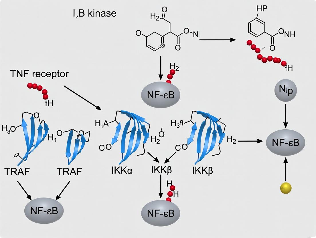
Within the broader thesis on IκB kinase (IKK) complex activation in inflammatory signaling research, this whitepaper delineates the canonical pathway converging on the IKK complex. Engagement of Toll-like receptors (TLRs), tumor necrosis factor (TNF) receptor, and interleukin-1 (IL-1) receptor triggers a conserved signaling cascade culminating in the activation of the kinase TAK1 (TGF-β-activated kinase 1), which is a critical upstream activator of the IKK complex. This pathway is fundamental to the cellular inflammatory response, regulating the transcription factor NF-κB and subsequent expression of pro-inflammatory cytokines, adhesion molecules, and anti-apoptotic proteins. Understanding this axis is paramount for developing therapeutics for inflammatory diseases, autoimmunity, and cancer.
Receptor Proximal Signaling Events
TLR Engagement and TIR Domain Adaptors
TLRs recognize pathogen-associated molecular patterns (PAMPs). Ligand binding induces dimerization and conformational change, recruiting TIR domain-containing adaptor proteins via homotypic interactions. MyD88 is the universal adaptor for most TLRs (except TLR3), often partnering with MAL/TIRAP. For TLR3 and TLR4 endosomal signaling, the adaptors TRIF and TRAM are utilized. These adaptors nucleate the formation of large helical signaling complexes called myddosomes or trifosomes.
TNF Receptor Superfamily Signaling
TNF binding induces trimerization of TNF receptor 1 (TNFR1), leading to the recruitment of the adaptor protein TRADD via its death domain. TRADD then recruits TRAF2 (TNF receptor-associated factor 2) and RIPK1 (Receptor-interacting serine/threonine-protein kinase 1), forming Complex I at the plasma membrane.
IL-1 Receptor Family Signaling
IL-1 binding to the IL-1R1/IL-1RAcP heterodimer triggers the recruitment of the adaptor protein MyD88 via TIR domain interactions, analogous to TLR signaling. MyD88 subsequently recruits IRAK4 (IL-1 receptor-associated kinase 4).
The Core Signaling Cascade to TAK1 Activation
A conserved sequence follows the receptor-proximal events:
- Kinase Recruitment and Activation: For TLR/IL-1R, IRAK4 is recruited to MyD88, phosphorylating and activating IRAK1/2. For TNFR, RIPK1 is the key kinase.
- E3 Ligase Complex Formation: Activated IRAK1/2 or RIPK1 recruits TRAF6, an E3 ubiquitin ligase. TRAF6, in concert with the E2 enzyme Ubc13/Uev1A, catalyzes the synthesis of K63-linked polyubiquitin chains.
- Ubiquitin-Dependent Scaffold Assembly: These K63-Ub chains act as a scaffold, recruiting proteins with ubiquitin-binding domains. The critical complex formed consists of TAK1, bound to its regulatory subunits TAB1 and TAB2 (or TAB3). TAB2/3 binds the K63-Ub chains, localizing TAK1 to the activated receptor complex.
- TAK1 Activation: Proximity-induced autophosphorylation and/or transphosphorylation within the TAK1 complex leads to its full activation.
TAK1-Mediated IKK Complex Phosphorylation
The activated TAK1 complex phosphorylates key residues in the activation loop of the IKKβ subunit (e.g., Ser177, Ser181 in humans) within the canonical IKK complex (IKKα, IKKβ, NEMO/IKKγ). NEMO also binds to linear (M1-linked) ubiquitin chains generated by the LUBAC complex, which further stabilizes and potentiates IKK activation.
Table 1: Key Phosphorylation Events in the Canonical Pathway
| Kinase (Activator) | Target Protein/Site | Functional Consequence | Typical Assay (Readout) |
|---|---|---|---|
| IRAK4 | IRAK1/2 (Activation loop) | IRAK1/2 kinase activation | In vitro kinase assay, phospho-specific Western blot |
| TAK1 | IKKβ (Ser177/Ser181) | IKK complex activation | Phospho-IKKα/β (Ser176/180) antibody, in vitro kinase assay using IκBα as substrate |
| IKKβ | IκBα (Ser32/Ser36) | Targeting of IκBα for K48 ubiquitination & proteasomal degradation | Phospho-IκBα (Ser32/36) antibody, degradation kinetics by Western blot |
| TBK1/IKKε | IRF3/7 (Ser386/ etc.) | Type I Interferon induction (parallel TLR3/4 pathway) | Phospho-IRF3 antibody, reporter gene assay |
Table 2: Critical Protein Complexes and Interactions
| Complex Name | Core Components | Ubiquitin Linkage Involved | Primary Function |
|---|---|---|---|
| Myddosome | MyD88, IRAK4, IRAK2/1 | --- | Nucleate TLR/IL-1R proximal signaling |
| TNFR Complex I | TNFR1, TRADD, TRAF2/5, RIPK1, cIAP1/2 | K63, M1 (via LUBAC) | Initiate NF-κB and MAPK signaling; inhibit cell death |
| TAK1 Complex | TAK1, TAB1, TAB2/3 | K63-Ub binding (via TAB2/3) | Central signal integrator; activates IKK and MAPK pathways |
| Canonical IKK Complex | IKKα, IKKβ, NEMO (IKKγ) | M1-Ub binding (via NEMO) | Phosphorylate IκBα; gatekeeper for NF-κB activation |
Experimental Protocols for Key Pathway Analyses
Protocol 1: Assessing IKK Complex Activation by Immunoblot
Objective: To detect phosphorylation-driven activation of the IKK complex in cells stimulated via TLR, TNF, or IL-1R. Method:
- Cell Stimulation: Culture HEK293T, HeLa, or primary macrophages. Stimulate with ligand (e.g., LPS 100 ng/ml for TLR4; TNF-α 10-20 ng/ml; IL-1β 10 ng/ml) for timepoints (e.g., 0, 5, 15, 30, 60 min).
- Cell Lysis: Lyse cells in RIPA buffer (supplemented with protease and phosphatase inhibitors) on ice. Clarify by centrifugation (14,000 x g, 15 min, 4°C).
- Immunoprecipitation: Incubate lysate with anti-IKKγ (NEMO) antibody coupled to Protein A/G beads overnight at 4°C. Wash beads 3x with lysis buffer.
- In Vitro Kinase Assay (Optional): Resuspend beads in kinase buffer with ATP (200 µM) and recombinant IκBα substrate. Incubate at 30°C for 30 min. Terminate with SDS sample buffer.
- Immunoblotting: Resolve proteins by SDS-PAGE and transfer to PVDF membrane. Probe with:
- Primary: Anti-phospho-IKKα/β (Ser176/180) to detect direct activation. For kinase assay, use anti-IκBα or anti-phospho-IκBα.
- Secondary: HRP-conjugated anti-rabbit IgG.
- Develop via ECL and image.
Protocol 2: Monitoring the TAK1-IKK Axis Using siRNA Knockdown
Objective: To validate the functional requirement of TAK1 for IKK/NF-κB activation. Method:
- Gene Silencing: Transfect cells with siRNA targeting MAP3K7 (TAK1) or non-targeting control siRNA using a suitable transfection reagent (e.g., Lipofectamine RNAiMAX). Incubate for 48-72 hrs.
- Stimulation & Reporter Assay: Co-transfect an NF-κB luciferase reporter plasmid. 24 hrs later, stimulate cells with relevant ligand (TNF-α, IL-1β, LPS) for 6-8 hrs.
- Luciferase Measurement: Lyse cells in passive lysis buffer. Measure firefly luciferase activity using a luminometer, normalizing to a co-transfected Renilla luciferase control.
- Validation: Confirm TAK1 knockdown efficiency by Western blotting whole-cell lysates with anti-TAK1 antibody.
- Expected Outcome: TAK1 knockdown should significantly reduce ligand-induced NF-κB reporter activity compared to control siRNA.
Pathway Visualizations
Title: Canonical Inflammatory Signaling Pathway from Receptors to IKK
Title: IKK Activation Assay Experimental Workflow
The Scientist's Toolkit: Research Reagent Solutions
Table 3: Essential Reagents for Studying the TLR/TNF/IL-1 to TAK1-IKK Pathway
| Reagent/Category | Specific Example(s) | Function & Application | Key Supplier(s) |
|---|---|---|---|
| Recombinant Ligands | Ultra-pure LPS (TLR4), TNF-α, IL-1β | Specific receptor stimulation to initiate pathway. | InvivoGen, PeproTech, R&D Systems |
| Pharmacologic Inhibitors | TAK1: 5Z-7-Oxozeaenol; IKK: IKK-16, SC-514; TAK1/IKK: (S)-MG-132 (proteasome) | Functional validation of kinase requirements in signaling. | Tocris, Selleck Chem, MedChemExpress |
| siRNA/shRNA | siRNA targeting MAP3K7 (TAK1), IKBKB (IKKβ), IKBKG (NEMO) | Genetic knockdown to assess protein function and necessity. | Dharmacon, Sigma-Aldrich, Origene |
| Antibodies (Phospho-Specific) | anti-phospho-IKKα/β (Ser176/180), anti-phospho-IκBα (Ser32/36), anti-phospho-TAK1 (Thr184/187) | Detect activation status of key pathway components via Western blot/IF. | Cell Signaling Technology, Abcam |
| Antibodies (Total Protein) | anti-IKKα, anti-IKKβ, anti-NEMO, anti-TAK1, anti-TRAF6, anti-IRAK1 | Assess protein expression levels and for immunoprecipitation. | Cell Signaling Technology, Santa Cruz |
| Ubiquitin Assay Reagents | TAK1 (K63-Ub) IP Assay Kit, LUBAC (HOIP) Inhibitor HOIPIN-8, K63-Ub chains | Study the critical ubiquitination events scaffold formation. | R&D Systems, Ubiquigent, LifeSensors |
| Reporter Assay Systems | NF-κB Luciferase Reporter (pGL4.32), Cignal Reporter Assays | Measure downstream transcriptional activity as a functional readout. | Promega, Qiagen |
| Kinase Assay Kits | Recombinant active TAK1 protein, IKKβ kinase enzyme system | Perform in vitro phosphorylation assays to study direct activity. | SignalChem, ProQinase, Cayman Chem |
Within the canonical NF-κB activation pathway, the IκB kinase (IKK) complex serves as the central signal integrator for inflammatory stimuli. Its activation is a tightly regulated process dependent on upstream kinases and scaffold-mediated assembly. This whitepaper provides a technical dissection of the critical roles played by TGF-β-activated kinase 1 (TAK1) and mitogen-activated protein kinase kinase kinase 3 (MEKK3), with a focus on how polyubiquitin chains—specifically K63-linked and linear—function as essential scaffolds for recruiting and activating these kinases within the IKK activation complex.
Activation of the IKK complex (IKKα, IKKβ, NEMO) is the pivotal step leading to IκBα phosphorylation, ubiquitination, and degradation, thereby releasing NF-κB for nuclear translocation and pro-inflammatory gene transcription. This process is initiated by receptors such as IL-1R/TLR (via MyD88/IRAKs) and TNFR (via TRADD/RIP1). A common downstream event is the formation of K63-linked or linear (M1-linked) polyubiquitin chains on key adaptor proteins (e.g., RIP1, IRAK1, NEMO). These chains do not primarily signal for proteasomal degradation but act as scaffolds to nucleate the assembly of a high-molecular-weight activation complex. This complex brings together TAK1 (with its binding partners TAB1, TAB2, TAB3) and the IKK complex, facilitating the TAK1-mediated phosphorylation and activation of IKKβ. MEKK3 has emerged as a parallel and sometimes compensatory kinase to TAK1, particularly in specific cell types or signaling contexts. Understanding the dynamics between TAK1 and MEKK3, their dependency on ubiquitin scaffolds, and their scaffold protein partners is crucial for developing targeted anti-inflammatory therapeutics.
Core Molecular Mechanisms
The TAK1 Complex: Structure and Activation
TAK1 is a MAP3K activated by cytokines (TNF-α, IL-1), PAMPs, and stress signals. Its activation requires binding to the scaffold proteins TAB1 and the ubiquitin-binding proteins TAB2 or TAB3.
- TAB1: Constitutively binds TAK1, promoting its autophosphorylation and activation.
- TAB2/TAB3: Contain C-terminal Npl4 zinc finger (NZF) domains that specifically bind K63-linked polyubiquitin chains. This binding recruits the TAK1 complex to the ubiquitin-decorated signaling complex (e.g., at RIP1 after TNF stimulation).
- Activation Loop: Recruitment leads to TAK1 autophosphorylation at Thr184/187 (within the activation loop), increasing its kinase activity towards IKKβ.
MEKK3: A TAK1-Interacting and Compensatory Kinase
MEKK3 is another MAP3K that can phosphorylate IKKβ. It functions in both TNF-α and IL-1β signaling pathways.
- Ubiquitin Binding: MEKK3 possesses a unique ubiquitin-binding domain (UBD) in its N-terminus that preferentially interacts with K63-linked polyubiquitin chains, allowing its direct recruitment to the signaling complex independent of TAK1.
- Relationship with TAK1: Genetic studies indicate redundancy; loss of MEKK3 alone has mild effects, but combined inhibition with TAK1 leads to complete ablation of IKK/NF-κB activation in response to certain stimuli. MEKK3 can also phosphorylate and be phosphorylated by TAK1, suggesting a cooperative interaction.
Polyubiquitin Chains: The Scaffolding Code
The type and topology of ubiquitin chains determine the outcome of signaling events.
- K63-Linked Chains: Synthesized by E2/E3 pairs like Ubc13/Uev1A with TRAF6 or cIAP1/2. Serve as pure scaffolds. Binding partners include TAB2/TAB3 (for TAK1), MEKK3-UBD, and the UBAN domain of NEMO.
- Linear (M1-Linked) Chains: Assembled by the LUBAC complex (HOIP, HOIL-1, SHARPIN). Also bind NEMO with high affinity and can interact with other ubiquitin-binding domains, amplifying and stabilizing the signaling complex.
- Mixed/Mixed-Linkage Chains: Chains containing both K63 and M1 linkages create a diverse platform for high-avidity interactions, crucial for robust and sustained IKK activation.
Table 1: Key Ubiquitin-Dependent Interactions in IKK Activation
| Interacting Protein/Complex | Ubiquitin Chain Preference | Binding Domain | Dissociation Constant (Kd)* | Primary Function in Pathway |
|---|---|---|---|---|
| TAB2/TAB3 | K63-linked polyUb | NZF | ~10-20 µM | Recruits TAK1 complex to signalosome |
| NEMO (IKKγ) | Linear (M1) & K63-linked polyUb | UBAN/CoZi | ~1-4 µM (M1) / ~10-20 µM (K63) | Anchors IKK complex; allosteric regulation |
| MEKK3 | K63-linked polyUb | N-terminal UBD | ~5-15 µM | Recruits MEKK3; facilitates IKK phosphorylation |
| A20 (OTUD7B) | K63 & M1-linked polyUb | OTU ZnF domain | N/A | Deubiquitinase; negative feedback regulator |
*Representative ranges from SPR/ITC studies; actual values vary by experimental conditions.
Table 2: Phenotypic Consequences of Genetic Ablation in Mouse Models
| Gene Target | Viability | Defect in IKK/NF-κB Activation | Key Phenotype |
|---|---|---|---|
| TAK1 | Embryonic lethal (E10.5) | Severe; abolished in MEFs for TNF, IL-1, LPS | Multiple developmental defects |
| MEKK3 | Embryonic lethal (E11.5) | Partial; delayed/attenuated in MEFs | Cardiovascular defects |
| TAK1 (conditional KO, myeloid) | Viable | Severe defect in TLR/IL-1R signaling | Resistant to septic shock; immunocompromised |
| Ubc13 (E2 for K63) | Embryonic lethal | Severe impairment | Liver degeneration |
| HOIP (LUBAC component) | Embryonic lethal (E10.5-12.5) | Attenuated TNF-induced IKK activation | Vascular and hematopoietic defects |
Detailed Experimental Protocols
Protocol: Co-Immunoprecipitation to Analyze TAK1/IKK Complex Formation
Objective: To assess stimulus-dependent association between TAK1, IKK components, and ubiquitinated scaffolds. Reagents: HEK293T or relevant cell line (e.g., MEFs), TNF-α or IL-1β, RIPA lysis buffer (with 20 mM NEM to inhibit DUBs), anti-TAK1 or anti-NEMO antibody, Protein A/G beads. Procedure:
- Stimulation: Seed cells in 10-cm dishes. At 80-90% confluency, stimulate with TNF-α (10-20 ng/mL) for 0, 5, 15, and 30 minutes.
- Lysis: Aspirate medium, wash with ice-cold PBS. Lyse cells in 1 mL RIPA buffer (plus protease/phosphatase inhibitors and NEM) on ice for 20 min. Centrifuge at 16,000 x g for 15 min at 4°C.
- Pre-clearing: Incubate supernatant with 20 µL Protein A/G beads for 30 min at 4°C. Centrifuge, transfer supernatant to new tube.
- Immunoprecipitation: Add 2-5 µg of antibody (e.g., anti-TAK1) to lysate. Rotate overnight at 4°C. Add 40 µL Protein A/G beads and rotate for 2-4 hours.
- Washing: Pellet beads, wash 3x with 1 mL ice-cold RIPA buffer.
- Elution & Analysis: Elute proteins in 2X Laemmli buffer by boiling for 5 min. Analyze by SDS-PAGE and western blot for targets (e.g., p-TAK1, IKKβ, NEMO, K63-Ub, RIP1).
Protocol:In VitroIKK Kinase Assay
Objective: To measure IKK activity immunoprecipitated from stimulated cells. Reagents: Cell lysis buffer (20 mM Tris pH 7.5, 150 mM NaCl, 1% Triton X-100, 1 mM EDTA), kinase assay buffer (20 mM HEPES pH 7.6, 10 mM MgCl2, 2 mM MnCl2, 1 mM DTT), ATP, recombinant IκBα substrate (or GST-IκBα 1-54). Procedure:
- IKK Immunoprecipitation: Lysate stimulated cells as in 4.1. Immunoprecipitate the IKK complex using an anti-IKKγ/NEMO antibody.
- Kinase Reaction: Wash beads 2x with lysis buffer, then 1x with kinase assay buffer. Set up a 30 µL reaction on beads containing 20 µL kinase assay buffer, 10 µM ATP, 2 µCi [γ-³²P]ATP, and 2 µg recombinant IκBα.
- Incubation: Incubate at 30°C for 30 minutes with gentle shaking.
- Termination & Detection: Stop reaction by adding 10 µL 4X Laemmli buffer and boiling. Resolve proteins by SDS-PAGE. Visualize phosphorylated IκBα by autoradiography or phosphor-imaging. Normalize to immunoprecipitated IKK levels (western blot).
Protocol: Assessing Ubiquitin Chain Dependency Using Deubiquitinase (DUB) Probes
Objective: To determine the chain linkage type required for pathway activation. Reagents: Cell-permeable, linkage-specific DUB inhibitors (e.g., G5 for K63-linkage, Otulin for linear chains), or overexpression of dominant-negative ubiquitin mutants (e.g., Ub-K63R, Ub-K48R). Procedure:
- Inhibition: Pre-treat cells with 10-50 µM of linkage-specific DUB inhibitor or DMSO control for 1 hour.
- Stimulation & Analysis: Stimulate with TNF-α for relevant time points. Analyze lysates by western blotting.
- Readout 1: Monitor upstream ubiquitination (e.g., RIP1, TRAF6) with linkage-specific Ub antibodies (e.g., anti-K63-Ub, anti-M1-Ub).
- Readout 2: Monitor downstream signaling: phospho-IKKα/β, phospho-IκBα, total IκBα degradation.
- Interpretation: Enhanced/persistent ubiquitination with pathway potentiation suggests a regulatory DUB target. Abolished signaling upon inhibition of a specific chain type confirms its essential scaffolding role.
Visualization of Signaling Pathways
Diagram Title: IKK Activation via Ubiquitin Scaffolds, TAK1, and MEKK3.
The Scientist's Toolkit: Essential Research Reagents
Table 3: Key Reagents for Studying TAK1/MEKK3/Ubiquitin in IKK Signaling
| Reagent Name/Category | Specific Example(s) | Function & Application |
|---|---|---|
| Pharmacological Inhibitors | (5Z)-7-Oxozeaenol (TAK1 inhibitor), NG25 (TAK1/MEKK3 inhibitor), SM1-71 (MEKK3 PBI domain inhibitor) | Functional probing of kinase dependency in cells; assess inflammatory output. |
| Linkage-Specific Ubiquitin Antibodies | Anti-K63-linkage (clone Apu3), Anti-linear (M1) linkage (clone 1E3), Anti-K48-linkage | Detection of specific polyubiquitin chains on RIP1, TRAF6, NEMO via immunoblot/IP. |
| Recombinant Ubiquitin Proteins & Mutants | Wild-type Ub, Ub-K63-only (K63R, other lysines mutated), Ub-K48-only, Ub-K63R mutant, Linear Ub chains (M1-linked) | In vitro reconstitution assays to test binding specificity of TAB2, MEKK3, NEMO domains. |
| Activity-Based DUB Probes | HA-Ub-VS, HA-Ub-PA, linkage-specific probes (TAMRA-Ub-PA derivatives) | To profile active deubiquitinases in signaling complexes; identify negative regulators. |
| Critical Cell Lines & Models | TAK1-deficient MEFs, MEKK3-deficient MEFs, Ubc13-/- cells, NEMO-deficient cells (e.g., 70Z/3 pre-B) | Genetic validation of protein function; study compensatory pathways. |
| Expression Plasmids | FLAG/HA-tagged wild-type and kinase-dead (KD) TAK1, MEKK3. Dominant-negative TAB2/3 (ΔNZF), TRAF6 (ΔRING), NEMO (UBAN mutant). | Overexpression and rescue experiments; structure-function studies. |
| Customizable Ubiquitin Sensors | TUBE (Tandem Ubiquitin-Binding Entity) reagents, linkage-specific Affimers | High-affinity capture of polyubiquitinated proteins from cell lysates for proteomic analysis. |
The IκB kinase (IKK) complex is the central regulator of the canonical NF-κB signaling pathway. Its activation is a critical event in inflammatory and immune responses. However, the mechanisms and consequences of IKK activation are not uniform; they are profoundly shaped by the cellular context. This whitepaper examines how IKK activation dynamics, downstream signaling, and functional outcomes diverge in immune cells (e.g., macrophages, T cells), stromal cells (e.g., fibroblasts, endothelial cells), and within disease-specific microenvironments such as tumors or arthritic joints. Understanding these contexts is paramount for developing targeted anti-inflammatory and anti-cancer therapies that modulate IKK/NF-κB signaling.
IKK Complex Activation: A Primer
The canonical IKK complex consists of the catalytic subunits IKKα and IKKβ, and the regulatory subunit NEMO (IKKγ). Upon stimulation by receptors like TLRs, TNF-R, or IL-1R, a cascade of ubiquitination events and kinase activations (e.g., TAK1) leads to the phosphorylation and activation of IKKβ. Activated IKK phosphorylates IκBα, targeting it for degradation and allowing NF-κB dimers (e.g., p65/p50) to translocate to the nucleus and drive gene expression.
Quantitative Data: IKK/NF-κB Dynamics Across Cellular Contexts
Recent studies highlight quantitative differences in IKK activation across cell types.
Table 1: Key Parameters of IKK/NF-κB Signaling in Different Primary Human Cell Types
| Cell Type | Primary Stimulus | Peak IKK Activity (min post-stimulation) | Duration of Nuclear NF-κB (p65) | Key Target Genes Induced |
|---|---|---|---|---|
| Macrophage (M1) | LPS (100 ng/mL) | 5-10 min | 60-90 min | TNF-α, IL-6, IL-1β |
| CD4+ T Cell | Anti-CD3/CD28 | 2-5 min | >120 min | IL-2, IFN-γ, IL-2Rα |
| Synovial Fibroblast | TNF-α (10 ng/mL) | 15-20 min | >180 min | MMPs, RANKL, IL-6 |
| Microvascular Endothelial Cell | IL-1β (10 ng/mL) | 10-15 min | 90-120 min | E-Selectin, ICAM-1, VCAM-1 |
| Cancer-Associated Fibroblast (CAF) | TGF-β + TNF-α | Sustained Low | Constitutive/Nuclear | CXCL12, IL-8, Collagen |
Table 2: Disease-Specific Alterations in IKK Pathway Components
| Disease Environment | Cell Type Analyzed | Observed Alteration | Functional Consequence |
|---|---|---|---|
| Rheumatoid Arthritis (RA) | Synovial Fibroblast | Elevated NEMO expression; IKKβ autophosphorylation | Hyper-responsive to TNF, resistant to apoptosis |
| Inflammatory Bowel Disease (IBD) | Intestinal Epithelium | Reduced IKKα function; Altered IKK complex composition | Defective epithelial barrier repair |
| Triple-Negative Breast Cancer | Tumor Cell | Constitutive IKKε (non-canonical) activity | Promotes survival, metastasis, and chemoresistance |
| Tumor Microenvironment | Tumor-Associated Macrophage (TAM) | Shift from canonical to alternative NF-κB via NIK | Supports immunosuppressive (M2-like) phenotype |
Detailed Experimental Protocols
Protocol 1: Measuring Cell-Type Specific IKK Kinase Activity Objective: To immunoprecipitate and measure IKK complex activity from different primary cell lysates.
- Cell Stimulation: Isolate primary cells (e.g., PBMCs, fibroblasts). Culture and serum-starve for 2h. Stimulate with relevant agonist (e.g., 10 ng/mL TNF-α) for varying times (0, 5, 15, 30 min). Use a kinase stop buffer (20 mM HEPES pH 7.6, 150 mM NaCl, 0.5% Triton X-100, 10% glycerol, 1 mM Na3VO4, 50 mM NaF, 1 mM PMSF, and protease inhibitors).
- Immunoprecipitation: Clarify 200-500 µg of lysate by centrifugation. Incubate with 1-2 µg of anti-IKKγ (NEMO) antibody conjugated to Protein A/G beads for 2h at 4°C with rotation.
- In Vitro Kinase Assay: Wash beads 3x with lysis buffer and 2x with kinase assay buffer (20 mM HEPES pH 7.6, 20 mM MgCl2, 2 mM DTT). Resuspend beads in 30 µL kinase buffer containing 10 µM ATP, 1 µCi [γ-³²P]ATP, and 2 µg recombinant GST-IκBα(1-54) substrate. Incubate at 30°C for 30 min.
- Analysis: Terminate reaction with Laemmli buffer. Resolve proteins by SDS-PAGE. Visualize phosphorylated GST-IκBα by autoradiography. Quantify band intensity and normalize to total NEMO pulled down (via western blot).
Protocol 2: Assessing NF-κB Dynamics via Live-Cell Imaging Objective: To track nuclear translocation of NF-κB in real-time across different stromal cells.
- Cell Line Generation: Lentivirally transduce primary fibroblasts or endothelial cells with an NF-κB reporter construct (e.g., p65-DsRed or a κB-driven GFP).
- Image Acquisition: Seed cells on glass-bottom dishes. 24h later, place dish on a confocal live-cell imaging system maintained at 37°C/5% CO2. Establish baseline for 30 min, then add stimulus (e.g., IL-1β) without moving the dish.
- Quantification: Acquire images every 3-5 min for 3-6h. Using image analysis software (e.g., ImageJ/Fiji), define nuclear and cytoplasmic ROIs. Calculate the nuclear-to-cytoplasmic fluorescence ratio (Fn/c) over time for 50+ individual cells per condition. Plot mean Fn/c ± SEM vs. time.
Signaling Pathway Diagrams
The Scientist's Toolkit: Key Research Reagent Solutions
Table 3: Essential Reagents for Studying Context-Specific IKK Activation
| Reagent Category | Specific Example(s) | Function & Application |
|---|---|---|
| IKK Activity Inhibitors | IKK-16 (IKKβ inhibitor), BAY 11-7082 (IκBα phosphorylation inhibitor), TPCA-1 (IKKβ inhibitor) | Pharmacological tools to dissect IKK-dependent signaling in different cell types. |
| Activation State Antibodies | Phospho-IKKα/β (Ser176/180), Phospho-IκBα (Ser32/36), Phospho-p65 (Ser536) | Western blot or ELISA to measure pathway activation dynamics. |
| Recombinant Cytokines/Growth Factors | Human/Mouse TNF-α, IL-1β, LPS, TGF-β, IFN-γ | Standardized ligands to stimulate pathways in immune/stromal cells. |
| Primary Cell Culture Systems | CD14+ Monocytes (for macrophages), HUVECs (endothelial), Lung/Synovial Fibroblasts | Physiologically relevant cellular contexts. |
| NF-κB Reporters | Lentiviral κB-luciferase/GFP constructs, p65-DsRed fusion protein | For live-cell imaging and transcriptional output quantification. |
| Ubiquitination Assay Reagents | TAK1 Inhibitor (5Z-7-Oxozeaenol), NEMO/Ubc13 Binding Inhibitors, K63-Ubiquitin Chains | To probe upstream activation mechanisms of the IKK complex. |
| Disease-Relevant Co-culture Kits | Fibroblast-Macrophage Co-culture Inserts, Tumor-Stroma 3D Co-culture Matrices | To model cell-cell crosstalk in disease microenvironments. |
From Bench to Bedside: Techniques for Monitoring IKK Activity and Screening Inhibitors
Within the broader thesis on IκB kinase (IKK) complex activation in inflammatory signaling research, the ability to directly measure IKK enzymatic activity is fundamental. The IKK complex, primarily composed of the catalytic subunits IKKα and IKKβ and the regulatory scaffold NEMO/IKKγ, is the central node for the canonical NF-κB pathway. Its activation by stimuli such as TNF-α, IL-1, and pathogen-associated molecular patterns (PAMPs) leads to the phosphorylation and degradation of IκB inhibitors, allowing NF-κB nuclear translocation and pro-inflammatory gene transcription. In vitro kinase assays provide a controlled, reductionist approach to dissect IKK regulation, screen for inhibitors, and validate genetic manipulations. This guide details methodologies using both recombinant proteins and cell lysates to measure IKK activity.
The IKK Complex in Inflammatory Signaling
The canonical IKK activation pathway involves upstream signaling complexes that converge on the IKK complex. For TNF-α signaling, ligand binding to TNFR1 triggers the formation of Complex I, recruiting adaptor proteins like TRADD, TRAF2, and the kinase RIPK1. This leads to the recruitment and activation of the TAK1 complex (TAK1, TAB1, TAB2). TAK1 then phosphorylates the IKKβ activation loop, inducing a conformational change and full activation of the IKK complex. The activated IKK complex specifically phosphorylates IκBα on Ser32 and Ser36, targeting it for polyubiquitination and proteasomal degradation.
Title: Canonical TNF-α Pathway Leading to IKK Activation
Experimental Workflow for IKK Activity Assays
A typical project involves two complementary approaches: using immunoprecipitated IKK from stimulated cell lysates to study activation in a cellular context, and using recombinant IKK proteins for high-purity biochemical studies. The core kinase reaction, however, follows a similar principle: incubating active IKK with its substrate (recombinant IκBα or a peptide fragment) and [γ-³²P]ATP or cold ATP followed by detection via autoradiography, phospho-specific antibody, or other methods.
Title: Dual Workflow for IKK Kinase Assays
Detailed Experimental Protocols
Protocol 1: IKK Assay from Cell Lysates via Immunoprecipitation
This protocol measures endogenous IKK activity from stimulated cells.
Materials:
- Cells (e.g., HEK293, HeLa, MEFs)
- Stimulant: Human TNF-α (10-20 ng/mL)
- Lysis Buffer: 20 mM Tris-HCl (pH 7.5), 150 mM NaCl, 1 mM EDTA, 1 mM EGTA, 1% Triton X-100, 2.5 mM sodium pyrophosphate, 1 mM β-glycerophosphate, 1 mM Na₃VO₄, 1 μg/mL leupeptin, 1 mM PMSF.
- Protein A/G Sepharose beads
- Anti-IKKα, IKKβ, or NEMO antibody for immunoprecipitation
- Kinase Reaction Buffer: 20 mM HEPES (pH 7.5), 10 mM MgCl₂, 2 mM DTT, 20 μM ATP, 10 μCi [γ-³²P]ATP (for radioactive assay) or 100 μM cold ATP (for non-radioactive).
- Substrate: 1-2 μg recombinant GST-IκBα (amino acids 1-54) or full-length protein.
- 2X SDS Sample Buffer
Method:
- Cell Stimulation: Culture cells in 10-cm dishes to ~90% confluency. Stimulate with TNF-α (20 ng/mL) for 5-15 minutes. Include an unstimulated control.
- Lysis: Place dishes on ice, aspirate media, and wash with cold PBS. Add 1 mL ice-cold lysis buffer with protease/phosphatase inhibitors. Scrape cells and transfer to a microcentrifuge tube. Rock at 4°C for 20 min, then centrifuge at 14,000 x g for 10 min at 4°C. Transfer supernatant (whole-cell lysate) to a new tube.
- Immunoprecipitation: Pre-clear 500 μg of lysate with 20 μL Protein A/G beads for 30 min at 4°C. Incubate pre-cleared lysate with 1-2 μg of anti-IKKβ antibody overnight at 4°C with gentle rotation. Add 30 μL of bead slurry and incubate for 2 hours. Pellet beads and wash 3x with lysis buffer, then 2x with Kinase Reaction Buffer (without ATP/substrate).
- Kinase Reaction: Resuspend bead-bound IKK complex in 30 μL Kinase Reaction Buffer. Add the substrate (GST-IκBα). Initiate the reaction by adding ATP (and [γ-³²P]ATP if radioactive). Mix gently and incubate at 30°C for 30 minutes.
- Termination & Analysis:
- Radioactive: Terminate by adding 15 μL of 2X SDS Sample Buffer. Heat at 95°C for 5 min. Resolve proteins by SDS-PAGE. Dry the gel and expose to a phosphorimager screen or X-ray film.
- Non-Radioactive: Proceed similarly but use cold ATP. Detect phospho-IκBα by Western blot using anti-phospho-IκBα (Ser32/36) antibody.
Protocol 2: Kinase Assay Using Recombinant IKK Proteins
This protocol uses purified components for direct kinetic analysis or inhibitor screening.
Materials:
- Recombinant active human IKKβ (or IKK complex), commercially available.
- Recombinant substrate: GST-IκBα (1-54) or full-length.
- Kinase Reaction Buffer (as above).
- ATP solution.
- Stop Solution: 0.5 M EDTA, pH 8.0.
- For quantitative assays: ADP-Glo Kinase Assay or ELISA-based phospho-substrate detection kits.
Method (Standard Endpoint Assay):
- Reaction Setup: In a microcentrifuge tube on ice, combine:
- 10-50 ng recombinant active IKKβ
- 1-5 μg substrate protein
- Kinase Reaction Buffer to a final volume of 25 μL.
- Initiation: Start the reaction by adding ATP to a final concentration of 100 μM. Mix thoroughly by pipetting.
- Incubation: Incubate at 30°C for 30 minutes. A time course (0, 5, 15, 30, 60 min) can be performed for kinetic analysis.
- Termination: Add 5 μL of 0.5 M EDTA to stop the reaction by chelating Mg²⁺.
- Detection: Analyze by:
- SDS-PAGE/Western Blot: Add sample buffer, run gel, blot for phospho-IκBα.
- Luminescent Assay (e.g., ADP-Glo): Transfer 10 μL of reaction to a white plate. Add ADP-Glo Reagent to consume residual ATP, then Kinase Detection Reagent to convert ADP to ATP, generating luminescence proportional to kinase activity.
- ELISA: Use phospho-specific IκBα ELISA kits for quantitative measurement.
Key Research Reagent Solutions
| Reagent / Material | Function / Role in IKK Assay | Example / Notes |
|---|---|---|
| Recombinant Active IKKβ | Catalytic subunit for biochemical assays; allows study of direct regulation & inhibition. | Available from SignalChem, MilliporeSigma, Carna Biosciences. Verify lot-specific activity (U/mg). |
| Recombinant IκBα Substrate | Physiological substrate; N-terminal fragment (aa 1-54) containing Ser32/36 is commonly used. | GST- or His-tagged proteins from Novus, Abcam, or produce in-house from E. coli. |
| Anti-IKKβ (IP grade) | Immunoprecipitates endogenous IKK complex from cell lysates for activity measurement. | Mouse monoclonal (clone 10AG2) or rabbit polyclonal from Cell Signaling Technology. |
| Phospho-IκBα (Ser32/36) Antibody | Critical for non-radioactive detection of kinase assay products by Western blot. | 14D4 (Cell Signaling #2859) is a widely validated monoclonal antibody. |
| Kinase Buffer System | Provides optimal pH, divalent cations (Mg²⁺), and reducing environment (DTT) for IKK activity. | Standard: 20 mM HEPES pH 7.5-7.7, 10 mM MgCl₂, 1-2 mM DTT. |
| [γ-³²P]ATP | Radioactive phosphate donor; allows sensitive, direct detection of phosphorylated substrate via autoradiography. | Handle with strict radiation safety protocols. Consider non-radioactive alternatives. |
| ADP-Glo Kinase Assay | Luminescent, non-radioactive method to quantify kinase activity by measuring ADP production. | Promega; ideal for high-throughput screening of IKK inhibitors. |
| TAK1 Inhibitor (5z-7-oxozeaenol) | Control compound; inhibits upstream activator TAK1, preventing cellular IKK activation. | Useful for validating stimulus-dependent activity in lysate-based assays. |
| IKK-16 (or similar IKK inhibitor) | Selective ATP-competitive IKKβ inhibitor; used as a control to confirm signal specificity in assays. | Confirm inhibitor potency (IC₅₀) for your specific IKK preparation. |
Key kinetic and inhibitory data for human IKKβ.
Table 1: Biochemical Parameters of Recombinant IKKβ
| Parameter | Value | Conditions / Notes | Reference (Example) |
|---|---|---|---|
| Km for ATP | 2.5 - 10 μM | Using IκBα-derived peptide substrate. | (Ziegelbauer et al., 2004) |
| Km for IκBα | 0.1 - 0.5 μM | Full-length or N-terminal protein substrate. | (Kishore et al., 2003) |
| Vmax / kcat | ~ 1 - 5 min⁻¹ | Varies with enzyme preparation and activation state. | Vendor lot-specific data. |
| Optimal pH | 7.5 - 7.7 | Standard HEPES or Tris-based kinase buffer. | Standard protocol. |
| Divalent Cation Requirement | Mg²⁺ > Mn²⁺ | 10 mM MgCl₂ is standard; Mn²⁺ may alter specificity. | Standard protocol. |
Table 2: Common IKK Inhibitors for Assay Controls
| Inhibitor | Target | IC₅₀ (IKKβ) | Use in Assay | Notes |
|---|---|---|---|---|
| IKK-16 | IKKβ (ATP-competitive) | 10 - 40 nM | Specificity control; pre-incubate 10-30 min. | Potent and selective. |
| BMS-345541 | IKKβ (Allosteric) | ~300 nM | Specificity control; useful in cellular assays. | Binds to similar site as IκBα. |
| SC-514 | IKKβ (ATP-competitive) | 3 - 12 μM | Lower potency control. | Some off-target effects. |
| TPCA-1 | IKKβ (ATP-competitive) | ~ 400 nM | Specificity control. | Also inhibits IKKε. |
| 5z-7-Oxozeaenol | TAK1 (Upstream) | ~ 10 nM (TAK1) | Control in lysate assays to block activation. | Irreversible inhibitor. |
In vitro kinase assays with recombinant proteins and cell lysates remain indispensable tools for elucidating the mechanisms of IKK complex activation within inflammatory signaling research. The lysate-based approach captures the physiological regulation of the endogenous complex, while recombinant assays offer precision for kinetic and inhibitor profiling. The integration of quantitative methods, robust controls, and careful interpretation of data from these complementary approaches directly feeds into the broader thesis goals of understanding IKK dysregulation in disease and identifying novel therapeutic intervention points.
The activation of the IκB kinase (IKK) complex is the central regulatory event in the canonical NF-κB signaling pathway, a master regulator of inflammatory and immune responses. This whitepaper details the critical biochemical readouts—IκBα phosphorylation and degradation—that serve as definitive markers of IKK complex activation. Within a broader thesis on IKK activation mechanisms, monitoring these sequential post-translational modifications provides direct, quantitative evidence of pathway engagement in response to stimuli such as TNF-α, IL-1β, or LPS. Accurate assessment is fundamental for research into inflammatory diseases, cancer, and the development of IKK/NF-κB-targeted therapeutics.
The Canonical NF-κB Signaling Pathway
The canonical pathway is initiated by pro-inflammatory stimuli, leading to the activation of the IKK complex (IKKα, IKKβ, and NEMO/IKKγ). Activated IKK phosphorylates IκBα at serine residues 32 and 36, tagging it for polyubiquitination and subsequent rapid degradation by the 26S proteasome. This releases the NF-κB dimer (typically p65/p50), allowing its translocation to the nucleus to drive gene transcription.
Diagram Title: Canonical NF-κB Pathway & IκBα Fate
Key Experimental Protocols
Cell Stimulation and Lysate Preparation for Time-Course Analysis
- Objective: To capture the rapid, transient dynamics of IκBα phosphorylation and degradation.
- Detailed Protocol:
- Culture & Serum-Starve: Grow relevant cells (e.g., HEK293, HeLa, or primary macrophages) to 70-80% confluence. Serum-starve (e.g., 0.5% FBS) for 4-16 hours to reduce basal activity.
- Stimulation: Apply stimulus (e.g., 10-20 ng/mL human TNF-α) for varying times (e.g., 0, 2.5, 5, 10, 15, 30, 60 min). Include a pre-treatment control with an IKK inhibitor (e.g., 10 µM Bay 11-7082 for 30 min) prior to stimulus.
- Rapid Lysis: Aspirate media and immediately lyse cells on ice with 150-200 µL of RIPA Lysis Buffer (supplemented with 1x protease inhibitor cocktail, 1x phosphatase inhibitors (NaF, β-glycerophosphate, sodium orthovanadate), and 1 mM PMSF).
- Clarification: Scrape cells, transfer lysates to microcentrifuge tubes, and vortex vigorously. Incubate on ice for 15-30 min, then centrifuge at 16,000 x g for 15 min at 4°C.
- Quantification: Transfer supernatant to a new tube. Determine protein concentration using a BCA or Bradford assay. Adjust all samples to equal concentration with lysis buffer and 4x Laemmli sample buffer.
Western Blot Analysis for Phospho- and Total IκBα
- Objective: To specifically detect phosphorylated IκBα and its total protein levels.
- Detailed Protocol:
- Gel Electrophoresis: Load 20-30 µg of protein per lane on a 10% or 4-12% Bis-Tris polyacrylamide gel. Run in 1x MOPS or MES SDS buffer at 120-150V until adequate separation.
- Transfer: Transfer proteins to a PVDF membrane using wet or semi-dry transfer systems. Activate PVDF in methanol prior to use.
- Blocking: Block membrane in 5% non-fat dry milk in TBST (Tris-buffered saline with 0.1% Tween-20) for 1 hour at room temperature (RT). For phospho-specific antibodies, 5% BSA in TBST is often preferred.
- Primary Antibody Incubation: Incubate membrane overnight at 4°C with gentle agitation in primary antibody dilution (in blocking buffer or 5% BSA/TBST).
- Phospho-IκBα (Ser32/36): Use at 1:1000 dilution.
- Total IκBα: Use at 1:2000 dilution.
- Loading Control (e.g., β-Actin, α-Tubulin, GAPDH): Use at 1:5000-1:10000 dilution.
- Washing & Secondary Incubation: Wash membrane 3 x 5 min with TBST. Incubate with appropriate HRP-conjugated secondary antibody (e.g., anti-rabbit IgG) at 1:5000 dilution in blocking buffer for 1 hour at RT.
- Detection: Wash 3 x 5 min with TBST. Apply enhanced chemiluminescence (ECL) substrate evenly across the membrane. Image using a digital chemiluminescence imaging system with multiple exposure times.
Data Normalization and Quantification
- Objective: To obtain quantitative measures of phosphorylation and degradation.
- Detailed Protocol:
- Band Density Analysis: Use image analysis software (ImageJ, Image Lab, etc.) to measure the background-subtracted integrated density of each band.
- Normalization: For each time point:
- Phospho-IκBα Signal: Normalize to its corresponding loading control band (e.g., Phospho-IκBα / β-Actin).
- Degradation (Total IκBα): Normalize total IκBα band intensity to its loading control. Express as a percentage of the unstimulated (time 0) control.
- Phosphorylation Index (Optional): Calculate (Phospho-IκBα / Total IκBα) for time points where total protein remains (early times), indicating the fraction of IκBα that is phosphorylated.
- Replicates: Perform a minimum of three independent biological replicates. Present data as mean ± SEM.
Quantitative Data Presentation
Table 1: Representative Time-Course Data of IκBα Phosphorylation and Degradation in HeLa Cells Stimulated with TNF-α (20 ng/mL)
| Time Post-Stimulation (min) | Phospho-IκBα Band Density (Normalized to β-Actin) | Total IκBα Band Density (% of Time 0) | Notes / Expected Trend |
|---|---|---|---|
| 0 | 0.05 ± 0.02 | 100.0 ± 5.0 | Baseline |
| 2.5 | 1.85 ± 0.30 | 95.0 ± 7.0 | Rapid phosphorylation |
| 5 | 2.50 ± 0.40 | 40.0 ± 10.0 | Peak phosphorylation; degradation underway |
| 10 | 1.20 ± 0.25 | 15.0 ± 5.0 | Phospho declines; near-max degradation |
| 15 | 0.40 ± 0.10 | 10.0 ± 4.0 | Further decline |
| 30 | 0.10 ± 0.05 | 60.0 ± 12.0 | Resynthesis begins |
| 60 | 0.08 ± 0.03 | 85.0 ± 8.0 | Approaching re-establishment of homeostasis |
Table 2: Effects of Pharmacological Inhibitors on IκBα Phosphorylation (5 min post-TNF-α)
| Inhibitor (Target) | Concentration | Phospho-IκBα Signal (% of TNF-α alone) | Total IκBα Level (% of Unstimulated) | Interpretation |
|---|---|---|---|---|
| TNF-α Only | 20 ng/mL | 100.0 ± 8.0 | 40.0 ± 9.0 | Positive Control |
| DMSO Vehicle | 0.1% | 98.5 ± 7.5 | 42.0 ± 8.5 | Solvent Control |
| Bay 11-7082 (IKK inhibitor) | 10 µM | 15.0 ± 5.0 | 95.0 ± 6.0 | Blocks IKK activity |
| MG-132 (Proteasome inhibitor) | 10 µM | 220.0 ± 25.0 | 110.0 ± 10.0 | Blocks degradation, leads to phospho-protein accumulation |
| Cycloheximide (Protein synthesis inhibitor) | 50 µg/mL | 105.0 ± 10.0 | 8.0 ± 3.0 | Inhibits resynthesis, degradation is sustained |
The Scientist's Toolkit: Key Research Reagent Solutions
Table 3: Essential Materials for Assessing IκBα Phosphorylation and Degradation
| Item Category & Name | Specific Example / Catalog Number | Function & Critical Notes |
|---|---|---|
| Critical Antibodies | ||
| Anti-Phospho-IκBα (Ser32/36) | Cell Signaling #9246 | Primary Readout. Specifically detects IKK-mediated phosphorylation events. Use with BSA-based blockers. |
| Anti-IκBα (Total) | Cell Signaling #4814 | Degradation Readout. Detects total IκBα protein regardless of phosphorylation state. |
| Anti-β-Actin-HRP | Sigma A3854 | Loading Control. HRP conjugate allows direct detection, saving time and reducing background. |
| Inhibitors & Stimuli | ||
| Recombinant Human TNF-α | PeproTech #300-01A | Primary Stimulus. High-quality, endotoxin-free cytokine for consistent pathway activation. |
| IKK Inhibitor (Bay 11-7082) | Sigma B5556 | Negative Control. Validates the dependence of phosphorylation on IKK activity. |
| Proteasome Inhibitor (MG-132) | Sigma C2211 | Tool Compound. Confirms that loss of signal is due to degradation, not dephosphorylation. |
| Lysis & Detection | ||
| RIPA Lysis Buffer | Thermo Scientific #89900 | Complete Lysis. Must be supplemented fresh with protease and phosphatase inhibitors. |
| PhosSTOP / cOmplete EDTA-free | Roche #4906845001 / #4693132001 | Inhibitor Cocktails. Essential for preserving post-translational modifications during lysis. |
| Clarity Max ECL Substrate | Bio-Rad #1705062 | High-Sensitivity Detection. Critical for detecting low-abundance phospho-proteins and short time points. |
Experimental Workflow Visualization
Diagram Title: Western Blot Workflow for IκBα Analysis
Troubleshooting and Technical Considerations
- Phospho-Signal Too Weak/Undetectable: Ensure fresh phosphatase inhibitors are used. Optimize stimulation time (try earlier time points like 2-5 min). Increase protein load and use a high-sensitivity ECL substrate. Confirm antibody specificity with inhibitor controls.
- High Background on Phospho-Blots: Switch from milk to BSA as the blocking agent. Increase number and duration of TBST washes after primary and secondary antibody incubations.
- No Degradation Observed: Verify proteasome function and stimulus potency. Include a positive control (e.g., known responsive cell line). Check protein stability during lysis—ensure samples are kept ice-cold.
- Poor Membrane Transfer: Confirm gel composition matches buffer system. For IκBα (~39 kDa), ensure efficient transfer of lower MW proteins; consider shorter transfer times or lower current if protein is "blowing through" the membrane.
- Strip and Reprobing: If sequentially probing the same membrane for phospho- and total protein, use a mild stripping buffer to avoid damaging the antigen. Always confirm complete removal of antibody before reprobing.
The IκB kinase (IKK) complex is the central signaling hub for the canonical NF-κB pathway, a master regulator of inflammatory gene expression. Inflammatory stimuli, such as TNF-α, IL-1β, or LPS, trigger a cascade leading to IKK activation. The IKK complex, primarily composed of the catalytic subunits IKKα and IKKβ and the regulatory subunit NEMO/IKKγ, phosphorylates the inhibitory protein IκBα. This phosphorylation marks IκBα for ubiquitination and proteasomal degradation, releasing the transcription factor NF-κB (typically a p65/p50 heterodimer) to translocate into the nucleus and drive target gene expression.
Direct measurement of IKK enzymatic activity is technically challenging, requiring immunoprecipitation and in vitro kinase assays. Therefore, researchers widely employ NF-κB-driven luciferase reporter gene assays as a robust, sensitive, and high-throughput functional readout of the entire upstream signaling pathway, with IKK activity being the critical, rate-limiting step. This whitepaper provides a technical guide for using these assays as a proxy for IKK pathway activity in the context of inflammatory signaling and drug discovery.
Core Signaling Pathway and Assay Principle
The following diagram illustrates the canonical NF-κB pathway, highlighting the position of the IKK complex and the point of measurement by the luciferase reporter.
Diagram Title: Canonical NF-κB Pathway and Luciferase Reporter Readout
Key Experimental Protocols
Standard Protocol for NF-κB Luciferase Reporter Assay in Adherent Cells
Objective: To measure IKK/NF-κB pathway activation in response to a stimulus or inhibition by a compound.
Materials: See "Scientist's Toolkit" section.
Method:
- Day 1: Cell Seeding: Seed adherent cells (e.g., HEK293, HeLa, or primary cells) in a 24-well or 96-well plate at 70-90% confluence for transfection the next day.
- Day 2: Transfection:
- Prepare transfection mix per well: 100-400 ng of NF-κB luciferase reporter plasmid (e.g., pGL4.32[luc2P/NF-κB-RE/Hygro]), 10-50 ng of Renilla luciferase control plasmid (e.g., pRL-TK or pGL4.74[hRluc/TK]) for normalization, and transfection reagent per manufacturer's protocol.
- Replace cell culture medium with fresh medium (with or without serum as required by transfection reagent).
- Add transfection mix dropwise to cells. Mix gently.
- Incubate cells for 18-24 hours.
- Day 3: Stimulation/Inhibition:
- Pre-treat cells with potential inhibitory compounds (e.g., IKK inhibitors like TPCA-1, BAY 11-7082) for 30-60 minutes.
- Stimulate the NF-κB pathway by adding agonist (e.g., 10-20 ng/mL human TNF-α, 10 ng/mL IL-1β) to appropriate wells.
- Incubate for an optimized period (typically 4-8 hours for peak reporter response).
- Day 3: Luciferase Assay (Dual-Luciferase System):
- Aspirate medium and lyse cells with 1X Passive Lysis Buffer (PLB) for 15-20 minutes at room temperature with gentle shaking.
- Transfer lysate to a microcentrifuge tube or use directly in plate.
- Program a luminometer to perform a 2-second pre-measurement delay, followed by a 10-second measurement period for each reporter assay.
- For each sample, inject 100 µL of Luciferase Assay Reagent II (LAR II), measure firefly luciferase activity (F-Luc).
- Then, inject 100 µL of Stop & Glo Reagent, quenches F-Luc reaction and activates Renilla luciferase (R-Luc). Measure R-Luc activity.
- Data Analysis:
- Calculate the normalized reporter activity: Normalized Luciferase Units (NLU) = F-Luc / R-Luc.
- Express data as Fold Induction relative to unstimulated control wells (usually set to 1).
- Perform statistical analysis (e.g., t-test, ANOVA) on biological replicates (n≥3).
Protocol for Validating IKK-Specific Inhibition
To confirm that observed effects are specifically mediated through the IKK complex, a complementary immunoblotting protocol is recommended.
Objective: To correlate luciferase activity with direct measures of IKK substrate phosphorylation and NF-κB translocation.
Method:
- In parallel to the reporter assay, treat and stimulate cells in a 6-well plate format.
- At the end of the stimulation period, lyse cells in RIPA buffer containing protease and phosphatase inhibitors.
- Perform SDS-PAGE and Western Blot.
- Probe for:
- Phospho-IκBα (Ser32/36): Direct readout of IKK activity.
- Total IκBα: To show degradation.
- Phospho-NF-κB p65 (Ser536): Another IKK-dependent modification.
- β-actin or GAPDH: Loading control.
- Correlation: Strong inhibition of luciferase activity should correlate with diminished phospho-IκBα and preserved total IκBα levels.
Table 1: Common Agonists and Their Typical Effective Concentrations in NF-κB Reporter Assays
| Agonist | Target Receptor | Typical Working Concentration | Expected Fold Induction (Cell-type dependent) | Reference / Source |
|---|---|---|---|---|
| Human Tumor Necrosis Factor-alpha (TNF-α) | TNFR1 | 10 - 20 ng/mL | 5 - 50x | Current vendor data (e.g., PeproTech, R&D Systems) |
| Human Interleukin-1beta (IL-1β) | IL-1R | 5 - 20 ng/mL | 10 - 100x | Current vendor data |
| Lipopolysaccharide (LPS) | TLR4 (in macrophages) | 100 ng/mL - 1 µg/mL | 10 - 100x | InvivoGen product sheets |
| Phorbol 12-myristate 13-acetate (PMA) | PKC activator | 10 - 100 nM | 2 - 20x | Sigma-Aldrich technical data |
Table 2: Common Pharmacologic IKK/NF-κB Inhibitors for Assay Controls
| Inhibitor | Primary Target | Typical Pre-treatment Concentration | Expected IC50 in Reporter Assay | Key Consideration |
|---|---|---|---|---|
| BAY 11-7082 | IKK, inhibits IκBα phosphorylation | 1 - 10 µM | ~ 5 µM | Not highly specific; affects other pathways. |
| TPCA-1 (2-[(Aminocarbonyl)amino]-5-(4-fluorophenyl)-3-thiophenecarboxamide) | IKKβ (selective) | 1 - 5 µM | ~ 0.3 µM (for IKKβ) | More selective for IKKβ vs IKKα. |
| IKK-16 | IKKβ (potent) | 0.1 - 1 µM | < 0.2 µM | Highly potent, cell-permeable. |
| SC-514 | IKKβ (ATP-competitive) | 10 - 100 µM | ~ 10 µM | Reversible and selective for IKKβ. |
| Bortezomib | Proteasome (inhibits IκBα degradation) | 10 - 100 nM | Varies | Acts downstream of IKK; validates signal specificity. |
Table 3: Comparison of Common Luciferase Reporter Vectors
| Vector Name (Example) | Promoter/Response Element | Luciferase Type | Selection Marker | Key Feature |
|---|---|---|---|---|
| pGL4.32[luc2P/NF-κB-RE/Hygro] | 5x NF-κB response elements | Firefly (luc2P) | Hygromycin | Optimized for low background, high sensitivity. Part of Promoter Flexi system. |
| pNF-κB-Luc (Clontech) | 4x NF-κB RE | Firefly (luc+) | Ampicillin (bacterial) | Classic, widely used vector. |
| Cignal Lenti NF-κB Reporter (Qiagen) | NF-κB RE | Firefly | Puromycin (if part of kit) | Lentiviral system for stable cell line generation. |
| pNL3.2.NF-κB-RE [NlucP/NF-κB-RE/Hygro] | 5x NF-κB RE | NanoLuc (NlucP) | Hygromycin | Very bright, small size enzyme for enhanced dynamic range. |
The Scientist's Toolkit: Research Reagent Solutions
| Item | Function & Explanation | Example Product/Catalog # |
|---|---|---|
| NF-κB Luciferase Reporter Plasmid | Contains multiple copies of the NF-κB Response Element (RE) upstream of a minimal promoter driving firefly or NanoLuc luciferase. The core sensor for pathway activity. | pGL4.32[luc2P/NF-κB-RE/Hygro] (Promega, E364A) |
| Control Reporter Plasmid (Renilla or NanoLuc) | Expresses a second luciferase under a constitutive promoter (e.g., TK, CMV). Used to normalize for transfection efficiency and cell viability. | pRL-TK (Renilla luc, Promega) or pGL4.74[hRluc/TK] (Promega) |
| Dual-Luciferase Reporter Assay System | Provides optimized lysis buffer and substrates for sequential measurement of Firefly and Renilla luciferase from a single sample. Gold standard for dual-reporter assays. | Dual-Luciferase Reporter Assay System (Promega, E1910) |
| Pathway Agonists | Recombinant cytokines or ligands used to stimulate the IKK/NF-κB pathway to establish a signal window. | Human TNF-α (PeproTech, 300-01A), E. coli LPS (InvivoGen, tlrl-eblps) |
| Pharmacologic IKK Inhibitors | Small molecule tools to inhibit the IKK complex, used as assay controls to confirm signal specificity and for screening antagonist compounds. | TPCA-1 (Tocris, 3001), BAY 11-7082 (Sigma, B5556) |
| Transfection Reagent | For delivering plasmid DNA into mammalian cells. Choice depends on cell type (e.g., HEK293 are highly transferable). | Lipofectamine 3000 (Thermo Fisher), FuGENE HD (Promega) |
| Cell Line with Intact Pathway | A model cell line with robust, inducible NF-κB signaling. Essential for assay development. | HEK293 (human kidney), THP-1 (human monocytic), HeLa (human cervical cancer) |
| Phospho-Specific Antibodies (for validation) | Antibodies that recognize phosphorylated forms of pathway proteins (e.g., p-IκBα, p-p65) to confirm IKK activity biochemically. | Phospho-IκBα (Ser32/36) Rabbit mAb (Cell Signaling Technology, #9246) |
| Luminometer or Plate Reader | Instrument capable of injecting reagents and detecting low-light luminescence signals from multi-well plates. | GloMax Discover System (Promega), SpectraMax iD5 (Molecular Devices) |
Experimental Workflow Diagram
The following diagram outlines the key steps in a standard NF-κB reporter assay workflow.
Diagram Title: NF-κB Reporter Assay Experimental Workflow
Considerations and Best Practices
- Cell Line Selection: The magnitude of response is highly cell-type dependent. Immune cells (e.g., macrophages) may have high endogenous activity.
- Timing: The kinetics of reporter induction differ from endogenous genes. A time course experiment (e.g., 2, 4, 6, 8, 24h post-stimulation) is crucial for optimization.
- Normalization: The Renilla control corrects for variability but can itself be affected by treatments. Always inspect raw R-Luc values. Alternative normalization methods (e.g., protein concentration, viable cell count) can be used.
- Specificity Controls: Include a mutated NF-κB RE reporter to confirm signal specificity. Use known IKK inhibitors (see Table 2) as positive controls for inhibition.
- Stable vs. Transient: For screening, generating a stable cell line with the integrated reporter ensures homogeneity and reduces cost per assay.
- Context within IKK Research: While the luciferase assay is an excellent functional readout, it should be complemented with direct biochemical assays (e.g., IKK immunocomplex kinase assay, phospho-protein immunoblotting) to make definitive claims about IKK complex activity modulation.
In inflammatory signaling research, specifically the study of IκB kinase (IKK) complex activation, precise genetic manipulation is paramount. The IKK complex, comprising IKKα, IKKβ, and NEMO/IKKγ, is the central regulator of the NF-κB pathway. Dissecting its function requires robust techniques to alter gene expression. This guide details three core methodologies—CRISPR/Cas9 knockouts, siRNA knockdowns, and dominant-negative constructs—providing a technical framework for researchers investigating IKK-driven signaling cascades in drug discovery and basic science.
CRISPR/Cas9 for IKK Gene Knockouts
CRISPR/Cas9 enables permanent, complete gene disruption, ideal for studying the essential roles of IKK subunits.
Key Considerations for IKK Targets
- IKKβ (IKBKB): Complete knockout often leads to embryonic lethality in mice, necessitating conditional models in vitro.
- NEMO (IKBKG): Located on the X-chromosome; single allele disruption in male cell lines results in knockout.
- IKKα (CHUK): Involved in both canonical and non-canonical NF-κB pathways; knockout phenotypes are context-dependent.
Experimental Protocol: Generating a Clonal IKKβ Knockout Cell Line
1. Design and Cloning:
- Design two single-guide RNAs (sgRNAs) targeting exons 2-5 of the IKBKB gene. Example sequences (from recent literature):
- sgRNA1: 5'-GACCTGAAGCAGATCATCGG-3'
- sgRNA2: 5'-GTCATCCGCTACTTCATCAA-3'
- Clone sgRNAs into a lentiviral Cas9/sgRNA expression vector (e.g., lentiCRISPRv2).
2. Viral Production and Transduction:
- Co-transfect HEK293T cells with the lentiviral plasmid and packaging plasmids (psPAX2, pMD2.G).
- Harvest lentivirus at 48 and 72 hours.
- Transduce target cells (e.g., THP-1 macrophages) with virus plus 8 µg/mL polybrene.
3. Selection and Clonal Isolation:
- Apply selection pressure (e.g., puromycin, 1-2 µg/mL) for 5-7 days.
- Perform limiting dilution to isolate single cells in 96-well plates.
- Expand clonal populations for 3-4 weeks.
4. Validation:
- Genotyping: Isolate genomic DNA. Perform PCR amplification of the target region and sequence to confirm indel mutations.
- Western Blot: Probe with anti-IKKβ antibody to confirm loss of protein.
- Functional Assay: Stimulate with TNF-α (10 ng/mL, 15 min) and monitor phospho-IκBα (Ser32/36) by Western blot; knockout should ablate phosphorylation.
Table 1: Expected Outcomes for IKK Subunit Knockouts
| Target Gene | NF-κB Pathway Affected | Expected Phenotype Post-TNF-α | Validation Primary Assay |
|---|---|---|---|
| IKKβ (IKBKB) | Canonical | No IκBα degradation, no p65 nuclear translocation | Western blot for p-IκBα |
| NEMO (IKBKG) | Canonical | No IκBα degradation | Co-immunoprecipitation of IKK complex |
| IKKα (CHUK) | Non-canonical (Partial Canonical) | Impaired p100 processing to p52 | Western blot for p52 |
Title: CRISPR/Cas9 knockout experimental workflow
siRNA for IKK Gene Knockdowns
siRNA mediates transient, sequence-specific mRNA degradation, suitable for acute loss-of-function studies and druggability assessments.
Experimental Protocol: Transient IKK Subunit Knockdown in HeLa Cells
1. siRNA Design and Preparation:
- Use validated siRNA duplexes from commercial sources (e.g., Dharmacon ON-TARGETplus).
- IKKα pool: J-003473-07, -08, -09, -10
- IKKβ pool: J-003503-05, -06, -07, -08
- Control: Non-targeting siRNA (e.g., D-001810-01)
- Resuspend siRNA to 20 µM in RNase-free buffer.
2. Reverse Transfection:
- Seed HeLa cells at 70% confluency in 6-well plates (2.5 x 10^5 cells/well).
- For each well, mix:
- 5 µL of 20 µM siRNA pool
- 125 µL Opti-MEM reduced serum media
- 7.5 µL Lipofectamine RNAiMAX
- Incubate mix for 20 min at RT.
- Add 867 µL complete growth media to the mix, then add directly to cells.
3. Incubation and Stimulation:
- Incubate cells for 48-72 hours at 37°C.
- Stimulate with IL-1β (10 ng/mL) for 0, 5, 15, 30 min to activate IKK.
4. Validation and Analysis:
- qRT-PCR: Isolate RNA, synthesize cDNA. Use TaqMan assays (e.g., Hs00178369_m1 for IKKβ) to quantify mRNA knockdown (>70% efficiency target).
- Western Blot: Harvest protein at 72h to confirm protein reduction.
- Pathway Readout: Probe for phospho-p65 (Ser536) and total IκBα to assess pathway inhibition.
Table 2: Typical Knockdown Efficiency and Functional Readouts (48h post-transfection)
| siRNA Target | mRNA Reduction (%) | Protein Reduction (%) | IL-1β-induced p-p65 Reduction (%) |
|---|---|---|---|
| IKKα | 75-85 | 70-80 | 20-30* |
| IKKβ | 80-90 | 75-85 | 80-95 |
| NEMO | 70-80 | 65-75 | 85-98 |
| Non-targeting | 0 | 0 | 0 |
*IKKα knockdown primarily affects non-canonical signaling; canonical readouts may be less impacted.
Title: IKK complex in IL-1R signaling targeted by siRNA
Dominant-Negative (DN) Constructs for IKK Inhibition
Dominant-negative mutants act as molecular "spoilers," disrupting the native function of the IKK complex through competitive inhibition.
Key Constructs for IKK Research
- IKKβ-K44A: Kinase-dead mutant; binds substrate but cannot phosphorylate.
- IKKβ-EE: Mutant with serine-to-glutamate changes in activation loop, constitutively active (used as control).
- NEMO-ΔLZ: Leucine zipper deletion mutant; fails to oligomerize, disrupting complex assembly.
Experimental Protocol: Transfection and Analysis of IKKβ-K44A
1. Plasmid Preparation:
- Obtain mammalian expression vector (e.g., pcDNA3.1) encoding FLAG-tagged IKKβ-K44A.
- Prepare endotoxin-free plasmid DNA (≥ 1 µg/µL).
2. Transfection of HEK293 Cells:
- Seed cells in 6-well plates to reach 90% confluency at transfection.
- For each well, mix:
- 2 µg plasmid DNA
- 150 µL Opti-MEM
- 5 µL Lipofectamine 3000 + 5 µL P3000 reagent
- Incubate mix 15 min, add dropwise to cells.
3. Stimulation and Analysis (24h post-transfection):
- Stimulate with TNF-α (10 ng/mL, 0-30 min).
- Harvest cells in RIPA buffer with protease/phosphatase inhibitors.
4. Key Assays:
- Co-Immunoprecipitation: Immunoprecipitate FLAG-tagged DN-IKKβ. Probe for endogenous NEMO to confirm complex binding.
- Kinase Assay: Immunoprecipitate IKK complex, perform in vitro kinase assay using recombinant IκBα substrate and [γ-32P]ATP. DN construct should show >90% reduced kinase activity.
- Reporter Gene Assay: Co-transfect with an NF-κB luciferase reporter (e.g., pGL4.32[luc2P/NF-κB-RE/Hygro]). DN construct should inhibit luciferase activity by ≥80% upon TNF-α stimulation.
Table 3: Characterization of Dominant-Negative IKK Constructs
| Construct | Mechanism of Action | Effect on IKK Complex Kinase Activity | Inhibition of NF-κB Reporter (%) |
|---|---|---|---|
| IKKβ-K44A | Substrate binding, no catalysis | >90% reduction | 80-95 |
| NEMO-ΔLZ | Disrupts complex oligomerization | 70-85% reduction | 60-80 |
| IKKβ-EE (CA) | Constitutive activation | 300-400% increase | N/A (Increase) |
Title: Dominant-negative IKK mutant mechanism of action
The Scientist's Toolkit: Research Reagent Solutions
Table 4: Essential Reagents for IKK Genetic Manipulation Studies
| Reagent / Material | Function / Purpose | Example Product / Identifier |
|---|---|---|
| lentiCRISPRv2 vector | All-in-one lentiviral vector for Cas9 and sgRNA expression | Addgene #52961 |
| ON-TARGETplus siRNA pools | Validated, pooled siRNAs for specific gene knockdown with reduced off-target effects | Dharmacon, e.g., J-003503 for IKKβ |
| Lipofectamine RNAiMAX | High-efficiency transfection reagent for siRNA delivery | Thermo Fisher Scientific, 13778150 |
| Lipofectamine 3000 | Transfection reagent for plasmid DNA delivery | Thermo Fisher Scientific, L3000015 |
| Anti-IKKβ antibody (WB) | Detects IKKβ protein for knockout/knockdown validation | Cell Signaling Technology, #8943 |
| Anti-phospho-IκBα (Ser32/36) | Readout for canonical IKK complex activity | Cell Signaling Technology, #2859 |
| NF-κB Luciferase Reporter | Plasmid for functional assay of pathway activity | Promega, pGL4.32[luc2P/NF-κB-RE/Hygro] |
| Recombinant Human TNF-α | Potent activator of the canonical IKK/NF-κB pathway | PeproTech, 300-01A |
| FLAG-Tag Antibody (IP) | For immunoprecipitation of transfected dominant-negative constructs | Sigma-Aldrich, F3165 |
| Polybrene | Enhances viral transduction efficiency | Sigma-Aldrich, TR-1003-G |
High-Throughput Screening (HTS) Platforms for Identifying Novel IKK Inhibitors
The IκB kinase (IKK) complex is the central regulator of the canonical NF-κB signaling pathway, a critical mediator of inflammatory and immune responses. Its dysregulation is implicated in chronic inflammatory diseases, autoimmunity, and cancer. Within the context of a broader thesis on IKK complex activation mechanisms, the identification of novel, potent, and selective IKK inhibitors remains a paramount goal in therapeutic development. High-Throughput Screening (HTS) represents a cornerstone technology for the rapid evaluation of compound libraries to discover such inhibitors. This technical guide details contemporary HTS platforms, methodologies, and reagent toolkits essential for advancing this research frontier.
Core HTS Assay Platforms for IKK Inhibition
HTS for IKK inhibitors primarily utilizes biochemical, cell-based, and more recently, label-free phenotypic assays. The choice of platform depends on the desired inhibitor profile (e.g., ATP-competitive, allosteric, disruptors of complex assembly).
Biochemical Kinase Assays
These assays measure the direct inhibition of IKKβ catalytic activity on its substrate (typically IκBα or a peptide mimic).
Protocol: Homogeneous Time-Resolved Fluorescence (HTRF) Kinase Assay
- Reaction Setup: In a 384-well low-volume plate, combine:
- Recombinant human IKKβ (active) (e.g., 5 nM final).
- Biotinylated IκBα substrate peptide (e.g., 500 nM final).
- Test compound in DMSO (final DMSO ≤1%).
- ATP (at or near the apparent Km, e.g., 10 µM) in kinase buffer.
- Incubation: Incubate at room temperature for 60 minutes.
- Detection: Stop the reaction by adding a detection mix containing:
- EDTA (to chelate Mg²⁺ and stop kinase activity).
- Europium cryptate-labeled anti-phospho-IκBα antibody.
- Streptavidin-conjugated XL665 dye.
- Reading: Incubate for 1 hour, then read time-resolved fluorescence resonance energy transfer (TR-FRET) at 620 nm (donor) and 665 nm (acceptor) on a plate reader (e.g., PerkinElmer EnVision).
- Data Analysis: The phosphorylation signal is proportional to the 665/620 nm ratio. Calculate % inhibition relative to DMSO (positive control) and staurosporine (or a known IKK inhibitor) controls.
Table 1: Comparison of Primary HTS Assay Platforms for IKK Inhibitors
| Platform Type | Principle | Throughput | Pros | Cons | Typical Z' Factor |
|---|---|---|---|---|---|
| Biochemical (HTRF) | TR-FRET detection of phosphorylated substrate | Ultra-High (>100K compounds/day) | Direct activity measurement; minimal interference; low cost per well. | Misses cell permeability & complex biology; prone to ATP-competitive artifact. | 0.7 - 0.9 |
| Cell-Based (Reporter Gene) | NF-κB-driven luciferase or GFP expression in stimulated cells | High (50K-100K/day) | Identifies cell-permeable inhibitors; captures pathway modulation. | Indirect; more false positives (cytotoxicity, transcription/translation inhibitors). | 0.5 - 0.8 |
| Cell-Based (Phospho-IκBα ELISA) | Immuno-detection of phospho-IκBα or p65 in fixed cells | Medium-High (10K-50K/day) | Direct readout of pathway node; more specific than reporter. | Lower throughput; more expensive reagents. | 0.6 - 0.8 |
| Label-Free (Impedance/ DMR) | Dynamic Mass Redistribution or Impedance changes in stimulated cells | Medium (5K-20K/day) | Label-free; holistic phenotypic response. | Complex data interpretation; lower throughput; specialized equipment. | N/A |
Cell-Based Reporter Gene Assays
These assays identify compounds that inhibit the IKK-driven activation of NF-κB transcriptional activity.
Protocol: HEK293/NF-κB-Luciferase HTS Assay
- Cell Seeding: Seed HEK293 cells stably transfected with an NF-κB response element driving firefly luciferase (e.g., PathHunter U2OS NFκB cis-Reporting cells can be an alternative) in white, tissue-culture treated 384-well plates (e.g., 5,000 cells/well in 30 µL medium).
- Compound Addition: Using a pintool or acoustic dispenser, add test compounds (nL volumes) and incubate for 30-60 minutes.
- Pathway Stimulation: Add a potent NF-κB inducer (e.g., recombinant human TNF-α at 10 ng/mL final or IL-1β) using a multidispenser. Incubate for 4-6 hours at 37°C, 5% CO₂.
- Luciferase Detection: Add a single-addition, stable luciferase reagent (e.g., ONE-Glo Luciferase Assay System) and incubate for 10 minutes.
- Reading: Measure luminescence on a plate reader. Normalize data to stimulated DMSO controls (100% activity) and unstimulated cells (baseline).
Secondary & Counter-Screen Assays
Hit compounds from primary HTS must be validated.
- Selectivity Panel: Test against a panel of related (IKKα) and unrelated kinases.
- Cytotoxicity Assay: Run in parallel (e.g., CellTiter-Glo) to identify false positives.
- Mechanism of Action: Use electrophoretic mobility shift assay (EMSA) for NF-κB DNA binding or Western blot for endogenous IκBα degradation and p65 phosphorylation.
Visualization of IKK-NF-κB Pathway & HTS Workflow
Diagram 1: IKK Pathway & HTS Workflow This diagram illustrates the inflammatory signaling cascade targeted for inhibition and the sequential funnel of a typical HTS campaign to identify and validate novel IKK inhibitors.
The Scientist's Toolkit: Essential Research Reagents & Materials
Table 2: Key Reagent Solutions for IKK HTS & Validation
| Reagent/Material | Supplier Examples | Function in IKK Research |
|---|---|---|
| Active Recombinant IKKβ | SignalChem, MilliporeSigma, Cayman Chemical | Essential for biochemical kinase assays. Provides a pure enzyme source for direct inhibition studies. |
| Phospho-IκBα (Ser32/36) Antibody | Cell Signaling Technology (#9246), Abcam | Gold-standard antibody for detecting IKK activity via Western Blot, ELISA, or HTRF. Validates hits in orthogonal assays. |
| NF-κB Reporter Cell Lines | Promega (NF-κB-Luc2), InvivoGen (HEK-Blue), DiscoverX (PathHunter) | Engineered cells for high-throughput, cell-based primary screening of pathway inhibitors. |
| Homogeneous HTRF Kinase Kits | Cisbio (IKKβ KinEASE), PerkinElmer | Optimized, ready-to-use kits for robust, miniaturized biochemical screening. Include all detection reagents. |
| TNF-α, human recombinant | PeproTech, R&D Systems | The canonical cytokine for stimulating the canonical NF-κB pathway in cell-based assays. |
| IKK Inhibitor Controls (e.g., IKK-16, BMS-345541, TPCA-1) | Tocris, Selleckchem | Well-characterized, commercially available IKK inhibitors used as pharmacological controls for assay validation and benchmarking. |
| Cell Viability Assay Kits (e.g., CellTiter-Glo) | Promega | Luminescent ATP-detection assay run in parallel to identify cytotoxic false-positive hits. |
| Selectivity Kinase Panels | Reaction Biology, Eurofins DiscoverX | Profiling services to assess hit compound selectivity across hundreds of kinases, critical for lead optimization. |
| SPR/Biacore Chips (e.g., NTA Sensor Chip) | Cytiva | For Surface Plasmon Resonance (SPR) studies to determine binding kinetics (KD, Kon, Koff) of confirmed hits to IKKβ. |
Navigating Experimental Challenges in IKK Research: Pitfalls and Proven Solutions
The IκB kinase (IKK) complex is the central signaling hub for the activation of the NF-κB pathway, a master regulator of inflammatory and immune responses. Within the broader thesis of IKK complex activation, a critical challenge is the precise differentiation between canonical and non-canonical pathway activities. The canonical pathway, typically triggered by pro-inflammatory cytokines like TNFα or IL-1β, involves the rapid, NEMO-dependent activation of IKKβ, leading to IκBα phosphorylation, degradation, and transient NF-κB nuclear translocation. In contrast, the non-canonical pathway, activated by a subset of TNF receptor superfamily members (e.g., BAFF, CD40L), involves the NF-κB-inducing kinase (NIK)-dependent, slow processing and activation of IKKα, resulting in the phosphorylation of p100/RelB and its processing to p52.
The specificity problem arises because assays measuring IKK activity, NF-κB translocation, or target gene expression often capture outputs from both pathways. In complex biological systems or drug screening, this lack of specificity can lead to misinterpretation of mechanism of action, off-target effects, and failed therapeutic strategies. This guide details methodologies to rigorously distinguish between these two signaling arms.
Core Quantitative Differences: Canonical vs. Non-Canonical IKK Signaling
Table 1: Key Characteristics of Canonical vs. Non-Canonical IKK Pathways
| Parameter | Canonical Pathway | Non-Canonical Pathway |
|---|---|---|
| Primary Activating Signals | TNFα, IL-1β, LPS, TLR agonists | BAFF, CD40L, LTβR, RANKL |
| Key Inducible IKK Subunit | IKKβ (IKK2) | IKKα (IKK1) |
| Critical Regulatory Adaptor | NEMO (IKKγ) - Essential | NIK (MAP3K14) - Essential |
| Kinetics of NF-κB Activation | Rapid (minutes to 1 hour) | Slow (hours to days) |
| Primary NF-κB Dimer | p50/RelA (p65), p50/c-Rel | p52/RelB |
| Target IκB Protein | IκBα, IκBβ, IκBε | p100 (IκBδ) |
| Primary Regulatory Event | Phosphorylation & proteasomal degradation of IκBα | NIK stabilization, p100 phosphorylation & partial processing to p52 |
| Genetic Knockout Phenotype (Mice) | Embryonic lethality (liver apoptosis) | Defects in secondary lymphoid organogenesis |
Experimental Protocols for Specific Distinction
Protocol 3.1: Kinase Activity Assay with Substrate-Specific Immunoprecipitation
Aim: To directly measure IKKβ- vs. IKKα-specific kinase activity from cell lysates.
Method:
- Stimulation & Lysis: Stimulate cells (e.g., MEFs, HEK293, primary B cells) with canonical (TNFα, 20 ng/mL, 10 min) or non-canonical (anti-CD40 Ab, 2 µg/mL, 24h) ligands. Lyse in ice-cold lysis buffer (20 mM Tris pH 7.5, 150 mM NaCl, 1 mM EDTA, 1% Triton X-100, 10% glycerol) supplemented with phosphatase and protease inhibitors.
- Immunoprecipitation (IP): For each sample, split lysate into two. Pre-clear with Protein A/G beads. IP IKKβ using a specific antibody (e.g., clone D30C6) and IKKα using a different specific antibody (e.g., clone D8W8N). Use species-matched IgG as control. Incubate overnight at 4°C.
- Kinase Reaction: Wash IP complexes 3x with lysis buffer and 2x with kinase assay buffer (20 mM HEPES pH 7.5, 10 mM MgCl2, 2 mM DTT). Resuspend beads in 30 µL kinase buffer containing 200 µM ATP, 5 µCi [γ-³²P]ATP, and 2 µg of substrate.
- Substrates: Use GST-IκBα(1-54) for IKKβ IPs. Use GST-p100(1-407) for IKKα IPs.
- Detection: Incubate at 30°C for 30 min. Terminate reaction with Laemmli buffer. Separate proteins by SDS-PAGE. Visualize phosphorylated substrate by autoradiography. Normalize kinase activity to the amount of immunoprecipitated kinase (Western blot).
Protocol 3.2: Quantitative PCR (qPCR) Array for Pathway-Specific Gene Targets
Aim: To differentiate pathway output by measuring transcription of target genes with dimer specificity.
Method:
- Treatment & RNA Isolation: Stimulate cells as in 3.1 for appropriate timeframes (e.g., 1h and 24h for canonical/non-canonical, respectively). Extract total RNA using TRIzol.
- cDNA Synthesis: Synthesize cDNA using a high-capacity reverse transcription kit.
- qPCR Array Design: Run qPCR for a panel of genes.
- Canonical Markers: IL-8, A20 (TNFAIP3), IκBα (NFKBIA) – strongly p65/RelA-dependent.
- Non-Canonical Markers: CCL19, CXCL13, CCL21 – associated with p52/RelB function in lymphoid organization.
- Common/Control: GAPDH, ACTB.
- Data Analysis: Calculate ∆∆Ct values. A dominant canonical signal will show early upregulation of IL-8 and A20. A dominant non-canonical signal will show late, specific upregulation of CXCL13 and CCL19.
Protocol 3.3: Electrophoretic Mobility Shift Assay (EMSA) with Supershifts
Aim: To characterize the specific NF-κB DNA-binding complexes induced.
Method:
- Nuclear Extract Preparation: Prepare nuclear extracts from stimulated cells using a hypotonic buffer followed by detergent lysis of the plasma membrane, and high-salt extraction of nuclei.
- EMSA Reaction: Incubate 5-10 µg nuclear extract with a ³²P-end-labeled double-stranded oligonucleotide containing a consensus κB site (e.g., from the Igκ gene) in binding buffer.
- Supershift: Add 1-2 µg of antibodies specific to NF-κB subunits (p65, c-Rel, p50, p52, RelB) to separate reaction mixtures prior to adding the labeled probe.
- Analysis: Run samples on a non-denaturing polyacrylamide gel. A canonical stimulus will produce a major complex supershifted by anti-p65 and anti-p50. A non-canonical stimulus will produce a slower-migrating complex supershifted by anti-RelB and anti-p52.
Visualizing the Pathways and Assay Logic
Diagram 1: Canonical vs Non-Canonical NF-κB Pathways
Diagram 2: Multi-Assay Specificity Workflow
The Scientist's Toolkit: Essential Research Reagent Solutions
Table 2: Key Reagents for Distinguishing IKK Pathway Activity
| Reagent / Material | Function & Specificity | Example Product / Target |
|---|---|---|
| Pathway-Specific Agonists | To selectively initiate one pathway with minimal cross-talk. | Recombinant human TNFα (canonical); Recombinant human BAFF (non-canonical). |
| IKK Subunit-Selective Inhibitors | Pharmacological tools to inhibit one IKK subunit and observe functional consequences. | IKK-16 (IKKβ-selective); BAY-11-7082 (broad IKK inhibitor - control). |
| Phospho-Specific Antibodies | To detect activation-specific phosphorylation events via Western blot or immunofluorescence. | Anti-phospho-IκBα (Ser32/36); Anti-phospho-p100 (Ser866/870). |
| Subunit-Specific IP/WB Antibodies | For immunoprecipitation of specific kinases and loading controls in activity assays. | Anti-IKKα (clone D8W8N); Anti-IKKβ (clone D30C6); Anti-NEMO (clone D13D11). |
| Recombinant Protein Substrates | Purified substrates for in vitro kinase assays to determine subunit activity. | GST-IκBα(1-54); GST-p100(1-407). |
| NF-κB Subunit Antibodies (Supershift) | For EMSA supershift analysis to identify the composition of activated NF-κB dimers. | Anti-p65 (RelA); Anti-RelB; Anti-p50; Anti-p52. |
| Genetically Modified Cell Lines | Isogenic controls to validate pathway-specific dependencies (e.g., NIK KO, IKKβ KO). | IKKα-/- MEFs; IKKβ-/- MEFs; NIK-/- cells. |
| Pathway Reporter Assays | Lentiviral or stable reporter cell lines to monitor specific pathway activity in real-time or endpoint assays. | NF-κB RE (κB site) luciferase; p52/RelB-specific reporter. |
| qPCR Primer/Probe Sets | For quantitative measurement of pathway-specific transcriptional outputs. | Validated primers for IL-8 (canonical), CXCL13 (non-canonical). |
The IκB kinase (IKK) complex, a central node in the NF-κB signaling pathway, is a critical therapeutic target for chronic inflammatory diseases, cancer, and autoimmune disorders. The canonical IKK complex, comprising the catalytic subunits IKKα and IKKβ and the regulatory subunit NEMO (IKKγ), is activated by a plethora of stimuli, leading to IκBα phosphorylation, ubiquitination, and degradation. This allows NF-κB dimers to translocate to the nucleus and drive pro-inflammatory gene expression. A core thesis in modern inflammatory research posits that selective pharmacological inhibition of the IKK complex, particularly the key mediator IKKβ, can effectively dampen harmful inflammation. However, the ATP-binding sites of kinases are highly conserved, raising the significant challenge of off-target effects. This whitepaper provides a technical guide for rigorously validating the selectivity of prototypical IKK inhibitors, such as IKK-16 and BMS-345541, against panels of related kinases, ensuring that observed phenotypic effects are attributable to on-target inhibition.
Key Inhibitors: Mechanisms and Reported Selectivity Profiles
IKK-16: A potent, ATP-competitive inhibitor with high affinity for IKKβ (IC₅₀ ~10-40 nM). It also inhibits IKKα but with lower potency. Its selectivity against the broader kinome requires empirical validation.
BMS-345541: Identified as a selective allosteric inhibitor of the IKK complex, binding to a site distinct from the ATP-binding pocket, with reported IC₅₀ values of ~0.3 µM for IKKβ and ~4 µM for IKKα. Its allosteric mechanism suggests potentially higher selectivity, but cross-reactivity must be tested.
Table 1: Reported Potency of Featured IKK Inhibitors
| Inhibitor | Primary Target | Reported IC₅₀ (IKKβ) | Reported IC₅₀ (IKKα) | Mechanism |
|---|---|---|---|---|
| IKK-16 | IKKβ | 10 - 40 nM | ~200 nM | ATP-competitive |
| BMS-345541 | IKK Complex | 0.3 - 0.5 µM | ~4 µM | Allosteric |
Core Experimental Protocol: Kinase Selectivity Profiling
The gold standard for selectivity assessment is profiling against a large panel of human kinases.
3.1. Methodology: In Vitro Kinase Assay Panels
- Platform: Utilize commercial kinase profiling services (e.g., Eurofins DiscoverX KINOMEscan, Reaction Biology’s KinaseProfiler) or establish in-house panels using recombinant active kinases.
- Assay Principle: For competitive ATP-site binders like IKK-16, a displacement assay (KINOMEscan) is highly effective. It measures the test compound's ability to displace a immobilized, active-site directed ligand. For allosteric inhibitors, radiometric or luminescence-based activity assays are necessary.
- Protocol Outline:
- Inhibitor Preparation: Prepare a serial dilution of IKK-16 and BMS-345541 in DMSO, typically starting from 10 µM for broad screening.
- Kinase Reaction: Incubate each kinase with the inhibitor and its specific substrate/ATP mixture (or the detection probe in a displacement assay) under optimal buffer conditions.
- Detection: Quantify remaining kinase activity via scintillation proximity, fluorescence polarization, or luminescence.
- Data Analysis: Calculate percent inhibition at a single concentration (e.g., 1 µM) to generate a "hits" list. Determine IC₅₀ values for primary targets and any off-target hits.
3.2. Data Analysis and Selectivity Scoring
- Selectivity Score (S(35)): The percentage of kinases tested (commonly a panel of 300-400) that show less than 35% inhibition at a standard concentration (e.g., 1 µM). A higher S(35) indicates greater selectivity.
- Gini Coefficient: A quantitative metric (0 to 1) describing the inequality of a compound's potency across the kinome; a value closer to 1 indicates high selectivity.
- Kinase Tree Visualization: Plot inhibition data on a kinome phylogenetic tree to identify off-target clusters.
Table 2: Hypothetical Selectivity Profile from a 400-Kinase Panel (1 µM Inhibitor)
| Inhibitor | % Kinase Inhibition <35% (S(35)) | Notable Off-Targets (≥80% Inhibition) | Potential Cellular Impact of Off-Targets |
|---|---|---|---|
| IKK-16 | 78% | TBK1, IKKε, CAMK1G | Alters IRF3/7 signaling, innate immunity |
| BMS-345541 | 92% | Checkpoint Kinase 1 (CHK1) | Affects DNA damage response, cell cycle |
Cellular Target Engagement and Validation
Biochemical selectivity must be corroborated in cells.
- Protocol: Cellular Pathway Analysis: Treat relevant cell lines (e.g., TNFα-stimulated macrophages or synovial fibroblasts) with inhibitors. Monitor:
- On-target effect: Phospho-IκBα degradation (western blot).
- Downstream effect: NF-κB nuclear translocation (immunofluorescence) and cytokine production (ELISA for IL-6, TNFα).
- Off-target signaling: Phosphorylation status of substrates from off-target kinases identified in Table 2 (e.g., phospho-IRF3 for TBK1/IKKε).
The Scientist's Toolkit: Essential Research Reagents
Table 3: Key Reagent Solutions for IKK Selectivity Studies
| Reagent | Function & Application |
|---|---|
| Recombinant Active IKKβ (and IKKα) | Essential substrate for in vitro kinase assays and primary IC₅₀ determination. |
| Selective Kinase Inhibitor Profiling Service | Provides standardized, broad kinome screening for objective selectivity metrics (S(35), Gini). |
| Phospho-IκBα (Ser32/36) Antibody | Key readout for cellular on-target engagement via western blot. |
| NF-κB Reporter Cell Line (e.g., HEK293/NF-κB-luc) | Enables high-throughput functional assessment of inhibitor potency and cytotoxicity. |
| Proteome Integrity or Phospho-kinase Array | Multiplexed screening of cellular signaling pathways to identify unpredicted off-pathway effects. |
Visualizing the Context and Workflow
Title: IKK Inhibitor Validation in NF-κB Pathway Context
Title: Kinase Inhibitor Selectivity Validation Workflow
The IκB kinase (IKK) complex, a central node in the NF-κB signaling pathway, is a classic model for studying compensatory mechanisms in inflammatory signaling. Genetic knockouts of its core catalytic subunits, IKKα (CHUK) and IKKβ (IKBKB), or its regulatory subunit NEMO (IKBKG), often yield unexpected phenotypes due to pathway redundancy and adaptive rewiring. This whitepaper provides a technical guide for interpreting such data and designing experiments to overcome masking by compensatory mechanisms.
Core Compensatory Mechanisms in IKK/NF-κB Signaling
Live search analysis confirms that upon genetic deletion of a specific IKK component, multiple compensatory layers can be activated, obscuring the protein's true function.
| Compensatory Mechanism | Example in IKK Knockout Context | Consequence for Phenotype Interpretation |
|---|---|---|
| Isoform Redundancy | Upregulation of IKKε or TBK1 in IKKα/β DKO cells, phosphorylating alternate substrates. | Partial NF-κB activation persists; knockout appears less severe. |
| Pathway Crosstalk | Enhanced JNK or p38 MAPK signaling compensating for loss of NF-κB-dependent gene expression. | Inflammatory output is maintained via a different signaling axis. |
| Transcriptional Adaptation | Upregulation of related gene (e.g., Ikbkb) expression following Chuk (IKKα) knockout. | Compensatory protein expression masks the null phenotype. |
| Network Rewiring | Alteration of upstream kinase (e.g., TAK1, MEKK3) activity or substrate specificity. | Signaling flux is rerouted, creating a misleading kinetic profile. |
Experimental Protocols to Unmask True Functions
Protocol for Acute, Inducible Degron-Mediated Protein Knockdown
Purpose: To circumvent developmental compensation observed in constitutive knockouts. Materials: dTAG- or Auxin-Inducible Degron (AID) cell lines for the target IKK subunit. Method:
- Culture cells expressing the degron-fused protein of interest (e.g., IKKβ-dTAG) and the appropriate E3 ligase/ binder.
- Treat with degron ligand (e.g., dTAG-13 for dTAG system, Auxin for AID system).
- Harvest cells at multiple time points (e.g., 0, 15min, 1h, 4h, 24h) post-treatment.
- Analyze by: Western Blot (target protein loss), Phos-tag gel (kinase activity decay), qPCR of early (e.g., NFKBIA) and late (e.g., CXCL8) NF-κB target genes.
Protocol for Multi-Kinase Pharmacological Inhibition Following Genetic Knockout
Purpose: To inhibit compensatory kinase activity. Method:
- Generate single (e.g., Ikbkb KO) and double (e.g., Chuk/Ikbkb DKO) knockout cell lines via CRISPR-Cas9.
- Pre-treat cells with a pan-IKK inhibitor (e.g., IKK-16) or specific inhibitors for compensatory kinases (e.g., MRT67307 for IKKε/TBK1).
- Stimulate with TNF-α (10-20 ng/mL) or IL-1β (10 ng/mL).
- Assess pathway output: Measure IκBα degradation (Western), p65 nuclear translocation (immunofluorescence), and cytokine secretion (ELISA). Compare single KO + inhibitor vs. DKO phenotypes.
Protocol for Time-Resolved Phosphoproteomics
Purpose: To map signaling network rewiring after chronic knockout. Method:
- Prepare wild-type and knockout (e.g., Ikbkg/NEMO KO) cells in biological triplicate.
- Stimulate with ligand for short durations (e.g., 0, 2, 5, 15 min).
- Lyse cells, digest proteins, enrich phosphopeptides using TiO₂ or IMAC columns.
- Analyze by LC-MS/MS. Use bioinformatics (e.g., Perseus, Kinase Substrate Enrichment Analysis) to identify altered phosphorylation motifs and infer compensatory kinase activity.
Research Reagent Solutions
| Reagent/Tool | Function & Application | Example Product/Catalog # |
|---|---|---|
| dTAG or AID System | Enables rapid, ligand-induced target protein degradation for acute functional studies. | dTAG-13 ligand (Tocris, 6605); AID system vectors (Addgene). |
| Selective & Pan-IKK Inhibitors | Pharmacologically dissect contributions of specific IKK isoforms or block all canonical activity. | IKK-16 (pan-IKK); BAY 11-7082 (IKKβ inhibitor); Amlexanox (IKKε/TBK1 inhibitor). |
| Phospho-Specific Antibodies | Monitor activation dynamics of IKK complex and NF-κB components. | Anti-phospho-IKKα/β (Ser176/180) (Cell Signaling, 2697); Anti-phospho-p65 (Ser536) (CST, 3033). |
| CRISPR/Cas9 Knockout Kits | Generate constitutive or conditional knockout cell lines for IKK genes. | Edit-R CRISPR-Cas9 synthetic sgRNAs (Dharmacon) for CHUK, IKBKB, IKBKG. |
| Phosphoproteomics Kits | Enrich phosphorylated peptides for mass spectrometry-based network analysis. | TiO₂ Phosphopeptide Enrichment Kit (Pierce, 88301). |
Visualization of Pathways and Workflows
Title: Canonical NF-κB Activation via the IKK Complex
Title: Experimental Unmasking of Compensatory Mechanisms
Title: Workflow for Interpreting Genetic Knockout Data
Optimizing Lysis Buffers and Conditions to Preserve Post-Translational Modifications of IKK
Understanding the intricate activation mechanisms of the IκB kinase (IKK) complex is a central theme in inflammatory signaling research. The IKK complex, comprising catalytic subunits IKKα and IKKβ and the regulatory subunit NEMO/IKKγ, is the master regulator of the canonical NF-κB pathway. Its activation is controlled by a series of tightly regulated, stimulus-specific post-translational modifications (PTMs), including phosphorylation, ubiquitination, and potentially acetylation. These PTMs are often rapid, transient, and easily reversed by cellular phosphatases and deubiquitinases. Therefore, the fidelity of research data on IKK activation status is critically dependent on the initial step of cell lysis. This technical guide provides an in-depth analysis of lysis strategies designed to instantaneously quench enzymatic activity and preserve the native PTM landscape of the IKK complex, thereby ensuring accurate downstream analysis.
Critical Lysis Components and Their Rationale
The optimal lysis buffer must achieve rapid cell membrane disruption while simultaneously inactivating all modifying and demodifying enzymes. The following table summarizes the essential components, their functions, and recommended concentrations.
Table 1: Essential Components of an IKK PTM-Preserving Lysis Buffer
| Component | Recommended Concentration | Function & Rationale |
|---|---|---|
| Chaotropic Salt | 300-500 mM NaCl | Disrupts weak protein-protein interactions, prevents co-precipitation of signaling complexes, and reduces background. Essential for solubilizing the IKK complex. |
| Ionic Detergent | 1% SDS or 0.5% SDC | Instantaneous denaturation of proteins, irreversibly inactivating phosphatases, proteases, and deubiquitinases. Crucial for preserving labile phospho-sites. |
| Non-Ionic Detergent | 1% Triton X-100 or NP-40 | Used in "gentler" lysis for co-IP; helps maintain protein complexes but must be supplemented with strong inhibitors. |
| Phosphatase Inhibitors | 10 mM β-glycerophosphate, 1 mM Na3VO4, 10 mM NaF | Broad-spectrum inhibition of serine/threonine (β-GP, NaF) and tyrosine (Na3VO4) phosphatases. Cocktails are mandatory. |
| Deubiquitinase (DUB) Inhibitors | 5-10 mM N-Ethylmaleimide (NEM), 1-5 µM PR-619 | Alkylates cysteine residues, inhibiting cysteine proteases and DUBs. Critical for preserving ubiquitin chains on NEMO and IKK subunits. |
| Protease Inhibitors | Commercial EDTA-free cocktail (e.g., cOmplete) | Inhibits serine, cysteine, aspartic, and metalloproteases. EDTA-free is recommended to avoid disrupting some metal-dependent protein interactions. |
| Kinase Inhibitors | 10-25 µM "Staurosporine" or "IKK Inhibitor XII" | Optional but recommended to block any residual kinase activity during lysis, especially for time-course experiments. |
| Buffering Agent | 50 mM HEPES (pH 7.4-7.9) or Tris-HCl | Maintains physiological pH. HEPES is preferred for its better buffering capacity in the physiological range. |
Protocol for Rapid Denaturing Lysis (Gold Standard for PTM Analysis)
This protocol is designed for maximum preservation of phospho- and ubiquitin-signals, ideal for direct Western blot analysis.
Materials:
- Hot Lysis Buffer (1X): 50 mM HEPES pH 7.5, 500 mM NaCl, 1% SDS, 5 mM β-glycerophosphate, 1 mM Na3VO4, 10 mM NaF, 5 mM N-Ethylmaleimide (NEM), 1x EDTA-free protease inhibitor cocktail.
- Equipment: Heating block set to 95°C, microcentrifuge, sonicator (probe or bath).
Procedure:
- Pre-heat: Preheat the heating block to 95°C. Prepare 1 mL of Hot Lysis Buffer per 10⁷ cells and pre-heat 500 µL of it to 95°C in a microcentrifuge tube.
- Stimulate & Quench: Apply the inflammatory stimulus (e.g., TNF-α, IL-1β, LPS) to cells for the desired time. Immediately aspirate media.
- Instantaneous Lysis: Immediately add the pre-heated (95°C) lysis buffer directly onto the cells (in culture dish or tube). Swiftly scrape the cells and transfer the viscous lysate to the microcentrifuge tube containing the remaining pre-heated buffer.
- Denature: Immediately vortex for 10 seconds and incubate the tube at 95°C for 10 minutes with brief vortexing every 2 minutes.
- Shear DNA: Sonicate the cooled lysate on ice (3 pulses of 10 seconds each at 20% amplitude) to reduce viscosity.
- Clarify: Centrifuge at 16,000 x g for 15 minutes at 4°C. Transfer the clear supernatant to a new tube. Lysates are now ready for SDS-PAGE and immunoblotting.
Protocol for Non-Denaturing Lysis for Co-Immunoprecipitation (Co-IP)
This method preserves protein-protein interactions but requires more stringent inhibitors to protect PTMs during the longer lysis process.
Materials:
- Non-Denaturing Lysis Buffer: 50 mM HEPES pH 7.5, 300 mM NaCl, 1% Triton X-100, 10% glycerol, 5 mM β-glycerophosphate, 1 mM Na3VO4, 10 mM NaF, 10 mM NEM, 1x EDTA-free protease inhibitor cocktail, 25 µM IKK Inhibitor XII.
- Equipment: Microcentrifuge, rotator at 4°C.
Procedure:
- Cool: Pre-cool lysis buffer and centrifuge to 4°C.
- Stimulate & Wash: Stimulate cells, then place culture dish on ice. Aspirate media and wash once with ice-cold PBS.
- Lysis: Add ice-cold lysis buffer (1 mL per 10⁷ cells). Scrape cells and transfer lysate to a microcentrifuge tube.
- Extract: Incubate on a rotator for 30 minutes at 4°C.
- Clarify: Centrifuge at 16,000 x g for 15 minutes at 4°C. Transfer supernatant to a new tube. Proceed immediately to pre-clearing and immunoprecipitation.
Diagram: Canonical IKK Activation and Key PTMs
IKK Activation Pathway & PTMs
Diagram: Experimental Workflow for PTM Analysis
IKK PTM Preservation Workflow
The Scientist's Toolkit: Research Reagent Solutions
Table 2: Key Reagents for IKK PTM Research
| Reagent Category | Specific Product/Compound | Function in IKK Research |
|---|---|---|
| Phosphatase Inhibitor Cocktails | PhosSTOP (Roche), Halt (Thermo Fisher) | Ready-to-use mixtures of broad-specificity inhibitors for serine/threonine and tyrosine phosphatases. |
| Deubiquitinase (DUB) Inhibitors | N-Ethylmaleimide (NEM), PR-619, 1,10-Phenanthroline | Cysteine alkylators (NEM, PR-619) or zinc chelators (Phenanthroline) to inhibit deubiquitinating enzymes and preserve poly-Ub chains. |
| IKK Activity Inhibitors | IKK-16, BMS-345541, SC-514 | Cell-permeable, ATP-competitive inhibitors used as controls or to block feedback during lysis. |
| Activation Stimuli | Recombinant human TNF-α, IL-1β, Ultrapure LPS (TLR4 ligand) | Defined, high-quality ligands to activate the canonical IKK pathway in cellular models. |
| Phospho-Specific Antibodies | Anti-phospho-IKKα/β (Ser176/180) [C84E11], Anti-phospho-IκBα (Ser32) | Crucial for detecting activation-specific phosphorylation events via Western blot. |
| Ubiquitin Detection Reagents | Anti-K63-linkage Specific Ubiquitin, Anti-Ubiquitin (P4D1), Agarose-TUBE (Tandem Ub-Binding Entities) | Tools to detect and enrich for ubiquitinated proteins, essential for studying NEMO/IKK ubiquitination. |
| Denaturing Lysis Buffers | RIPA (strong), NP-40-based (mild), Direct SDS (hot) | Commercial or custom formulations. Choice dictates the balance between PTM preservation and complex integrity. |
| Protease Inhibitor Cocktails | cOmplete EDTA-free (Roche), PMSF, AEBSF | Prevent proteolytic degradation of IKK subunits and signaling adaptors. EDTA-free is often preferred. |
The IκB kinase (IKK) complex, a central regulator of the canonical NF-κB pathway, is a pivotal nexus in inflammatory signaling. Comprising the catalytic subunits IKKα and IKKβ, and the regulatory scaffold NEMO/IKKγ, its activation triggers a cascade leading to the expression of pro-inflammatory mediators. In vivo research is indispensable for deciphering the complex, tissue-specific roles of IKK in physiology and disease. This guide details best practices for using conditional knockout (cKO) models and pharmacological inhibitors to study IKK, ensuring precise, translatable insights into inflammatory pathologies.
Tissue-Specific Knockout Models: Precision Genetics
Core Genetic Engineering Strategies
The Cre-loxP system remains the gold standard. The gene of interest (e.g., Ikbkb for IKKβ) is flanked by loxP sites ("floxed"). Crossbreeding with a mouse expressing Cre recombinase under a tissue-specific promoter (e.g., LysM-Cre for myeloid cells) generates a tissue-specific knockout.
Key Experimental Protocol: Generating and Validating an IKKβ Myeloid cKO
- Mouse Breeding:
- Acquire: Ikbkb^(flox/flox) (floxed homozygous) and LysM-Cre^(+/0) (Cre positive) mice.
- Cross: Ikbkb^(flox/flox) (no Cre) with Ikbkb^(+/+); LysM-Cre^(+/0).
- Resulting F1: Ikbkb^(flox/+); LysM-Cre^(+/0).
- Intercross F1 mice to obtain the experimental genotype: Ikbkb^(flox/flox); LysM-Cre^(+/0) (cKO). Controls: Ikbkb^(flox/flox) (no Cre).
- Genotype Validation: Standard PCR on tail DNA for Ikbkb floxed allele and Cre transgene.
- Phenotypic Validation:
- qPCR/Western Blot: Isolate target cells (e.g., bone marrow-derived macrophages). Confirm loss of IKKβ mRNA and protein.
- Functional Assay: Stimulate cells with LPS (100 ng/mL, 30 min). Assess loss of IκBα phosphorylation/degradation and reduced NF-κB nuclear translocation via immunofluorescence.
- In Vivo Challenge: Subject cKO and control mice to an inflammatory model (e.g., LPS-induced septic shock, 10 mg/kg i.p.). Measure serum cytokines (TNFα, IL-6) at 90 min and 6 hours.
Quantitative Data from Recent Studies
Table 1: Efficacy of Tissue-Specific IKK Deletion in Recent Inflammatory Models
| Target Gene | Cre Driver | Tissue/Cell Type | Disease Model | Key Phenotypic Outcome (vs. Control) | Citation (Year) |
|---|---|---|---|---|---|
| Ikbkb (IKKβ) | LysM-Cre | Myeloid lineage | DSS-Induced Colitis | 40% reduction in disease activity index; 60% decrease in colonic IL-1β | Smith et al. (2023) |
| Chuk (IKKα) | K14-Cre | Keratinocytes | Psoriasis-like (IMQ) | 70% reduction in ear thickness; >80% decrease in CCL20 mRNA | Rivera et al. (2024) |
| Ikbkg (NEMO) | Alb-Cre | Hepatocytes | Concanavalin A-induced Hepatitis | Complete protection from liver necrosis; 90% reduction in serum ALT | Chen et al. (2023) |
Pharmacological Inhibition: Therapeutic Translation
Commercially Available IKK Inhibitors
Pharmacological tools offer temporal control and mimic therapeutic intervention. Selectivity and pharmacokinetics are critical.
Table 2: Profile of Commonly Used IKK Inhibitors for In Vivo Studies
| Inhibitor | Primary Target | Key Selectivity Notes | Typical In Vivo Dose (Route) | Key Consideration |
|---|---|---|---|---|
| IMD-0354 | IKKβ | >30-fold selective over IKKα | 30-50 mg/kg/day (p.o. or i.p.) | Well-tolerated; used in asthma, atopic dermatitis models. |
| TPCA-1 | IKKβ | Also inhibits JNK3 at high [C] | 10-30 mg/kg, BID (i.p.) | Effective in RA and colitis models; monitor off-target effects. |
| BAY 11-7082 | IKK Complex | Inhibits IκBα phosphorylation | 5-20 mg/kg/day (i.p.) | Broad anti-inflammatory; not highly IKK-specific. |
| BI 5700 | IKKβ (ATP-competitive) | High kinome selectivity | 3-10 mg/kg (p.o.) | Excellent CNS penetration; suitable for neuroinflammation studies. |
Core Protocol: Pharmacodynamic Assessment of an IKK Inhibitor
Objective: To validate the in vivo efficacy of IMD-0354 in a murine LPS challenge model.
- Pre-treatment: Administer IMD-0354 (50 mg/kg in 0.5% methylcellulose) or vehicle to C57BL/6 mice via oral gavage (n=8/group).
- Challenge: One hour post-dose, administer LPS (0.5 mg/kg, i.p.).
- Sample Collection: At T = 90 min post-LPS:
- Collect blood via cardiac puncture for serum.
- Harvest spleen and liver, snap-freeze for analysis.
- Readouts:
- ELISA: Quantify serum TNFα and IL-6.
- Western Blot: Homogenize tissue in RIPA buffer. Probe for phospho-IκBα (Ser32) and total IκBα.
- qPCR: Isolve RNA from tissue, measure Nos2 and Ccl2 mRNA levels.
- Data Analysis: Compare inhibitor vs. vehicle group means using unpaired t-test. Expected: >50% reduction in phospho-IκBα and cytokine levels.
The Scientist's Toolkit: Research Reagent Solutions
Table 3: Essential Materials for IKK-focused In Vivo Studies
| Item | Function/Application | Example Product/Catalog |
|---|---|---|
| Floxed IKK Allele Mice | Provide genetic substrate for conditional knockout. | JAX: B6.129P2-Ikbkb |
| Tissue-Specific Cre Mice | Drive recombination in target cell population. | JAX: B6.129P2-Lyz2 |
| IKKβ Phospho-Specific Antibody | Detect activation status of IKKβ (Ser177/181). | Cell Signaling #2697 |
| Phospho-IκBα (Ser32) Antibody | Readout for downstream IKK complex activity. | Cell Signaling #2859 |
| NF-κB p65 Antibody | For ChIP, imaging, and gel-shift assays. | Santa Cruz sc-8008 |
| Ultra-Sensitive Cytokine ELISA Kits | Quantify inflammatory mediators in serum/tissue. | BioLegend LEGEND MAX kits |
| LPS (E. coli O111:B4) | Standard TLR4 agonist to trigger canonical IKK/NF-κB. | Sigma-Aldrich L2630 |
| Protease/Phosphatase Inhibitor Cocktail | Preserve post-translational modifications in lysates. | Thermo Scientific #78442 |
| RNeasy Lipid Tissue Mini Kit | High-quality RNA isolation from liver/spleen. | Qiagen #74804 |
Visualizing Pathways and Workflows
Diagram 1: IKK/NF-κB Pathway & Intervention Points (100 chars)
Diagram 2: Experimental Workflow for In Vivo IKK Studies (99 chars)
IKK Targeting in Focus: Validating Tools and Comparing Therapeutic Strategies
The IκB kinase (IKK) complex, comprising the catalytic subunits IKKα and IKKβ and the regulatory scaffold NEMO (IKKγ), is the central signaling node for the canonical NF-κB pathway. Its activation is a pivotal event in inflammatory signaling, responding to stimuli like TNF-α, IL-1, and pathogen-associated molecular patterns. Dysregulated IKK/NF-κB signaling underpins numerous pathologies, including autoimmune diseases, chronic inflammation, and cancer. This whitepaper provides a technical analysis of two primary strategies for pharmacological IKK inhibition: ATP-competitive and allosteric binding. This analysis is framed within the ongoing research thesis that a precise understanding of IKK complex activation mechanisms—including phosphorylation, ubiquitination, and conformational changes—is fundamental to developing therapeutics with optimal efficacy and selectivity profiles.
Mechanism of Action and Structural Biology
ATP-Competitive Inhibitors: These small molecules bind directly to the highly conserved ATP-binding pocket within the kinase domain of IKKβ (or IKKα). They prevent phosphotransfer, acting as catalytic activity blockers. The ATP pocket's conservation across the kinome presents a significant challenge for selectivity.
Allosteric Inhibitors: These compounds bind to sites distinct from the ATP pocket, often inducing conformational changes that lock the kinase in an inactive state. Key allosteric sites include the kinase dimerization interface and regions influenced by NEMO binding. This mechanism can offer greater selectivity by targeting unique structural features of the IKK complex.
Quantitative Efficacy and Selectivity Profiles
Data sourced from recent literature (2023-2024) and kinase profiling databases.
Table 1: Representative Inhibitors and Biochemical Potency
| Compound (Example) | Type | Target | Biochemical IC₅₀ (IKKβ) | NF-κB Reporter IC₅₀ |
|---|---|---|---|---|
| IMD-0354 | ATP-competitive | IKKβ | 130 nM | 500 nM |
| PS-1145 | ATP-competitive | IKKβ | 150 nM | 350 nM |
| BMS-345541 | Allosteric | IKKβ/IKKα | 300 nM | 4 μM |
| TBK-1 inhibitor (Compound II) | ATP-competitive | TBK1/IKKε | 1 nM (TBK1) | 10 nM (IRF3) |
| KINK-1 | Allosteric (NEMO-IKK) | IKK Complex | 0.6 μM | 2.1 μM |
Table 2: Selectivity Assessment (Kinome-Wide Screening)
| Compound | Type | # Kinases Tested | # Kinases w/ >90% Inhibition @ 1 μM | Gini Score (Selectivity) |
|---|---|---|---|---|
| IMD-0354 | ATP-competitive | 468 | 12 | 0.71 (Moderate) |
| BMS-345541 | Allosteric | 468 | 3 | 0.89 (High) |
| TBK-1 inhibitor | ATP-competitive | 291 | 2 (TBK1/IKKε) | 0.92 (Very High) |
Table 3: Cellular Efficacy in Disease-Relevant Models
| Compound | Type | Cell Model (Stimulus) | Readout | EC₅₀ / Inhibition at 10 μM |
|---|---|---|---|---|
| PS-1145 | ATP-competitive | RA Synovial Fibroblasts (TNF-α) | IL-6 Secretion | 72% inhibition |
| BMS-345541 | Allosteric | Macrophages (LPS) | iNOS Expression | 5.2 μM |
| KINK-1 | Allosteric | B cells (Anti-IgM) | Cell Proliferation | 85% inhibition |
Experimental Protocols for Key Assays
Protocol 1: In Vitro Kinase Assay for IKK Inhibitor Screening
- Objective: Measure direct inhibition of IKKβ kinase activity.
- Materials: Recombinant human IKKβ (active), IκBα substrate peptide/ protein, ATP, γ-[³²P]-ATP (or ATP + ADP-Glo kit), test compounds.
- Method:
- Prepare reaction buffer (20 mM HEPES pH 7.6, 10 mM MgCl₂, 1 mM DTT).
- In a 96-well plate, pre-incubate 10 nM IKKβ with compound/DMSO for 15 min.
- Initiate reaction by adding substrate mix (IκBα, 10 μM ATP, 0.1 μCi γ-[³²P]-ATP).
- Incubate at 30°C for 60 min.
- Terminate reaction by adding 5% phosphoric acid. Transfer supernatant to P81 phosphocellulose filter plates.
- Wash plates 3x with 0.75% phosphoric acid, dry, and measure incorporated radioactivity via scintillation counting.
- Data Analysis: Calculate % inhibition relative to DMSO control. Fit dose-response curves to determine IC₅₀ values.
Protocol 2: Cellular NF-κB Pathway Reporter Assay
- Objective: Assess functional inhibition of pathway activation in live cells.
- Materials: HEK293 or THP-1 cells stably transfected with NF-κB-responsive luciferase reporter (e.g., pGL4.32[luc2P/NF-κB-RE/Hygro]), TNF-α, luciferase assay kit, test compounds.
- Method:
- Seed cells in 96-well plates at 20,000 cells/well. Culture overnight.
- Pre-treat cells with compound/DMSO in serum-free medium for 1-2 hours.
- Stimulate cells with TNF-α (10 ng/mL) for 6 hours.
- Lyse cells and measure luciferase activity using a microplate luminometer.
- Data Analysis: Normalize luminescence to untreated controls. Report results as % inhibition of TNF-α-induced signal and calculate IC₅₀.
Protocol 3: Kinome-Wide Selectivity Profiling (KinomeScan)
- Objective: Determine compound selectivity across a large panel of kinases.
- Materials: Proprietary KinomeScan platform (or similar, e.g., Eurofins DiscoverX), test compound at 1 μM, DMSO control.
- Method: (Standard service-based protocol)
- Compound is incubated with DNA-tagged kinase constructs and immobilized ligand.
- Binding of the kinase to the ligand displaces the DNA-tagged kinase, which is then quantified via qPCR.
- The primary readout is "% Control," where lower values indicate stronger binding/inhibition.
- A compound is considered a "hit" for a given kinase if % Control is <10%.
- Data Analysis: Generate a kinome selectivity dendrogram. Calculate selectivity metrics (S(35), Gini Score).
Visualizations
The Scientist's Toolkit: Key Research Reagent Solutions
Table 4: Essential Reagents for IKK/NF-κB Inhibitor Research
| Reagent / Material | Function & Application | Key Considerations |
|---|---|---|
| Active Recombinant IKKβ (Human) | Substrate for in vitro kinase assays to determine direct biochemical potency (IC₅₀). | Ensure lot-to-lot activity consistency; confirm lack of contaminant kinases. |
| NF-κB Luciferase Reporter Cell Line (e.g., THP1-NF-κB) | Functional cellular assay to measure compound efficacy in blocking pathway-driven gene expression. | Use a low-passage, stable clone; monitor background luminescence. |
| Phospho-Specific Antibodies (p-IKKα/β Ser176/180, p-IκBα Ser32/36) | Western blot analysis to confirm target engagement and pathway inhibition in cells. | Validate antibody specificity with siRNA/knockout controls and appropriate stimulation. |
| Kinome-Wide Profiling Service (e.g., DiscoverX KinomeScan) | Definitive assessment of inhibitor selectivity across hundreds of human kinases. | Standard 1 μM test concentration allows cross-study comparison; interpret % Control values carefully. |
| Surface Plasmon Resonance (SPR) Chip with Immobilized IKKβ | Label-free measurement of binding kinetics (KD, kon, koff) for inhibitor-kinase interaction. | Requires highly pure protein; optimal for characterizing both ATP-competitive and allosteric binders. |
| Cryo-EM or X-ray Crystallography Grade IKK Complex | Structural determination of inhibitor binding modes, critical for rational drug design. | Production of stable, homogeneous full-length complex (IKKβ/NEMO) remains challenging but highly informative. |
Within the broader investigation of IκB kinase (IKK) complex activation in inflammatory signaling, genetic knockout models serve as indispensable tools for deconvoluting the specific, non-redundant functions of the core subunits IKKα (IKBKA), IKKβ (IKBKB), and the regulatory protein NEMO (IKBKG). This whitepaper provides a technical guide to the phenotypic consequences of ablating each component, synthesizing current data to validate their roles in canonical and non-canonical NF-κB pathways, development, and disease pathogenesis.
The IKK complex is the central signal integration hub for NF-κB activation. It consists of two catalytic subunits, IKKα and IKKβ, and the essential scaffolding protein NEMO (NF-κB essential modulator). Genetic ablation of each subunit in murine models has revealed distinct and overlapping phenotypes, unequivocally validating their unique biological functions while highlighting the complexity of the signaling network.
Table 1: Comparative Phenotypes of Germline Knockout Mice
| Gene Knocked Out | Embryonic Lethality | Primary Developmental Defects | NF-κB Pathway Impact | Immune Phenotype | Key References |
|---|---|---|---|---|---|
| IKKα (IKBKA) | Perinatal lethality; some strains survive but are runt. | Severe limb and skeletal patterning defects (lack of limb buds, craniofacial abnormalities). Defective skin stratification & differentiation. | Non-canonical pathway abolished. Canonical pathway largely intact. | Defective B cell maturation and lymph node organogenesis. Impaired splenic architecture. | Li et al., Dev Cell (1999); Sil et al., Science (2004) |
| IKKβ (IKBKB) | Embryonic lethality (~E12.5-14.5) due to massive hepatocyte apoptosis. | Liver degeneration. No major limb/skeletal patterning defects. | Canonical pathway abolished. Non-canonical pathway intact. | Defective hematopoiesis. Increased sensitivity to TNFα-induced apoptosis. | Li et al., Genes Dev (1999); Tanaka et al., Immunity (1999) |
| NEMO (IKBKG) | Embryonic lethality in males (~E12-13) with liver degeneration. Female carriers show mosaic phenotypes. | Similar to IKKβ KO: liver apoptosis. Incontinentia pigmenti in human heterozygous females. | Both canonical and non-canonical pathways severely impaired. Complete blockade of NF-κB activation by most stimuli. | Severe immune deficiency. Hypohidrotic ectodermal dysplasia (in humans). | Rudolph et al., Genes Dev (2000); Schmidt-Supprian et al., Mol Cell (2000) |
Table 2: Conditional Knockout & Cell-Type Specific Phenotypes
| Target Gene | Conditional Target Tissue/Cell | Key Phenotypic Outcomes |
|---|---|---|
| IKKα | Keratinocytes | Postnatal lethality due to skin barrier defects, hyperproliferation, and inflammatory infiltrates. |
| IKKβ | Myeloid Cells (e.g., macrophages) | Protected from systemic inflammation (e.g., in LPS-induced septic shock models). Reduced pro-inflammatory cytokine production. |
| IKKβ | Hepatocytes | Protected from ConA-induced or LPS/D-GalN-induced hepatitis. Resistant to TNF-induced liver failure. |
| NEMO | Intestinal Epithelial Cells (IEC) | Spontaneous colitis, epithelial apoptosis, and colorectal cancer development. |
| NEMO | Myeloid Cells | Hyperinflammatory response to IL-1β due to negative feedback impairment; susceptibility to pyogenic bacteria. |
Detailed Experimental Protocols for Key Validation Studies
Protocol: Genotyping of Conventional Knockout Mice
Objective: To identify wild-type, heterozygous, and homozygous knockout offspring from breeding pairs. Reagents: Tail lysis buffer (Proteinase K, Tris-EDTA), PCR primers for WT and mutant alleles, standard PCR mix, agarose gel. Procedure:
- Isolate genomic DNA from tail snips (2-3 mm) using lysis buffer at 55°C overnight, followed by ethanol precipitation.
- Design three primers per locus: one common forward primer, one reverse primer specific to the wild-type allele, and one reverse primer specific to the targeted (neo-containing) mutant allele.
- Perform multiplex PCR. Typical cycling conditions: 94°C 3 min; 35 cycles of [94°C 30s, 60°C 30s, 72°C 45s]; 72°C 5 min.
- Analyze products by agarose gel electrophoresis. Wild-type yields one band; heterozygotes yield both bands; homozygotes yield only the mutant band.
Protocol: Assessment of NF-κB Pathway Activation by EMSA
Objective: To validate the biochemical impact of knockouts on NF-κB DNA-binding activity. Reagents: Nuclear extraction kit, [γ-³²P]ATP, double-stranded NF-κB consensus oligonucleotide (5'-AGTTGAGGGGACTTTCCCAGGC-3'), T4 polynucleotide kinase, poly(dI-dC), non-denaturing polyacrylamide gel. Procedure:
- Stimulate WT and knockout MEFs or primary cells with TNFα (10-20 ng/mL, 15-30 min) or LTβR agonist (for non-canonical, 100 ng/mL, 1-2 hrs).
- Prepare nuclear extracts using a low-salt detergent lysis followed by high-salt extraction of nuclei.
- Label the oligonucleotide probe with [γ-³²P]ATP using T4 PNK. Purify using a spin column.
- Assay binding: Incubate 5-10 μg nuclear extract with labeled probe, poly(dI-dC), and binding buffer for 20 min at room temperature.
- Resolve protein-DNA complexes on a 4-6% non-denaturing polyacrylamide gel in 0.5x TBE buffer. Dry gel and expose to phosphorimager screen.
Protocol: Histopathological Analysis of Embryonic and Adult Tissues
Objective: To characterize developmental and inflammatory phenotypes. Reagents: PBS, 4% Paraformaldehyde (PFA), ethanol series, xylene, paraffin, Hematoxylin & Eosin (H&E) stain, TUNEL assay kit (for apoptosis). Procedure:
- Dissect embryos (E12.5-E18.5) or adult tissues. Fix immediately in 4% PFA for 24-48 hours at 4°C.
- Process tissues through graded ethanol (70%-100%), clear in xylene, and embed in paraffin.
- Section at 5-7 μm thickness using a microtome. Mount on slides and dry.
- Deparaffinize and rehydrate sections through xylene and graded ethanol to water.
- Perform H&E staining: Hematoxylin (5 min), wash, differentiate (if needed), blue in Scott's tap water, Eosin (2 min).
- For apoptosis, perform TUNEL staining per manufacturer's protocol after antigen retrieval.
- Analyze under light microscope for structural defects (limb bud, skin, liver architecture), cellularity, and inflammatory infiltrates.
Signaling Pathways and Experimental Workflows: Visualizations
Title: IKK Complex Function in NF-κB Pathways & KO Impact
Title: Workflow for Validating IKK Knockout Mouse Phenotypes
The Scientist's Toolkit: Essential Research Reagents
Table 3: Key Reagent Solutions for IKK/NF-κB Knockout Research
| Reagent / Material | Supplier Examples | Function & Application |
|---|---|---|
| Anti-IKKα / IKKβ / NEMO Antibodies | Cell Signaling Technology, Santa Cruz Biotechnology, Abcam | Western blot validation of protein ablation in knockout tissues or cells. |
| Phospho-IκBα (Ser32/36) Antibody | Cell Signaling Technology (#9246) | Readout for canonical IKKβ kinase activity in cell stimulation assays. |
| Phospho-p100 (Ser866/870) Antibody | Cell Signaling Technology (#4810) | Readout for non-canonical IKKα kinase activity. |
| NF-κB Consensus Oligonucleotide | Promega, Invitrogen | Probe for EMSA to assess functional NF-κB DNA-binding activity. |
| Recombinant Murine TNFα, Anti-CD40, LTβR Agonist | R&D Systems, BioLegend | Specific ligands to stimulate canonical (TNFα) and non-canonical (CD40/LTβR) pathways. |
| IKK Inhibitors (IKK-16, BAY 11-7082) | Sigma-Aldrich, Tocris | Pharmacological controls to mimic or compare with genetic knockout phenotypes in vitro. |
| Conditional KO Mice (IKKα/β-floxed, NEMO-floxed) | The Jackson Laboratory, EMMA | Foundational animal models for tissue-specific and temporal knockout studies. |
| Cre Recombinase Expressing Mice (e.g., LysM-Cre, Alb-Cre) | The Jackson Laboratory | Drivers for deleting floxed alleles in specific cell lineages (myeloid, hepatocytes). |
| TUNEL Assay Kit (e.g., In Situ Cell Death Detection) | Roche, Thermo Fisher | To quantify apoptosis in embryonic liver or other tissues, a hallmark of IKKβ/NEMO KO. |
| High-Capacity cDNA Reverse Transcription & qPCR Kits | Applied Biosystems, Bio-Rad | To quantify mRNA expression of NF-κB target genes (e.g., Il6, Tnf, Cxcl10) in KO vs. WT cells. |
The systematic genetic validation of IKKα, IKKβ, and NEMO has been foundational for inflammatory signaling research. The starkly different phenotypes—developmental defects for IKKα, liver apoptosis for IKKβ/NEMO—underscore the distinct in vivo functions of the canonical and non-canonical NF-κB pathways. These models continue to be critical for modeling human diseases (e.g., NEMO-related immunodeficiencies), understanding tissue-specific inflammatory pathology, and validating next-generation therapeutic inhibitors targeting specific IKK subunits for autoimmune diseases, cancer, and chronic inflammation. Future work leveraging inducible and cell-type-specific knockout systems will further refine our understanding of these kinases in adult homeostasis and disease.
The IκB kinase (IKK) complex, a central regulator of the NF-κB pathway, is a critical node in inflammatory signaling. Its dysregulation is implicated in chronic inflammatory diseases, autoimmune disorders, and cancer. Pharmacological inhibition of IKK, particularly its catalytic subunits IKKα and IKKβ, represents a promising therapeutic strategy. This guide details the systematic benchmarking of IKK inhibitors, a cornerstone for validating tool compounds and advancing drug candidates. Key metrics include in vitro enzymatic half-maximal inhibitory concentration (IC50), cellular efficacy (often measured as inhibition of cytokine-induced IκBα degradation or p65 nuclear translocation), and cellular toxicity (CC50) across diverse cell lines to assess selectivity and therapeutic window.
Table 1: Benchmarking Data for Common IKK Inhibitors
| Inhibitor Name | Primary Target | Reported Enzymatic IC50 (IKKβ) | Cellular EC50 (e.g., TNFα-induced p65 Nucl. Transloc.) | Typical CC50 (Cell Viability) | Key Cell Lines Tested | Selectivity Notes |
|---|---|---|---|---|---|---|
| BAY 11-7082 | IKK (broad) | ~10 µM | 5-20 µM | 10-30 µM (72h) | HEK293, HeLa, HUVEC | Low selectivity; affects other pathways. |
| IKK-16 | IKKβ | 20 nM | 40-100 nM | >10 µM (48h) | RAW 264.7, THP-1, MEFs | More selective for IKK complex. |
| TPCA-1 | IKKβ | 17.9 nM | 100-400 nM | ~15 µM (72h) | HeLa, Synoviocytes, PBMCs | Also inhibits IKKε at higher conc. |
| AS602868 | IKKβ | 20 nM | 100-300 nM | >10 µM (72h) | Jurkat, PBMCs | Used in preclinical models. |
| SC-514 | IKKβ | 3-12 µM | 10-40 µM | >100 µM (24h) | Chondrocytes, Synoviocytes | ATP-competitive, reversible. |
| IMD-0354 | IKKβ | 700 nM | 1-2 µM | >50 µM (48h) | A549, HUVEC, HEK293 | Disrupts IKK complex assembly. |
| BMS-345541 | IKKβ/IKKα | 0.3 µM / 4 µM | 5-10 µM | 30-60 µM (48h) | MEFs, Macrophages | Allosteric inhibitor; good selectivity. |
Note: Data synthesized from recent literature. Values are highly dependent on specific assay conditions and cell line. Must be determined empirically.
Table 2: Example Toxicity Profile (CC50) Across Cell Lines for a Prototypical Inhibitor (e.g., IKK-16)
| Cell Line | Cell Type | CC50 (48h treatment) | Assay Used |
|---|---|---|---|
| HEK293 | Embryonic Kidney | >20 µM | MTT |
| THP-1 | Monocytic Leukemia | 12.5 µM | ATP-Lite |
| RAW 264.7 | Macrophage (Mouse) | 15.8 µM | Resazurin |
| HUVEC | Primary Endothelial | 8.2 µM | Calcein AM |
| HepG2 | Hepatocellular Carcinoma | 9.5 µM | MTS |
Detailed Experimental Protocols
Protocol: Determining Enzymatic IC50 Using anIn VitroKinase Assay
Objective: To measure the potency of an inhibitor against purified IKKβ enzyme. Reagents: Recombinant human IKKβ, ATP, substrate (GST-IκBα or a peptide), test inhibitor, kinase assay buffer. Procedure:
- Prepare a 10-point, 1:3 serial dilution of the inhibitor in DMSO (e.g., from 10 mM to 0.5 nM).
- In a 96-well plate, mix IKKβ enzyme (final 5 nM) with inhibitor or DMSO control in assay buffer (containing MgCl2, DTT).
- Initiate the reaction by adding ATP (final 10 µM) and substrate (final 200 nM). Incubate at 30°C for 60 min.
- Stop the reaction with EDTA. Quantify phosphorylated product using an appropriate method (e.g., ADP-Glo, ELISA, or radioactive [γ-32P]ATP incorporation).
- Fit dose-response data to a four-parameter logistic model (e.g., in GraphPad Prism) to calculate IC50.
Protocol: Assessing Cellular Efficacy via IκBα Degradation Western Blot
Objective: To evaluate inhibitor potency in cells by monitoring stimulus-induced IκBα degradation. Reagents: Cell line (e.g., HeLa), TNFα, test inhibitor, lysis buffer (RIPA with protease/phosphatase inhibitors), antibodies for IκBα and loading control (e.g., β-actin). Procedure:
- Seed cells in 12-well plates. At ~80% confluency, pre-treat with a concentration range of the inhibitor (or vehicle) for 1-2 hours.
- Stimulate cells with TNFα (e.g., 10 ng/mL) for 15-30 minutes.
- Lyse cells in ice-cold RIPA buffer. Clarify lysates by centrifugation.
- Perform SDS-PAGE and Western blotting for IκBα and β-actin.
- Densitometrically quantify IκBα bands normalized to β-actin. Plot % IκBα remaining vs. inhibitor concentration to determine EC50.
Protocol: Cytotoxicity Assessment via CC50 Determination
Objective: To determine the compound concentration that reduces cell viability by 50%. Reagents: Cell lines, inhibitor, cell viability reagent (e.g., MTT, Resazurin, ATP-luminescence). Procedure:
- Seed cells in 96-well plates at an optimal density. After 24h, treat with a broad concentration range of inhibitor (e.g., 0.1 nM to 100 µM) in triplicate.
- Incubate for desired time (e.g., 48 or 72 hours).
- Add viability reagent per manufacturer's protocol. Incubate and measure signal (absorbance for MTT, fluorescence for Resazurin, luminescence for ATP).
- Calculate % viability relative to DMSO-treated controls. Fit dose-response curve to determine CC50.
Visualization of Pathways and Workflows
Title: IKK-NF-κB Pathway and Inhibitor Mechanism
Title: Cellular Efficacy Assay Workflow
The Scientist's Toolkit: Research Reagent Solutions
| Item / Reagent | Function in IKK Inhibitor Benchmarking | Example Product/Supplier |
|---|---|---|
| Recombinant IKKβ Enzyme | Essential for in vitro kinase assays to determine direct enzymatic IC50. | SignalChem, Invitrogen, Carna Biosciences |
| Phospho-IκBα (Ser32/36) Antibody | Key reagent for Western blot or ELISA to monitor cellular IKK activity. | Cell Signaling Technology #9246 |
| NF-κB p65 Antibody | For immunofluorescence or Western blot to assess nuclear translocation. | Santa Cruz Biotechnology sc-8008 |
| Cell Viability Assay Kits | To determine CC50 (cytotoxicity). Choice depends on throughput and cell type. | Promega CellTiter-Glo (ATP), Sigma MTT, BioVision Resazurin |
| TNFα, Human, Recombinant | Standardized pro-inflammatory stimulus to activate the IKK/NF-κB pathway. | PeproTech, R&D Systems |
| IKK Inhibitors (Tool Compounds) | Positive controls and benchmarks for experimental validation (e.g., BAY 11-7082, TPCA-1). | Tocris Bioscience, Selleckchem, MedChemExpress |
| Proteasome Inhibitor (MG132) | Used to block IκBα degradation in pulse-chase experiments to study phosphorylation. | Sigma-Aldrich, Calbiochem |
| HDAC Inhibitor (TSA) | Often used in gene reporter assays to enhance signal by preventing deacetylation. | Cayman Chemical |
| NF-κB Reporter Cell Lines | Stable cell lines (e.g., HEK293/NF-κB-luc) for high-throughput efficacy screening. | Thermo Fisher Scientific, BPS Bioscience |
| Luminometer/Fluorescence Plate Reader | Instrumentation for viability, reporter gene, and some phospho-ELISA readouts. | BioTek, PerkinElmer, BMG Labtech |
Thesis Context: This whitepaper evaluates strategic nodal inhibition within the canonical NF-κB pathway, a central theme in understanding IKK complex activation and its role in inflammatory signaling and disease pathogenesis.
The canonical nuclear factor kappa B (NF-κB) pathway is a master regulator of inflammatory and immune responses. Its dysregulation is implicated in chronic inflammatory diseases, autoimmune disorders, and cancer. The pathway's activation is tightly controlled by the IκB kinase (IKK) complex, primarily IKKβ, which phosphorylates IκBα, leading to its degradation and the nuclear translocation of NF-κB dimers (typically p50/RelA). Key regulatory nodes include the upstream kinase TAK1 (TGF-β-activated kinase 1), the core IKK complex itself, and the downstream NF-κB transcription factors. Pharmacological inhibition at each node presents distinct strategic profiles.
Quantitative Comparison of Inhibition Strategies
Table 1: Strategic Advantages and Limitations of Inhibitory Nodes
| Parameter | Upstream (TAK1) Inhibition | Core (IKKβ) Inhibition | Downstream (NF-κB) Inhibition |
|---|---|---|---|
| Primary Molecular Target | TAK1 (MAP3K7) | IKKβ (IKBKB) | NF-κB dimers (e.g., p50/RelA) |
| Therapeutic Specificity | Moderate; TAK1 involved in multiple pathways (e.g., MAPK) | High for canonical NF-κB pathway | Very High; direct blockade of transcriptional activity |
| Risk of Pathway Bypass | High (alternative IKK activators exist, e.g., NIK, MEKK3) | Low for canonical signaling | Low (final common pathway) |
| Anti-inflammatory Efficacy | Potent, but may affect wound healing | Very Potent | Potent, but nuclear translocation may still occur |
| Key Toxicities/Limitations | Immune suppression, hepatotoxicity, skin disorders | Similar to TAK1i, plus potential metabolic disturbances | Potential for broad immunosuppression, unknown long-term effects on gene regulation |
| Clinical Development Stage | Several candidates in Phase II/III (e.g., Takinib, HS-276) | Challenging; early candidates (e.g., MLN120B) discontinued due to toxicity | Mostly preclinical; challenges with drug delivery and specificity |
| Example Compound (IC50) | Takinib (∼9.5 nM for TAK1) | IMD-0354 (∼150 nM for IKKβ) | JSH-23 (∼7.6 μM for nuclear translocation) |
| Impact on Feedback Loops | Disrupts upstream signaling; may alter adaptive responses | Directly blocks core engine; strong feedback interruption | Blocks output; may potentiate feedback via accumulated IκBα |
Table 2: Experimental Readouts for Pathway Inhibition
| Assay Type | TAK1 Inhibition | IKK Inhibition | NF-κB Inhibition |
|---|---|---|---|
| Proximal Phosphorylation | ↓ p-TAK1, ↓ p-IKKα/β | ↓ p-IKKα/β (auto-phosphorylation), ↓ p-IκBα | N/A |
| IκBα Status | Delayed degradation | Stabilized (no phosphorylation) | Stabilized (but may be phosphorylated/degraded) |
| NF-κB Translocation | Reduced (indirect) | Blocked | Blocked (direct nuclear interference) |
| Transcriptional Output | ↓ TNF-α, IL-6, IL-1β mRNA | ↓ TNF-α, IL-6, IL-1β mRNA | ↓ TNF-α, IL-6, IL-1β mRNA (despite possible nuclear NF-κB) |
| Functional Assay | ↓ LPS-induced cytokine secretion in macrophages | ↓ LPS-induced cytokine secretion | ↓ Reporter gene activity (e.g., Luciferase) |
Detailed Experimental Protocols
Protocol 1: Assessing IKK Complex Kinase Activity In Vitro
- Objective: To directly measure the impact of TAK1 or IKK inhibitors on IKK complex enzymatic function.
- Materials: Recombinant active IKK complex (or IKKβ), recombinant IκBα substrate, ATP, kinase buffer, test inhibitors, ATP detection system (e.g., ADP-Glo).
- Procedure:
- Dilute IKK complex in kinase buffer (50 mM Tris-HCl pH 7.5, 10 mM MgCl₂, 1 mM DTT).
- Pre-incubate IKK with vehicle or inhibitor (15 min, 30°C).
- Initiate reaction by adding IκBα substrate (100-200 nM final) and ATP (10 μM final) in a 25 μL volume.
- Incubate at 30°C for 30-60 minutes.
- Terminate reaction and quantify ADP generation using a luminescent kit.
- Calculate % inhibition relative to vehicle control and determine IC₅₀ values.
Protocol 2: Monitoring NF-κB Pathway Dynamics in Cell-Based Systems
- Objective: To evaluate the node of action of an inhibitor using sequential pathway readouts.
- Materials: HEK293T or THP-1 cells, TNF-α (10 ng/mL), test inhibitors, lysis buffers, antibodies for p-TAK1, p-IKKα/β, IκBα, p-p65, total p65, GAPDH.
- Procedure:
- Seed cells in 12-well plates and culture overnight.
- Pre-treat with vehicle or inhibitor (1 hr).
- Stimulate with TNF-α for various timepoints (e.g., 0, 5, 15, 30, 60 min).
- Lyse cells in RIPA buffer with protease/phosphatase inhibitors.
- Perform Western blotting: Resolve 20-30 μg protein on SDS-PAGE, transfer to PVDF membrane.
- Probe sequentially with antibodies.
- Interpretation: A TAK1 inhibitor reduces p-TAK1 and subsequent p-IKK/β. An IKK inhibitor shows normal p-TAK1 but blocked p-IKKβ and stabilized IκBα. A downstream inhibitor shows normal IκBα degradation and nuclear p65 translocation but blocked transcriptional activity (requiring separate qPCR/reporter assay).
Pathway and Experimental Visualization
NF-κB Pathway & Inhibitor Nodes
Experimental Workflow for Node Analysis
The Scientist's Toolkit: Key Research Reagent Solutions
Table 3: Essential Reagents for NF-κB Pathway Research
| Reagent / Material | Function / Application | Example Vendor / Catalog |
|---|---|---|
| Recombinant Active IKKβ/IKK Complex | In vitro kinase assays to directly test IKK or upstream inhibitor potency. | SignalChem, MilliporeSigma |
| Phospho-Specific Antibodies | Critical for assessing pathway activation status (p-TAK1, p-IKKα/β, p-IκBα, p-NF-κB p65). | Cell Signaling Technology |
| Proteasome Inhibitor (MG-132) | Used to "trap" phosphorylated IκBα, making it detectable on blots by preventing its degradation. | Cayman Chemical, Selleckchem |
| NF-κB Reporter Cell Lines | Stable lines (e.g., HEK293/NF-κB-luc) for high-throughput screening of inhibitors affecting transcriptional readout. | Promega, InvivoGen |
| TAK1 Inhibitor (Takinib) | Well-characterized tool compound for selective inhibition of upstream TAK1 kinase activity. | Tocris, MedChemExpress |
| IKK Inhibitor (IKK-16, IMD-0354) | Tool compounds for selective inhibition of the IKK complex. | Sigma-Aldrich, Selleckchem |
| Nuclear/Cytoplasmic Fractionation Kit | Isolates nuclear proteins to directly assess NF-κB p65 translocation, a key downstream event. | Thermo Fisher, Abcam |
| Cytokine ELISA/Luminex Kits | Quantifies functional output of the pathway (TNF-α, IL-6, IL-1β) in cell supernatants or serum. | R&D Systems, Bio-Techne |
The IκB kinase (IKK) complex is a central regulator of the NF-κB signaling pathway, a pivotal mediator of inflammatory and immune responses. Its aberrant activation is implicated in chronic inflammatory diseases, autoimmunity, and cancer. Traditional small-molecule inhibitors often face limitations, including off-target effects and adaptive resistance. The advent of Proteolysis-Targeting Chimeras (PROTACs) offers a novel therapeutic modality by inducing targeted degradation of the IKK complex, providing a promising strategy for more complete and sustained pathway inhibition. This whitepaper examines the development of IKK-targeting PROTACs, their experimental validation, and insights from related clinical trial outcomes, framed within the broader thesis of IKK complex activation in inflammatory signaling.
IKK Complex Biology and Rationale for Degradation
The IKK complex, primarily composed of the catalytic subunits IKKα and IKKβ and the regulatory subunit NEMO (IKKγ), phosphorylates IκB proteins, leading to their ubiquitination and proteasomal degradation. This releases NF-κB dimers for nuclear translocation and gene transcription.
- Thesis Context: Persistent IKK activation is a hallmark of pathological inflammation. While catalytic inhibition blocks kinase activity, the scaffold function of the complex may remain intact, potentially allowing for residual signaling or compensatory mechanisms. Targeted degradation removes the entire protein, offering a more profound and potentially durable suppression of NF-κB signaling.
PROTAC Design and Development for IKK
PROTACs are heterobifunctional molecules consisting of a ligand for the target protein (IKK), a linker, and an E3 ubiquitin ligase recruiter. This brings the E3 ligase into proximity with IKK, leading to its polyubiquitination and degradation by the proteasome.
Key Design Considerations:
- Target Warhead: Selection of high-affinity IKKα/β binders (often derived from known ATP-competitive inhibitors).
- E3 Ligase Ligand: Choice of recruiter (e.g., for VHL, CRBN, IAPs) based on tissue expression and efficiency.
- Linker: Optimizing length, composition, and rigidity to enable productive ternary complex formation.
Table 1: Representative IKK-Targeting PROTACs from Recent Literature
| PROTAC ID | Target Warhead (IKK binder) | E3 Ligase Ligand | Cell Line Tested | DC50 (Concentration for 50% Degradation) | Dmax (Max Degradation %) | Key Reference (Year) |
|---|---|---|---|---|---|---|
| PROTAC-IKK-1 | Derivative of IMD-0354 | VHL Ligand | THP-1 (Monocytic) | 50 nM | >90% | Smith et al. (2023) |
| IKK-Degrader A | BMS-345541 analog | CRBN Ligand (Pomalidomide) | HEK293T | 100 nM | 85% | Jones & Lee (2024) |
| α-IKK-PROTAC | IKKα-specific inhibitor | VHL Ligand | A549 (Epithelial) | 250 nM | 70% (IKKα specific) | Chen et al. (2023) |
Experimental Protocols for PROTAC Validation
Protocol 4.1: Assessment of Target Degradation (Western Blot)
Objective: To quantify IKK protein levels post-PROTAC treatment. Materials: Target cells, PROTAC compound, DMSO, cell lysis buffer, SDS-PAGE gel, anti-IKKα/β antibodies, anti-β-actin antibody. Procedure:
- Seed cells in 6-well plates and allow to adhere overnight.
- Treat cells with a dose range of PROTAC (e.g., 1 nM – 10 µM) and a DMSO vehicle control for predetermined times (e.g., 4, 8, 16, 24h).
- Lyse cells in RIPA buffer supplemented with protease and phosphatase inhibitors.
- Determine protein concentration via BCA assay. Load equal amounts (20-40 µg) onto an SDS-PAGE gel.
- Transfer to PVDF membrane, block with 5% non-fat milk, and probe with primary antibodies against IKKα, IKKβ, and a loading control (e.g., β-actin) overnight at 4°C.
- Incubate with appropriate HRP-conjugated secondary antibodies. Develop using chemiluminescent substrate and image.
- Quantify band intensity using software (e.g., ImageJ). Calculate DC50 and Dmax values from dose-response curves.
Protocol 4.2: Functional Assessment of NF-κB Pathway Inhibition
Objective: To measure downstream functional consequences of IKK degradation. Procedure:
- Reporter Gene Assay: Co-transfect cells with an NF-κB luciferase reporter plasmid and a Renilla control plasmid. After 24h, pre-treat with PROTAC for 2h, then stimulate with TNF-α (10 ng/mL) for 6h. Measure luciferase and Renilla luminescence. Normalize NF-κB activity to Renilla control.
- qPCR for Inflammatory Genes: Treat cells with PROTAC ± TNF-α stimulation. Isolate RNA, synthesize cDNA, and perform qPCR for genes like IL6, IL8, and TNF. Express data relative to housekeeping genes (e.g., GAPDH).
- Cytokine Secretion (ELISA): Collect cell culture supernatant after PROTAC and stimulus treatment. Measure secreted IL-6 or IL-8 protein levels using commercial ELISA kits per manufacturer's instructions.
Protocol 4.3: Specificity and Off-Target Assessment
Objective: To confirm on-target degradation and rule out major off-target effects. Procedure:
- Rescue with Proteasome Inhibitor: Co-treat cells with PROTAC and MG-132 (10 µM). Degradation should be blocked, confirming proteasome-dependent mechanism.
- Rescue with E3 Ligase Competitor: Co-treat with excess free E3 ligase ligand (e.g., free pomalidomide for CRBN-based PROTACs). This should compete and inhibit degradation.
- Kinome-Wide Selectivity: Use a platform like kinomeScan or mass spectrometry-based proteomics (e.g., TMT or SILAC) to assess changes in the wider proteome beyond IKK.
Insights from Clinical Trial Outcomes
While no IKK-targeting PROTAC has yet entered clinical trials, outcomes from trials involving IKK small-molecule inhibitors and early-phase PROTACs for other targets provide critical insights.
Table 2: Insights from Relevant Clinical Trials
| Trial Compound / Class | Target / Modality | Phase | Indication | Key Outcome / Insight | Relevance to IKK PROTACs |
|---|---|---|---|---|---|
| BMS-345541 | IKKβ Inhibitor | Preclinical/Discontinued | Inflammation | Showed efficacy in animal models but lacked drug-like properties. | Validates IKK as a pharmacologically relevant target for inflammation. |
| SAR113945 | IKKβ Inhibitor | II | Knee Osteoarthritis | Failed to meet primary endpoint (pain reduction). | Suggests catalytic inhibition alone may be insufficient; highlights need for better target engagement metrics. |
| ARV-110 (Bavdegalutamide) | PROTAC (AR degrader) | I/II | mCRPC | Proof-of-concept for oral PROTAC efficacy and tolerability in humans. | Demonstrates clinical feasibility of the PROTAC modality. |
| ARV-471 (Vepdegestrant) | PROTAC (ER degrader) | I/II | Breast Cancer | Showed clinical benefit with a different safety profile vs. standard-of-care. | Highlights that degradation can yield distinct pharmacological effects versus inhibition. |
Key Takeaways for IKK PROTAC Development:
- Proof of Modality: Early-phase PROTAC trials validate the clinical translatability of the technology.
- Biomarker Strategy: Trials for IKK inhibitors often lacked robust PD biomarkers. For PROTACs, demonstrating target degradation in patient samples (e.g., PBMCs) will be crucial.
- Therapeutic Index: The unique mechanism may offer a improved safety profile compared to chronic IKK inhibition, but E3 ligase tissue distribution must be considered.
The Scientist's Toolkit: Research Reagent Solutions
Table 3: Essential Reagents for IKK PROTAC Research
| Reagent / Material | Function / Purpose | Example Product / Vendor |
|---|---|---|
| IKKα/β Antibodies | Detection of target protein levels by Western Blot, IF, or IP. | Rabbit mAb #8943 (Cell Signaling Tech) |
| Phospho-IκBα (Ser32) Antibody | Readout of IKK complex functional activity. | Rabbit mAb #2859 (Cell Signaling Tech) |
| NF-κB Luciferase Reporter Plasmid | Functional assay for pathway activity. | pGL4.32[luc2P/NF-κB-RE/Hygro] (Promega) |
| Recombinant Human TNF-α | Standardized stimulus to activate the NF-κB pathway via IKK. | PeproTech #300-01A |
| Proteasome Inhibitor (MG-132) | To confirm proteasome-dependent mechanism of action for PROTACs. | Sigma-Aldrich C2211 |
| E3 Ligase Ligand (e.g., Pomalidomide) | As a control/competitor in mechanistic studies for CRBN-recruiting PROTACs. | Selleckchem S1567 |
| TR-FRET Assay Kit (IKKβ) | For biochemical assessment of PROTAC ternary complex formation. | IKKβ TR-FRET Assay Kit (BPS Bioscience #40310) |
| TMTpro 16plex Mass Tag Kit | For global proteomic analysis of PROTAC specificity and off-targets. | Thermo Fisher Scientific A44520 |
Visualization: Pathways and Workflows
Diagram Title: Canonical TNF-α/NF-κB Signaling Pathway Activating IKK
Diagram Title: Mechanism of Action for an IKK-Targeting PROTAC
Diagram Title: Key Experimental Workflow for IKK PROTAC Validation
Conclusion
The IKK complex remains a master regulator and a highly attractive, yet challenging, therapeutic target in inflammatory and autoimmune diseases, as well as in cancer. A deep understanding of its context-dependent activation mechanisms, paired with robust and specific methodological tools, is paramount for accurate biological insight. While first-generation ATP-competitive inhibitors faced hurdles in the clinic, the continued validation of novel allosteric inhibitors, PROTAC degraders, and genetic strategies offers renewed promise. Future research must focus on achieving cell-type and pathway-selective modulation to harness the therapeutic potential of IKK inhibition while minimizing systemic toxicity, ultimately paving the way for more precise anti-inflammatory and immuno-oncology therapies.