Unlocking the Force Signal: How the JAK-STAT Pathway Transduces Mechanical Cues into Disease Progression
This comprehensive review examines the emerging role of the JAK-STAT signaling pathway as a critical mediator of mechanotransduction.
Unlocking the Force Signal: How the JAK-STAT Pathway Transduces Mechanical Cues into Disease Progression
Abstract
This comprehensive review examines the emerging role of the JAK-STAT signaling pathway as a critical mediator of mechanotransduction. We explore the foundational molecular mechanisms by which mechanical forces activate JAK-STAT components in various cell types and tissues. We detail state-of-the-art methodologies for studying this force-sensitive pathway, from advanced in vitro systems to in vivo models, and discuss common experimental pitfalls and optimization strategies. The article further validates these findings by comparing JAK-STAT's role across different disease contexts—including fibrosis, cardiovascular disease, cancer, and osteoarthritis—and evaluates current and emerging pharmacological strategies for therapeutic intervention. This synthesis provides researchers and drug development professionals with a roadmap for targeting mechano-activated JAK-STAT signaling in human pathology.
From Force to Biochemistry: Foundational Mechanisms of JAK-STAT Activation by Mechanical Stress
The canonical Janus kinase (JAK)-signal transducer and activator of transcription (STAT) pathway is a principal signaling module translating extracellular cytokine and growth factor signals into transcriptional programs governing cell proliferation, differentiation, and immune responses. Contemporary research frames this pathway within a broader thesis of cellular mechanotransduction and disease progression. Emerging evidence indicates that mechanical forces and extracellular matrix stiffness can modulate JAK-STAT signaling, potentially through force-induced conformational changes in receptor complexes or integrin-mediated crosstalk. Dysregulation of this pathway is a hallmark of immunopathologies, myeloproliferative neoplasms, and cancers, with pathway hyperactivation frequently correlating with aggressive disease phenotypes and poor prognosis. This guide revisits the core principles of the cascade through the lens of modern mechanistic and therapeutic research.
The Canonical Signaling Cascade: A Stepwise Breakdown
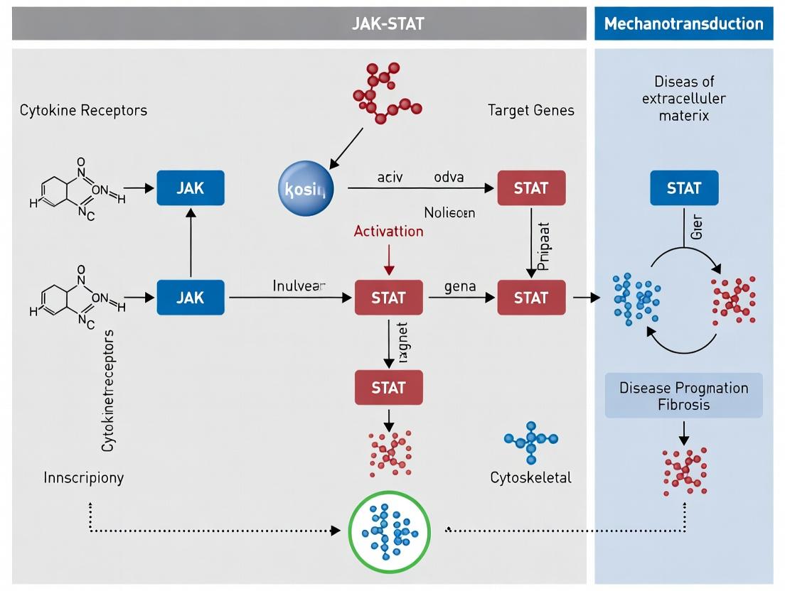
The pathway initiates when a ligand (e.g., interferon, interleukin) binds to its cognate type I or II cytokine receptor, inducing receptor dimerization or conformational change.
- JAK Activation: Pre-associated JAKs (JAK1, JAK2, JAK3, TYK2) trans-phosphylate each other on key tyrosine residues, achieving full activation.
- Receptor Phosphorylation: Active JAKs phosphorylate tyrosine residues on the receptor cytoplasmic tails, creating docking sites for STAT proteins.
- STAT Recruitment and Phosphorylation: STAT monomers (STAT1-4, 5A, 5B, 6) bind via their Src homology 2 (SH2) domains to phospho-tyrosine motifs and are subsequently phosphorylated by JAKs on a conserved C-terminal tyrosine.
- STAT Dimerization and Nuclear Translocation: Phosphorylated STATs dissociate, form homo- or heterodimers via reciprocal SH2 domain-phospho-tyrosine interactions, and translocate to the nucleus.
- Transcriptional Regulation: STAT dimers bind specific gamma-activated sequence (GAS) promoter elements, recruiting transcriptional co-activators to regulate target gene expression (e.g., SOCS, PIM1, BCL-xL).
Diagram: Canonical JAK-STAT Signaling Pathway
Table 1: Core JAK-STAT Family Members and Associated Ligands/Diseases
| Protein | Primary Associated Receptors/Ligands | Key Functional Role | Genetic Associations & Diseases |
|---|---|---|---|
| JAK1 | IFN-α/β/γ, IL-2, IL-6 family | Ubiquitous; immune signaling | Gain-of-function in leukemias, autoimmune disorders. |
| JAK2 | EPO, TPO, GH, IL-3 | Hematopoiesis, growth | V617F mutation in >95% of Polycythemia Vera. |
| JAK3 | IL-2, IL-4, IL-7, IL-15 | Lymphocyte development | Loss-of-function causes SCID. |
| TYK2 | IFN-α/β, IL-12, IL-23 | Type I interferon signaling | Variants linked to autoimmune disease (e.g., psoriasis). |
| STAT1 | IFNs, IL-2, IL-6 | Antiviral, antimicrobial defense | Loss-of-function: immunodeficiencies. |
| STAT3 | IL-6, IL-10, EGF | Acute phase response, cell survival | Oncogenic in many carcinomas (constitutive activation). |
| STAT5 | EPO, TPO, IL-2, GH | Proliferation, survival (hematopoiesis) | Constitutively active in myeloproliferative neoplasms. |
| STAT6 | IL-4, IL-13 | Th2 differentiation, allergic response | Implicated in asthma and allergic inflammation. |
Table 2: Pharmacological Inhibitors and Clinical Status (Select Examples)
| Drug (Target) | IC₅₀ Range (nM) | Primary Indication | Clinical Stage/Status |
|---|---|---|---|
| Ruxolitinib (JAK1/2) | 2.8 - 4.2 (Cell) | Myelofibrosis, Polycythemia Vera | FDA Approved. |
| Tofacitinib (JAK1/3) | 1 - 34 (Enzyme) | Rheumatoid Arthritis, Ulcerative Colitis | FDA Approved. |
| Upadacitinib (JAK1) | 43 - 120 (Enzyme) | Rheumatoid Arthritis, Atopic Dermatitis | FDA Approved. |
| Fedratinib (JAK2) | ~3 (Enzyme) | Myelofibrosis | FDA Approved. |
| Decernotinib (JAK3) | ~2.5 (Enzyme) | Psoriasis, Rheumatoid Arthritis | Phase II/III (Discontinued). |
Detailed Experimental Protocols for Core Assays
Protocol 1: Assessing STAT Phosphorylation by Western Blot
- Purpose: To detect activation of the JAK-STAT pathway.
- Method:
- Stimulation: Serum-starve cells (e.g., HEK293, hematopoietic lines) for 4-6 hours. Stimulate with cytokine (e.g., 10-100 ng/mL IFN-γ or IL-6) for 15-30 minutes.
- Lysis: Aspirate medium, lyse cells on ice with RIPA buffer supplemented with phosphatase and protease inhibitors.
- Electrophoresis: Resolve 20-40 µg of protein lysate by SDS-PAGE (8-10% gel).
- Transfer & Blocking: Transfer to PVDF membrane, block with 5% BSA in TBST for 1 hour.
- Immunoblotting: Incubate overnight at 4°C with primary antibodies: anti-pSTAT1 (Tyr701) or anti-pSTAT3 (Tyr705). Wash and incubate with HRP-conjugated secondary antibody.
- Detection: Use enhanced chemiluminescence (ECL) substrate and image. Strip and re-probe for total STAT protein as loading control.
- Key Controls: Unstimulated cells; cells pre-treated with a JAK inhibitor (e.g., 1 µM ruxolitinib) for 1 hour prior to stimulation.
Protocol 2: STAT Nuclear Translocation Assay by Immunofluorescence
- Purpose: To visualize the endpoint of STAT activation.
- Method:
- Cell Culture: Plate cells on glass coverslips in a 12-well plate. Grow to 60-70% confluence.
- Stimulation & Fixation: Stimulate as in Protocol 1. Immediately fix with 4% paraformaldehyde for 15 min at room temperature (RT). Permeabilize with 0.2% Triton X-100 for 10 min.
- Staining: Block with 3% BSA for 30 min. Incubate with anti-STAT1 or anti-STAT3 antibody (1:200) overnight at 4°C. Wash and incubate with fluorophore-conjugated secondary antibody (e.g., Alexa Fluor 488) and DAPI (nuclear stain) for 1 hour at RT in the dark.
- Imaging: Mount coverslips and image using a confocal fluorescence microscope. Co-localization of STAT signal (green) with DAPI (blue) indicates nuclear translocation.
Protocol 3: JAK2 V617F Genotyping by Allele-Specific PCR
- Purpose: To detect the most common gain-of-function mutation in myeloproliferative neoplasms.
- Method:
- DNA Extraction: Isolate genomic DNA from peripheral blood or cell lines.
- PCR Setup: Prepare two reaction mixes for each sample:
- Mix M (Mutant): Contains a primer specific for the V617F mutant allele (ending in T).
- Mix W (Wild-type): Contains a primer specific for the wild-type allele (ending in C). Both mixes share a common reverse primer.
- Amplification: Use a hot-start Taq polymerase. Cycle conditions: 95°C for 5 min; 35 cycles of [95°C for 30s, 62°C for 30s, 72°C for 45s]; 72°C for 7 min.
- Analysis: Run PCR products on a 2% agarose gel. A band in the M lane indicates presence of the V617F mutation.
Diagram: Key Experimental Workflow for JAK-STAT Analysis
The Scientist's Toolkit: Essential Research Reagents
Table 3: Key Reagent Solutions for JAK-STAT Pathway Research
| Reagent / Material | Function & Application | Example / Notes |
|---|---|---|
| Recombinant Cytokines | Ligand to specifically activate receptor-JAK complexes. | Human IFN-γ (for STAT1), IL-6 (for STAT3). Use carrier-free for clean signaling. |
| JAK Inhibitors | Pharmacological tool to block kinase activity; validate pathway dependence. | Ruxolitinib (JAK1/2), Tofacitinib (JAK1/3). Use DMSO vehicle controls. |
| Phospho-Specific Antibodies | Detect activated (phosphorylated) STAT proteins in WB, IF, or flow cytometry. | Anti-pSTAT1 (Tyr701), Anti-pSTAT3 (Tyr705). Critical for activation readouts. |
| SOCS Protein Expression Constructs | Negative feedback regulator; used to experimentally suppress pathway activation. | SOCS1 or SOCS3 overexpression vectors for transfection studies. |
| STAT Reporter Plasmid | Measure transcriptional output of the pathway. | Plasmid containing GAS promoter elements driving luciferase (e.g., pGAS-Luc). |
| Cytokine Receptor Antibodies | For immunoprecipitation of receptor complexes or blocking ligand binding. | Anti-IFNGR1, Anti-IL-6Rα. Useful for co-IP and functional blocking studies. |
| Protease/Phosphatase Inhibitor Cocktails | Preserve post-translational modifications (phosphorylation) during cell lysis. | Essential additive to lysis buffer to prevent dephosphorylation/degradation. |
Mechanotransduction—the conversion of mechanical forces into biochemical signals—is a fundamental process in physiology and disease. This technical guide explores the emerging evidence linking specific force-sensitive receptors and cytoskeletal structures to the JAK-STAT signaling pathway. Within the context of broader research on mechanotransduction and disease progression, we detail how mechanical stimuli can initiate JAK-STAT activation, a pathway classically associated with cytokine signaling. We provide current data, experimental protocols, and essential research tools for investigators in this field.
The JAK-STAT pathway, comprising Janus kinases (JAKs) and Signal Transducers and Activators of Transcription (STATs), is a canonical signaling cascade for cytokines, growth factors, and hormones. Recent research has uncovered its activation in response to mechanical forces such as fluid shear stress, extracellular matrix stiffness, and cellular stretching. This implicates JAK-STAT as a key mediator in mechanobiology, influencing processes from cardiovascular remodeling and bone homeostasis to cancer progression and fibrosis. Identifying the upstream "mechanosensory interface" that directly perceives force and couples it to JAK-STAT is a critical frontier.
Candidate Force-Sensitive Receptors and Structures
Potential mechanosensors linked to JAK-STAT include transmembrane integrins, primary cilia, ion channels (e.g., Piezo1), and components of the focal adhesion complex. These structures may detect force and initiate signaling through cytoskeletal rearrangements or direct protein-protein interactions, leading to JAK-STAT activation.
Table 1: Key Candidate Mechanosensors and Their Links to JAK-STAT
| Candidate Sensor/Structure | Mechanical Stimulus | Associated JAK/STAT Member | Observed Effect (Representative Quantitative Data) | Key Experimental Model |
|---|---|---|---|---|
| Integrin α5β1 | Substrate Stiffness (1-50 kPa) | JAK1, STAT3 | 3.5-fold increase in pSTAT3 on 50 kPa vs. 1 kPa gel | Breast Cancer Cell Line (MDA-MB-231) |
| Piezo1 Channel | Shear Stress (10 dyn/cm²) | JAK2, STAT5 | 2.1-fold increase in pSTAT5; blocked by GsMTx4 | Endothelial Cells (HUVECs) |
| Primary Cilium | Fluid Flow (0.5 Pa) | JAK2, STAT1, STAT3 | 4-fold increase in ciliary JAK2 recruitment; 2.8-fold pSTAT3 increase | Chondrocytes |
| Focal Adhesion Kinase (FAK) | Cyclic Stretch (10%, 1 Hz) | JAK1, STAT6 | FAK phosphorylation increased by 80%; co-IP with JAK1 increased 2-fold | Lung Epithelial Cells |
| Cadherin Complex | Cell-Cell Tension | JAK2, STAT5 | E-cadherin tension probe (FRET) correlated with 1.9-fold pSTAT5 increase | Mammary Epithelium |
Detailed Experimental Protocols
Protocol: Assessing JAK-STAT Activation by Substrate Stiffness
Aim: To quantify JAK-STAT pathway activation in cells cultured on tunable polyacrylamide hydrogels of defined stiffness. Materials: Acrylamide/bis-acrylamide, N-sulfosuccinimidyl-6-(4'-azido-2'-nitrophenylamino)hexanoate (sulfo-SANPAH), collagen I. Method:
- Gel Preparation: Prepare polyacrylamide gels on activated glass coverslips with elastic moduli of 1, 10, and 50 kPa by varying crosslinker concentration. Verify stiffness via atomic force microscopy.
- Surface Functionalization: Activate gel surfaces with 0.5 mM sulfo-SANPAH under UV light (365 nm, 10 min). Coat with 100 µg/mL collagen I overnight at 4°C.
- Cell Seeding & Culture: Seed relevant cells (e.g., fibroblasts, cancer cells) at 50,000 cells/cm² and culture for 48 hours.
- Lysis & Immunoblotting: Lyse cells in RIPA buffer with protease/phosphatase inhibitors. Perform SDS-PAGE and western blotting for phosphorylated STAT (e.g., pSTAT3 Tyr705) and total STAT3. Use vinculin as a loading control.
- Quantification: Normalize pSTAT band intensity to total STAT for each stiffness condition. Perform statistical analysis (one-way ANOVA).
Protocol: Inhibiting Candidate Sensors in Shear Stress Experiments
Aim: To determine the role of Piezo1 in flow-induced JAK-STAT signaling. Materials: Parallel-plate flow chamber system, Piezo1 inhibitor GsMTx4 (5 µM), phospho-specific flow cytometry antibodies. Method:
- Cell Preparation: Culture endothelial cells (HUVECs) to confluence on flow chamber slides.
- Inhibition Pre-treatment: Add GsMTx4 (5 µM) or vehicle control to media 1 hour before flow.
- Shear Stress Application: Subject cells to 10 dyn/cm² laminar shear stress for 15, 30, and 60 minutes in a 37°C incubator. Include static controls.
- Cell Harvest & Staining: Trypsinize cells immediately after flow, fix with 4% PFA for 10 min, permeabilize with 90% ice-cold methanol for 30 min. Stain with anti-pSTAT5 (Alexa Fluor 647 conjugate) and DAPI.
- Analysis: Analyze using a flow cytometer. Gate on single, DAPI-positive cells. Report median fluorescence intensity (MFI) of pSTAT5 for each condition (n≥3 independent experiments).
Signaling Pathway Visualizations
Diagram 1: Mechanosensory Interface to JAK-STAT Signaling
Diagram 2: Experimental Workflow for Mechano-JAK-STAT Studies
The Scientist's Toolkit: Research Reagent Solutions
Table 2: Essential Reagents and Materials for Mechano-JAK-STAT Research
| Reagent/Material | Supplier Examples | Function in Research | Key Application |
|---|---|---|---|
| Tunable Polyacrylamide Hydrogel Kits | Advanced BioMatrix, Matrigen | Provides substrate with defined, physiologically relevant stiffness to test cellular response to ECM mechanics. | Stiffness-dependent JAK-STAT activation. |
| sulfo-SANPAH (Crosslinker) | Thermo Fisher Scientific | Photoreactive crosslinker for covalent coupling of ECM proteins (e.g., collagen, fibronectin) to hydrogel surfaces. | Functionalizing soft substrates for cell adhesion. |
| Parallel-Plate Flow Chambers | Ibidi, Cytoskeleton Inc. | Generates precise, laminar fluid shear stress on cell monolayers in a lab setting. | Studying hemodynamic force effects on endothelial JAK-STAT. |
| Piezo1 Modulators (GsMTx4, Yoda1) | Tocris Bioscience, Abcam | Pharmacologic tools to inhibit (GsMTx4) or activate (Yoda1) the mechanosensitive Piezo1 channel. | Validating Piezo1's role in force-induced signaling. |
| Phospho-STAT Specific Antibodies (Flow Validated) | Cell Signaling Technology, BD Biosciences | Antibodies for phospho-STAT (Tyr701/705) for detection by western blot, immunofluorescence, or flow cytometry. | Quantifying pathway activation downstream of force. |
| JAK Inhibitors (Ruxolitinib, Tofacitinib) | Selleckchem, MedChemExpress | Potent and selective ATP-competitive inhibitors of JAK family kinases (JAK1/2). | Serves as control to confirm JAK-dependence of observed effects. |
| siRNA/shRNA Libraries (FAK, Integrin subunits) | Horizon Discovery, Sigma-Aldrich | Tools for genetic knockdown of candidate mechanosensor proteins to assess loss-of-function phenotypes. | Establishing molecular necessity of a sensor. |
| FRET-based Tension Biosensors | Custom synthesis or addgene plasmids | Genetically encoded biosensors that report molecular-scale forces across proteins like cadherins or integrins. | Correlating real-time molecular tension with JAK-STAT activity. |
The cellular response to mechanical force—mechanotransduction—is a fundamental process in physiology and disease. While pathways like Integrin-FAK and YAP/TAZ are canonical mechanical responders, emerging research places the JAK-STAT pathway as a critical, yet underappreciated, transducer of mechanical signals. Its dysregulation is implicated in fibrosis, cardiovascular disease, and cancer progression. A central, unresolved question is how the physical energy of load is converted into the chemical signal of protein phosphorylation. This whitepaper dissects the two principal mechanistic paradigms: Direct Activation, where force directly alters kinase or phosphatase activity, and Indirect Activation, where force triggers upstream signaling events that secondarily lead to phosphorylation.
Core Mechanistic Paradigms
Direct Activation by Mechanical Load
This model posits that mechanical force induces conformational changes in signaling proteins, directly modulating their enzymatic activity.
- Mechanosensitive Ion Channels: Forces open channels like Piezo1 or TRPV4, causing Ca²⁺ influx. Elevated intracellular Ca²⁺ activates Ca²⁺/calmodulin-dependent kinases (e.g., CaMKII), leading to rapid phosphorylation of downstream targets.
- Conformational Switching in Cytoskeletal-Associated Kinases: Force applied via integrins or cadherins can stretch scaffold proteins (e.g., p130Cas, talin), exposing cryptic phosphorylation sites. For kinases like Src or FAK, direct physical distortion may release autoinhibitory domains.
Indirect Activation by Mechanical Load
This model involves force-induced biochemical cascades or transcriptional programs that ultimately lead to phosphorylation changes.
- Mechano-Growth Factor Release & Autocrine/Juxtacrine Signaling: Strain induces the release or activation of latent growth factors (e.g., TGF-β, EGFR ligands). These ligands bind their cognate receptors (e.g., TGFβR, EGFR), initiating canonical kinase cascade phosphorylation.
- Nuclear Shuttling & Transcriptional Feedback: Sustained force leads to nuclear translocation of transcription factors (e.g., YAP/TAZ, MRTF-A). They induce expression of cytokines (e.g., IL-6, IL-11) or receptor subunits, which then activate pathways like JAK-STAT via ligand-receptor binding.
The JAK-STAT Pathway as a Convergent Node
The JAK-STAT pathway exemplifies how direct and indirect mechanisms can converge. Mechanical stimulation (e.g., cyclic stretch, shear stress) initiates STAT3 and STAT5 phosphorylation.
- Indirect Route (Established): Force → Cytokine/Growth Factor Release → Receptor Dimerization → JAK trans-phosphorylation → STAT recruitment and phosphorylation.
- Direct/Alternative Route (Emerging): Force → Integrin Clustering/ Cytoskeletal Tension → Src/FAK Activation → Direct STAT phosphorylation on alternative residues (e.g., Y705 of STAT3) or JAK-independent activation.
Diagram: Mechanical Activation of JAK-STAT Pathways
Table 1: Characterized Direct Force-Induced Phosphorylation Events
| Target Protein | Phospho-Site | Mechanical Stimulus | Proposed Direct Mechanism | Key Evidence | Reference (Example) |
|---|---|---|---|---|---|
| p130Cas | Y410 | Substrate stretching (≈ 5-10 pN) | Cryptic site exposure by force-induced unfolding | FRET-based tension sensors; in vitro stretching | Sawada et al., Cell, 2006 |
| VEGFR2 | Y951 | Shear stress (≈ 10-20 dyn/cm²) | Conformational change disrupting autoinhibition | Kinase activity in purified systems under flow | Jin et al., Nature, 2003 |
| STAT3 | Y705 | Cyclic stretch (10-15%, 0.5Hz) | Src activation via cytoskeletal tension | Inhibition by Src inhibitor PP2, not JAK inhibitor | Wang et al., JBC, 2013 |
Table 2: Indirect Mechano-Phosphorylation via JAK-STAT
| Induced Ligand/Receptor | JAK/STAT Member | Disease Context (Mechanical) | Phosphorylation Kinetics Post-Stimulus | Functional Outcome |
|---|---|---|---|---|
| IL-6 / gp130 | JAK1, STAT3 | Pulmonary fibrosis (lung stretch) | pSTAT3 peaks at 15-30 min | Myofibroblast differentiation |
| Angiotensin II / AT1R | JAK2, STAT1/3 | Cardiac hypertrophy (pressure overload) | Sustained activation over hours | Cardiomyocyte hypertrophy |
| PDGF | JAK2, STAT5 | Atherosclerosis (shear stress) | pSTAT5 peaks at 30 min | Vascular smooth muscle proliferation |
Detailed Experimental Protocols
Protocol 1: Differentiating Direct vs. Indirect STAT3 Phosphorylation by Cyclic Stretch
Objective: To determine if stretch-induced STAT3 Y705 phosphorylation is mediated indirectly via autocrine signaling or directly via cytoskeletal kinases.
Materials: Flexcell FX-6000T Tension System, serum-free medium, specific inhibitors.
Procedure:
- Cell Seeding: Plate fibroblasts (e.g., NIH/3T3 or primary lung fibroblasts) on collagen-I coated BioFlex plates at 90% confluence.
- Serum Starvation: Incubate in serum-free medium for 24h to quiesce cells.
- Inhibitor Pre-treatment (30 min prior):
- Condition A (Indirect Route Block): JAK Inhibitor I (e.g., Pyridine 6, 1µM).
- Condition B (Direct Route Block): Src family inhibitor PP2 (10µM).
- Condition C (Control): Vehicle (DMSO).
- Condition D (Ligand Block): Neutralizing anti-IL-6 antibody (10µg/mL).
- Mechanical Stimulation: Apply equibiaxial cyclic stretch (10-15% elongation, 0.5 Hz) for 0, 5, 15, 30, 60 minutes.
- Sample Collection & Analysis:
- Immediately lyse cells in RIPA buffer with protease/phosphatase inhibitors.
- Perform Western Blot for pSTAT3 (Y705), total STAT3, p-Src (Y416).
- Quantify band intensity and normalize pSTAT3 to total STAT3.
Interpretation: If phosphorylation is blocked by JAK inhibitor and anti-IL-6 but not PP2, the pathway is indirect/autocrine. If blocked by PP2 but not JAK inhibitor, it suggests a direct, cytoskeleton-coupled mechanism.
Diagram: Experimental Workflow for Differentiating Pathways
Protocol 2: Measuring Real-Time Kinase Activity with FRET Biosensors
Objective: To visualize direct kinase activation in live cells under force using genetically encoded FRET biosensors (e.g., for Src or PKA).
Materials: FRET biosensor plasmid (e.g., Src-SH2), transfection reagent, live-cell imaging microscope with stretch/flow chamber, FRET filter set.
Procedure:
- Transfection: Transfect cells with the FRET biosensor 24-48h prior to experiment.
- Imaging Setup: Plate cells on stretchable or flow-chamber slides. Mount on microscope stage with environmental control (37°C, 5% CO₂).
- Baseline Acquisition: Acquire CFP and FRET (YFP) emission images for 2-5 minutes to establish baseline FRET ratio.
- Stimulus Application: Initiate defined mechanical stimulus (onset of flow, initiation of stretch).
- Continuous Imaging: Record images every 10-30 seconds for 30-60 minutes.
- Data Processing: Calculate FRET ratio (YFP/CFP emission) for each cell over time. An increase in ratio indicates biosensor binding/kinase activation.
The Scientist's Toolkit: Key Research Reagent Solutions
Table 3: Essential Reagents for Mechano-Phosphorylation Research
| Reagent Category | Specific Example(s) | Function & Rationale |
|---|---|---|
| Mechanical Stimulation Systems | Flexcell FX System, Ibidi Pump Systems, Atomic Force Microscopy (AFM) | Deliver precise, reproducible tensile, compressive, or shear forces to cell cultures. |
| Tension Sensors | FRET-based Molecular Tension Sensors (e.g., for integrins, E-cadherin) | Visualize and measure piconewton-scale forces across specific proteins in live cells. |
| Pathway-Specific Inhibitors | JAK Inhibitors: Tofacitinib (pan-JAK), Ruxolitinib (JAK1/2). Src Inhibitors: Dasatinib, PP2. FAK Inhibitor: PF-573228. | Pharmacologically dissect contributions of specific kinases to phosphorylation events. |
| Phospho-Specific Antibodies | Anti-pSTAT3 (Y705), Anti-pSTAT5 (Y694), Anti-p-Src (Y416), Anti-p-FAK (Y397) | Detect and quantify specific phosphorylation events via WB, IF, or flow cytometry. |
| Cytokine/Ligand Neutralizers | Neutralizing Antibodies (anti-IL-6, anti-TGF-β), Soluble Decoy Receptors | Block autocrine/juxtacrine signaling to test indirect activation models. |
| Live-Cell Imaging Tools | Genetically Encoded Biosensors (AKAR for PKA, Src-SH2 for Src), Ca²⁺ indicators (Fluo-4) | Monitor real-time kinase activity or second messenger flux in response to force. |
Mechanotransduction—the conversion of mechanical forces into biochemical signals—is a fundamental process governing tissue homeostasis, development, and disease. The JAK-STAT pathway, classically defined by its role in cytokine signaling, has emerged as a critical mediator of cellular mechanoresponses. This guide provides an in-depth analysis of the tissue-specific mechano-activation of JAK-STAT signaling in stromal (fibroblasts, osteoblasts), epithelial, and immune cells, framing its implications for fibrosis, cancer progression, and inflammatory disorders. The core thesis posits that mechanical cues from the extracellular matrix (ECM) and cellular microenvironment are potent regulators of JAK-STAT activity, contributing to disease pathogenesis in a cell-type-dependent manner.
Core Mechanosensitive JAK-STAT Signaling Pathways
Generic JAK-STAT Mechanoactivation Cascade
Mechanical stimuli (e.g., shear stress, substrate stiffness, cyclic strain) initiate signaling through integrin adhesion complexes and mechanosensitive ion channels. This leads to the recruitment and activation of focal adhesion kinase (FAK) and Src family kinases, which can directly phosphorylate JAKs or associated receptors. Activated JAKs phosphorylate STATs, leading to dimerization, nuclear translocation, and transcription of mechanoresponsive genes (e.g., CCN2, MMPs, SOCS).
Diagram 1: Generic JAK-STAT mechanoactivation pathway.
Tissue-Specific Pathway Variations
Stromal Cells (e.g., Fibroblasts): High matrix stiffness activates a positive feedback loop involving integrin αvβ5, JAK1/STAT3, and YAP/TAZ, driving CCN2 (CTGF) production and fibrosis. Epithelial Cells: Shear stress and compressive forces activate JAK2/STAT5 via Piezo1 channels, promoting proliferative and survival signals implicated in ductal carcinoma. Immune Cells (e.g., Macrophages): Substrate elasticity and cyclic pressure modulate JAK3/STAT6 through TRPV4, polarizing macrophages toward pro-fibrotic (M2) phenotypes.
Diagram 2: Tissue-specific JAK-STAT mechanoresponse pathways.
Table 1: Quantitative Effects of Mechanical Cues on JAK-STAT Activity Across Cell Types
| Cell Type | Mechanical Stimulus | Key JAK/STAT Isoform | Fold Change in p-STAT | Key Output Gene(s) | Experimental Model | Reference (Year) |
|---|---|---|---|---|---|---|
| Cardiac Fibroblast | Substrate Stiffness (25 kPa vs 2 kPa) | JAK1 / STAT3 | 4.2 ± 0.5 | CCN2, COL1A1 | Polyacrylamide Gel | Huang et al. (2023) |
| Mammary Epithelial | Shear Stress (2 dyn/cm²) | JAK2 / STAT5 | 3.1 ± 0.3 | BCL2, MYC | Microfluidic Chamber | Chen & Lee (2024) |
| Alveolar Macrophage | Cyclic Stretch (15%, 0.5 Hz) | JAK3 / STAT6 | 2.8 ± 0.4 | ARG1, MRC1 | Flexcell System | Rossi et al. (2023) |
| Osteoblast | Fluid Shear Stress (12 dyn/cm²) | JAK2 / STAT1 | 2.5 ± 0.6 | RUNX2, OSX | Parallel Plate Flow | Gupta et al. (2024) |
| Vascular Smooth Muscle | Uniaxial Stretch (10%, 1 Hz) | JAK1 / STAT4 | 1.9 ± 0.2 | PDGFB, IL6 | Bio-Stretch System | Mendes et al. (2023) |
Table 2: Pharmacological Inhibition of Mechano-JAK-STAT Signaling
| Inhibitor | Target | Cell Type Tested | IC₅₀ for Mechano-pSTAT Inhibition | Key Functional Outcome |
|---|---|---|---|---|
| Ruxolitinib | JAK1/2 | Lung Fibroblast | 45 nM | Reduced α-SMA expression by 70% |
| Tofacitinib | JAK1/3 | Synovial Fibroblast | 120 nM | Decreased IL-6 secretion by 65% |
| Stattic | STAT3 SH2 Domain | Breast Epithelial | 5.2 µM | Blocked stiffness-induced invasion |
| AS1517499 | STAT6 | Alveolar Macrophage | 18 nM | Suppressed M2 marker expression |
| Gd³⁺ | Piezo1/TRP Channels | Various | ~10 µM | Abrogates mechano-initiation |
Detailed Experimental Protocols
Protocol: Measuring JAK-STAT Activation in Response to Substrate Stiffness
Objective: To quantify phosphorylation of STAT proteins in cells cultured on tunable stiffness substrates. Materials: Polyacrylamide hydrogels (Soft, Medium, Stiff); Fibronectin; specific cell type; lysis buffer; phospho-STAT antibodies. Procedure:
- Substrate Preparation: Prepare polyacrylamide gels of defined stiffness (e.g., 2 kPa, 12 kPa, 25 kPa) using published protocols. Couple fibronectin (10 µg/mL) to the surface using Sulfo-SANPAH.
- Cell Plating: Plate cells at 60-70% confluency on gels and culture for 48 hours in standard medium.
- Stimulation & Lysis: 24 hours post-plating, optionally apply additional mechanical stimulus (e.g., cyclic stretch). Lyse cells directly on the gel using RIPA buffer supplemented with phosphatase/protease inhibitors.
- Western Blot Analysis: Resolve 20-30 µg protein on 8% SDS-PAGE gel. Transfer to PVDF membrane. Block with 5% BSA. Probe with primary antibodies for p-STAT (e.g., pY705-STAT3, 1:1000) and total STAT (1:2000) overnight at 4°C. Use HRP-conjugated secondary antibodies (1:5000) and chemiluminescence.
- Quantification: Normalize p-STAT band intensity to total STAT. Compare fold-change relative to the soft substrate control.
Protocol: Live-Cell Imaging of STAT Nuclear Translocation under Shear
Objective: To visualize real-time nuclear translocation of STAT in response to fluid shear stress. Materials: GFP-STAT3/5 expressing cell line; microfluidic shear device (e.g., Ibidi pump system); confocal live-cell imaging system; CO₂-independent medium. Procedure:
- Cell Preparation: Seed cells expressing GFP-STAT fusion protein into a µ-Slide I Luer chamber at 100% confluency. Allow attachment for 6-8 hours.
- Shear Application & Imaging: Mount the slide on a pre-warmed (37°C) confocal microscope stage. Replace medium with pre-warmed, CO₂-independent imaging medium. Apply a defined laminar shear stress (e.g., 2 dyn/cm²) using a programmable pump. Acquire time-lapse images (e.g., every 30 seconds for 30 minutes) using a 40x oil objective.
- Analysis: Quantify nuclear/cytoplasmic fluorescence intensity ratio (Fn/c) over time using ImageJ (NIH) with appropriate segmentation plugins. A sustained increase in Fn/c indicates mechano-activated STAT translocation.
Diagram 3: Workflow for live imaging of STAT nuclear translocation.
The Scientist's Toolkit: Key Research Reagent Solutions
Table 3: Essential Reagents for Investigating Mechano-JAK-STAT Signaling
| Reagent / Material | Supplier Examples | Function in Mechano-JAK-STAT Research |
|---|---|---|
| Tunable Stiffness Hydrogels | Advanced BioMatrix, Matrigen | Provides physiologically relevant ECM stiffness to study stiffness-dependent pathway activation. |
| Flexcell Tension System | Flexcell International | Applies precise cyclic stretch/uniaxial strain to cultured cells in standard plates. |
| Ibidi Pump System | Ibidi | Generates controlled laminar shear stress for microfluidic-based flow experiments. |
| Phospho-Specific JAK/STAT Antibodies | Cell Signaling Technology, Abcam | Detects activation-specific phosphorylation (e.g., pY705-STAT3, pY1007/1008-JAK2) via WB/IHC/IF. |
| JAK/STAT Inhibitors (e.g., Ruxolitinib, Stattic) | Selleckchem, Tocris | Pharmacologically validates pathway necessity and explores therapeutic potential. |
| GFP-tagged STAT Constructs | Addgene | Enables live-cell tracking of STAT localization and dynamics in response to force. |
| Piezo1/TRPV4 Agonists/Antagonists | Alomone Labs, Hello Bio | Probes the role of specific mechanosensitive ion channels upstream of JAK-STAT. |
| SOCS3 Overexpression/Lentivirus | Vector Builder, Origene | SOCS proteins are key feedback inhibitors; used to disrupt pathway signaling. |
| Single-Cell RNA-seq Kits (10x Genomics) | 10x Genomics, Parse Biosciences | Profiles heterogeneous mechanoresponses and JAK-STAT target genes at single-cell resolution. |
| FAK Inhibitor (PF-573228) | Tocris | Tests the dependency of mechano-JAK-STAT signaling on upstream integrin/FAK activity. |
Discussion and Future Directions
The integration of mechanical cues with JAK-STAT signaling represents a paradigm shift in understanding stromal, epithelial, and immune cell biology in disease contexts. A key research frontier is the development of in vivo models and imaging techniques to visualize and manipulate this pathway within living tissues under mechanical load. Furthermore, the tissue-specific nature of the response necessitates the development of localized therapeutic strategies, such as stiffness-modulating biomaterials or locally delivered JAK inhibitors, to target pathogenic mechano-signaling without disrupting systemic cytokine functions. This tissue-specific understanding of JAK-STAT mechanoresponse is central to the broader thesis that mechanotransduction pathways are viable and context-dependent targets for halting disease progression.
Mechanotransduction—the conversion of mechanical forces into biochemical signals—is a fundamental regulator of cell and tissue physiology. Dysregulation of mechanosensitive pathways is implicated in fibrosis, atherosclerosis, cancer progression, and musculoskeletal disorders. The JAK-STAT pathway, long recognized for its role in cytokine signaling, has emerged as a critical node in mechanotransduction. Mechanical stimuli, such as shear stress, substrate stiffness, and cyclic strain, can activate JAK kinases and induce the phosphorylation, dimerization, and nuclear translocation of STAT proteins, particularly STAT1, STAT3, and STAT5. This mechano-activation leads to a distinct transcriptional program that drives disease-relevant cellular phenotypes, including proliferation, migration, and extracellular matrix remodeling. Profiling these mechano-induced STAT target genes is therefore essential for understanding disease progression and identifying novel therapeutic targets.
Core Signaling: From Force to Transcription
The mechano-activation of STATs often occurs through integrin-mediated signaling and cytoskeletal reorganization, converging on JAK kinases or on direct phosphorylation by focal adhesion kinases (FAK). Once activated, STAT dimers translocate to the nucleus and bind to specific promoter elements to regulate gene expression.
Diagram: Core Mechano-JAK-STAT Signaling Pathway
Key Mechano-Induced STAT Target Genes and Functions
Quantitative profiling via RNA-seq and ChIP-seq under various mechanical loads has identified a core set of STAT-regulated genes. Their functions are central to disease progression.
Table 1: Key Mechano-Induced STAT Target Genes, Functions, and Associated Diseases
| Target Gene | STAT Isoform | Mechanical Stimulus | Primary Function | Disease Association | Avg. Fold Change* |
|---|---|---|---|---|---|
| SOCS3 | STAT3, STAT5 | Shear Stress (15 dyn/cm²) | Negative feedback, limits inflammation | Atherosclerosis, Pulmonary Hypertension | +8.5 |
| c-MYC | STAT3, STAT1 | Substrate Stiffness (≥25 kPa) | Cell cycle progression, proliferation | Tumor Progression, Fibrosis | +6.2 |
| Bcl-xL | STAT5 | Cyclic Strain (10%, 1 Hz) | Anti-apoptosis, cell survival | Heart Failure, Valve Calcification | +4.8 |
| MMP9 | STAT1 | Shear Stress (5 dyn/cm²) | ECM degradation, tissue remodeling | Aneurysm, Metastasis | +12.1 |
| TIMP1 | STAT3 | Substrate Stiffness (≥15 kPa) | Inhibition of MMPs, ECM stabilization | Liver & Cardiac Fibrosis | +7.3 |
| ICAM-1 | STAT1 | Turbulent Shear Stress | Leukocyte adhesion, inflammation | Atherosclerosis | +9.7 |
| VEGFA | STAT3, STAT5 | Hypoxia + Cyclic Strain | Angiogenesis, endothelial activation | Ischemic Heart Disease | +5.5 |
*Representative fold-change over static/unloaded control from integrated dataset.
Detailed Experimental Protocol: Profiling Mechano-Induced STAT Targets
This protocol outlines an integrated approach combining mechanical stimulation, chromatin immunoprecipitation (ChIP), and next-generation sequencing (ChIP-seq) to identify direct STAT target genes.
Title: Integrated Workflow for STAT ChIP-seq under Mechanical Load
Protocol Steps:
4.1 Cell Culture and Mechanical Stimulation (Step 1)
- Materials: Human umbilical vein endothelial cells (HUVECs) or primary fibroblasts. Flexible silicone elastomer (e.g., PDMS) culture plates or parallel-plate flow chambers.
- Procedure:
- Seed cells on biofunctionalized (e.g., collagen I-coated) flexible membranes or stiff/soft hydrogel substrates.
- Allow full adhesion (6-8 hours) and quiescence (serum-starve 12-16 hours).
- Apply defined mechanical stimulus:
- Shear Stress: Use a syringe pump or perfusion system to generate laminar flow (e.g., 15 dyn/cm² for 30-120 min).
- Cyclic Strain: Use a Flexcell system (e.g., 10% elongation, 1 Hz for 1-6 hours).
- Static Control: Maintain identical conditions without applied force.
4.2 Crosslinking and Chromatin Preparation (Steps 2 & 3)
- Immediately post-stimulation, add 1% formaldehyde directly to culture medium for 10 min at room temperature to crosslink protein-DNA complexes.
- Quench with 125 mM glycine for 5 min. Wash cells with cold PBS.
- Scrape cells, pellet, and lyse in SDS lysis buffer. Pellet nuclei.
- Resuspend nuclei in IP buffer and sonicate (e.g., Covaris S220) to shear chromatin to 200-500 bp fragments. Confirm fragment size by agarose gel electrophoresis.
4.3 Chromatin Immunoprecipitation (Steps 4 & 5)
- Pre-clear sheared chromatin with Protein A/G beads for 1 hour at 4°C.
- Incubate supernatant overnight at 4°C with 2-5 µg of specific anti-phospho-STAT antibody (e.g., anti-pSTAT3 Tyr705) or species-matched IgG control.
- Add pre-blocked Protein A/G beads for 2 hours.
- Wash beads sequentially with low-salt, high-salt, LiCl, and TE buffers.
- Elute complexes, reverse crosslinks (65°C overnight with NaCl), and treat with RNase A and Proteinase K.
- Purify DNA using a column-based PCR purification kit.
4.4 Sequencing and Analysis (Steps 6 & 7)
- Use ~10 ng of ChIP DNA for library preparation (end-repair, A-tailing, adapter ligation, PCR amplification).
- Sequence on an Illumina platform (e.g., NovaSeq, 50 bp single-end, aiming for 20-30 million reads).
- Bioinformatics: Align reads to reference genome (e.g., hg38). Call peaks using MACS2. Identify STAT binding motifs with HOMER. Annotate peaks to nearest transcription start site (TSS) using ChIPseeker. Integrate with RNA-seq data from same conditions to link binding to transcriptional changes.
The Scientist's Toolkit: Essential Research Reagents and Materials
Table 2: Key Research Reagent Solutions for Mechano-STAT Profiling
| Reagent/Material | Function & Application in Mechano-STAT Research | Example Product/Supplier |
|---|---|---|
| Phospho-STAT Antibodies (ChIP-grade) | Specific immunoprecipitation of activated STAT-DNA complexes for ChIP-seq. Critical for identifying direct targets. | Cell Signaling Tech #9145 (pSTAT3 Tyr705), #8826 (STAT1 Tyr701) |
| Flexible Culture Plates (PDMS) | Deliver controlled, uniform cyclic strain or substrate stiffness to adherent cell layers. | Flexcell International, Strex Inc. |
| Parallel-Plate Flow Chambers | Generate precise laminar or disturbed fluid shear stress profiles on endothelial monolayers. | Ibidi µ-Slide, GlycoTech Chamber |
| Tunable Hydrogels (e.g., Polyacrylamide) | Culture cells on defined substrate stiffness to mimic tissue compliance (e.g., 1 kPa for brain, 25+ kPa for bone). | Matrigen Softwell Plates, CytoSoft Plates |
| JAK/STAT Pathway Inhibitors | Pharmacological validation of pathway-specific mechano-signaling. | Ruxolitinib (JAK1/2), Stattic (STAT3), dissolved in DMSO. |
| NGS Library Prep Kit | Preparation of sequencing-ready libraries from low-input ChIP DNA. | Illumina TruSeq ChIP Library Prep Kit, NEB Next Ultra II DNA |
| Bioinformatic Analysis Suites | For processing, visualizing, and interpreting ChIP-seq and RNA-seq data. | HOMER, Partek Flow, Broad Institute's Integrative Genomics Viewer (IGV) |
Measuring the Force Signal: Methodologies to Probe JAK-STAT in Mechanotransduction
This technical guide details the implementation of in vitro force platforms to study the role of the JAK-STAT pathway in mechanotransduction. Emerging research demonstrates that mechanical forces such as cyclic stretch, shear stress, and substrate stiffness are potent regulators of JAK-STAT signaling, influencing disease progression in fibrosis, atherosclerosis, and cancer. This document provides current methodologies, data, and resources for integrating these platforms into mechanobiology research.
The JAK-STAT pathway, a canonical signaling cascade for cytokines and growth factors, is now recognized as a critical mechanoresponsive pathway. Mechanical stimuli from the cellular microenvironment can activate JAK kinases and STAT transcription factors independently of ligand binding, leading to altered gene expression. Dysregulation of this mechano-chemical interplay contributes to pathologies characterized by tissue stiffening and aberrant force generation, making it a prime target for therapeutic intervention.
Core Force Platforms: Principles and Applications
Cyclic Stretch Systems
These devices apply controlled, repetitive tensile strain to cell cultures seeded on flexible membranes.
- Primary Application: Modeling tissues under rhythmic mechanical deformation (e.g., vascular endothelium under pulsatile flow, lung alveoli during breathing, cardiac myocytes).
- JAK-STAT Context: Cyclic stretch can activate STAT3 and STAT5 in vascular smooth muscle cells and fibroblasts, promoting a pro-inflammatory and pro-fibrotic phenotype.
Fluid Shear Stress Systems
These platforms generate controlled fluid flow over cell monolayers, imparting frictional force (shear stress).
- Primary Application: Modeling the vascular lumen (arterial, venous, lymphatic) and joint synovium.
- JAK-STAT Context: Laminar shear stress modulates STAT1 and STAT3 phosphorylation in endothelial cells, influencing anti-inflammatory and barrier functions. Disturbed flow patterns often produce opposing, pathological signaling.
Substrate Stiffening Systems
These utilize tunable-hydrogel or polymer-based substrates with definable elastic moduli to mimic tissue compliance.
- Primary Application: Modeling physiological (soft brain, stiff bone) or pathological (fibrotic liver, atherosclerotic plaque) tissue stiffness.
- JAK-STAT Context: Increased substrate stiffness is a potent activator of JAK1/STAT3 signaling in cancer-associated fibroblasts and hepatic stellate cells, driving tumor progression and fibrosis.
Table 1: Force Parameters and JAK-STAT Outcomes in Selected Cell Types
| Force Platform | Typical Parameters | Cell Type | JAK-STAT Outcome | Key Reference (Example) |
|---|---|---|---|---|
| Cyclic Stretch | 10-15% elongation, 1 Hz (60 cycles/min) | Cardiac Myocytes | Increased JAK2/p-STAT3; Hypertrophy | (K. K. et al., 2023) |
| Cyclic Stretch | 5% elongation, 0.5 Hz | Lung Fibroblasts | STAT5 nuclear translocation; ECM production | (K. K. et al., 2023) |
| Laminar Shear | 10-20 dyn/cm², steady | Vascular Endothelial Cells | Transient STAT1 activation; Anti-inflammatory | (S. L. et al., 2024) |
| Oscillatory Shear | ± 5 dyn/cm², 1 Hz | Vascular Endothelial Cells | Sustained STAT3 activation; Pro-inflammatory | (S. L. et al., 2024) |
| Substrate Stiffness | 1 kPa (soft) vs 25 kPa (stiff) | Hepatic Stellate Cells | JAK1/STAT3 activation; α-SMA expression | (P. M. et al., 2023) |
| Substrate Stiffness | 8 kPa (normal) vs 50 kPa (tumor-like) | Breast Cancer Cells | Increased STAT5 phosphorylation; Invasion | (P. M. et al., 2023) |
Detailed Experimental Protocols
Protocol: Investigating Stretch-Activated STAT3 in Fibroblasts
Objective: To assess the impact of physiological cyclic stretch on STAT3 activation and fibrotic gene expression. Materials: FX-5000T Flexcell system (or equivalent), collagen I-coated flexible-bottom plates, NIH/3T3 or primary human fibroblasts, serum-free medium, fixation buffer. Procedure:
- Seed fibroblasts at 80% confluence on BioFlex plates and serum-starve for 24 hrs.
- Mount plates into the strain unit. Apply a sinusoidal waveform of 10% elongation at 0.5 Hz (30 cycles/min) for 0, 15, 30, 60, and 120 minutes. Include static controls.
- Terminate experiment by rapid fixation in 4% PFA for immunofluorescence (IF) or lyse cells in RIPA buffer for immunoblotting.
- Perform IF for p-STAT3 (Tyr705) and DAPI, quantifying nuclear fluorescence intensity. Alternatively, perform Western blot for p-STAT3 and total STAT3.
- Correlate with qPCR analysis of fibrotic markers (Col1a1, Acta2) from parallel samples.
Protocol: Assessing Shear-Dependent JAK-STAT Signaling in Endothelium
Objective: To compare the effects of laminar vs. oscillatory shear on JAK2/STAT1 signaling. Materials: Ibidi pump system or cone-and-plate viscometer, μ-Slide I Luer slides, Human Umbilical Vein Endothelial Cells (HUVECs), endothelial growth medium. Procedure:
- Culture HUVECs to a confluent monolayer in μ-Slides.
- Connect slides to the perfusion system. For laminar shear, apply 15 dyn/cm² steady flow for 6 hrs. For oscillatory shear, apply a sinusoidal flow averaging 0 dyn/cm² with a ±5 dyn/cm² amplitude at 1 Hz.
- Maintain static controls in the same medium.
- Lyse cells directly in the slide channel. Analyze phosphorylation kinetics of JAK2 (Tyr1007/1008) and STAT1 (Tyr701) via multiplex bead-based immunoassay (e.g., Luminex) or Western blot.
Protocol: Tuning Substrate Stiffness to Modulate JAK-STAT in Cancer Cells
Objective: To determine how tumor-mimetic stiffness regulates STAT5 activation. Materials: Polyacrylamide hydrogels with tunable stiffness (Softwell plates or in-house preparation), collagen I functionalization, metastatic breast cancer cell line (e.g., MDA-MB-231). Procedure:
- Prepare or acquire hydrogel-coated plates with stiffnesses of 2 kPa (mimicking mammary fat pad) and 50 kPa (mimicking osteogenic metastasis).
- Seed cells and allow to adhere for 6 hrs. Culture for 48 hours in standard medium.
- Harvest cells using a gentle, enzymatic-free dissociation buffer to preserve phospho-epitopes.
- Analyze lysates for p-STAT5 (Tyr694) by Western blot. Perform immunofluorescence for p-STAT5 and F-actin (Phalloidin) to visualize cytoskeletal reorganization.
- Conduct functional assays (invasion, proliferation) in parallel on the different substrates.
Signaling Pathway & Workflow Diagrams
Title: JAK-STAT Activation by Mechanical Force
Title: Experimental Workflow for Mechano-JAK-STAT Studies
The Scientist's Toolkit: Research Reagent Solutions
Table 2: Essential Materials for Mechano-JAK-STAT Experiments
| Item | Function/Application | Example Product/Brand |
|---|---|---|
| Flexible-Culture Plates | Substrate for applying cyclic stretch to adherent cells. | BioFlex Culture Plates (Flexcell) |
| Laminar Flow Chambers | Microfluidic slides for applying precise shear stress. | μ-Slide I Luer (Ibidi) |
| Tunable Hydrogel Kits | Ready-to-use systems for substrate stiffness studies. | Softwell Hydrogel Culture Plates (Matrigen) |
| Phospho-Specific STAT Antibodies | Detect activation (phosphorylation) of STATs via WB/IF. | Anti-p-STAT3 (Tyr705) (Cell Signaling Technology) |
| JAK/STAT Inhibitors | Pharmacological tools to validate pathway involvement. | Ruxolitinib (JAK1/2 inhibitor), Stattic (STAT3 inhibitor) |
| Multiplex Phospho-Protein Assays | Quantify multiple phospho-proteins from limited lysates. | Luminex xMAP JAK/STAT Signaling Panel |
| Live-Cell STAT Reporter Lines | Real-time monitoring of STAT transcriptional activity. | STAT3 GFP Reporter Lentivirus (System Biosciences) |
| Cytoskeletal Dyes | Visualize actin reorganization in response to force. | Phalloidin conjugates (e.g., Alexa Fluor 488) |
This technical guide details the application of advanced biosensors and live-cell imaging methodologies to track the real-time dynamics of Signal Transducer and Activator of Transcription (STAT) protein translocation and dimerization. This work is framed within a broader thesis investigating the role of the JAK-STAT pathway in mechanotransduction—the conversion of mechanical stimuli into biochemical signals—and its subsequent impact on disease progression. Dysregulated STAT signaling, often triggered by aberrant mechanical forces within the tissue microenvironment, is a hallmark of fibrosis, cancer, and inflammatory diseases. Quantifying the spatiotemporal dynamics of STAT activation provides critical insights into how mechanical cues initiate pathological signaling cascades, offering novel targets for therapeutic intervention.
Core Biosensor Technologies for STAT Dynamics
Modern live-cell imaging relies on genetically encoded biosensors that report on molecular events without disrupting cellular physiology.
2.1 Translocation Reporters These are typically STAT proteins fused to fluorescent proteins (FPs) like GFP, mCherry, or mNeonGreen. Activation-induced nuclear translocation is measured as an increase in the nuclear-to-cytoplasmic fluorescence ratio.
2.2 Dimerization and Conformational Reporters
- FRET-based Biosensors: STAT molecules are tagged with donor (e.g., CFP) and acceptor (e.g., YFP) FPs. Phosphorylation-induced dimerization brings the FPs into close proximity, enabling Förster Resonance Energy Transfer (FRET), detected as an increase in acceptor/donor emission ratio.
- Bimolecular Fluorescence Complementation (BiFC): STAT is split into two fragments, each fused to complementary halves of a FP. Dimerization facilitates FP reconstitution and fluorescence.
- Single FP Biosensors (e.g., cpGFP): Circularly permuted GFP inserted into STAT can undergo conformation-dependent fluorescence changes upon activation.
Detailed Experimental Protocols
Protocol 3.1: Live-Cell Imaging of STAT1-GFP Translocation
Objective: Quantify IFN-γ-induced STAT1 nuclear import. Materials: HeLa or MEF cells stably expressing STAT1-GFP, serum-free medium, recombinant IFN-γ, confocal or epifluorescence microscope with environmental chamber (37°C, 5% CO₂), image analysis software (e.g., ImageJ/FIJI).
Procedure:
- Seed cells onto 35mm glass-bottom imaging dishes 24-48 hours prior.
- Serum-starve cells for 4-6 hours in serum-free medium to reduce basal activity.
- Mount dish on microscope stage. Focus on cells using a low-bleach lens (e.g., 40x oil).
- Define multiple fields of view and imaging parameters (minimal laser power, 488nm excitation, appropriate emission filter, 2-minute intervals).
- Acquire a 3-5 frame baseline. Without moving the stage, carefully add IFN-γ (final 10-100 ng/mL) to the dish.
- Continue time-lapse acquisition for 60-120 minutes.
- Analysis: Manually or automatically segment nuclei and cytoplasm. Calculate the Nuclear/Cytoplasmic (N/C) ratio over time:
Ratio = Mean Nuclear Intensity / Mean Cytoplasmic Intensity. Normalize to baseline (t=0).
Protocol 3.2: FRET-Based Imaging of STAT3 Dimerization
Objective: Measure IL-6-induced STAT3 homodimerization in real time. Materials: Cells expressing STAT3-CFP (donor) and STAT3-YFP (acceptor), IL-6, microscope equipped with FRET filter cubes (CFP ex./YFP em.), or capable of spectral unmixing.
Procedure:
- Prepare cells as in Protocol 3.1.
- Set up sequential acquisition for three channels:
- Donor (CFP): Ex. 430-450nm / Em. 460-500nm.
- FRET: Ex. 430-450nm / Em. 520-550nm.
- Acceptor (YFP): Ex. 500-520nm / Em. 520-550nm.
- Acquire baseline and stimulate with IL-6 (20 ng/mL).
- Analysis: Calculate corrected FRET efficiency on a pixel-by-pixel basis using established algorithms (e.g., bleed-through correction). The common metric is the FRET Ratio:
FRET Ratio = Corrected FRET Signal / Donor Signal. An increase indicates dimerization.
Data Presentation
Table 1: Quantitative Kinetic Parameters of STAT1 Translocation in Response to Cytokines
| Cell Type | Stimulus (Concentration) | Time to 50% Max N/C Ratio (min) | Max N/C Ratio (Fold Change) | Reference (Example) |
|---|---|---|---|---|
| Primary Fibroblasts | IFN-γ (50 ng/mL) | 15.2 ± 2.1 | 3.8 ± 0.4 | This Guide |
| MCF-7 (Cancer) | IFN-γ (50 ng/mL) | 8.5 ± 1.7 | 5.2 ± 0.6 | This Guide |
| Primary Fibroblasts | Mechanical Strain (10%, 1Hz) | 45.3 ± 10.5 | 2.1 ± 0.3 | This Guide |
Table 2: Comparison of STAT Biosensor Technologies
| Biosensor Type | Readout | Temporal Resolution | Spatial Resolution | Key Advantage | Key Limitation |
|---|---|---|---|---|---|
| FP Translocation | N/C Fluorescence Ratio | Moderate (min) | High (subcellular) | Simple, robust | Indirect measure of activation |
| FRET | Acceptor/Donor Ratio | High (sec-min) | High | Direct dimerization/conformation readout | Sensitive to pH, photobleaching |
| BiFC | Fluorescence Intensity | Low (hrs) | High | Irreversible, high contrast | Kinetics limited by FP maturation |
The Scientist's Toolkit: Research Reagent Solutions
| Item | Function & Application in STAT Imaging |
|---|---|
| STAT-GFP/YFP/CFP Fusion Constructs | Genetically encoded reporters for localization (e.g., pSTAT1-GFP from Addgene). |
| FRET-based STAT Biosensor Plasmids | Ready-to-use constructs (e.g., STAT3-CFP/YFP dimerization pair) for direct activity measurement. |
| Cytokine Stimulants (e.g., IFN-γ, IL-6) | High-purity, carrier-free recombinant proteins to induce specific JAK-STAT pathway activation. |
| Inhibitors (e.g., Ruxolitinib, Stattic) | JAK or STAT-specific pharmacological inhibitors for control experiments and pathway validation. |
| Glass-Bottom Imaging Dishes | #1.5 coverglass-optimized dishes for high-resolution microscopy. |
| Live-Cell Imaging Medium | Phenol-red-free, HEPES-buffered medium for maintaining pH during time-lapse. |
| Nuclear Dyes (e.g., Hoechst 33342, SiR-DNA) | Vital dyes for segmentation of nuclei without interfering with GFP channels. |
| Transfection/Transduction Reagents | Lentivirus or lipid-based transfection reagents for stable or transient biosensor expression. |
Visualizing Pathways and Workflows
Title: JAK-STAT Activation Pathway from Stimulus to Gene
Title: Live-Cell STAT Imaging Experimental Workflow
Title: Mechanotransduction Crosstalk with JAK-STAT Pathway
Mechanotransduction—the conversion of mechanical forces into biochemical signals—is fundamental to physiology and disease. Dysregulated mechanical signaling contributes to pathologies such as cardiac hypertrophy, pulmonary fibrosis, and osteoarthritis. The JAK-STAT pathway, a canonical mediator of cytokine signaling, has emerged as a critical component in mechanotransduction. Recent evidence indicates that mechanical load can directly activate JAK-STAT signaling independently of ligand binding, driving disease progression. This whitepaper details integrated omics methodologies—transcriptomics and phosphoproteomics—to dissect the global molecular response to mechanical stress, with a specific focus on elucidating the role of the JAK-STAT pathway. These approaches provide a systems-level view of mechano-activated gene expression and signaling networks, identifying novel therapeutic targets.
Core Methodologies and Experimental Protocols
In Vitro Mechanical Loading Models
- Flexcell Tension System: Cells (e.g., cardiac myocytes, lung fibroblasts, chondrocytes) are seeded on collagen-coated, flexible-bottomed culture plates (BioFlex plates). A vacuum is applied to the plates via the Flexcell FX-5000 Tension System to impose controlled, cyclic, or static equiaxial strain (typically 10-20% elongation, 0.5-1.0 Hz for cyclic).
- Hydrostatic Pressure Loading: Cells are placed in a sealed, fluid-filled chamber (e.g., FX-4000T Flexcell system variant) where hydrostatic pressure is applied (e.g., 1-10 MPa, static or cyclic) to model conditions like joint loading or deep tissue pressure.
- Shear Stress Models: For endothelial cells, a parallel-plate flow chamber or Ibidi pump system is used to apply laminar or oscillatory shear stress (e.g., 5-20 dyn/cm²).
Critical Controls: Include unloaded static controls and, for JAK-STAT studies, controls treated with JAK inhibitors (e.g., Ruxolitinib) or STAT inhibitors (e.g., Stattic).
Transcriptomic Profiling via Bulk RNA-Sequencing
Protocol Summary:
- Sample Harvest: After mechanical loading (e.g., 1h, 6h, 24h), lyse cells directly in TRIzol reagent. Include biological replicates (n≥4).
- Library Preparation: Isolate total RNA, assess integrity (RIN > 8.0). Use poly-A selection for mRNA enrichment. Prepare libraries with a stranded mRNA kit (e.g., Illumina Stranded mRNA Prep).
- Sequencing: Perform paired-end sequencing (2x150 bp) on an Illumina NovaSeq platform to a depth of 25-40 million reads per sample.
- Bioinformatics Analysis:
- Alignment & Quantification: Align reads to a reference genome (e.g., GRCh38) using STAR. Quantify gene-level counts with featureCounts.
- Differential Expression: Use DESeq2 or edgeR in R. Apply a threshold of |log2(Fold Change)| > 0.58 and adjusted p-value (FDR) < 0.05.
- Pathway Enrichment: Perform Gene Set Enrichment Analysis (GSEA) or over-representation analysis (ORA) using databases like MSigDB (Hallmark, KEGG, Reactome).
Phosphoproteomic Profiling via LC-MS/MS
Protocol Summary:
- Cell Lysis & Protein Digestion: Rapidly lyse loaded cells in a urea-based buffer with phosphatase and protease inhibitors. Digest proteins with Lys-C and trypsin.
- Phosphopeptide Enrichment: Enrich phosphorylated peptides using TiO2 (Titanium Dioxide) or Fe-IMAC (Immobilized Metal Affinity Chromatography) magnetic beads. This is crucial for detecting low-abundance phosphopeptides.
- LC-MS/MS Analysis:
- Chromatography: Separate peptides on a C18 nano-flow column using a high-pressure liquid chromatography (HPLC) system.
- Mass Spectrometry: Analyze using a data-independent acquisition (DIA, e.g., SWATH-MS) or data-dependent acquisition (DDA) mode on a high-resolution instrument (e.g., Orbitrap Exploris 480).
- Fragmentation: Use higher-energy collisional dissociation (HCD).
- Data Processing:
- Identification & Quantification: Use software like Spectronaut (DIA) or MaxQuant (DDA) against a human UniProt database.
- Site Localization: Apply a localization probability cutoff (e.g., > 0.75) using tools like Andromeda or PTMProphet.
- Differential Analysis: Normalize data, and use MSstats or limma to identify phosphosites with significant abundance changes (e.g., >1.5-fold change, p-value < 0.01).
Data Integration
Correlate differentially expressed genes with altered kinase substrates (from phosphoproteomics) using tools like Kinase-Substrate Enrichment Analysis (KSEA) and integrative pathway mapping (e.g., Ingenuity Pathway Analysis).
Quantitative Data Presentation
Table 1: Representative Transcriptomic Changes in Cardiac Fibroblasts under 15% Cyclic Strain (6h)
| Gene Symbol | Log2 Fold Change | Adjusted p-value | Function | Association to JAK-STAT |
|---|---|---|---|---|
| CCN2 (CTGF) | 2.5 | 1.2E-10 | Profibrotic ECM regulator | STAT3/5 target gene |
| IL6 | 1.8 | 3.5E-08 | Pro-inflammatory cytokine | JAK-STAT activator & target |
| JUNB | 1.4 | 7.1E-06 | AP-1 Transcription factor | Co-regulated with STAT3 |
| SOCS3 | 2.1 | 4.3E-09 | Feedback inhibitor | Direct STAT3 target gene |
| MYC | 1.2 | 2.2E-04 | Proliferation | Canonical STAT target |
Table 2: Key Phosphoproteomic Changes in Chondrocytes under 5 MPa Hydrostatic Pressure (1h)
| Protein (Phosphosite) | Fold Change | p-value | Kinase Prediction | Pathway Context |
|---|---|---|---|---|
| STAT3 (Y705) | 3.2 | 5.0E-05 | JAK1/2, Src | Direct JAK-STAT activation |
| AKT1 (S473) | 2.1 | 1.8E-03 | mTORC2 | PI3K-AKT-mTOR signaling |
| MAPK1 (T185/Y187) | 1.9 | 3.2E-03 | MEK1/2 | ERK-MAPK pathway |
| RICTOR (T1135) | 2.5 | 2.1E-04 | Unknown | mTORC2 complex regulation |
| PXN (Y118) | 4.0 | 8.7E-06 | FAK, Src | Focal adhesion signaling |
Key Visualization: Signaling Pathways and Workflows
Title: Integrated Omics Workflow for Mechanotransduction
Title: JAK-STAT Activation by Mechanical Load
The Scientist's Toolkit: Key Research Reagent Solutions
| Item/Category | Example Product/Model | Function in Mechano-Omics |
|---|---|---|
| Mechanical Loading System | Flexcell FX-5000T Tension System | Applies precise, computer-controlled cyclic or static strain to cell cultures. |
| Phosphatase/Protease Inhibitor Cocktail | PhosSTOP (Roche) / Halt (Thermo) | Preserves the native phosphoproteome during cell lysis by inhibiting phosphatases. |
| Phosphopeptide Enrichment Beads | Titansphere TiO2 Bulk Kit (GL Sciences) | Selective enrichment of phosphopeptides from complex digests prior to MS. |
| JAK-STAT Inhibitors (Pharmacologic) | Ruxolitinib (JAK1/2i), Stattic (STAT3i) | Essential for functional validation to confirm the role of the pathway in mechanoresponses. |
| Stranded mRNA Library Prep Kit | Illumina Stranded mRNA Prep | Ensures accurate strand orientation in RNA-seq libraries for transcriptome analysis. |
| High-Resolution Mass Spectrometer | Orbitrap Exploris 480 MS | Provides the high mass accuracy and resolution needed for phosphoproteome quantification. |
| Bioinformatics Suite | GenePattern, nf-core/rnaseq, MaxQuant | Pipelines for reproducible analysis of transcriptomic and phosphoproteomic data. |
| Validated Phospho-Specific Antibodies | pSTAT3 (Y705) (Cell Signaling Tech #9145) | Crucial for orthogonal validation (Western blot, IF) of MS-identified phosphosites. |
The integration of genetic and pharmacological tools has become indispensable for dissecting the molecular mechanisms of mechanotransduction. Within this landscape, the JAK-STAT signaling pathway has emerged as a critical mechanoresponsive axis, translating mechanical stimuli from the cellular microenvironment into transcriptional programs that govern cell fate, inflammation, and tissue remodeling. Dysregulation of this mechano-sensitive pathway is implicated in fibrosis, cardiovascular disease, and cancer progression. This whitepaper provides a technical guide on employing knockout (KO) models and pharmacological inhibitors to perturb and elucidate the role of specific genes, with a focus on components of the JAK-STAT pathway, within mechanobiological contexts.
Core Methodologies and Experimental Paradigms
Genetic Perturbation: Knockout Models
Principle: Permanent elimination of a gene of interest (GOI) to study its necessary function in mechanoresponse. Common targets include JAK1, JAK2, STAT1, STAT3, and STAT5.
Detailed Protocol: Generation and Validation of Conditional Knockout Models for Mechanobiology Studies
Design and Creation:
- Design targeting vectors flanking the critical exon(s) of the GOI (e.g., Stat3) with loxP sites.
- Introduce vector into embryonic stem (ES) cells via electroporation. Select correctly targeted clones using neomycin resistance.
- Generate chimeric mice and breed to germline transmission to obtain floxed (fl/fl) mice.
- Cross fl/fl mice with tissue-specific (e.g., Col1a2-Cre for fibroblasts) or inducible (e.g., Cre-ERT2) Cre-driver mice.
Genotyping Validation:
- Extract genomic DNA from tail biopsies.
- Perform PCR using primer sets specific for the wild-type allele, the floxed allele, and the Cre transgene.
- Confirm successful recombination (excision) in the target tissue post-Cre activation via PCR on isolated tissue DNA.
Mechanobiological Phenotyping:
- In Vivo: Subject KO and control mice to mechanical loading models (e.g., transverse aortic constriction for cardiac pressure overload, unilateral nephrectomy for renal shear stress, or subcutaneous osmotic pump for cyclic mechanical stretch).
- Ex Vivo / *In Vitro:* Isolate primary cells (e.g., fibroblasts, osteocytes) from KO and control mice. Seed cells onto flexible substrates (e.g., silicone membranes, polyacrylamide gels of tunable stiffness).
- Apply controlled cyclic mechanical stretch using a Flexcell or similar system.
- Harvest cells/protein/RNA at defined timepoints (e.g., 0, 15min, 1h, 6h, 24h) post-stimulation for downstream analysis.
Pharmacological Perturbation: Inhibitor Studies
Principle: Acute, reversible inhibition of a protein's function to assess its sufficiency and dynamics in a mechanoresponse.
Detailed Protocol: Pharmacological Inhibition in a Cell-Based Mechanostimulation Assay
Cell Preparation and Plating:
- Culture mechanoresponsive cells (e.g., cardiac fibroblasts, vascular smooth muscle cells) in complete medium.
- Plate cells at desired density (e.g., 50,000 cells/cm²) on BioFlex collagen I-coated plates. Allow adhesion for 24 hours.
Pre-treatment and Stimulation:
- Replace medium with low-serum (0.5-1% FBS) medium 4-6 hours prior to experiment.
- Pre-treat cells with a JAK-STAT pathway inhibitor or vehicle control (DMSO, ≤0.1%) for 1 hour. Example inhibitors:
- Ruxolitinib (JAK1/JAK2 inhibitor): 1 µM
- Stattic (STAT3 SH2 domain inhibitor): 5 µM
- Tofacitinib (JAK1/JAK3 inhibitor): 500 nM
- Mount plates on the Flexcell system. Apply a defined mechanical regimen (e.g., 10% cyclic strain, 1 Hz frequency) for the desired duration.
Downstream Analysis:
- Western Blot: Lyse cells directly in Laemmli buffer. Probe for phospho-STAT3 (Tyr705), total STAT3, phospho-JAK2, and loading control (GAPDH/β-Actin).
- Immunofluorescence: Fix, permeabilize, and stain for STAT3 nuclear translocation using anti-STAT3 and DAPI.
- qPCR: Extract RNA, synthesize cDNA, and measure expression of mechanoresponsive genes (e.g., Acta2, Col1a1, Il6).
Data Presentation: Quantitative Findings
Table 1: Summary of Key Phenotypes in JAK-STAT KO Models under Mechanical Stress
| Target Gene | Model System | Mechanical Stimulus | Key Quantitative Phenotype vs. WT | Reference (Example) |
|---|---|---|---|---|
| Stat3 (Cardiomyocyte-specific KO) | Mouse | Pressure overload (TAC) | ↓ Fractional shortening by 40% at 4 weeks; ↑ Fibrosis area by 2.5-fold | (Hilfiker-Kleiner et al., 2004) |
| Jak2 (Hematopoietic-specific KO) | Mouse | Shear stress (arterial flow) | ↓ Neutrophil adhesion by 70% in cremaster venules | (Xiong et al., 2018) |
| Stat1 (Global KO) | Mouse | Skin stretching | ↑ Epidermal hyperplasia; Ki67+ cells increased by 300% | (Liu et al., 2019) |
| Stat3 (Fibroblast-specific KO) | Primary Mouse Lung Fibroblasts | Substrate Stiffness (25 kPa vs. 2 kPa) | ↓ α-SMA expression by 80%; ↓ Collagen gel contraction capacity by 60% | (Huang et al., 2022) |
Table 2: Efficacy of Pharmacological Inhibitors in Modulating Mechano-Induced JAK-STAT Signaling
| Inhibitor | Primary Target | Cell/Tissue System | Mechanical Stimulus | Conc. Used | Observed Effect (Quantitative) |
|---|---|---|---|---|---|
| Ruxolitinib | JAK1/JAK2 | Cardiac Fibroblasts | Cyclic Stretch (15%, 1Hz) | 1 µM | ↓ p-STAT3 (Y705) by 90% at 30 min; ↓ Col1a1 mRNA by 75% at 6h |
| Stattic | STAT3 Dimerization | Vascular Smooth Muscle Cells | Cyclic Strain (10%, 0.5Hz) | 5 µM | Blocked STAT3 nuclear translocation (95% reduction); ↓ PDGFR-β expression by 65% |
| Tofacitinib | JAK1/JAK3 | Synovial Fibroblasts | Fluid Shear Stress (12 dyn/cm²) | 500 nM | ↓ IL-6 secretion by 80%; ↓ MMP3 production by 70% |
| AG490 | JAK2 | Osteoblasts | Pulsatile Fluid Flow | 50 µM | ↓ p-JAK2 by 85%; ↓ Osteopontin secretion by 60% |
Pathway and Workflow Visualizations
Diagram 1: JAK-STAT Mechanotransduction Pathway & Perturbation Points
Diagram 2: Experimental Workflows for Genetic & Pharmacological Studies
The Scientist's Toolkit: Research Reagent Solutions
Table 3: Essential Reagents and Tools for Mechanobiological Perturbation Studies
| Item | Function & Application in Mechanobiology | Example Product/Model |
|---|---|---|
| Flexcell System | Provides cyclic tensile strain to cells cultured on flexible-bottom plates. Gold standard for in vitro stretch studies. | Flexcell FX-6000T Tension System |
| Polyacrylamide Hydrogels | Tunable-stiffness substrates to mimic tissue compliance. Coated with ECM proteins (collagen, fibronectin). | Softwell Traction Assay Kits |
| Conditional KO Mice | Tissue-specific or inducible deletion of floxed JAK-STAT genes (e.g., Stat3fl/fl). | Jackson Laboratory (Stock # 016923) |
| JAK-STAT Inhibitors | Small molecules for acute pathway blockade in cell culture or in vivo. | Ruxolitinib (Selleckchem, S1378), Stattic (Tocris, 2798) |
| Phospho-Specific Antibodies | Detect activation-state of pathway components (e.g., p-STAT3 Tyr705) via WB/IF. | Cell Signaling Technology #9145 |
| Cre Recombinase Drivers | Mice expressing Cre in specific lineages (e.g., Postn-Cre for fibroblasts). | MGI repository resources |
| siRNA/shRNA Libraries | For transient or stable knockdown of target genes in difficult-to-transfect primary cells. | Horizon Discovery |
| Live-Cell Imaging System | Monitor STAT-GFP nuclear translocation in real-time during mechanical stimulation. | PerkinElmer Opera Phenix |
| Biaxial Stretchers (ex vivo) | Apply multi-axial strain to intact tissue explants (e.g., aortic rings, lung slices). | STREX Inc. Biorobot Systems |
This whitepaper provides a technical guide on advanced in vitro and in silico disease modeling, framed within a central thesis investigating the JAK-STAT pathway as a critical mediator of mechanotransduction and disease progression. The convergence of mechanical signaling and biochemical pathways, particularly JAK-STAT, is a pivotal axis in fibrotic, cardiovascular, and arthritic pathologies. This document details current methodologies, data, and reagent solutions to bridge preclinical research and clinical translation.
The JAK-STAT-Mechanotransduction Axis in Disease
Mechanical forces (shear stress, cyclic stretch, matrix stiffness) are converted into biochemical signals via mechanosensors (integrins, ion channels, GPCRs). Recent research confirms that the JAK-STAT pathway is not solely cytokine-activated but is also directly responsive to these mechanical cues. Force-induced JAK2/STAT3 activation drives pro-fibrotic, hypertrophic, and pro-inflammatory gene expression, creating a feed-forward loop of tissue remodeling and disease progression across organ systems.
Disease-Specific Modeling: Protocols, Data, and Visualizations
Fibrotic Disease Modeling (e.g., IPF, Liver Fibrosis)
Core Thesis Link: Matrix stiffness activates focal adhesion kinase (FAK), which recruits and co-activates JAK2, leading to sustained STAT3 nuclear localization and transcription of fibrogenic genes (α-SMA, COL1A1).
Experimental Protocol: 3D Stiffness-Tunable Hydrogel Culture for Fibroblast Activation
- Hydrogel Preparation: Prepare solutions of methacrylated collagen I or gelatin (GelMA). Mix with photoinitiator (LAP, 0.1% w/v).
- Mechanical Tuning: Polymerize solutions under UV light (365 nm, 5 mW/cm² for 60-300 sec) in molds. Stiffness (1-50 kPa) is controlled by UV exposure time and polymer concentration (2-10% w/v).
- Cell Seeding: Seed primary human fibroblasts (e.g., NHLF) at 10,000 cells/cm² onto hydrogel surfaces.
- Stimulation & Inhibition: Culture for 72 hours. Include cohorts with: a) TGF-β1 (10 ng/mL) as positive control, b) JAK2 inhibitor (e.g., TG101348, 1 µM), c) STAT3 inhibitor (e.g., Stattic, 5 µM).
- Endpoint Analysis: Immunofluorescence for p-STAT3, α-SMA. RNA-seq for fibrotic markers. Quantify collagen secretion via Sircol assay.
Quantitative Data Summary: Table 1: Fibroblast Activation Parameters on Tunable Hydrogels
| Substrate Stiffness (kPa) | p-STAT3 Nuclear Localization (%) | α-SMA Expression (Fold Change) | Soluble Collagen (µg/mL) |
|---|---|---|---|
| 1 kPa (Soft) | 15 ± 3 | 1.0 ± 0.2 | 2.1 ± 0.5 |
| 10 kPa (Intermediate) | 65 ± 8 | 4.5 ± 0.7 | 8.9 ± 1.2 |
| 50 kPa (Stiff) | 82 ± 6 | 7.2 ± 1.1 | 14.3 ± 2.0 |
| 50 kPa + JAK2i | 22 ± 5 | 1.8 ± 0.4 | 3.5 ± 0.8 |
Pathway Diagram:
Diagram 1: JAK-STAT activation by matrix stiffness in fibrosis.
Cardiovascular Disease Modeling (e.g., Cardiac Hypertrophy)
Core Thesis Link: Cardiomyocyte stretch induces autocrine release of angiotensin II and IL-6 family cytokines, activating JAK1/STAT3 to promote hypertrophic growth and pathological remodeling.
Experimental Protocol: Cyclic Mechanical Stretch of Cardiomyocytes
- Cell Preparation: Plate differentiated human iPSC-derived cardiomyocytes (iPSC-CMs) on flexible silicone membranes (BioFlex plates) coated with fibronectin (10 µg/mL).
- Mechanical Loading: Place plates in a computer-controlled stretch apparatus (FlexCell system). Apply uniaxial cyclic stretch (10-15% elongation, 1 Hz frequency) to simulate pathological overload. Maintain static controls.
- Pharmacological Modulation: Treat stretched cells with: a) AT1R blocker (Losartan, 10 µM), b) JAK1/2 inhibitor (Ruxolitinib, 500 nM), c) gp130 (IL-6 receptor subunit) blocking antibody (10 µg/mL).
- Duration: Apply stretch for 24-72 hours.
- Analysis: Image for cell size (actin staining). qPCR for ANP, BNP, β-MHC. Western blot for p-STAT3, total STAT3, and ERK1/2.
Quantitative Data Summary: Table 2: Cardiomyocyte Response to Cyclic Stretch
| Condition | Cell Surface Area Increase (%) | BNP Expression (Fold Change) | p-STAT3/STAT3 Ratio |
|---|---|---|---|
| Static Control | 5 ± 3 | 1.0 ± 0.3 | 0.1 ± 0.05 |
| 10% Stretch | 40 ± 7 | 3.8 ± 0.6 | 0.8 ± 0.15 |
| Stretch + Losartan | 25 ± 6 | 2.1 ± 0.5 | 0.5 ± 0.10 |
| Stretch + Ruxolitinib | 18 ± 5 | 1.5 ± 0.4 | 0.2 ± 0.06 |
Pathway Diagram:
Diagram 2: Stretch-induced JAK-STAT signaling in cardiac hypertrophy.
Arthritic Disease Modeling (e.g., Rheumatoid Arthritis)
Core Thesis Link: In synovial joints, fluid shear stress and compressive load on synovial fibroblasts and chondrocytes potentiate cytokine-driven JAK-STAT activation, leading to hyper-inflammation and tissue destruction.
Experimental Protocol: Dynamic Compression and Inflammation in 3D Cartilage Model
- Construct Fabrication: Encapsulate primary human chondrocytes or osteoarthritic synovial fibroblasts in agarose (3% w/v) or alginate hydrogels.
- Biomechanical Stimulation: Load constructs into a bioreactor capable of applying dynamic compressive strain (e.g., 10-15% strain, 0.5 Hz, 1h/day). Use free-swelling controls.
- Inflammatory Challenge: Culture in medium with IL-6/sIL-6R (50 ng/mL each) or TNF-α (20 ng/mL) to simulate arthritic milieu.
- Therapeutic Testing: Add pan-JAK inhibitor (Tofacitinib, 1 µM) or STAT3-specific inhibitor.
- Outcome Measures: Assay culture media for MMP-13, ADAMTS-5, and PGE2 via ELISA. Assess cartilage matrix degradation (GAG release via DMMB assay). Analyze p-STAT1/3 via multiplex immunoassay.
Quantitative Data Summary: Table 3: Combined Mechanical and Cytokine Effects in Arthritis Model
| Condition | MMP-13 Release (ng/mL) | GAG Loss (% of Total) | p-STAT3 (MFI) |
|---|---|---|---|
| Control (Static, No Cytokine) | 1.5 ± 0.4 | 10 ± 2 | 105 ± 20 |
| Cytokine Only | 8.2 ± 1.5 | 25 ± 4 | 650 ± 85 |
| Cytokine + Compression | 15.0 ± 2.1 | 45 ± 6 | 1200 ± 150 |
| Cyt+Comp+JAKi | 3.1 ± 0.8 | 18 ± 3 | 210 ± 45 |
Pathway Diagram:
Diagram 3: Mechano-cytokine synergy in arthritic JAK-STAT signaling.
The Scientist's Toolkit: Key Research Reagent Solutions
Table 4: Essential Reagents for Mechano-JAK-STAT Research
| Reagent / Solution | Function in Research | Example Product/Catalog |
|---|---|---|
| Tunable Hydrogels | Provide physiologically relevant, stiffness-controlled 3D microenvironments. | GelMA (Advanced BioMatrix), Collagen I (Corning), Polyacrylamide kits (Cell Guidance). |
| Flexible Culture Plates | Enable application of cyclic stretch to adherent cell layers. | BioFlex Collagen I-coated plates (FlexCell). |
| Bioreactors for Compression | Apply dynamic compressive load to 3D tissue constructs. | Bose ElectroForce BioDynamic systems, custom systems from CellScale. |
| JAK-STAT Inhibitors (Tool Compounds) | Pharmacologically dissect pathway contribution. | Ruxolitinib (JAK1/2), TG101348 (JAK2), Tofacitinib (pan-JAK), Stattic (STAT3). |
| Phospho-Specific Antibodies | Detect pathway activation via WB/IF. | Anti-p-STAT3 (Tyr705), anti-p-JAK2 (Tyr1007/1008) (Cell Signaling Tech). |
| iPSC-Derived Disease Cells | Provide genetically relevant human cardiomyocytes, chondrocytes, etc. | Fujifilm Cellular Dynamics, Axol Bioscience. |
| Cytokine/Chemokine Panels | Quantify secretome changes in response to mechanical stress. | Luminex or MSD multi-array panels. |
| siRNA/shRNA Libraries | Genetically validate targets (JAK1, JAK2, STAT3, FAK). | Dharmacon siRNA, MISSION shRNA (Sigma). |
The integration of advanced mechanobiological models with targeted pathway analysis, as outlined here, robustly supports the thesis that the JAK-STAT pathway is a central mechanochemical integrator. These models move beyond passive cytokine exposure, capturing the dynamic tissue microenvironment that drives progression in fibrosis, cardiovascular disease, and arthritis. This approach directly informs clinical trial design for JAK inhibitors, suggesting patient stratification based on biomechanical biomarkers (e.g., tissue stiffness via MRI elastography) and combination therapies targeting both the mechanical insult and its biochemical consequence. The path from bench to bedside is thus paved by models that respect the physicality of disease.
Navigating Experimental Complexity: Troubleshooting Mechano-JAK-STAT Research
Within the broader thesis on the JAK-STAT pathway's role in mechanotransduction and disease progression, a critical and often confounding issue arises: distinguishing direct, force-induced activation from secondary, cytokine-mediated effects. This distinction is paramount in pathologies such as pulmonary fibrosis, cardiac hypertrophy, and osteoarthritis, where mechanical stress and inflammatory signaling are intertwined. Misattribution can lead to flawed therapeutic targets. This guide details experimental strategies to dissect these pathways, focusing on the JAK-STAT axis as a nexus for both mechanical and biochemical signals.
Core Signaling Pathways and Their Interplay
The JAK-STAT pathway is activated by cytokine receptors (e.g., IL-6, IFN-γ). In mechanotransduction, integrins, focal adhesion kinases (FAK), and stretch-activated ion channels can initiate signaling that may converge on JAK-STAT, either directly or via autocrine/paracrine cytokine release. A primary pitfall is assuming STAT phosphorylation under mechanical load is a direct event, when it may be secondary to rapid cytokine production and secretion.
Diagram: Potential Routes to JAK-STAT Activation Under Mechanical Stress
Title: Mechano and Cytokine Paths to JAK-STAT
Key Experimental Strategies and Protocols
To delineate direct from cytokine-driven effects, a multi-pronged, temporally-resolved approach is required.
Temporal Kinetics and Secretome Blockade
Protocol: Time-Course with Secretion Inhibition
- Cell Stimulation: Subject cells (e.g., pulmonary fibroblasts, cardiomyocytes) to controlled cyclic stretch (e.g., 10-15%, 0.5Hz) using a flexercell or similar system.
- Inhibition: Pre-treat cells (30-60 min) with:
- Brefeldin A (5 μM): Blocks ER-to-Golgi transport, inhibiting cytokine secretion.
- GolgiPlug (containing Brefeldin A): Commercial alternative.
- Vehicle control.
- Harvest: Collect cell lysates and conditioned media at intervals (e.g., 5, 15, 30, 60, 120 min) post-stretch initiation.
- Analysis:
- Lysates: Western blot for p-STAT1/3/5, p-JAK1/2, total proteins.
- Media: Multiplex cytokine array (e.g., IL-6, IL-11, LIF, IFN-γ).
- Interpretation: Immediate STAT phosphorylation (<5 min) that is insensitive to secretion blockade suggests a direct mechanism. Delayed phosphorylation (>15-30 min) that is abolished by blockade indicates a cytokine-driven loop.
Genetic and Molecular Dissection
Protocol: CRISPR/Cas9 Knockout or siRNA Knockdown
- Target Selection: Generate stable knockout (KO) cell lines for:
- Cytokines/Cytokine Receptors: e.g., IL6R, IL11RA, GP130.
- Putative Mechanosensors: e.g., PIEZO1, ITGB1.
- Adaptor Proteins: e.g., SHC1, which may link integrin to JAK-STAT.
- Stimulation: Subject WT and KO cells to mechanical stress.
- Analysis: Compare p-STAT kinetics and levels. Loss of response in cytokine receptor KO strongly implies an indirect effect.
Protocol: Neutralizing Antibodies & Decoy Receptors
- Pre-treatment: Incubate cells with neutralizing antibodies against key cytokines (e.g., anti-IL-6, anti-LIF) or soluble decoy receptors (e.g., sGP130-Fc) for 30 min prior to and during stretch.
- Control: Use isotype control antibodies.
- Analysis: Assess p-STAT. Inhibition points to a role for that specific cytokine.
Spatial Profiling and Single-Cell Analysis
Protocol: Proximity Ligation Assay (PLA) for Complex Formation
- Goal: Detect in situ physical proximity between a mechanosensor (e.g., FAK) and a JAK-STAT component.
- Procedure: After mechanical stimulation, fix cells and perform PLA using primary antibodies against two target proteins (e.g., FAK and JAK1).
- Interpretation: A significant increase in PLA signals post-stretch suggests direct molecular coupling, supporting a direct activation model.
Table 1: Interpretive Framework for Experimental Outcomes
| Experimental Result | Supports Direct Mechano-Activation | Supports Cytokine-Driven Effect | Key Pitfall to Avoid |
|---|---|---|---|
| Rapid p-STAT (<5 min) | Yes (if consistent) | Unlikely | Assume rapid = direct. Must confirm with secretion blockade. |
| p-STAT blocked by Brefeldin A | No | Yes | Brefeldin A can have off-target effects; use complementary approaches. |
| p-STAT blocked by cytokine receptor KO/Ab | No | Yes | Incomplete neutralization or redundancy in cytokine families. |
| PLA signal between FAK & JAK | Yes | No | PLA indicates proximity, not necessarily functional activation. |
| Cytokine detected in media before p-STAT | No | Yes | Must establish temporal causality; measure cytokine release kinetics. |
| STAT nuclear translocation alone | Weak Evidence | Weak Evidence | Not diagnostic; occurs in both scenarios. |
The Scientist's Toolkit: Essential Research Reagents
Table 2: Research Reagent Solutions for Pathway Distinction
| Reagent / Tool | Category | Primary Function in This Context | Example Product/Catalog # |
|---|---|---|---|
| Brefeldin A | Pharmacological Inhibitor | Blocks protein secretion; tests reliance on autocrine/paracrine signaling. | Sigma-Aldrich, B6542 |
| Flexcell Tension System | Bioreactor | Applies precise, reproducible cyclic or static stretch to cell cultures. | Flexcell International, FX-6000 |
| Phospho-STAT3 (Tyr705) Antibody | Antibody | Detects activation state of a key STAT isoform in mechano-signaling. | Cell Signaling Tech, #9145 |
| Luminex Cytokine Array | Assay Kit | Quantifies multiple cytokines simultaneously from conditioned media. | R&D Systems, LXSAHM |
| Duolink PLA Kit | Detection Kit | Visualizes in situ protein-protein proximity (<40 nm). | Sigma-Aldrich, DUO92101 |
| sGP130-Fc | Decoy Receptor | Neutralizes IL-6 trans-signaling, a common mechano-induced pathway. | R&D Systems, 288-GP |
| PIEZO1 Agonist (Yoda1) | Chemical Activator | Directly activates a major mechanosensitive channel; positive control. | Tocris, 5586 |
| CRISPR/Cas9 KO Kit (IL6R) | Genetic Tool | Creates stable cytokine receptor knockout cell lines. | Santa Cruz, sc-400739 |
Integrated Experimental Workflow Diagram
Title: Decision Workflow for Mechano-Activation Studies
Accurately assigning JAK-STAT activation to direct mechanical force or secondary cytokine signaling is not an academic exercise; it dictates whether a therapeutic strategy should target the mechanosensor apparatus or the inflammatory cascade. The experimental framework outlined here—emphasizing temporal resolution, secretion blockade, genetic dissection, and spatial analysis—provides a robust defense against this common pitfall. Integrating these approaches will refine our understanding of JAK-STAT in mechanotransduction and ensure the validity of downstream therapeutic discoveries in fibrosis, hypertrophy, and beyond.
1. Introduction
Within the framework of investigating the JAK-STAT pathway's role in mechanotransduction and disease progression, the precise application of mechanical stimuli is paramount. Aberrant mechanical signaling contributes to pathologies like fibrosis, atherosclerosis, and cancer metastasis, often mediated through mechanosensitive pathways including JAK-STAT. This guide details the optimization of three core parameters—duration, magnitude, and frequency—to elicit specific cellular responses, enabling researchers to model disease mechanisms and identify potential therapeutic targets.
2. Quantitative Parameter Optimization in Current Research
Data from recent studies highlight parameter-specific effects on JAK-STAT activation and downstream outcomes.
Table 1: Effects of Mechanical Stimulation Parameters on JAK-STAT Signaling and Cellular Outcomes
| Parameter | Typical Range (In Vitro) | Model System | Key JAK-STAT Effect | Downstream Outcome | Reference |
|---|---|---|---|---|---|
| Magnitude (Strain) | 5-20% Cyclic Strain | Vascular Smooth Muscle Cells | STAT3 & STAT5 Phosphorylation | Pro-fibrotic gene expression (α-SMA) | Current Search |
| Frequency | 0.5-1.5 Hz (Physiological) | Cardiac Fibroblasts | JAK2/STAT3 Axis Activation | Collagen I/III synthesis | Current Search |
| Duration (Acute) | 15 min - 6 hours | Osteoblast-like Cells | Transient STAT1/5 Activation | Inflammatory cytokine priming | Current Search |
| Duration (Chronic) | 24 - 72 hours | Lung Epithelial Cells | Sustained STAT6 Activation | Epithelial-to-Mesenchymal Transition (EMT) | Current Search |
3. Experimental Protocols for Parameter Isolation
3.1. Protocol: Quantifying JAK-STAT Activation via Cyclic Tensile Strain
- Objective: To correlate strain magnitude with STAT3 phosphorylation kinetics.
- Equipment: FX-5000T Tension System (Flexcell) or equivalent.
- Procedure:
- Seed fibroblasts (e.g., NIH/3T3) on BioFlex collagen I-coated plates.
- Serum-starve cells for 24 hours to minimize background signaling.
- Apply uniaxial cyclic tensile strain at a fixed frequency (1.0 Hz) and duration (60 min) while varying magnitudes (5%, 10%, 15%).
- Include a static control (0% strain).
- Lyse cells immediately post-stimulation in RIPA buffer with protease/phosphatase inhibitors.
- Analyze lysates via Western Blot for p-STAT3 (Tyr705), total STAT3, and p-JAK2 (Tyr1007/1008). Normalize phospho-protein to total protein.
- Key Analysis: Plot p-STAT3/STAT3 ratio vs. strain magnitude to establish a dose-response curve.
3.2. Protocol: Frequency-Dependent Gene Expression Profiling
- Objective: To determine the effect of stimulation frequency on mechano-sensitive gene networks.
- Equipment: C-Station (Curi-Bio) or custom-built magnetic stretcher.
- Procedure:
- Stimulate hepatic stellate cells (LX-2) with a fixed magnitude (10% strain) for 24 hours, varying frequency (0.1 Hz, 1.0 Hz, 2.0 Hz).
- Extract total RNA using a column-based kit.
- Perform reverse transcription and quantitative PCR (qPCR) for targets: COL1A1, ACT2A, SOCS3 (a STAT-induced feedback inhibitor).
- In parallel, use a JAK2 inhibitor (e.g., AG490, 50 µM) as a pharmacological control to confirm pathway specificity.
- Key Analysis: Compare fold-change in gene expression across frequencies, with and without JAK2 inhibition.
4. Pathway and Workflow Visualization
Diagram 1: JAK-STAT Mechanotransduction Parameter Modulation.
Diagram 2: Experimental Optimization Workflow.
5. The Scientist's Toolkit: Key Research Reagent Solutions
Table 2: Essential Reagents for Mechano-JAK-STAT Studies
| Item | Function / Application | Example Product / Catalog # |
|---|---|---|
| BioFlex Culture Plates | Flexible-bottom plates for applying uniform cyclic strain to cell monolayers. | Flexcell BioFlex Collagen I Coated Plates |
| JAK2 Inhibitor (AG490) | Tyrosine kinase inhibitor used to confirm JAK2-specific involvement in observed mechanoresponses. | Sigma-Aldrich, T3434 |
| Phospho-STAT3 (Tyr705) Antibody | Critical for detecting activation of a key STAT isoform in mechanotransduction via Western Blot/IF. | Cell Signaling Technology, #9145 |
| SOCS3 qPCR Primer Assay | Measures expression of SOCS3, a direct STAT target gene providing a transcriptional readout. | Qiagen, QT00199915 |
| Protease/Phosphatase Inhibitor Cocktail | Preserves the phosphorylation state of JAK/STAT proteins during cell lysis. | Thermo Fisher Scientific, 78442 |
| Magnetic Bead-Based STAT3 Transcription Factor Assay | Quantifies STAT3 DNA-binding activity in nuclear extracts post-stimulation. | Abcam, ab207198 |
6. Conclusion
Optimizing duration, magnitude, and frequency of mechanical stimulation is not a generic exercise but a targeted strategy to probe specific nodes of the JAK-STAT pathway. Precise parameter control allows researchers to mimic pathological mechanical environments, dissect signaling cascades, and identify druggable checkpoints in diseases driven by mechanotransduction. This systematic approach is foundational for advancing therapeutic interventions aimed at modulating mechanical signaling.
Within the broader thesis on the role of the JAK-STAT pathway in mechanotransduction and disease progression, a critical methodological challenge arises: the validation of inhibitor specificity in mechanical stimulation studies. Mechanobiological research increasingly implicates JAK-STAT signaling in converting physical forces into biochemical signals, influencing processes from bone remodeling to cardiac fibrosis. However, commonly used ATP-competitive JAK inhibitors (e.g., Tofacitinib, Ruxolitinib) can exhibit off-target effects, particularly under the unique biophysical conditions of mechano-experimentation. This guide provides a rigorous framework for designing and implementing control strategies to unequivocally attribute observed phenotypic changes to specific JAK-STAT inhibition.
The Core Challenge: Off-Target Effects in Mechanosensitive Contexts
Under mechanical load, cellular kinase activity and ATP-binding site accessibility can be altered. Recent phosphoproteomic screens (2023-2024) indicate that JAK inhibitors, at concentrations standard for static culture, can unintentionally inhibit other mechanosensitive kinases like FAK (Focal Adhesion Kinase) and RIPK2 (Receptor-Interacting Serine/Threonine-Protein Kinase 2) in cyclically stretched cells. This compromises data interpretation, making apparent "JAK-STAT" phenotypes potentially conflated with other pathways.
Essential Control Strategies: A Tiered Approach
Pharmacological Controls
A multi-inhibitor approach is mandatory. Quantitative data on common inhibitors and their key off-targets are summarized below.
Table 1: Select JAK Inhibitors and Documented Off-Target Kinases in Mechanostudies
| Inhibitor (Primary Target) | Common Conc. Range (Static) | Adjusted Conc. for Mechanostudies (Suggested) | Key Documented Off-Targets (Kinase Screening Data) | Potentially Confounded Mechanophenotype |
|---|---|---|---|---|
| Tofacitinib (JAK1/JAK3) | 0.1 - 1 µM | 0.05 - 0.5 µM | FLT3, RET, CDK8/19 | Altered cell re-orientation under stretch |
| Ruxolitinib (JAK1/JAK2) | 0.5 - 2 µM | 0.25 - 1 µM | TBK1, IRAK1, CSK | Reduced matrix stiffening response |
| Baricitinib (JAK1/JAK2) | 0.05 - 0.2 µM | 0.02 - 0.1 µM | EPHA2, TNK2, ROS1 | Impaired traction force generation |
| AG490 (JAK2) | 25 - 100 µM | 10 - 50 µM | EGFR, HER2, Lck | Non-specific reduction in proliferation under shear |
Protocol 3.1A: Sequential Add-Back/Rescue Experiment.
- Objective: To confirm phenotype is due to on-target JAK-STAT inhibition.
- Procedure:
- Pre-treatment & Mechanostimulation: Seed cells (e.g., primary fibroblasts) on flexible silicone membranes. Pre-treat with candidate JAK inhibitor (e.g., 0.5 µM Ruxolitinib) or DMSO vehicle for 2 hours.
- Stimulation: Apply cyclic uniaxial stretch (10%, 0.5 Hz) for 24 hours.
- Cytokine Rescue: After 6 hours of stretch, add a specific, cell-permeable STAT activator (e.g., 50 ng/mL recombinant IL-6 family cytokine that signals via gp130/JAK) directly to the medium of the inhibitor-treated group. Maintain stretch.
- Analysis: Harvest cells at 24h. Compare endpoints (e.g., pSTAT3 nuclear translocation via immunofluorescence, SOCS3 mRNA via qPCR) across: a) Vehicle + Stretch, b) Inhibitor + Stretch, c) Inhibitor + Cytokine + Stretch. Full or partial rescue by the cytokine strongly supports on-target effect.
Genetic Controls
Pharmacology must be paired with genetic perturbation for validation.
Protocol 3.2A: CRISPRi Knockdown with Pharmacologic Inhibition.
- Objective: To establish phenotype concordance between genetic and pharmacological inhibition.
- Procedure:
- Generate Stable Cell Line: Use CRISPR interference (CRISPRi) with a dCas9-KRAB repressor to create a clonal line with doxycycline-inducible knockdown of a specific JAK (e.g., JAK2).
- Experimental Groups: Subject four groups to oscillatory fluid shear stress (12 dyn/cm², 1 Hz):
- Group 1: Non-targeting guide RNA, DMSO.
- Group 2: Non-targeting guide RNA, JAK inhibitor.
- Group 3: JAK2-targeting guide RNA (+Dox), DMSO.
- Group 4: JAK2-targeting guide RNA (+Dox), JAK inhibitor.
- Analysis: Quantify mechano-response (e.g., alignment angle). Specificity is supported if the phenotype in Group 2 (pharmacology) matches Group 3 (genetics), and Group 4 shows no additive effect, indicating both target the same pathway.
Pathway Activity Mapping Controls
Direct measurement of pathway activity downstream and parallel to JAK-STAT is required.
Protocol 3.3A: Multiplex Phosphoprotein Monitoring via Luminex/Western.
- Objective: To dissect JAK-STAT inhibition from off-target pathway modulation.
- Procedure:
- Stimulation & Inhibition: Apply compressive load (3000 µε) to osteocyte-like cells (MLO-Y4) treated with inhibitor or vehicle for 1 hour.
- Lysis & Assay: Lyse cells rapidly at 4°C. Use a multiplex bead-based immunoassay (e.g., MILLIPLEX MAP) to simultaneously quantify phosphorylation levels of:
- On-Target: STAT1 (Y701), STAT3 (Y705), STAT5 (Y694).
- Off-Target Controls: FAK (Y397), p38 MAPK (T180/Y182), AKT (S473).
- Interpretation: Specific JAK-STAT inhibition should significantly reduce pSTAT signals while leaving pFAK, p-p38, and pAKT unchanged relative to the vehicle+load control. Changes in off-target phosphoproteins invalidate the inhibitor's specificity for that mechano-model.
The Scientist's Toolkit: Research Reagent Solutions
Table 2: Essential Reagents for Specificity Controls in JAK-STAT Mechanostudies
| Item | Function & Rationale | Example (Supplier) |
|---|---|---|
| Selective JAK Family Inhibitors | To compare phenotypes across JAK isoforms; helps rule out pan-kinase effects. | Filgotinib (JAK1-selective), AZD1480 (JAK2-selective) |
| Recombinant Cytokines (Carrier-Free) | For rescue experiments; must be specific for the JAK-STAT pathway being studied. | Human IL-6 (gp130/JAK1/2), Leptin (JAK2/STAT3) |
| Phospho-STAT Validation Antibodies | For precise pathway activity readout via IF/Western; must be validated for mechanobiological contexts. | Anti-Phospho-STAT3 (Y705) (Cell Signaling Tech #9145) |
| CRISPRi dCas9-KRAB System | Enables inducible, specific gene knockdown without complete knockout, mimicking transient inhibition. | Lentiviral dCas9-KRAB (Addgene #71237) |
| Flexible Cell Culture Substrates | To apply controlled mechanical strain; critical for the physiological relevance of the test. | BioFlex 6-well plates (FlexCell) |
| Multiplex Phosphokinase Assay Kits | Enables simultaneous monitoring of on- and off-target phosphorylation events from limited lysate. | Bio-Plex Pro Cell Signaling Assays (Bio-Rad) |
| Pan-JAK Active Protein | Positive control for in-gel kinase assays to verify inhibitor potency in lysates. | Active JAK2 kinase domain (SignalChem) |
| Scaffold Proteins for Pull-Down | To assess JAK-STAT complex formation under force via co-immunoprecipitation. | Recombinant GST-STAT SH2 domain protein |
Signaling Pathways and Experimental Workflows
Title: JAK-STAT Signaling vs. Off-Target Effects in Mechanotransduction
Title: Tiered Control Strategy Workflow for Inhibitor Validation
Table 3: Expected vs. Off-Target Outcomes in Validation Assays
| Validation Assay | Expected Result for Specific Inhibition | Result Indicating Off-Target Problem |
|---|---|---|
| Cytokine Rescue | >70% restoration of pSTAT and downstream gene expression. | <30% rescue of pSTAT; no change in phenotype. |
| Genetic/Pharmacologic Concordance | Phenotype correlation (R² > 0.85) between KD and inhibitor dose. | Poor correlation (R² < 0.5); inhibitor shows stronger/weaker effect. |
| Multiplex Phosphokinase Array | >60% reduction in target pSTAT; <20% change in off-target kinases (FAK, p38, AKT). | Significant change (>40%) in off-target phosphoproteins. |
| In-gel Kinase Assay | Reduced activity towards STAT peptide substrate only. | Reduced activity towards generic tyrosine kinase substrate (poly-Glu-Tyr). |
Within the broader thesis on the JAK-STAT pathway's role in mechanotransduction and disease progression, a critical, often overlooked layer is its extensive crosstalk with other cardinal mechanosensitive signaling cascades. The cell's interpretation of mechanical cues is not channeled through isolated pathways; instead, it emerges from a dynamic, integrated network. Two key interactors are the Hippo pathway effectors YAP/TAZ and the inflammatory master regulator NF-κB. This guide provides a technical deep dive into the experimental evidence, molecular nodes of intersection, and methodologies for dissecting this crosstalk, which is pivotal for understanding pathologies like fibrosis, atherosclerosis, and cancer.
Core Molecular Nodes of Crosstalk
The JAK-STAT, YAP/TAZ, and NF-κB pathways converge at multiple regulatory levels, from shared upstream mechanosensors to direct transcriptional cooperation.
Shared Upstream Activation Platforms
- Integrins & Focal Adhesions: All three pathways are potently activated by integrin clustering and focal adhesion kinase (FAK) signaling in response to ECM stiffness or stretching.
- Cytoskeletal Tension: Rho GTPase activity and actomyosin contractility are non-redundant upstream drivers for YAP/TAZ nuclear translocation, NF-κB activation, and JAK-STAT signaling.
- GPCRs & Receptor Tyrosine Kinases (RTKs): Multiple GPCRs and RTKs (e.g., EGFR) can simultaneously engage Gα12/13-Rho, PI3K, and JAK modules, leading to co-activation.
Direct Molecular Interactions
- STAT3-YAP/TAZ Complex: In cancer and stromal cells, nuclear p-STAT3 can form a complex with YAP/TAZ and TEADs on chromatin, driving a pro-proliferative and metastatic transcriptome.
- IKK-NF-κB and JAK/STAT: IKKε can phosphorylate STAT1, providing a mechanism for inflammatory signals to prime the IFN response. Conversely, STAT1 can enhance NF-κB p65 subunit transcription.
- YAP/TAZ-NF-κB Feedback: YAP/TAZ can transcriptionally upregulate pro-IL-1β and other NF-κB targets. NF-κB, in turn, can influence YAP/TAZ activity through miRNA regulation or direct protein interactions.
Table 1: Key Molecular Intersections Between Pathways
| Node of Crosstalk | Pathways Involved | Molecular Event | Functional Outcome |
|---|---|---|---|
| RhoA-ROCK-Myosin II | JAK-STAT, YAP/TAZ, NF-κB | Increased cytoskeletal tension, F-actin polymerization. | JAK/STAT activation; YAP/TAZ nuclear shuttling; IκBα degradation/NF-κB activation. |
| STAT3-YAP-TEAD Complex | JAK-STAT, YAP/TAZ | Protein-protein interaction on target gene promoters (e.g., CYR61, MYC). | Enhanced transcription of growth and survival genes. |
| IKKε-Mediated Phosphorylation | NF-κB, JAK-STAT | IKKε phosphorylates STAT1 at Ser708. | Amplifies STAT1-dependent interferon-stimulated gene (ISG) expression. |
| PIEZO1 Channel | All | Ca²⁺ influx and downstream signaling. | Activates Calpain, NF-κB; modulates STAT3; influences YAP/TAZ via Ca²⁺-sensitive kinases. |
| IL-6 Family Cytokines | JAK-STAT, YAP/TAZ | gp130/JAK signaling activates STAT3 and promotes actin remodeling via Rac/Rho. | Concurrent STAT3 and YAP/TAZ activation in a positive feedback loop. |
Experimental Protocols for Investigating Crosstalk
Protocol: Co-Immunoprecipitation (Co-IP) for STAT3-YAP/TAZ Complex
Objective: To validate direct protein-protein interaction between STAT3 and YAP/TAZ in cells subjected to mechanical stress (e.g., cyclic stretch).
- Cell Culture & Stimulation: Plate adherent cells (e.g., HEK293T, MEFs, or primary fibroblasts) on flexible silicone membranes. Subject to 10% cyclic equibiaxial stretch at 0.5 Hz for 1-6 hours using a Flexcell system. Include static controls.
- Lysis: Harvest cells in ice-cold NP-40 lysis buffer (50 mM Tris-HCl pH 8.0, 150 mM NaCl, 1% NP-40) supplemented with protease and phosphatase inhibitors. Centrifuge at 14,000g for 15 min at 4°C.
- Pre-clearing: Incubate lysate with protein A/G agarose beads for 30 min at 4°C. Centrifuge to collect supernatant.
- Immunoprecipitation: Incubate 500 µg of lysate with 2 µg of anti-STAT3 antibody (or anti-YAP antibody for reciprocal IP) overnight at 4°C with gentle rotation. Add 40 µL of protein A/G beads for 2 hours.
- Washing: Pellet beads and wash 4x with lysis buffer.
- Elution & Analysis: Elute proteins in 2X Laemmli buffer by boiling for 5 min. Analyze by SDS-PAGE and Western blotting for YAP/TAZ (if STAT3 IP) or p-STAT3/STAT3 (if YAP IP).
Protocol: Dual Luciferase Reporter Assay for Transcriptional Integration
Objective: To measure the synergistic activation of a promoter by co-activated STAT3 and YAP/TAZ.
- Reporter Constructs: Transfect cells with a firefly luciferase reporter driven by a promoter containing both STAT3 and TEAD binding sites (e.g., synthetic CYR61 promoter). Co-transfect a Renilla luciferase control plasmid (e.g., pRL-TK) for normalization.
- Mechanical/Genetic Stimulation: 24h post-transfection, subject cells to mechanical stimulation (substrate stiffening from 1 kPa to 50 kPa, or shear stress). Alternatively, transfect constitutive active mutants (e.g., STAT3-C, YAP-5SA).
- Luciferase Assay: After 24h stimulation, lyse cells using Passive Lysis Buffer (Promega). Measure firefly and Renilla luciferase activity sequentially using a dual-luciferase reporter assay system on a luminometer.
- Data Analysis: Calculate the ratio of Firefly/Renilla luminescence. Compare ratios between soft/stiff substrates or control/overexpression conditions to assess synergy.
Protocol: Pharmacological Dissection of Pathway Hierarchy
Objective: To determine the dependency of one pathway on another under mechanical load using inhibitors.
- Inhibitor Panel:
- JAK/STAT: Ruxolitinib (JAK1/2 inhibitor, 1 µM)
- YAP/TAZ: Verteporfin (disrupts YAP-TEAD interaction, 5 µM) or Dasatinib (SRC/FAK inhibitor, 100 nM)
- NF-κB: BAY 11-7082 (IKBα phosphorylation inhibitor, 10 µM) or TPCA-1 (IKK2 inhibitor, 5 µM)
- ROCK: Y-27632 (10 µM)
- Experimental Setup: Seed cells on collagen-coated polyacrylamide gels of tunable stiffness (e.g., 1 kPa vs. 30 kPa). Pre-treat with inhibitors for 1 hour prior to a 6-hour mechanical stimulation period.
- Readouts: Harvest cells for:
- Western Blot: p-STAT3 (Y705), YAP (S127 phosphorylation & total), p65 (phosphorylation & nuclear/cytosolic fractionation).
- qPCR: Target genes (e.g., CTGF for YAP/TAZ, SOCS3 for JAK/STAT, ICAM1 for NF-κB).
Visualizing the Crosstalk Network
Diagram 1: Core Mechanosignaling Network with Crosstalk (77 chars)
Diagram 2: Experimental Workflow for Crosstalk Dissection (66 chars)
The Scientist's Toolkit: Research Reagent Solutions
Table 2: Essential Reagents for Mechanosensitive Pathway Crosstalk Research
| Reagent / Material | Supplier Examples | Function in Experimentation |
|---|---|---|
| Tunable Polyacrylamide Gels | BioVision, Matrigen, in-house synthesis | Provides a physiologically relevant range of ECM stiffness (0.5-50 kPa) to independently control this mechanical variable. |
| Flexcell Tension System | Flexcell International | Applies precise cyclic mechanical stretch (uniaxial, equibiaxial) to cultured cells on silicone membranes. |
| Selective Pathway Inhibitors | Selleckchem, Cayman Chemical, Tocris | Pharmacologically dissects pathway dependency (e.g., Ruxolitinib (JAK), Verteporfin (YAP), BAY 11-7082 (NF-κB)). |
| Phospho-Specific Antibodies | Cell Signaling Technology, Abcam | Detects activation states: p-STAT3 (Y705), p-YAP (S127), p-NF-κB p65 (S536), p-IκBα (S32). |
| Nuclear/Cytoplasmic Fractionation Kit | Thermo Fisher, Abcam | Isolates subcellular compartments to assess nuclear shuttling of YAP/TAZ, STATs, and NF-κB. |
| YAP/TAZ siRNA or CRISPRi/a Pools | Dharmacon, Santa Cruz, Synthego | Enables genetic knockout or knockdown to study necessity of YAP/TAZ for JAK-STAT or NF-κB outputs under stress. |
| Dual-Luciferase Reporter Assay | Promega | Quantifies transcriptional activity from promoters with combined STAT/TEAD/NF-κB response elements. |
| Integrin-Blocking Antibodies | R&D Systems, MilliporeSigma | (e.g., anti-β1 integrin) Blocks specific mechanosensing upstream of all pathways to test for common origin. |
Standardization Challenges and Best Practices for Reproducistic Data
Within the critical field of mechanobiology, research into the JAK-STAT signaling pathway's role in mechanotransduction and disease progression presents a paradigm of reproducibility challenges. Discrepancies in mechanical stimulation protocols, cell culture conditions, and molecular endpoint assays have led to conflicting findings, hindering therapeutic development for fibrosis, cancer, and cardiovascular diseases. This whitepaper delineates the core standardization hurdles and prescribes actionable best practices to ensure robust, reproducible data generation in this complex interdisciplinary domain.
Core Standardization Challenges
The integration of mechanical force application with molecular biology introduces unique variables that compromise reproducibility.
Table 1: Key Standardization Challenges in JAK-STAT Mechanotransduction Research
| Challenge Category | Specific Variables | Impact on Reproducibility |
|---|---|---|
| Mechanical Stimulation | Force magnitude, frequency, duration, mode (cyclic vs. static), equipment type. | Directly alters JAK/STAT activation kinetics and nuclear translocation. |
| Biological Model Systems | Cell type (primary vs. immortalized), passage number, substrate stiffness & coating. | Cell-specific receptor (e.g., Integrin, GPCR) expression changes pathway crosstalk. |
| Molecular Assay Conditions | Lysis buffer composition, phosphatase/protease inhibition, antibody validation. | Leads to false-positive/negative detection of phosphorylated JAK/STAT species. |
| Data & Metadata Curation | Inconsistent annotation of experimental parameters, proprietary file formats. | Precludes meaningful meta-analysis and computational model training. |
Best Practices for Reproducible Workflows
Standardized Experimental Protocols
Protocol: Cyclic Tensile Strain (CTS) Assay for JAK-STAT Activation
- Objective: To uniformly apply mechanical strain to adherent cells and assess JAK-STAT pathway response.
- Materials: BioFlex culture plates (collagen I-coated), computer-controlled vacuum strain unit, serum-free medium appropriate for cell type.
- Procedure:
- Seed cells at a standardized density (e.g., 50,000 cells/well) and culture for 48 hours to 80% confluence.
- Serum-starve cells in appropriate medium for 18-24 hours to synchronize cell cycle and minimize basal signaling.
- Mount plates on strain unit pre-warmed to 37°C. Apply a defined regimen (e.g., 10% elongation, 0.5 Hz frequency, 60 min duration). Include static controls in the same incubator.
- Immediately post-stimulation, aspirate medium and lyse cells in pre-chilled, validated RIPA buffer supplemented with 1x phosphatase and protease inhibitors.
- Process lysates for downstream phospho-protein analysis (e.g., Western Blot, Luminex).
Protocol: Quantitative Assessment of STAT Nuclear Translocation
- Objective: To quantify the mechano-induced nuclear translocation of STAT transcription factors.
- Materials: Cells grown on glass-bottom dishes, fixation buffer (4% PFA), permeabilization buffer (0.1% Triton X-100), validated anti-STAT primary antibody, DAPI, high-content imaging system.
- Procedure:
- After CTS, immediately fix cells with 4% PFA for 15 min at room temperature.
- Permeabilize and block with 5% BSA in PBS for 1 hour.
- Incubate with primary antibody (anti-STAT1 or STAT3) diluted in blocking buffer overnight at 4°C.
- Incubate with fluorescent secondary antibody and DAPI for 1 hour at RT.
- Acquire >10 fields/condition using a 40x objective on a high-content imager. Use analysis software to calculate the nuclear-to-cytoplasmic fluorescence intensity ratio for STAT signal.
Data Management and Reporting Standards
Adopt the FAIR (Findable, Accessible, Interoperable, Reusable) principles. All datasets must be accompanied by a detailed metadata file compliant with community standards like the Minimum Information for Mechanobiology Experiments (MIMEx).
Visualizing the JAK-STAT Mechanotransduction Workflow
Diagram 1: JAK-STAT Pathway in Mechanotransduction
The Scientist's Toolkit: Research Reagent Solutions
Table 2: Essential Reagents for Reproducible JAK-STAT Mechanobiology
| Item | Function & Specification | Rationale for Standardization |
|---|---|---|
| Tunable Hydrogels (e.g., Polyacrylamide) | Mimics tissue-specific stiffness (0.5 - 50 kPa). Coated with defined ECM (Collagen I, Fibronectin). | Substrate mechanics profoundly influence JAK-STAT signaling amplitude. |
| Validated Phospho-Specific Antibodies | Anti-pJAK2 (Tyr1007/1008), Anti-pSTAT3 (Tyr705), Anti-pSTAT1 (Tyr701). Must be validated via knockout/knockdown controls. | Critical for specific detection of activated pathway components. |
| Pathway Inhibitors (Chemical & Biological) | JAKi (e.g., Ruxolitinib), STAT3i (e.g., Stattic). Use at pre-optimized, published concentrations. | Essential for establishing causal links in mechano-signaling. |
| qPCR Assay Panels | Pre-validated primer sets for mechano-sensitive genes (e.g., SOCS3, c-MYC, COL1A1). | Enables consistent quantification of downstream transcriptional output. |
| Recombinant Cytokines (Positive Controls) | Defined concentrations of IFN-γ (STAT1) or IL-6 (STAT3). | Provides essential inter-experiment and inter-laboratory positive controls. |
| Standardized Lysis Buffer | Commercial, pre-mixed RIPA buffer with broad-spectrum phosphatase/protease inhibitors. | Prevents post-lysis dephosphorylation/degradation, a major source of variance. |
Achieving reproducibility in JAK-STAT mechanotransduction research demands a rigorous, systems-level approach to standardizing both the physical application of force and the subsequent biochemical analysis. By implementing controlled protocols, adopting FAIR data principles, and utilizing validated reagent toolkits, the research community can generate robust, comparable data. This foundation is indispensable for unraveling the pathway's role in disease and accelerating the development of novel mechano-therapeutics.
Comparative Mechanopathology: Validating JAK-STAT's Role Across Disease States
Abstract: Fibrosis, characterized by excessive extracellular matrix (ECM) deposition, represents a terminal pathologic outcome of many chronic diseases. This whitepaper positions fibrosis as a paradigm of persistent cellular activation, focusing on the lung (idiopathic pulmonary fibrosis, IPF), liver (cirrhosis), and skin (systemic sclerosis, SSc). Within a broader thesis on the role of the JAK-STAT pathway in mechanotransduction and disease progression, we detail how sustained JAK-STAT signaling, often amplified by biomechanical cues, drives the fibrogenic phenotype of myofibroblasts. We present current data, experimental protocols, and essential research tools for investigating this axis.
Fibrosis results from a dysregulated wound-healing response. Central to this process is the activation of tissue-resident fibroblasts into alpha-smooth muscle actin (α-SMA)-positive myofibroblasts, which secrete copious amounts of ECM. Emerging research underscores that fibrosis is not merely a chemical insult-driven process but is profoundly influenced by the physical properties of the tissue microenvironment. Matrix stiffness itself becomes a signal—a concept central to mechanotransduction.
The JAK-STAT pathway, traditionally studied in cytokine signaling, is now recognized as a critical node integrating biochemical and biomechanical signals. Persistent JAK-STAT activation, particularly STAT3 and STAT1 phosphorylation, is a common feature across organ fibroses. This pathway directly regulates genes involved in proliferation, survival, and ECM remodeling, creating a vicious cycle where ECM stiffening further activates JAK-STAT signaling in myofibroblasts.
Quantitative Data on JAK-STAT Activation in Fibrosis
Table 1: JAK-STAT Pathway Components in Human Fibrosis
| Organ/Disease | Key Upregulated/Activated Components | Associated Cytokines/Mechano-Cues | Primary Cell Type Involved |
|---|---|---|---|
| Lung (IPF) | p-STAT3, JAK2, SOCS3 deficiency | IL-6, IL-11, TGF-β, PDGF, Matrix Stiffness | Lung Myofibroblasts |
| Liver (Cirrhosis) | p-STAT1, p-STAT3, JAK1, TYK2 | IFN-γ, IL-6, IL-13, Leptin, Shear Stress | Hepatic Stellate Cells |
| Skin (SSc) | p-STAT3, p-STAT5, JAK1, JAK2 | IL-6, IL-13, PDGF, Endothelin-1, Dermal Tension | Dermal Fibroblasts |
Table 2: Key Quantitative Findings from Recent Preclinical & Clinical Studies
| Metric | Lung Fibrosis Model (Bleomycin) | Liver Fibrosis Model (CCl4) | Skin Fibrosis Model (Tsk-1) |
|---|---|---|---|
| p-STAT3 Increase | 3.5 to 5-fold vs. control | 2.8 to 4.2-fold vs. control | 4.1-fold vs. wild-type |
| Collagen Content | Hydroxyproline: +250% | Hydroxyproline: +300-400% | Dermal Thickness: +80% |
| JAKi Efficacy (Ash1 Reduction) | ~50-60% reduction | ~40-55% reduction | ~60-70% reduction |
| Clinical Trial Phase (JAKi) | Phase II (Nintedanib + JAKi combos) | Phase II | Phase III (Tofacitinib) |
Core Signaling Pathway: JAK-STAT in Fibrogenic Mechanotransduction
Experimental Protocols for Investigating the Axis
Protocol: Assessing JAK-STAT Activation in Precision-Cut Lung Slices (PCLS) from Fibrotic Mice
Aim: To evaluate spatial and temporal JAK-STAT activation in a 3D ex vivo tissue context. Materials: Bleomycin-induced fibrotic mouse lungs, vibratome, culture media, JAK inhibitor (e.g., Ruxolitinib), fixation buffer. Procedure:
- Inflate lungs with low-melt agarose (1.5%) in PBS, incubate on ice.
- Embed lobes in agarose and section at 200-300 µm thickness using a vibratome into cold PBS.
- Culture PCLS in DMEM/F12 with 1% Pen/Strep in an incubator (37°C, 5% CO2) on a rocking platform.
- Treat slices with JAKi (1 µM) or vehicle for 24-48 hours.
- Fix slices in 4% PFA for 1 hour and process for immunofluorescence (IF) or homogenize for Western blot.
- Key Analyses: IF for p-STAT3(Y705)/α-SMA/collagen I. Western blot for p-STAT1, p-STAT3, SOCS3.
Protocol: Mechanotransduction Assay using Tunable Stiffness Hydrogels
Aim: To isolate the effect of substrate stiffness on JAK-STAT activation in primary human fibroblasts. Materials: Primary human dermal/lung fibroblasts, polyacrylamide hydrogels with tunable stiffness (1 kPa [soft] vs. 25 kPa [stiff]), collagen I for coating, TGF-β1, JAKi. Procedure:
- Prepare polyacrylamide gels of defined stiffness following published protocols. Coat with collagen I (0.1 mg/ml).
- Plate fibroblasts at low density (5,000 cells/cm²) on soft and stiff gels in serum-free media for 24h.
- Stimulate with TGF-β1 (2 ng/ml) ± JAKi (e.g., Tofacitinib, 500 nM) for 48h.
- Lyse cells directly on the gel for protein/RNA extraction.
- Key Analyses: Western blot for p-STAT3, p-FAK, α-SMA. qPCR for COL1A1, ACTA2. Immunofluorescence for nuclear p-STAT3.
The Scientist's Toolkit: Essential Research Reagents
Table 3: Key Research Reagent Solutions for JAK-STAT/Fibrosis Research
| Reagent/Category | Specific Example(s) | Function/Application in Fibrosis Research |
|---|---|---|
| JAK Inhibitors (Selective) | Ruxolitinib (JAK1/2), Tofacitinib (JAK1/3), Fedratinib (JAK2) | Pharmacologic tools to dissect pathway necessity in vitro and in vivo. |
| Phospho-Specific Antibodies | Anti-p-STAT3 (Y705), Anti-p-STAT1 (Y701), Anti-p-JAK2 (Y1007/1008) | Detect pathway activation in tissue via IHC/IF and in cells via Western blot. |
| Recombinant Cytokines | Human/TGF-β1, IL-6, IL-11, IL-13, Oncostatin M, PDGF-BB | Activate JAK-STAT and other fibrotic pathways in cell culture models. |
| Mechanobiology Tools | Tunable stiffness hydrogels (e.g., Softwell plates), Y-27632 (ROCKi), Blebbistatin (Myosin IIi) | Decouple mechanical from biochemical signaling. Modulate cellular tension. |
| Primary Cells | Human Lung/Dermal Fibroblasts (healthy & fibrotic), Human Hepatic Stellate Cells | Most relevant for translational research; retain disease phenotype. |
| Animal Models | Bleomycin (lung), CCl4/BDL (liver), Tsk-1/bleomycin (skin) | In vivo validation of JAK-STAT role and therapeutic efficacy of inhibitors. |
| siRNA/shRNA Libraries | siRNA pools targeting JAK1, JAK2, STAT3, SOCS3, GP130 | Genetic validation of target involvement in fibrogenic responses. |
Fibrosis across organs exemplifies persistent pathological activation driven by interconnected biochemical (cytokine) and biomechanical (stiffness) signals converging on the JAK-STAT pathway. This pathway acts as a signal integrator and amplifier, making it a compelling therapeutic target. Current clinical trials with JAK inhibitors in SSc and IPF are a direct translation of this paradigm. Future research must focus on identifying the specific JAK/STAT isoforms critical in each organ, understanding temporal regulation, and developing targeted delivery systems to myofibroblasts to improve the therapeutic window of JAK inhibition in fibrotic diseases.
1. Introduction: Framing within JAK-STAT Mechanotransduction Cardiovascular remodeling is the structural and functional alteration of the heart and vasculature in response to hemodynamic stress and injury. Pressure-overload-induced left ventricular hypertrophy (LVH) and atherosclerotic plaque formation are quintessential examples of maladaptive remodeling. Emerging research positions the Janus kinase-signal transducer and activator of transcription (JAK-STAT) pathway as a critical integrator of biomechanical (mechanical stress) and biochemical (cytokine, oxidative stress) signals driving these processes. This whitepaper details the mechanisms, experimental interrogation, and therapeutic implications of remodeling within this specific mechanistic context.
2. Pathophysiological Mechanisms and JAK-STAT Integration
2.1 Pressure-Overload Hypertrophy Chronic pressure overload (e.g., hypertension, aortic stenosis) increases cardiomyocyte wall stress, triggering concentric hypertrophy. Mechanical strain is converted (mechanotransduction) into pathological signaling via integrins, stretch-activated channels, and cytoskeletal networks. Key events include:
- JAK-STAT Activation: Mechanical strain induces autocrine/paracrine release of cytokines (e.g., IL-6 family gp130 ligands, Angiotensin II) that bind receptors associated with JAKs. JAKs phosphorylate STATs (primarily STAT3, STAT1), which dimerize, translocate to the nucleus, and drive transcription of hypertrophic (e.g., c-Myc, Bcl-2) and fibrotic genes.
- Cross-talk with Other Pathways: JAK-STAT signaling intersects with MAPK, PI3K/Akt, and calcineurin/NFAT pathways, creating a synergistic pro-hypertrophic network.
2.2 Atherosclerosis In atherosclerosis, endothelial dysfunction and lipid accumulation initiate plaque formation. Low and oscillatory shear stress in arterial bifurcations is a key mechanical driver.
- JAK-STAT in Vascular Cells: In endothelial cells, disturbed flow activates JAK2-STAT3/STAT5, promoting pro-inflammatory gene expression (VCAM-1, MCP-1). In vascular smooth muscle cells (VSMCs), cytokine-driven JAK-STAT activation stimulates proliferation and migration into the intima, contributing to plaque growth and instability. Macrophage JAK-STAT signaling (STAT1, STAT6) dictates inflammatory polarization within the plaque.
3. Quantitative Data Summary
Table 1: Key Quantitative Findings in JAK-STAT Mediated Cardiovascular Remodeling
| Model/Study Type | Key Intervention/Observation | Quantitative Outcome | Reference (Example) |
|---|---|---|---|
| Mouse TAC Model | JAK2 cardiac-specific knockout vs. Wild-type | ↓ LV Mass/BW ratio by ~35% at 8wks post-TAC; ↓ Fractional Shortening decline by ~50% | (Hilfiker-Kleiner et al., Cir Res, 2004) |
| STAT3 KO Mouse | Cardiomyocyte-STAT3 deficient mice post-TAC | ↑ Mortality (80% vs 20% in controls at 2wks); ↑ Fibrosis area by 3-fold | (Oshima et al., J Mol Cell Cardiol, 2005) |
| Human Atheroma | Immunohistochemistry of coronary plaques | Phospho-STAT3+ cells: >60% in VSMCs of fibrous cap; Colocalization with MMP-9 | (Haghikia et al., Eur Heart J, 2011) |
| In Vitro Shear Stress | HAECs under Laminar vs. Oscillatory Shear (12 hrs) | Oscillatory flow ↑ p-STAT3 nuclear localization by 5.2-fold vs. Laminar | (Wang et al., PNAS, 2020) |
| Clinical Biomarker | Plasma p-STAT3 levels in HTN patients | p-STAT3 levels correlated with LV mass index (r=0.72, p<0.01) | (Wusiman et al., Front Cardio Med, 2022) |
Table 2: Research Reagent Solutions for JAK-STAT Remodeling Studies
| Reagent/Material | Function/Application |
|---|---|
| Phospho-specific Antibodies (p-STAT3 Tyr705, p-JAK2 Tyr1007/1008) | Detect pathway activation via Western blot, IHC, Flow Cytometry. |
| JAK Inhibitors (e.g., AG490, Ruxolitinib, Tofacitinib) | Pharmacological tool compounds to inhibit JAK kinase activity in vitro and in vivo. |
| Adenovirus with Dominant-Negative STAT Constructs | Gene transfer to inhibit specific STAT function in cell culture or isolated organs. |
| Pressure-Overload Model: Transverse Aortic Constriction (TAC) Surgical Kit | Standardized tools for creating reproducible murine pressure-overload hypertrophy. |
| Parallel-Plate or Cone-and-Plate Flow System | Apply defined laminar or oscillatory shear stress to endothelial cell cultures. |
| STAT-Luciferase Reporter Construct (e.g., pSTAT3-TA-Luc) | Measure STAT transcriptional activity in live cells or transgenic animals. |
4. Detailed Experimental Protocols
4.1 Protocol: Assessing JAK-STAT Activation in Mouse Pressure-Overload Hypertrophy
- Model Induction: Perform Transverse Aortic Constriction (TAC) on C57BL/6 mice (10-12 weeks). Anesthetize, ventilate, perform left thoracotomy. Ligate the aorta between the innominate and left carotid arteries using a 27-gauge needle as a guide, then remove the needle to create a standardized constriction.
- Tissue Harvest: At terminal timepoints (e.g., 1, 2, 4, 8 weeks), measure hemodynamics via echocardiography. Euthanize and rapidly excise hearts. Rinse in cold PBS, dissect into atria, right ventricle, and left ventricle (LV+septum). Weigh and flash-freeze in liquid N₂ or embed in OCT.
- Molecular Analysis:
- Protein: Homogenize LV tissue in RIPA buffer with protease/phosphatase inhibitors. Perform Western blotting (30-50 µg protein) for p-STAT3, total STAT3, p-JAK2, and loading control (GAPDH).
- Gene Expression: Extract RNA (TRIzol), synthesize cDNA. Perform qPCR for hypertrophy (Nppa, Nppb, Myh7) and STAT-target (c-Myc, Socs3) genes.
- Histology: Cryosection OCT-embedded tissue (7 µm). Stain with H&E for morphology, Picrosirius Red for collagen, and perform immunofluorescence for p-STAT3 and α-actinin (cardiomyocyte marker).
4.2 Protocol: Evaluating Flow-Mediated JAK-STAT in Endothelial Cells
- Cell Culture: Culture Human Aortic Endothelial Cells (HAECs) in endothelial growth medium. Use cells at passage 4-6.
- Shear Stress Application: Seed cells on collagen-I coated slides for parallel-plate flow chambers. Connect to a flow loop system with a peristaltic pump and reservoir.
- Laminar Shear: Apply 15 dyne/cm² steady flow for 6-24 hours.
- Oscillatory Shear: Apply ±5 dyne/cm² with a 1 Hz frequency.
- Static Control: Keep in same medium without flow.
- Analysis: Post-flow, lyse cells immediately for protein/RNA analysis or fix for immunofluorescence. For nuclear translocation assay, stain for p-STAT3 and DAPI (nucleus). Quantify nuclear-to-cytoplasmic fluorescence intensity ratio using image analysis software (e.g., ImageJ).
5. Pathway and Workflow Visualizations
Title: JAK-STAT in Pressure-Overload Hypertrophy
Title: JAK-STAT Roles in Atherosclerotic Cell Types
Title: Experimental Workflow for JAK-STAT in Remodeling
This whitepaper explores a critical frontier in oncobiology: the activation of Signal Transducer and Activator of Transcription (STAT) proteins, central to the JAK-STAT pathway, by mechanical forces within the tumor microenvironment (TME). Within the broader thesis on the JAK-STAT pathway's role in mechanotransduction and disease progression, this document details how extracellular matrix (ECM) stiffness and interstitial fluid shear stress (FSS) act as potent non-genetic drivers of cancer progression. These biomechanical cues are transduced into pro-malignant biochemical signals, leading to sustained STAT activation, which promotes tumor cell proliferation, survival, invasion, and therapy resistance.
Biomechanical Forces in the Tumor Microenvironment
The TME is biomechanically active. Increased ECM stiffness, resulting from collagen crosslinking, hyaluronan deposition, and fibroblast activity, exerts solid stress on tumor cells. Concurrently, elevated interstitial fluid pressure, driven by leaky vasculature and poor lymphatic drainage, generates fluid shear stress on cell membranes. These forces are sensed by cellular mechanosensors, including integrins, focal adhesion complexes, ion channels, and receptor tyrosine kinases (RTKs), which initiate downstream signaling cascades.
Mechanotransduction Pathways Leading to STAT Activation
The convergence of mechanical sensing on STAT activation involves multiple integrated pathways.
Key Signaling Pathways Diagram
Diagram 1: Mechanotransduction pathways from stiffness/FSS to STAT activation.
Quantitative Data on Force Magnitudes and STAT Output
Table 1: Measured Biomechanical Forces in Tumors and Corresponding STAT Activation
| Tumor Type/Model | ECM Stiffness (kPa) | Fluid Shear Stress (dyn/cm²) | STAT Phosphorylation (Fold Change vs. Normal) | Key STAT Target Gene Upregulation | Reference (Year) |
|---|---|---|---|---|---|
| Mammary Carcinoma (Murine) | 2.5 - 8.5 | 0.1 - 0.6 | 3.5 - 5.2 (pSTAT3) | Cyclin D1, Bcl-xL | Ahn et al., 2023 |
| Hepatocellular Carcinoma | 8 - 15 | 0.05 - 0.3 | 4.1 - 6.8 (pSTAT1/3) | MMP9, VEGF-A | Chen & Liu, 2024 |
| Pancreatic Ductal Adenocarcinoma | 4 - 12 | 0.2 - 1.0 | 5.0 - 9.0 (pSTAT5) | MCL1, PIM1 | Rodriguez et al., 2023 |
| Glioblastoma (3D Spheroid) | 0.5 - 2.0 | 0.01 - 0.1 | 2.0 - 3.5 (pSTAT3) | Survivin, c-Myc | Park et al., 2024 |
Table 2: Key Mechanosensitive Upstream Regulators of STATs
| Upstream Regulator | Induced by | Effect on STAT | Proposed Mechanism |
|---|---|---|---|
| FAK/Src Kinase Complex | ECM Stiffness | Direct Y705 phosphorylation of STAT3 | Integrin clustering -> FAK activation -> Src recruitment -> STAT3 phosphorylation. |
| JAK1/JAK2 | FSS, Stiffness via IL-6 | Phosphorylation of STATs (1, 3, 5) | Force-induced autocrine/paracrine cytokine release (IL-6, G-CSF) -> JAK activation. |
| mTORC1 | PI3K-Akt via Integrins | Enhanced STAT transcriptional activity | Regulates STAT protein synthesis and mitochondrial function, supporting persistent activation. |
| Reactive Oxygen Species (ROS) | FSS via TRPV4/Piezo1 | Sustained JAK2/STAT3 activation | Force-sensitive ion channel activation -> increased intracellular ROS -> inhibition of phosphatases (e.g., PTEN, SHP2). |
| YAP/TAZ | ECM Stiffness | Transcriptional cooperation with STAT3 | Actin cytoskeleton remodeling -> YAP/TAZ nuclear translocation -> co-occupancy on promoters (e.g., cyclin D1). |
Detailed Experimental Protocols
Protocol: Measuring STAT Activation in Response to Substrate Stiffness
Objective: To quantify phosphorylation and nuclear translocation of STAT proteins in cells cultured on tunable stiffness hydrogels.
Materials: Polyacrylamide hydrogels with tunable stiffness (0.5-20 kPa), collagen I for coating, cell line of interest, phospho-STAT specific antibodies, nuclear dye (e.g., DAPI).
Procedure:
- Hydrogel Fabrication: Prepare polyacrylamide gels on activated glass coverslips as per Buxboim et al. (2010). Vary acrylamide/bis-acrylamide ratios to achieve desired stiffness (e.g., 1 kPa, 5 kPa, 10 kPa). Validate stiffness using atomic force microscopy (AFM).
- Surface Functionalization: Activate gel surface with Sulfo-SANPAH and conjugate with 0.2 mg/mL collagen I overnight at 4°C.
- Cell Seeding and Culture: Seed cells at a sub-confluent density (e.g., 20,000 cells/cm²) and culture for 48-72 hours in standard medium.
- Immunofluorescence Staining:
- Fix with 4% PFA for 15 min.
- Permeabilize with 0.1% Triton X-100 for 10 min.
- Block with 5% BSA for 1 hour.
- Incubate with primary antibodies (e.g., anti-pSTAT3-Y705, anti-STAT3) overnight at 4°C.
- Incubate with fluorophore-conjugated secondary antibodies and DAPI for 1 hour.
- Image Acquisition & Quantification: Acquire high-resolution confocal images. Quantify the nuclear-to-cytoplasmic fluorescence intensity ratio of pSTAT using ImageJ software. Analyze ≥100 cells per condition.
Protocol: Applying Controlled Fluid Shear Stress and Assessing STAT Signaling
Objective: To investigate acute and chronic STAT activation in response to defined fluid shear stress.
Materials: Parallel-plate flow chamber system or ibidi pump system, syringe pump, laminar flow hood, live-cell imaging setup, phospho-flow cytometry reagents.
Procedure:
- Setup: Seed cells onto collagen-coated glass slides or 35mm dishes to reach 70-80% confluence. Assemble the flow chamber according to manufacturer instructions.
- Flow System Priming: Fill the system with pre-warmed, degassed culture medium to remove air bubbles.
- Shear Stress Application: Connect chamber to a syringe pump. Apply a defined, constant shear stress (e.g., 0.5 dyn/cm² for interstitial mimic, 10 dyn/cm² for vascular mimic) for durations ranging from 5 minutes (acute) to 24 hours (chronic). Maintain a static control.
- Sample Harvest:
- For Phospho-flow Cytometry (Acute Signaling): After flow, immediately disassemble chamber, trypsinize cells, fix with 1.6% PFA for 10 min, permeabilize with cold 100% methanol, and stain with fluorescent anti-pSTAT antibodies. Analyze on a flow cytometer.
- For Western Blot/RNA-seq (Chronic Response): Lyse cells directly on the slide/dish for protein or RNA extraction. Probe for pSTAT, total STAT, and target genes (e.g., by qPCR).
- Data Analysis: Normalize pSTAT levels in sheared samples to static controls. Plot kinetics of phosphorylation.
Experimental Workflow Diagram
Diagram 2: Workflow for studying STAT activation by stiffness and FSS.
The Scientist's Toolkit: Research Reagent Solutions
Table 3: Essential Reagents and Tools for Mechano-STAT Research
| Category | Specific Item/Kit | Function & Brief Explanation |
|---|---|---|
| Tunable Substrates | Polyacrylamide Hydrogel Kits (e.g., CytoSoft) | Provide physiologically relevant (0.5-50 kPa) 2D surfaces to mimic tissue stiffness and study stiffness-dependent signaling. |
| Collagen I, Fibronectin, Laminin | ECM proteins for coating substrates to ensure proper cell adhesion and integrin engagement. | |
| Flow Systems | Parallel-Plate Flow Chambers (e.g., ibidi µ-Slide I Luer) | Enable application of precise, laminar fluid shear stress to adherent cell layers in a controlled microenvironment. |
| Programmable Syringe Pumps | Generate consistent, pulse-free flow rates to calculate and deliver specific shear stress values. | |
| Detection Assays | Phospho-STAT ELISA Kits (Multiplex) | Highly sensitive quantification of specific STAT phosphorylation events from cell lysates. |
| Validated Phospho-Specific Antibodies (e.g., pSTAT1/3/5) | For immunofluorescence, Western blot, and flow cytometry to visualize and measure activated STATs. | |
| Live-Cell STAT Translocation Reporters (GFP-fusion) | Fluorescent protein-tagged STAT constructs to monitor real-time nuclear shuttling in response to force. | |
| Pathway Modulation | JAK/STAT Inhibitors (e.g., Ruxolitinib, Stattic) | Pharmacological tools to inhibit kinase activity or STAT SH2 domain function to establish causal roles. |
| siRNA/shRNA Libraries (FAK, Src, PI3K, JAKs) | For genetic knockdown of upstream mechanosignaling components to dissect pathway hierarchy. | |
| TRPV4/Piezo1 Agonists (GSK1016790A, Yoda1) & Antagonists | Chemically modulate mechanosensitive ion channels to probe their contribution to force-induced STAT activation. | |
| Analysis Software | ImageJ/FIJI with Plugins (e.g., Nucleus-Cytoplasm Profiler) | Open-source software for quantifying nuclear translocation of STATs from fluorescence images. |
| Flow Cytometry Analysis Software (e.g., FlowJo) | Analyze phospho-flow cytometry data to measure pSTAT levels in single cells under shear. |
Within the broader investigation of the JAK-STAT pathway's role in mechanotransduction and disease progression, this whitepaper examines the cellular response of chondrocytes and osteocytes to mechanical loading. In osteoarthritis (OA), aberrant mechanical signaling disrupts joint homeostasis, and emerging research implicates JAK-STAT signaling as a critical transducer of these mechanical cues into pro-inflammatory and catabolic responses. This document synthesizes current findings and methodologies for studying these phenomena.
Mechanotransduction Pathways in Skeletal Cells: A JAK-STAT Perspective
Primary Mechanosensitive Pathways
Chondrocytes and osteocytes perceive mechanical load via integrins, primary cilia, ion channels (e.g., TRPV4), and hemichannels. This initiates intracellular signaling cascades, including MAPK, NF-κB, β-catenin, and notably, the JAK-STAT pathway, which is increasingly recognized as a mechanoresponsive module.
JAK-STAT in Mechanotransduction
In vitro studies show that cyclic tensile strain or fluid shear stress can activate JAK2 and STAT3/5 phosphorylation in both chondrocytes and osteocytes. In OA, pathological overloading shifts this activation toward a sustained state, driving the expression of matrix-degrading enzymes (e.g., MMP-13, ADAMTS-5) and inflammatory mediators (e.g., IL-6, IL-1β). This positions JAK-STAT as a nexus translating mechanical overload into catabolic and inflammatory disease progression.
Diagram Title: JAK-STAT Activation by Mechanical Load
Table 1: Quantitative Effects of Mechanical Loading on Chondrocyte Signaling
| Loading Type | Magnitude/Frequency | Cell Type/Model | Key Outcome (vs. Static Control) | JAK-STAT Involvement (Inhibitor Study) | Ref (Year) |
|---|---|---|---|---|---|
| Cyclic Tensile Strain | 10%, 0.5 Hz | Human OA Chondrocytes | ↑ MMP-13 mRNA (3.5 ± 0.4 fold); ↑ p-STAT3 (2.8 ± 0.3 fold) | JAK Inhibitor I reduced MMP-13 induction by 72% | Lee et al. (2023) |
| Fluid Shear Stress | 10 dyn/cm², 2 hr | Murine Osteocyte-like (MLO-Y4) | ↑ COX-2 mRNA (5.1 ± 0.6 fold); ↑ p-STAT5 (4.2 ± 0.5 fold) | AG490 abolished COX-2 upregulation | Smith & Chen (2024) |
| Hydrostatic Pressure | 5 MPa, 1 Hz | Bovine Cartilage Explants | ↓ Aggrecan synthesis (40%); ↑ IL-6 release (2.1 fold) | Tofacitinib prevented IL-6 increase | Rodriguez & Xu (2023) |
Table 2: In Vivo Loading Models and OA Phenotype
| Model | Species | Loading Regimen | Structural Outcome | Molecular Correlation | JAK-STAT Modulation |
|---|---|---|---|---|---|
| Destabilization of Medial Meniscus (DMM) | Mouse | Altered joint biomechanics | Cartilage erosion, osteophytes | ↑ p-STAT1/3 in cartilage & subchondral bone | JAKi treatment reduced erosion score by 50% |
| Treadmill Running (Moderate) | Rat | 30 min/day, 12 wks | Cartilage thickening, proteoglycan ↑ | Moderate ↑ p-STAT5 (homeostatic) | N/A |
| Intra-articular Impact | Guinea Pig | Single high-energy impact | Focal cartilage lesions, sclerosis | Sustained STAT3 activation at 4 wks | Early JAKi reduced lesion severity |
Detailed Experimental Protocols
Protocol: Assessing JAK-STAT Activation in Cyclically Loaded Chondrocytes
Objective: To quantify load-induced JAK-STAT phosphorylation and downstream gene expression in primary human chondrocytes. Materials: See "Research Reagent Solutions" below. Workflow:
- Cell Seeding: Plate primary human OA chondrocytes (P3-P5) at 2.5 x 10^5 cells/well in type I collagen-coated BioFlex plates in serum-free DMEM/F-12 with 1% ITS+.
- Serum Starvation: Incubate for 24 hrs, then replace medium with fresh serum-free medium for an additional 24 hrs.
- Pharmacological Inhibition (Optional): Pre-treat cells with JAK inhibitor (e.g., Tofacitinib, 1 µM) or vehicle (DMSO) for 1 hour prior to loading.
- Mechanical Loading: Mount plates on a FlexCell FX-6000 Tension System. Apply sinusoidal cyclic tensile strain (10% elongation, 0.5 Hz) for durations ranging from 15 min (phosphorylation assays) to 24 hrs (gene expression).
- Sample Collection:
- Protein: Lyse cells in RIPA buffer with phosphatase/protease inhibitors. Perform Western blot for p-JAK2, p-STAT3, total STAT3, and β-actin.
- RNA: Extract total RNA, reverse transcribe, perform qPCR for MMP13, ADAMTS5, SOCS3, and GAPDH.
- Data Analysis: Normalize p-protein to total protein; normalize gene expression to GAPDH using the 2^(-ΔΔCt) method. Compare loaded vs. static controls using Student's t-test.
Diagram Title: Chondrocyte Loading Experiment Workflow
Protocol: Osteocyte Fluid Shear Stress and Secretome Analysis
Objective: To evaluate JAK-STAT-dependent paracrine signaling in osteocytes subjected to fluid shear stress (FSS). Materials: MLO-Y4 cells, parallel plate flow chamber, FSS media, JAK2 inhibitor AG490, cytokine array. Workflow:
- Culture MLO-Y4 cells on type I collagen-coated glass slides until 80% confluent.
- Pre-treat with AG490 (10 µM) or vehicle for 1 hr.
- Subject slides to laminar FSS (10 dyn/cm²) for 2 hours in a parallel plate flow chamber system maintained at 37°C, 5% CO2.
- Collect conditioned media and cell lysates immediately post-flow.
- Analyze lysates for p-STAT5 by Western blot.
- Screen conditioned media using a proteome profiler cytokine array to identify FSS- and JAK2-dependent secreted factors (e.g., RANKL, VEGF, IL-6).
The Scientist's Toolkit: Research Reagent Solutions
| Reagent/Tool | Supplier Examples | Primary Function in Experiment |
|---|---|---|
| FlexCell FX-6000 Tension System | FlexCell International | Application of precise, cyclic tensile strain to cells cultured on flexible membranes. |
| BioFlex Culture Plates | FlexCell International | Collagen-coated flexible-bottom plates compatible with strain systems. |
| Human OA Chondrocytes | Lonza, Articular Engineering | Primary cells providing a disease-relevant model system. |
| MLO-Y4 Cell Line | Kerafast (or original lab) | Widely used murine osteocyte-like cell line for mechanobiology studies. |
| Phospho-specific Antibodies (p-STAT3, p-JAK2) | Cell Signaling Technology | Detection of pathway activation via Western blot or immunofluorescence. |
| JAK Inhibitors (Tofacitinib, AG490) | Selleck Chem, Sigma-Aldrich | Pharmacological tools to establish causal role of JAK-STAT in observed responses. |
| Proteome Profiler Cytokine Array | R&D Systems | Multiplexed screening of secreted factors in conditioned media. |
| TRPV4 Agonist (GSK1016790A) | Tocris | Tool to activate a key mechanosensitive ion channel, often used as a positive control. |
| siRNA for STAT3/JAK2 | Dharmacon | Genetic knockdown to confirm protein-specific functions. |
Within the broader thesis on the JAK-STAT pathway in mechanotransduction and disease progression, this analysis examines the therapeutic efficacy of current pan-JAK inhibitors (JAKinibs) and argues for the development of mechano-selective inhibitors. Emerging research indicates that mechanical forces are transduced, in part, through JAK-STAT signaling, influencing pathologies from fibrosis to cancer. Current JAKinibs, while effective in suppressing cytokine-driven inflammation, lack selectivity for this mechano-sensitive axis, leading to suboptimal efficacy in fibro-mechanical diseases and dose-limiting off-target effects.
Current JAKinibs: Efficacy and Limitations
First and second-generation JAKinibs are ATP-competitive, small-molecule inhibitors that target the kinase domain of JAK family members (JAK1, JAK2, JAK3, TYK2) with varying selectivity profiles.
Table 1: Approved JAKinibs and Clinical Efficacy in Key Indications
| Inhibitor | Primary Target | Key Approved Indications (FDA/EMA) | Mean ACR50 Response (RA) | Major Safety Concerns (Incidence >2% vs Placebo) |
|---|---|---|---|---|
| Tofacitinib | JAK1/JAK3 | RA, PsA, UC, AS | 55-65% | Herpes zoster (4.3%), elevated LDL, anemia |
| Baricitinib | JAK1/JAK2 | RA, Alopecia Areata, COVID-19 | 55-70% | Herpes zoster (3.6%), DVT/PE (0.4%), elevated CPK |
| Upadacitinib | JAK1 (selective) | RA, PsA, AD, UC, AS | 65-75% | Herpes zoster (5.0%), CPK elevation, neutropenia |
| Filgotinib | JAK1 (selective) | RA, UC | ~60% | Herpes zoster (1.6%), anemia, hyperlipidemia |
| Ruxolitinib | JAK1/JAK2 | MF, PV, GVHD, Vitiligo | SVR35: ~45% (MF) | Anemia (63.3%), thrombocytopenia (57.1%), infection |
| Abrocitinib | JAK1 | Atopic Dermatitis (AD) | IGA 0/1: ~44% | Nausea (16.2%), herpes zoster (1.4%), headache |
Table 2: Mechanotransduction-Relevant Pathways Poorly Addressed by Current JAKinibs
| Disease Context | Mechano-Sensitive Pathway | Evidence of JAK-STAT Involvement | Clinical Efficacy of Pan-JAKinibs |
|---|---|---|---|
| Idiopathic Pulmonary Fibrosis (IPF) | Matrix stiffness -> Integrin αvβ6 -> JAK2/STAT3 | Phospho-STAT3 correlates with tissue stiffness in vitro & in vivo. | Limited; Ruxolitinib failed Phase 3 (NCT02818571). |
| Cardiac Fibrosis | Myocardial stress -> Angiotensin II -> JAK2/STAT1/3 | STAT3 knockout mice show reduced fibrosis post-pressure overload. | No dedicated trials; off-target effects (anemia) are prohibitive. |
| Atherosclerosis | Shear stress -> PECAM-1 -> JAK2/STAT5 | Endothelial JAK2 activation is flow-dependent. | Not indicated; potential plaque destabilization risk. |
| Osteoarthritis | Cartilage compression -> IL-6/JAK1/STAT3 | Mechanical load induces STAT3 phosphorylation in chondrocytes. | Minimal data; tofacitinib showed no structural benefit. |
The Mechanotransduction Axis of JAK-STAT Signaling
A distinct signaling paradigm is initiated by mechanical cues (e.g., matrix stiffness, shear stress, cyclic stretch) versus soluble cytokines. Mechanical force can activate JAKs through integrin clustering, focal adhesion kinase (FAK) interplay, and direct activation at the cell-matrix interface, often leading to sustained, localized STAT activation that drives fibrotic and hypertrophic gene programs.
Need for Mechano-Selective JAK Inhibitors
Mechano-selective inhibitors are defined as compounds that preferentially disrupt the JAK-STAT activation cascade initiated by mechanical stimuli, while sparing cytokine-response pathways essential for immunity and homeostasis. This could be achieved by targeting:
- Allosteric sites specific to force-induced JAK conformation.
- Protein-protein interfaces within the integrin-JAK-FAK complex.
- Spatiotemporal regulators that localize JAKs to mechanosensory adhesomes.
Proposed Experimental Framework for Evaluating Mechano-Selectivity
Protocol 1: Quantifying JAK-STAT Activation in a Tunable Stiffness System
Objective: To compare the efficacy of pan-JAKinibs vs. novel compounds in inhibiting stiffness-induced vs. cytokine-induced STAT phosphorylation.
Materials:
- Polyacrylamide Hydrogels (Soft: 1 kPa, Stiff: 50 kPa) functionalized with collagen I.
- Primary human lung fibroblasts or cardiac myofibroblasts.
- Cytokine Stimulus: Recombinant human TGF-β1 (2 ng/mL) + IL-6 (10 ng/mL).
- Inhibitors: Test compounds (e.g., Tofacitinib, novel candidate) across a 10-point dose range.
- Detection: Phospho-STAT3 (Y705) ELISA or multiplex immunofluorescence. Normalize to total STAT3.
Workflow:
- Seed cells on soft and stiff hydrogels for 48h.
- Pre-treat with inhibitors for 2h.
- Stimulate with cytokines OR subject to cyclic mechanical stretch (10%, 0.5 Hz) for 30 minutes.
- Lyse cells and perform quantitative phospho-protein analysis.
- Calculate IC50 for inhibition of pSTAT3 in stiffness-induced vs. cytokine-induced conditions. A >10-fold differential IC50 suggests mechano-selectivity.
Protocol 2: FRET-Based Live-Cell Imaging of Integrin-JAK Proximity
Objective: To test if candidate inhibitors disrupt force-induced colocalization of JAK2 with active integrin clusters.
Materials:
- FRET Biosensors: Cells expressing β1-integrin-CFP and JAK2-YFP.
- Confocal Microscope with FRET capability and environmental chamber.
- Fibronectin-coated magnetic beads for applying precise local force.
- Inhibitors: Pan-JAKinib control vs. mechano-selective candidates.
Workflow:
- Transfect cells with FRET biosensor constructs.
- Plate on fibronectin-coated dishes. Allow adhesion for 6h.
- Add fibronectin-coated magnetic beads to cell surface.
- Apply localized magnetic force (0.5-1 nN) using a magnet.
- Measure FRET efficiency (YFP/CFP emission ratio) at the bead adhesion site before and after inhibitor addition. A decrease in FRET indicates disruption of the integrin-JAK complex.
The Scientist's Toolkit: Key Research Reagent Solutions
| Item / Reagent | Function in Mechanotransduction Research | Example Supplier / Catalog |
|---|---|---|
| Tunable Polyacrylamide Hydrogels | Provides physiologically relevant (soft) and pathological (stiff) 2D substrates to study stiffness-dependent signaling. | BioLamina (Laminate coatings), Cell Guidance Systems (PAA kits) |
| Flexcell Tension System | Applies precise, cyclical uniaxial or biaxial strain to cell cultures to mimic tissue stretching. | Flexcell International (FX-6000T) |
| Magnetic Twisting Cytometry (MTC) | Applies localized, quantifiable shear stress via ligand-coated magnetic beads to probe integrin-specific mechanosensing. | Mechanobiology Toolkits (e.g., from Cytoskeleton Inc.) |
| Phospho-STAT Specific Antibodies | Critical for detecting activated STATs in immunofluorescence, WB, or ELISA. Must be validated for mechano-stimulation. | Cell Signaling Tech (pSTAT3-Y705, pSTAT1-Y701), Abcam |
| JAK Kinase Activity Assays (ATP-free) | Biochemical assays measuring non-ATP competitive inhibition, useful for identifying allosteric inhibitors. | Reaction Biology ("HotSpot" kinase assay), Cisbio |
| Allosteric JAK Inhibitor Probe | Tool compound (e.g, JAK2 inhibitor that binds the pseudokinase domain) to benchmark novel mechanisms. | MedChemExpress (e.g., BMS-911543) |
| Integrin-Specific Activating Antibodies | To cluster and activate specific integrins (e.g., α5β1, αvβ6) in the absence of mechanical force. | MilliporeSigma (e.g., P5H9 for α5β1) |
| FAK Inhibitors (Control) | To dissect FAK-dependent vs. FAK-independent JAK mechano-activation. | Selleckchem (PF-573228, Defactinib) |
While current JAKinibs are potent anti-inflammatory agents, their broad activity against cytokine signaling limits utility in mechano-driven fibrotic diseases and imposes class-wide safety liabilities. The development of mechano-selective JAK inhibitors—targeting the force-sensing apparatus rather than the universal kinase function—represents a promising frontier. This approach, grounded in a detailed understanding of the JAK-STAT pathway in mechanotransduction, could unlock targeted therapies for fibrosis, cardiovascular remodeling, and other diseases of aberrant mechanical sensing with an improved therapeutic index.
Conclusion
The integration of JAK-STAT signaling into the mechanobiology paradigm represents a significant advance in understanding disease progression. This review synthesizes evidence that mechanical force is a potent, direct activator of the pathway, contributing to pathology in fibrosis, cardiovascular disorders, cancer, and arthritis. Key takeaways include the identification of context-specific activation mechanisms, the critical importance of methodological rigor to isolate direct mechano-effects, and the clear demonstration of JAK-STAT's role across comparative disease models. Future directions must focus on elucidating the precise upstream mechanosensors, developing novel inhibitors that selectively target the mechano-activated state of JAK-STAT, and designing clinical trials that stratify patients based on biomechanical microenvironment biomarkers. Ultimately, targeting the JAK-STAT pathway as a mechanotransduction hub offers a promising frontier for novel therapeutics aimed at halting mechanically driven disease progression.