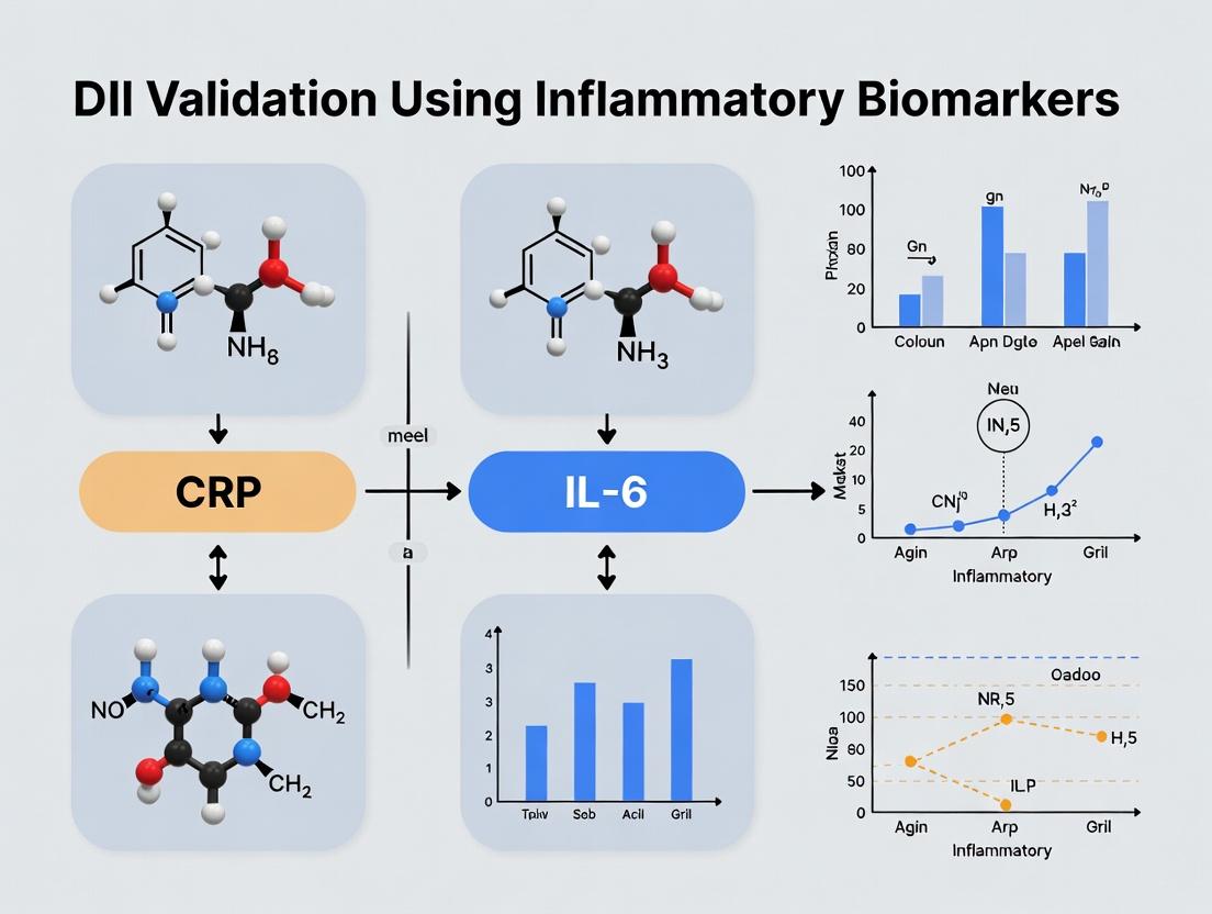
Validating the Dietary Inflammatory Index: CRP and IL-6 as Key Biomarkers in Chronic Disease Research
This article provides a comprehensive analysis for researchers and drug development professionals on validating the Dietary Inflammatory Index (DII) using the inflammatory biomarkers C-reactive protein (CRP) and interleukin-6 (IL-6).
Validating the Dietary Inflammatory Index: CRP and IL-6 as Key Biomarkers in Chronic Disease Research
Abstract
This article provides a comprehensive analysis for researchers and drug development professionals on validating the Dietary Inflammatory Index (DII) using the inflammatory biomarkers C-reactive protein (CRP) and interleukin-6 (IL-6). We explore the foundational relationship between diet-induced inflammation and these biomarkers, detail robust methodological approaches for DII validation in clinical and epidemiological studies, address common analytical challenges, and present comparative analyses of DII's performance against other dietary assessment tools. This resource synthesizes current evidence to support the integration of validated DII scores in mechanistically-driven nutrition and pharmacology research.
The Science Behind Diet and Inflammation: Unpacking the DII, CRP, and IL-6 Connection
Technical Support Center: Troubleshooting DII Validation Experiments with CRP & IL-6
This support center addresses common technical and methodological challenges encountered when validating the Dietary Inflammatory Index (DII) against inflammatory biomarkers like C-reactive protein (CRP) and Interleukin-6 (IL-6) in clinical and epidemiological research.
FAQs & Troubleshooting Guides
Q1: During DII score calculation, how should I handle missing dietary data for specific food parameters in the FFQ? A: The DII framework is designed to be robust to missing data. The standard operating procedure is to use the global comparative database mean for that food parameter as a substitute for the missing individual value. This ensures the individual’s score is centered on a standard global mean, preserving the comparative nature of the DII. Document the percentage and type of parameters substituted in your methods.
Q2: Why is my observed correlation between DII scores and serum CRP levels weak or non-significant, contrary to literature? A: Consider these troubleshooting steps:
- Biomarker Measurement Timing: CRP has a short half-life (~19 hours) and is highly variable. A single serum measurement may not reflect habitual inflammation. Protocol: Use high-sensitivity CRP (hs-CRP) assays and consider averaging values from two fasting blood draws taken one week apart.
- Confounding Variables: Inadequate adjustment for confounders can obscure relationships. Protocol: Statistically adjust for BMI, smoking status, prevalent infection (rule out CRP >10 mg/L), use of anti-inflammatory drugs, and comorbidities (e.g., cardiovascular disease).
- Population Homogeneity: If your cohort is very homogeneous (e.g., all healthy, young individuals), the range of both DII scores and CRP may be restricted, attenuating correlation. Ensure your sample has sufficient variance in dietary habits.
Q3: What is the optimal method for log-transforming IL-6 data prior to regression analysis with DII? A: IL-6 data is typically right-skewed and often contains values below the detection limit.
- Issue: Non-detectable Values. Simply substituting with half the detection limit can bias results.
- Recommended Protocol: Use multiple imputation or Tobit regression models specifically designed for censored data. For simpler approaches, use natural log transformation after adding a constant (e.g., 1) to all values, or use non-parametric tests (Spearman correlation). Consistently report your handling method.
Q4: How do I interpret a positive DII score in the context of my regression model with IL-6? A: The DII is scored such that more positive values indicate a more pro-inflammatory diet, and more negative values indicate a more anti-inflammatory diet. In a linear regression model where log(IL-6) is the dependent variable:
- A positive beta coefficient (β) for the DII variable means that for each 1-unit increase in DII (more pro-inflammatory), there is a corresponding increase in log(IL-6) levels. This provides direct evidence for the index's predictive validity.
Q5: Are there specific "anti-inflammatory" food parameters in the DII that most strongly drive associations with biomarkers? A: Yes, validation studies often identify key drivers. Component-wise analysis can be performed. A summary from recent meta-analyses is provided below:
Table 1: Key DII Food Parameters and Their Typical Association Strength with CRP/IL-6
| Food Parameter | Direction in DII | Typical Association with CRP/IL-6 | Relative Strength |
|---|---|---|---|
| Fiber (especially from grains, fruits) | Anti-inflammatory (lowers score) | Inverse correlation | Strong |
| Flavonoids & Polyphenols (e.g., in tea, berries) | Anti-inflammatory (lowers score) | Inverse correlation | Moderate to Strong |
| Saturated Fatty Acids (SFA) | Pro-inflammatory (raises score) | Positive correlation | Moderate |
| Beta-Carotene | Anti-inflammatory (lowers score) | Inverse correlation | Moderate |
| Trans Fat | Pro-inflammatory (raises score) | Positive correlation | Strong |
Experimental Protocol: Core Protocol for Validating DII against hs-CRP and IL-6
1. Objective: To assess the construct validity of the DII by examining its cross-sectional association with plasma concentrations of hs-CRP and IL-6.
2. Dietary Assessment & DII Calculation:
- Tool: Administer a validated, quantitative Food Frequency Questionnaire (FFQ) designed for your population, covering the previous 3-12 months.
- Calculation: Link FFQ-derived daily intake of ~40 food parameters to a global reference database (worldwide intake mean and standard deviation). Calculate the individual’s standardized intake (z-score) for each parameter, convert to a centered percentile score, and sum all values to derive the overall DII score. Use established software (e.g.,
r-diiin R) to ensure accuracy.
3. Biomarker Assessment:
- Sample Collection: Collect fasting (>8hr) venous blood into appropriate tubes (e.g., EDTA for plasma). Process within 2 hours (centrifuge at 1000-2000 x g for 10-15 mins at 4°C). Aliquot and store at -80°C.
- Assay: Use commercially available, high-sensitivity ELISA kits for hs-CRP (detection limit <0.1 mg/L) and IL-6 (detection limit <0.5 pg/mL). Analyze all samples in duplicate, on the same plate where possible, to minimize inter-assay variability. Include manufacturer-provided controls.
4. Statistical Analysis:
- Clean biomarker data (address outliers, non-detects).
- Use natural log transformation for hs-CRP and IL-6 to normalize distributions.
- Perform multiple linear regression with log(hs-CRP) or log(IL-6) as the dependent variable, DII score as the primary independent variable, and adjust for age, sex, BMI, energy intake, smoking, and medication use.
The Scientist's Toolkit: Key Research Reagent Solutions
Table 2: Essential Materials for DII Validation Studies
| Item | Function | Example/Note |
|---|---|---|
| Validated FFQ | Captures habitual intake of food parameters required for DII computation. | Must be culturally appropriate and include portion size images. |
| Global Nutrient Database | Provides the world mean and SD for each food parameter to standardize DII scores. | The original Shivappa et al. (2014) global database is the standard reference. |
| hs-CRP ELISA Kit | Precisely quantifies low levels of C-reactive protein in serum/plasma. | Choose kits with wide dynamic range (e.g., 0.01-50 mg/L) and high specificity. |
| IL-6 ELISA Kit | Quantifies basal levels of Interleukin-6. | Opt for high-sensitivity kits. Consider multiplex panels if measuring other cytokines. |
| Statistical Software (R, SAS, Stata) | For DII score calculation and complex regression modeling. | The r-dii package in R automates DII calculation. |
| Luminex/Multiplex Assay Panel | Alternative to ELISA for simultaneous measurement of IL-6, TNF-α, CRP, etc. | Increases throughput; validate against gold-standard ELISA. |
Visualization: DII Validation Experimental Workflow
Title: Workflow for DII and Biomarker Validation
Visualization: Key Confounding Factors in DII-Biomarker Analysis
Title: Confounders in DII-Biomarker Association
Technical Support Center: Troubleshooting & FAQs
FAQ Section: Common Conceptual & Biological Questions
Q1: Why are CRP and IL-6 considered the "gold-standard" systemic biomarkers, and can they be used interchangeably? A: No, they are complementary but not interchangeable. CRP is an acute-phase protein produced by the liver primarily in response to IL-6. IL-6 is a pleiotropic cytokine released early from immune and other cells (e.g., adipocytes). CRP offers a stable, amplified signal of systemic inflammation with a longer half-life (~19 hours). IL-6 provides earlier, more dynamic information but has a shorter half-life (~1-4 hours) and can be more challenging to measure reliably due to diurnal variation and sensitivity to handling.
Q2: My cell culture model shows high IL-6 gene expression but low secreted protein. What could be the issue? A: This is a common discrepancy. Refer to the troubleshooting guide below for a systematic approach.
Q3: How should I handle plasma/serum samples for CRP and IL-6 measurement to ensure stability? A: See the detailed protocol in the Experimental Protocols section. Key points: Process blood samples within 30-60 minutes. For IL-6, aliquot and freeze at -80°C immediately; avoid repeated freeze-thaw cycles (>2 cycles can degrade signal). CRP is more stable but should follow the same stringent protocol for consistency.
Troubleshooting Guide: IL-6 and CRP Assays
| Issue | Possible Cause | Recommended Solution |
|---|---|---|
| High CVs in ELISA | Inconsistent sample thawing/handling. | Thaw all samples on ice, vortex gently, and centrifuge before assay. Use a single, calibrated pipette for critical steps. |
| IL-6 levels below detection | Protein degradation or adsorption. | Use collection tubes with protein stabilizers. Use low-protein-binding tubes for aliquots. Try a high-sensitivity assay. |
| CRP results skewed high | Rheumatoid factor or heterophilic antibody interference. | Re-run with a kit including blocking agents. Use a dilutional linearity test; if non-linear, interference is likely. |
| Discrepancy between ELISA platforms | Differing antibody epitopes or standard calibrators. | Always compare using the same sample set in the same run. Establish lab-specific reference ranges for each platform. |
| Poor spike recovery in complex matrix | Matrix effects (e.g., lipids, hemoglobin). | Re-assay with a higher sample dilution if within dynamic range. Use a kit validated for your specific sample type (serum vs. plasma). |
Experimental Protocols
Protocol 1: Standardized Pre-Analytical Handling for Serum/Plasma Biomarker Analysis
- Collection: Draw blood into serum separator tubes (for CRP) or EDTA tubes (for IL-6, minimizes ex vivo platelet release). Note tube type consistently.
- Processing: Allow serum tubes to clot for 30 mins at room temp (RT). Centrifuge all tubes at 1,500-2,000 x g for 10 mins at 4°C.
- Aliquoting: Immediately transfer clear supernatant to pre-chilled, low-protein-binding microtubes.
- Storage: Freeze aliquots at -80°C. Avoid frost-free freezers. Record freeze-thaw cycle count.
Protocol 2: High-Sensitivity ELISA for Human IL-6 (Detailed Workflow) Principle: Sandwich ELISA. Steps:
- Coating: Coat 96-well plate with capture anti-human IL-6 antibody (in carbonate-bicarbonate buffer, pH 9.6). Incubate overnight at 4°C.
- Blocking: Wash 3x with PBS/0.05% Tween-20. Block with 1% BSA in PBS for 1-2 hours at RT.
- Sample Incubation: Wash 3x. Add standards (serial dilution from recombinant protein) and samples (suggested starting dilution 1:2). Incubate 2 hours at RT on orbital shaker.
- Detection Antibody: Wash 5x. Add biotinylated detection antibody. Incubate 1 hour at RT.
- Streptavidin-Enzyme Conjugate: Wash 5x. Add streptavidin-HRP. Incubate 30 mins at RT, protected from light.
- Signal Development: Wash 7x. Add TMB substrate. Incubate 10-15 mins in dark.
- Stop & Read: Add stop solution (e.g., 1M H2SO4). Read absorbance at 450 nm with 570 nm or 620 nm correction.
Data Presentation: Reference Ranges and Assay Performance
Table 1: Typical Reference Ranges & Assay Characteristics for Systemic Biomarkers
| Biomarker | Healthy Adult Range (Serum/Plasma) | Common Assay Methods | Typical Sensitivity (Lower Limit) | Key Interfering Factors |
|---|---|---|---|---|
| C-Reactive Protein (CRP) | < 3 mg/L (low-grade: 3-10 mg/L) | Immunoturbidimetry, ELISA, hsCRP assays | ~0.1 mg/L (hsCRP) | Rheumatoid factor, high triglycerides, heterophilic antibodies |
| Interleukin-6 (IL-6) | 1-5 pg/mL | ELISA, ECLIA, multiplex bead arrays | ~0.1 pg/mL (high-sensitivity) | Platelet activation ex vivo, repeated freeze-thaw, hemolysis |
Table 2: Comparison of Major Measurement Platforms
| Platform | Throughput | Dynamic Range | Sample Volume | Suitability for DII Studies |
|---|---|---|---|---|
| Standard ELISA | Medium | Wide (e.g., CRP: 1-200 mg/L) | 50-100 µL | Good for targeted, high-precision analysis. |
| High-Sensitivity ELISA | Medium | Focused low-end (e.g., IL-6: 0.1-10 pg/mL) | 50-100 µL | Essential for measuring baseline levels in healthy cohorts. |
| Multiplex Bead Array | High | Variable, can be narrower | 25-50 µL | Excellent for multi-biomarker panels alongside CRP/IL-6. |
| Clinical Chemistry Analyzer | Very High | Optimal for CRP >1 mg/L | <10 µL | Ideal for large-scale validation studies of elevated CRP. |
Diagrams
Title: CRP and IL-6 Signaling Pathway
Title: Biomarker Validation Workflow
The Scientist's Toolkit: Research Reagent Solutions
| Item | Function in CRP/IL-6 Research | Key Considerations |
|---|---|---|
| High-Sensitivity ELISA Kits | Quantifying low, baseline levels of IL-6 and CRP critical for DII studies. | Verify validated sample matrices (serum/plasma). Check range against expected values. |
| Multiplex Panels | Measuring IL-6, CRP, and other cytokines (e.g., TNF-α, IL-1β) simultaneously to contextualize inflammation. | Ensure CRP is on a separate, appropriate scale. Assess cross-reactivity. |
| Recombinant Protein Standards | Generating standard curves for ELISAs; positive controls. | Must match kit species/reactivity. Aliquot to avoid degradation. |
| Low-Protein-Binding Microtubes | Storing serum/plasma aliquots to minimize analyte adsorption. | Critical for low-abundance IL-6. |
| EDTA Plasma Tubes | Preferred collection for IL-6 to inhibit ex vivo release from platelets. | Use consistent anticoagulant across study. |
| Protein Stabilizer Cocktails | Added to samples to prevent degradation of cytokines during storage. | May interfere with some assays; validate first. |
| Assay Diluent (Matrix-Matched) | Diluting samples that read above range while minimizing matrix effects. | Kit-provided diluent is optimal. |
Troubleshooting Guide & FAQs
Q1: In our cell culture model, we observe high baseline IL-6 secretion even in the control group, obscuring the effect of our dietary metabolite treatment. What are the primary troubleshooting steps?
A1: High baseline inflammation often stems from endotoxin contamination or stressful culture conditions.
- Action 1: Test for LPS Contamination. Use a Limulus Amebocyte Lysate (LAL) assay on your media, serum, and treatment solutions. Ensure all reagents are certified endotoxin-free.
- Action 2: Review Cell Handling. Minimize passaging stress and avoid over-confluency. Use low-passage-number cells and confirm mycoplasma negativity via PCR.
- Action 3: Optimize Serum. Heat-inactivate FBS (56°C for 30 min) and consider testing with charcoal-stripped serum to reduce confounding factors.
Q2: Our human intervention study shows inconsistent correlations between the Dietary Inflammatory Index (DII) score and plasma CRP levels. What factors could be confounding our biomarker measurement?
A2: CRP is an acute-phase reactant highly sensitive to non-dietary factors.
- Checklist:
- Subclinical Infection: Exclude subjects with CRP >10 mg/L from analysis.
- Circadian Rhythm: Standardize blood collection time (e.g., 7-9 AM).
- Assay Variability: Use a high-sensitivity (hs-)CRP assay. Run all samples from a single subject in the same batch.
- Covariates: Statistically adjust for BMI, age, smoking status, and statin use in your analysis.
Q3: When stimulating peripheral blood mononuclear cells (PBMCs) with palmitic acid to model saturated fat intake, we get variable IL-6 responses. How can we standardize this protocol?
A3: Variability often arises from fatty acid preparation and donor selection.
- Protocol Standardization:
- Conjugate Palmitic Acid: Complex palmitic acid to fatty acid-free BSA at a 5:1 molar ratio. Dissolve in warm serum-free medium with gentle agitation. Filter sterilize (0.2 µm).
- Control Conjugation: Include a BSA-only vehicle control.
- Donor Pooling: For in vitro studies, use PBMCs pooled from at least 5 healthy donors to mitigate donor-specific immune responses.
- Stimulation Time: A 24-hour stimulation is standard for cytokine production; perform a time course (6, 12, 24, 48h) to optimize.
Q4: We are trying to validate the DII in a specific cohort. Which experimental design is more robust: a cross-sectional analysis or a controlled feeding study?
A4: The choice depends on your research question and resources.
- Cross-Sectional Analysis: Efficient for establishing associations in large, existing cohorts. Prone to confounding. Use it for initial validation and hypothesis generation.
- Controlled Feeding Study: The gold standard for establishing causality. Provides precise dietary control but is expensive, short-term, and has limited generalizability. Best for elucidating precise mechanisms.
Key Experimental Protocols
Protocol 1: Assessing NF-κB Nuclear Translocation in Macrophages (THP-1 cells) Treated with Pro-Inflammatory Dietary Components.
- Differentiation: Culture THP-1 cells with 100 nM PMA for 48 hours to differentiate into macrophages.
- Treatment: Stimulate with treatment (e.g., 200 µM palmitic acid-BSA conjugate, 1 µg/mL LPS positive control, BSA vehicle control) for 1 hour.
- Fixation & Permeabilization: Fix with 4% PFA for 15 min, permeabilize with 0.1% Triton X-100 for 10 min.
- Immunofluorescence: Block with 3% BSA. Incubate with primary antibody against p65 RelA (1:500) overnight at 4°C. Incubate with Alexa Fluor 488-conjugated secondary antibody (1:1000) for 1 hour. Use DAPI for nuclear counterstain.
- Imaging & Analysis: Visualize with confocal microscopy. Quantify the nuclear-to-cytosolic fluorescence intensity ratio of p65 using image analysis software (e.g., ImageJ).
Protocol 2: Measuring IL-6 and hs-CRP in Human Serum/Plasma for DII Validation Studies.
- Sample Collection: Collect fasting blood into serum separator or EDTA tubes. Process within 2 hours (centrifuge at 1500-2000xg for 15 min at 4°C). Aliquot and store at -80°C. Avoid freeze-thaw cycles.
- hs-CRP Measurement: Use a validated, high-sensitivity immunoturbidimetric or ELISA assay. Typical dynamic range: 0.1-20 mg/L. Run samples in duplicate.
- IL-6 Measurement: Use a high-sensitivity ELISA kit (detection limit <0.5 pg/mL). As IL-6 can be unstable, ensure consistent thawing and assay all samples from one subject on the same plate.
Table 1: Effect of Selected Pro-Nutrient Components on Inflammatory Biomarkers in In Vitro Models
| Pro-Nutrient Component | Model System | Concentration | Exposure Time | CRP Effect | IL-6 Effect (Fold Change vs. Control) | Key Signaling Pathway |
|---|---|---|---|---|---|---|
| Palmitic Acid (SFA) | THP-1 Macrophages | 200 µM | 24 h | N/A | 3.5 - 5.2 ↑ | TLR4/MyD88 → NF-κB |
| Glucose (High) | HUVECs | 25 mM | 48 h | N/A | 2.8 ↑ | ROS → NLRP3 Inflammasome |
| Advanced Glycation End Products (AGEs) | RAW 264.7 Macrophages | 100 µg/mL | 24 h | N/A | 4.1 ↑ | RAGE → p38 MAPK/NF-κB |
| Reference: LPS | THP-1 Macrophages | 100 ng/mL | 24 h | N/A | 10 - 15 ↑ | TLR4/TRIF/MyD88 → NF-κB |
Table 2: Association of DII Scores with Inflammatory Biomarkers in Recent Observational Studies (2022-2024)
| Study Cohort (Year) | Sample Size (n) | DII Score Range | Correlation with hs-CRP (β / r) | Correlation with IL-6 (β / r) | Adjusted Covariates |
|---|---|---|---|---|---|
| NHANES Subset (2023) | 4,892 | -4.5 to +4.8 | β = 0.12, p<0.01 | β = 0.08, p<0.05 | Age, Sex, BMI, Smoking, Diabetes |
| Mediterranean Cohort (2022) | 1,245 | -6.1 to +3.9 | r = 0.21, p<0.001 | r = 0.15, p=0.002 | Age, Energy Intake, Physical Activity |
| Asian Cohort (2024) | 2,117 | -3.8 to +5.2 | β = 0.18, p<0.001 | β = 0.11, p<0.01 | BMI, Hypertension, Medication Use |
Diagrams
Title: Pro-Inflammatory Diet to CRP/IL-6 Signaling Overview
Title: DII Validation Study Workflow
The Scientist's Toolkit: Research Reagent Solutions
| Item | Function in DII/Inflammation Research |
|---|---|
| High-Sensitivity ELISA Kits (hs-CRP & IL-6) | Quantify low-level circulating biomarkers critical for correlating with dietary scores. |
| Fatty Acid-Free BSA | Essential for solubilizing and delivering free fatty acids (e.g., palmitic acid) to cells in culture without non-specific cytotoxicity. |
| TLR4 Inhibitor (TAK-242/CLI-095) | Pharmacological tool to confirm the role of TLR4 signaling in nutrient-induced inflammation. |
| NF-κB Reporter Cell Line | Genetically engineered cells (e.g., THP-1-NF-κB-luc) to screen dietary compounds for NF-κB activation. |
| LAL Endotoxin Assay Kit | Critical quality control to rule out LPS contamination in cell culture reagents, which can confound results. |
| Multiplex Cytokine Panels | Measure a suite of inflammatory cytokines (TNF-α, IL-1β, IL-8) alongside IL-6 from a single sample to profile the inflammatory response. |
| RAGE Antagonist (e.g., FPS-ZM1) | Tool to investigate the contribution of Advanced Glycation End-products (AGEs) in dietary inflammation. |
| p65/NF-κB Phospho-Antibody | For Western blot or immunofluorescence to assess NF-κB pathway activation in tissue or cell samples. |
This technical support center is designed to assist researchers within the broader thesis context of validating the Dietary Inflammatory Index (DII) against inflammatory biomarkers, specifically C-reactive protein (CRP) and Interleukin-6 (IL-6). The FAQs and guides below address common experimental and analytical challenges.
Frequently Asked Questions (FAQs) & Troubleshooting
Q1: Our calculated DII scores do not show the expected correlation with serum CRP levels. What are the primary sources of error? A: Common issues include:
- Dietary Data Granularity: The DII requires intake data for up to 45 food parameters. Using a Food Frequency Questionnaire (FFQ) not validated for all these parameters leads to misclassification. Solution: Use or adapt an FFQ specifically designed for DII calculation.
- Biomarker Measurement Timing: CRP is an acute-phase reactant with high intra-individual variability. Solution: Use high-sensitivity CRP (hs-CRP) assays and collect multiple blood samples over time, preferably when participants are free from acute infection.
- Confounding: Failing to adequately control for smoking, BMI, physical activity, and medication use (e.g., statins, NSAIDs) can obscure true associations. Solution: Use multivariate regression models with careful confounder selection.
Q2: When analyzing IL-6, what are the key considerations for sample handling and assay selection? A: IL-6 is labile and can be influenced by experimental procedures.
- Pre-analytical Variables: Delay in processing, number of freeze-thaw cycles, and type of blood collection tube can affect levels. Solution: Process plasma/serum within 2 hours of collection, aliquot samples, and minimize freeze-thaw cycles to ≤2.
- Assay Sensitivity: Standard ELISA kits may lack sensitivity for detecting physiological levels in healthy cohorts. Solution: Use high-sensitivity (hs) IL-6 assays. Verify the kit's lower limit of detection (LLOD) is suitable for your population's expected range.
Q3: How should we handle non-normally distributed biomarker data (CRP/IL-6) in statistical analyses? A: CRP and IL-6 typically follow a right-skewed distribution.
- Standard Practice: Apply natural log (ln) or base-10 log transformation to approximate a normal distribution before parametric testing (e.g., Pearson correlation, linear regression).
- Alternative: Use non-parametric tests (Spearman's rank correlation) for unadjusted associations.
- Reporting: In tables, present geometric means (for log-transformed data) or medians with interquartile ranges.
Q4: What is the recommended statistical model to assess the DII-biomarker relationship? A: Use multiple linear regression with the biomarker (log-transformed) as the dependent variable and the DII score as the primary independent variable.
- Model Structure:
ln(CRP) = β0 + β1(DII Score) + β2(Covariate1) + ... + βn(Covariaten) + ε - Interpretation: The coefficient β1 represents the change in the log-transformed biomarker per one-unit increase in DII. Exponentiate β1 to express the multiplicative change in the original biomarker unit.
Key Experimental Protocols
Protocol 1: Calculation of the Dietary Inflammatory Index (DII)
- Data Collection: Administer a validated FFQ designed to capture intake of all ~45 DII food parameters (e.g., nutrients, flavonoids, spices).
- Standardization: Link individual dietary intake data to a global representative database (provided by the DII developers) to create a worldwide comparative z-score for each parameter:
z = (individual intake - global mean) / global standard deviation. - Inflammatory Effect Adjustment: Multiply each z-score by its respective "inflammatory effect score" (derived from literature review).
- Summation: Sum all adjusted z-scores to obtain the overall DII score for each participant. A higher, more positive score indicates a more pro-inflammatory diet.
Protocol 2: Measurement of High-Sensitivity CRP (hs-CRP) via ELISA
- Sample: Fasting serum or plasma (EDTA). Centrifuge within 2 hours and store at -80°C.
- Principle: Sandwich ELISA. A microplate is pre-coated with an anti-CRP antibody. Sample CRP is captured and detected by a second, enzyme-linked anti-CRP antibody.
- Steps: a. Load standards, controls, and samples in duplicate. b. Incubate, wash, and add detection antibody. c. Incubate, wash, and add enzyme substrate (e.g., TMB). d. Stop the reaction with acid and read absorbance at 450nm. e. Generate a standard curve (4-parameter logistic) to interpolate sample concentrations.
- Quality Control: Acceptable intra- and inter-assay CVs are typically <10%.
Summarized Quantitative Evidence
Table 1: Selected Epidemiological Studies on DII, CRP, and IL-6
| Study (Cohort) | Sample Size | DII Score Range (Mean) | Correlation with CRP (β, p-value) | Correlation with IL-6 (β, p-value) | Key Adjustment Factors |
|---|---|---|---|---|---|
| NHANES (US Adults) | ~10,000 | -5.7 to +5.1 | β=0.04 per DII unit, p<0.001 | β=0.02 per DII unit, p<0.001 | Age, sex, race, education, BMI, smoking, activity |
| PREDIMED (Mediterranean) | ~7,000 | ~ -0.5 (Pro-inflammatory arm) | Significant positive association (p=0.004) | Not Reported | Age, sex, smoking, BMI, diabetes, medication |
| Women's Health Initiative | ~2,500 | Not specified | β for highest vs. lowest DII quintile = 0.14, p-trend=0.01 | β for highest vs. lowest DII quintile = 0.12, p-trend=0.03 | Energy intake, demographics, lifestyle, BMI |
Table 2: Research Reagent Solutions Toolkit
| Item | Function/Application in DII-Biomarker Research |
|---|---|
| Validated DII FFQ | Standardized tool to collect intake data for all required food parameters reliably. |
| Global DII Database | Reference values (mean & std. dev.) for standardizing individual dietary data. |
| hs-CRP ELISA Kit | Quantifies low levels of CRP in serum/plasma with high sensitivity. |
| hs-IL-6 ELISA Kit | Measures physiological levels of IL-6; essential for non-clinical populations. |
| CRP Latex Reagents | For rapid, immunoturbidimetric analysis of CRP on clinical chemistry analyzers. |
| Cytokine Multiplex Panel | Allows simultaneous measurement of IL-6, TNF-α, IL-1β, etc., from a single sample. |
| RNAlater Stabilizer | Preserves RNA for downstream gene expression analysis of inflammatory pathways. |
| Statistical Software (R, SAS, Stata) | For complex multivariate regression analysis and data transformation. |
Visualizations
Diagram 1: DII Calculation & Validation Workflow
Diagram 2: Inflammatory Pathway Linking Diet to Biomarkers
Technical Support Center: Troubleshooting Guides & FAQs for DII Validation with CRP & IL-6
FAQ 1: Inconsistent CRP (C-Reactive Protein) ELISA Results in Dietary Intervention Samples Q: Why are my CRP ELISA results from human serum/plasma samples after a dietary intervention showing high intra-assay variability or values below the detection limit?
A: This is a common issue in nutritional studies. CRP is an acute-phase reactant with a wide dynamic range (0.1-200 µg/mL). Common causes and solutions:
- Cause 1: Improper Sample Handling. CRP can degrade if samples undergo multiple freeze-thaw cycles or are not separated from cells promptly.
- Solution: Process blood samples within 2 hours of collection. Aliquot serum/plasma to avoid repeated freeze-thaws. Store at -80°C.
- Cause 2: Assay Range Mismatch. High-sensitivity CRP (hs-CRP) assays are required for the lower end of the physiological range relevant to chronic inflammation (0.1-10 µg/mL). Standard assays may not detect subtle changes.
- Solution: Validate and use an hs-CRP ELISA kit specifically. Confirm the limit of detection (LOD) and quantitation (LOQ) in your lab.
- Cause 3: Matrix Effects from Lipemic Samples. High-fat meals can interfere with assay antibodies.
- Solution: Ensure participants fasted for an appropriate period (e.g., 10-12 hours) pre-blood draw. If lipemia is suspected, note it and consider dilutional linearity tests.
FAQ 2: IL-6 Measurement Challenges in DII Studies Q: Interleukin-6 (IL-6) levels in our cohort are often undetectable or show no significant change post-intervention, despite a hypothesized effect. What are the potential reasons?
A: IL-6 presents distinct challenges due to its biology:
- Cause 1: Short Half-life and Pulsatile Secretion. IL-6 has a short plasma half-life (~1 hour) and is secreted in pulses, leading to high temporal variability.
- Solution: Standardize time of day for blood collection. Consider measuring soluble IL-6 receptor (sIL-6R) or glycoprotein 130 (sgp130), which are more stable and modulate IL-6 signaling.
- Cause 2: Insensitive Assay. Many standard ELISA kits have LODs around 1-4 pg/mL, above the basal level in many healthy individuals.
- Solution: Use an ultra-sensitive or digital ELISA platform (e.g., Simoa, Singulex) capable of detecting IL-6 in the fg/mL to low pg/mL range.
- Cause 3: Cellular Source vs. Systemic Circulation. IL-6 may act locally (paracrine/autocrine) without significantly elevating systemic levels.
- Solution: Complement plasma/serum measures with ex vivo immune cell challenge assays (e.g., LPS-stimulated PBMC IL-6 production) to assess immune cell responsiveness.
FAQ 3: Validating the Dietary Inflammatory Index (DII) in a New Population Q: We are calculating DII scores for a new ethnic population. What is the gold-standard experimental protocol to validate that the DII correlates with inflammatory biomarkers like CRP and IL-6 in our cohort?
A: Validation requires a controlled dietary intervention or rigorous observational study.
Experimental Protocol for DII Validation Study:
- Study Design: A controlled feeding study (preferred for causality) or a detailed longitudinal observational study with repeated 24-hour dietary recalls/Food Frequency Questionnaires (FFQs).
- Participants: Recruit a representative sample (n ≥ 100 for observational studies) of your target population.
- DII Calculation: Use standardized, population-adjusted global nutrient intake values to calculate each participant's DII score from dietary data.
- Biomarker Measurement:
- Sample Collection: Collect fasting blood at baseline and endpoint.
- Primary Biomarkers: Measure hs-CRP and ultra-sensitive IL-6.
- Secondary/Confirmatory Biomarkers: Include TNF-α, IL-1β, and fibrinogen to strengthen inference.
- Assay Validation: For each batch, run kit controls and a pooled human plasma QC sample. Ensure intra- and inter-assay CVs are <10% and <15%, respectively.
- Statistical Analysis: Use linear regression or mixed models, adjusting for confounders (age, BMI, smoking, etc.), to assess the relationship between DII score change and biomarker change.
Data Presentation
Table 1: Common Inflammatory Biomarkers for DII Validation
| Biomarker | Full Name | Sample Type | Typical Healthy Range | Key Consideration for Nutritional Studies |
|---|---|---|---|---|
| CRP | C-Reactive Protein | Serum, Plasma | 0.1-3.0 µg/mL (hs-CRP) | Acute-phase reactant; sensitive to infection, injury. Use hs-CRP assay. |
| IL-6 | Interleukin-6 | Serum, Plasma, Cell Culture Supernatant | 0.5-5.0 pg/mL (varies) | Short half-life; consider ultra-sensitive assays. |
| TNF-α | Tumor Necrosis Factor-Alpha | Serum, Plasma | 0.5-5.0 pg/mL | Often low/undetectable in healthy; requires stimulation assays for robustness. |
| IL-1β | Interleukin-1 Beta | Serum, Plasma | <5 pg/mL | Similar challenges to TNF-α. |
| Fibrinogen | Factor I | Plasma | 200-400 mg/dL | Coagulation factor; stable but less specific. |
Table 2: Troubleshooting Matrix for Common Biomarker Assay Issues
| Symptom | Possible Causes | Recommended Validation Experiment |
|---|---|---|
| High Inter-assay CV | Reagent lot variability, operator drift, instrument calibration. | Run a panel of stored QC samples (low, mid, high) in each assay batch. |
| Values below LOD | Insensitive assay, inappropriate sample dilution, biomarker truly absent. | Spike-and-recovery experiment: Spike a known amount of recombinant protein into participant sample and measure recovery (target 80-120%). |
| Poor correlation between DII and biomarker | Inaccurate dietary assessment, unmeasured confounding, incorrect biomarker choice. | Sensitivity analysis: Calculate DII with alternative dietary input methods. Measure a panel of biomarkers to see if any correlate. |
Experimental Protocols
Protocol: High-Sensitivity CRP (hs-CRP) ELISA Principle: Solid-phase sandwich ELISA. Reagents: Commercial hs-CRP ELISA kit, microplate reader. Procedure:
- Preparation: Bring all reagents, samples, and standards to room temperature (RT).
- Loading: Add 100 µL of standard, control, or pre-diluted sample to appropriate wells. Cover and incubate 2 hours at RT.
- Washing: Aspirate and wash wells 4 times with 300 µL wash buffer.
- Detection Antibody: Add 100 µL of biotin-conjugated anti-CRP antibody. Incubate 1 hour at RT. Wash as step 3.
- Streptavidin-Enzyme: Add 100 µL of streptavidin-HRP solution. Incubate 30 minutes at RT. Wash as step 3.
- Substrate: Add 100 µL of TMB substrate. Incubate 10-20 minutes in the dark.
- Stop: Add 100 µL of stop solution. Read absorbance at 450 nm immediately.
- Calculation: Generate a standard curve (4-parameter logistic) and interpolate sample concentrations.
Protocol: Ex Vivo LPS Stimulation of PBMCs for IL-6 Production Principle: Assess immune cell responsiveness as a functional inflammatory readout. Reagents: Ficoll-Paque PLUS, RPMI-1640, LPS (E. coli 055:B5), human IL-6 ELISA kit. Procedure:
- PBMC Isolation: Isolate PBMCs from fresh heparinized blood via density-gradient centrifugation using Ficoll-Paque.
- Culture: Resuspend PBMCs at 1x10^6 cells/mL in complete RPMI. Seed 1 mL/well in a 24-well plate.
- Stimulation: Add LPS to experimental wells (final conc. 100 ng/mL). Include unstimulated control wells. Incubate at 37°C, 5% CO2 for 24 hours.
- Harvest: Centrifuge plates at 300 x g for 5 min. Carefully collect supernatant.
- Assay: Store supernatants at -80°C. Measure IL-6 concentration using an ultra-sensitive ELISA (protocol similar to above).
Mandatory Visualization
Title: DII to Biomarker Signaling Pathway
Title: DII Validation Experimental Workflow
The Scientist's Toolkit: Research Reagent Solutions
Table 3: Essential Materials for DII Biomarker Validation Studies
| Item | Function & Rationale | Example/Supplier Consideration |
|---|---|---|
| High-Sensitivity CRP ELISA Kit | Quantifies low levels of CRP in serum/plasma critical for assessing chronic, low-grade inflammation in nutritional studies. | R&D Systems Quantikine ELISA HS, Alpco hs-CRP ELISA. Ensure LOD <0.1 µg/mL. |
| Ultra-Sensitive IL-6 Assay Kit | Measures very low basal levels of IL-6, overcoming the limitation of standard ELISA sensitivity. | Quanterix Simoa, Meso Scale Discovery (MSD) U-PLEX, R&D Systems HS IL-6. |
| Multiplex Cytokine Panel Assay | Allows simultaneous measurement of a profile (CRP, IL-6, TNF-α, IL-1β) from a single small sample, conserving volume and reducing variability. | MSD V-PLEX, Luminex xMAP, Olink Target 96. |
| Recombinant Protein Standards | Used for assay calibration, spike-and-recovery experiments, and as positive controls to validate kit performance. | Ensure they match the analyte (e.g., human CRP, human IL-6). NIBSC provides WHO international standards. |
| LPS (from E. coli 055:B5) | Used in ex vivo PBMC stimulation experiments to trigger innate immune response and measure IL-6 production capacity. | Sigma-Aldrich, InvivoGen. Use a consistent source and lot for comparable results. |
| Ficoll-Paque PLUS | Density gradient medium for the isolation of high-quality, viable PBMCs from whole blood for functional assays. | Cytiva. |
| Stable, Low-Inflammatory Pooled Human Serum/Plasma | Serves as a critical Quality Control (QC) sample across assay runs to monitor inter-assay precision and drift. | BioIVT, SeraCare. Or prepare in-house from characterized donors. |
A Protocol for DII Validation: Designing Studies and Analyzing CRP/IL-6 Data
Troubleshooting Guides and FAQs for DII Validation with CRP & IL-6
Q1: In our cross-sectional study validating the Dietary Inflammatory Index (DII), we found a weak correlation (r ~0.2) between DII scores and serum CRP. What are the primary confounding factors we should re-examine? A: A weak correlation in a cross-sectional design often points to unmeasured confounding or measurement timing issues. Key factors to troubleshoot include:
- Acute Inflammation: Ensure participants were free from recent infection, injury, or vaccination (within 2-4 weeks) at blood draw, as these acutely elevate CRP independently of diet.
- Biomarker Half-life: CRP has a 19-hour half-life and reflects recent inflammatory stimuli. Reconcile the DII assessment period (typically 1-3 months via FFQ) with the CRP measurement window. Consider adding IL-6, which may have a more stable baseline.
- Medication Use: Statins, NSAIDs, and corticosteroids profoundly suppress CRP. Re-analyze data with stringent exclusion or stratification for these medications.
- Adiposity: BMI is a major confounder. Ensure multivariate models appropriately adjust for waist circumference or body fat percentage, not just BMI.
Q2: Our prospective cohort study found a significant association between DII and IL-6, but not with CRP. Is this a valid finding, or does it suggest an issue with our assay protocol? A: This is a plausible and valid finding. IL-6 is a primary inducer of CRP production in the liver. Discrepancies can arise from:
- Biological Sensitivity: IL-6 is a more proximal cytokine in the inflammatory cascade and may be more directly responsive to dietary patterns than the downstream acute-phase protein CRP.
- Assay Precision: Verify that your high-sensitivity CRP (hsCRP) assay had sufficient sensitivity (detection limit <0.1 mg/L) for a population-level study. Standard CRP assays are not adequate.
- Sample Handling: IL-6 is less stable than CRP. Review your protocol: was plasma/serum separated and frozen at -80°C within 2 hours of collection? Repeated freeze-thaw cycles (>2) can degrade IL-6.
Q3: We are designing a dietary intervention trial to validate the DII. What is the minimum intervention duration required to observe significant changes in CRP and IL-6? A: Based on current meta-analyses of dietary interventions, a minimum of 4 weeks is required to observe changes in IL-6, and 6-8 weeks for changes in CRP to stabilize. Longer trials (≥12 weeks) show more consistent and significant effects.
Q4: During our intervention trial, the control group's CRP levels decreased unexpectedly. What are common pitfalls in control group design? A: This is a frequent issue (the "Hawthorne effect" or contamination).
- Pseudo-Control: Ensure the control intervention is truly inert (e.g., wait-list, general health advice not focused on inflammation). Providing control participants with healthy but non-anti-inflammatory foods can cause bias.
- Behavioral Contamination: Participants in different arms may communicate and share dietary tips. Use cluster randomization or emphasize adherence to assigned protocol.
- Seasonal Variation: If the trial spans different seasons, dietary patterns and inflammation levels can shift in all groups. Account for this in randomization and analysis.
Table 1: Typical Effect Sizes and Timelines for Inflammatory Biomarker Response in DII-Related Studies
| Study Design | Primary Biomarker | Typical Effect Size (per unit DII change) | Minimum Meaningful Duration | Key Confounding Factors to Control |
|---|---|---|---|---|
| Cross-Sectional | hsCRP | β: 0.10 - 0.15 log(mg/L) | N/A (Single time point) | Age, Sex, BMI, Medication, Smoking, Comorbidity |
| Cross-Sectional | IL-6 | β: 0.08 - 0.12 log(pg/mL) | N/A (Single time point) | Age, BMI, Physical Activity, Recent Infection |
| Prospective Cohort | hsCRP | HR: 1.10 - 1.25 (for top vs. bottom DII tertile) | ≥ 1 Year Follow-up | Baseline Inflammation, Incident Disease, Weight Change |
| Prospective Cohort | IL-6 | HR: 1.15 - 1.30 (for top vs. bottom DII tertile) | ≥ 1 Year Follow-up | Baseline Inflammation, Incident Disease, Weight Change |
| Randomized Controlled Trial | hsCRP | Δ: -0.50 to -1.20 mg/L (vs. control) | 6-8 Weeks | Medication Changes, Weight Change, Adherence <80% |
| Randomized Controlled Trial | IL-6 | Δ: -0.50 to -1.50 pg/mL (vs. control) | 4-6 Weeks | Medication Changes, Weight Change, Adherence <80% |
Experimental Protocols
Protocol 1: High-Sensitivity CRP (hsCRP) & IL-6 Measurement from Serum
Principle: Quantification via solid-phase, sandwich ELISA or chemiluminescent immunoassay. Procedure:
- Sample Collection: Draw fasting venous blood into a serum separator tube (SST).
- Clotting & Separation: Allow blood to clot at room temperature for 30 minutes. Centrifuge at 1,500-2,000 x g for 15 minutes at 4°C.
- Aliquoting: Immediately transfer clarified serum to cryovials. Store at -80°C. Avoid repeated freeze-thaw cycles (>2).
- Assay: Use commercial high-sensitivity kits. For hsCRP, ensure detection limit ≤0.1 mg/L. For IL-6, typical detection limit is ≤0.5 pg/mL.
- Dilution: If sample concentrations exceed the standard curve's top point, dilute with the provided assay diluent.
- Quality Control: Run in duplicate. Include kit controls and a laboratory-pooled serum sample on each plate. Inter-assay CV should be <10%.
Protocol 2: DII Calculation and Alignment with Biomarker Timing
Principle: The DII is computed based on dietary intake data relative to a global standard mean. Procedure:
- Dietary Assessment: Administer a validated, quantitative food frequency questionnaire (FFQ) covering the past 1-3 months.
- Data Processing: Convert food consumption to daily intake of ~45 food parameters (e.g., nutrients, flavonoids).
- Z-score Calculation: For each parameter, subtract the global mean intake and divide by the global standard deviation.
- Inflammatory Effect Score: Multiply the z-score by the respective literature-derived inflammatory effect score for that parameter.
- Summation: Sum all food parameter scores to obtain the overall DII score for each participant.
- Temporal Alignment: For cross-sectional studies, blood draw must fall within the FFQ recall period. For cohort studies, update FFQ at each follow-up. For trials, collect FFQ at baseline, midpoint, and endpoint to calculate adherence-adjusted DII.
Visualizations
Diagram 1: Inflammatory Signaling Pathway from DII to CRP
Diagram 2: Workflow for Validating DII Across Study Designs
The Scientist's Toolkit: Research Reagent Solutions
Table 2: Essential Materials for DII Validation Studies
| Item | Function in DII/CRP/IL-6 Research | Example/Specification |
|---|---|---|
| Validated FFQ | Captures habitual dietary intake to compute the DII score. Must be culturally appropriate and include all DII components. | Diet History Questionnaire II, EPIC-Norfolk FFQ. |
| High-Sensitivity CRP (hsCRP) Assay Kit | Precisely quantifies low levels of circulating CRP in serum/plasma. Essential for population studies. | R&D Systems Quantikine ELISA, Siemens Atellica IM hsCRP. Detection limit: ≤0.1 mg/L. |
| IL-6 Immunoassay Kit | Quantifies circulating Interleukin-6 levels. Prefer electrochemiluminescence for wider dynamic range. | Meso Scale Discovery V-PLEX, Abcam SimpleStep ELISA. Detection limit: ≤0.5 pg/mL. |
| Serum Separator Tubes (SST) | Collects and clarifies blood samples for stable serum isolation for biomarker analysis. | BD Vacutainer SST tubes. |
| Cryogenic Vials | For long-term storage of serum/plasma aliquots at -80°C to preserve biomarker integrity. | Nunc, Corning; internally threaded, O-ring seal. |
| Statistical Software | For complex multivariate regression, longitudinal data analysis (Cox regression), and ANCOVA for trials. | R, SAS, Stata with appropriate dietary analysis packages. |
Technical Support Center
Troubleshooting Guides & FAQs
FAQ 1: Sample Handling & Pre-Analytics
- Q: Our hsCRP values are inconsistently high across sample batches. What are the most likely pre-analytical variables?
- A: hsCRP is stable but highly sensitive to improper handling. Key variables are:
- Hemolysis: Red blood cell lysis can interfere with immunoturbidimetric assays.
- Number of Freeze-Thaw Cycles: Limit to ≤3 cycles for both hsCRP and IL-6. Aliquot samples upon collection.
- Sample Type Consistency: Use either serum or lithium heparin plasma consistently. EDTA plasma can chelate calcium required for some hsCRP assays.
- Delay in Processing: Separate plasma/serum within 2 hours of collection for optimal IL-6 stability.
- A: hsCRP is stable but highly sensitive to improper handling. Key variables are:
FAQ 2: Assay Sensitivity & Detection Limits
- Q: For validating a Dietary Inflammatory Index (DII), we need to measure low-grade inflammation. Is our assay's lower limit of detection (LLD) sufficient?
- A: The assay's LLD must be below the established physiological cut-points. Refer to the table below for required performance characteristics.
Table 1: Key Analytical Performance Requirements for DII Validation Studies
| Biomarker | Recommended Assay Methodology | Required Lower Limit of Detection (LLD) | Key Clinical Cut-Points (for reference) | Sample Volume (Typical) |
|---|---|---|---|---|
| hsCRP | Particle-enhanced immunonephelometry or immunoturbidimetry | <0.10 mg/L | Low Risk: <1.0 mg/LAverage Risk: 1.0-3.0 mg/LHigh Risk: >3.0 mg/L | 5-20 µL |
| IL-6 | Electrochemiluminescence (ECLIA) or High-Sensitivity ELISA | <0.50 pg/mL | Normal: <1.8-2.0 pg/mLElevated: >2.0-3.0 pg/mL | 50-100 µL |
FAQ 3: Assay Interference & Discrepancies
- Q: Our IL-6 ELISA results show high background or non-parallel dilution curves. What could cause this?
- A: This suggests interference or matrix effects.
- Heterophilic Antibodies: Use an assay buffer containing blocking agents or employ a heterophilic antibody blocking tube prior to analysis.
- Rheumatoid Factor (RF): RF can cause false elevation. Ensure your chosen kit includes RF absorbent.
- Matrix Effects: Always use the matrix specified by the kit calibrator (e.g., human serum) for standard dilution. Do not use buffer alone.
- A: This suggests interference or matrix effects.
Experimental Protocols
Protocol 1: Serum hsCRP Quantification via Immunoturbidimetry
- Principle: Sample CRP agglutinates with latex particle-bound anti-CRP antibodies, increasing turbidity measured at 540-550 nm.
- Procedure:
- Reagent Preparation: Reconstitute/prepare latex reagent and diluent per manufacturer instructions. Equilibrate to 20-25°C.
- Assay Setup: Pipette 2 µL of sample (calibrator, control, or unknown) into appropriate cuvette.
- Dilution: Add 180 µL of assay diluent and mix.
- Initial Reading: Measure absorbance (A1).
- Reaction: Add 80 µL of latex reagent. Mix and incubate 5 minutes at 37°C.
- Final Reading: Measure absorbance again (A2).
- Calculation: CRP concentration is derived from ΔA (A2 - A1) using a 5-parameter logistic curve generated from calibrators.
Protocol 2: Plasma IL-6 Quantification via High-Sensitivity Electrochemiluminescence (ECLIA)
- Principle: A biotinylated capture antibody and a ruthenium-labeled detection antibody form a sandwich complex with IL-6, captured on streptavidin-coated magnetic beads. Voltage application induces chemiluminescence.
- Procedure:
- Plate/Strip Preparation: Add 50 µL of assay buffer to each well.
- Sample Addition: Add 50 µL of standard, control, or plasma sample. Seal and incubate with shaking (1200 rpm) for 2 hours at room temperature.
- Washing: Aspirate and wash each well 3x with 300 µL wash buffer.
- Detection Antibody Incubation: Add 50 µL of detection antibody. Seal and incubate with shaking for 1 hour.
- Washing: Repeat wash step.
- Reading: Add 150 µL of reading buffer and read immediately on an ECLIA-compatible analyzer.
Diagrams
Biomarker Analysis Workflow for DII Validation
IL-6 Induced CRP Synthesis Signaling Pathway
The Scientist's Toolkit: Key Research Reagent Solutions
Table 2: Essential Materials for hsCRP & IL-6 Analysis
| Item | Function & Specification | Example/Note |
|---|---|---|
| High-Sensitivity CRP Assay Kit | Quantifies CRP in range 0.1-20 mg/L. Method: immunoturbidimetry/nephelometry. | Must include calibrators traceable to WHO/IFCC reference material. |
| HS IL-6 Immunoassay Kit | Quantifies IL-6 with LLD <0.5 pg/mL. Method: ECLIA or ELISA. | Prefer kits with pre-coated plates & included blockers for heterophilic interference. |
| Matrix-Matched Calibrators | Provides accurate standard curve in correct biological matrix (human serum/plasma). | Critical for minimizing matrix effects. |
| Quality Control Sera | Multi-level (low, mid, high) QC material for monitoring assay precision & accuracy. | Run with each batch. |
| Heterophilic Antibody Blocking Reagent | Suppresses interference from human anti-animal antibodies. | Use if suspecting false highs. |
| Low-Binding Microtubes/Pipette Tips | Prevents analyte loss due to adhesion to plastic surfaces. | Essential for low-concentration IL-6. |
| Calibrated Micropipettes (2-1000 µL) | Ensures accurate and precise liquid handling for small volumes. | Regular servicing is mandatory. |
Troubleshooting Guide & FAQs
Q1: When performing correlation analysis between CRP and IL-6 in our DII validation cohort, we obtain a statistically significant but low Pearson's r (e.g., 0.25). Does this invalidate the inflammatory biomarker relationship? A: No. A low correlation coefficient is common and does not invalidate the relationship. CRP and IL-6 have different biological half-lives and regulatory mechanisms. CRP is a downstream acute-phase protein synthesized by the liver in response to IL-6, but its production is influenced by other cytokines (e.g., IL-1β). The correlation, while significant, is often moderate. Consider the following:
- Check for non-linear relationships using scatter plots and Spearman's rank correlation.
- Account for covariates like BMI, age, and sex using partial correlation analysis.
- Evaluate if data transformation (e.g., log-transformation due to right-skewed distributions) is appropriate before analysis.
Q2: Our linear regression model, predicting CRP levels from DII scores, shows high variance in residuals (heteroscedasticity). How do we proceed? A: Heteroscedasticity is common with biomarker data like CRP. To address this:
- Verify Assumptions: Plot residuals vs. fitted values. A funnel shape indicates heteroscedasticity.
- Apply Transformation: Use a natural log transformation of the CRP values. Re-run the model and re-check residuals.
- Robust Standard Errors: If transformation doesn't fully resolve the issue, use regression with Huber-White robust standard errors, which provide valid inference even with heteroscedasticity.
- Alternative Modeling: Consider Quantile Regression (see below), which does not assume constant variance.
Q3: Why would we use quantile regression instead of standard linear regression for DII validation? A: Quantile regression is superior for several reasons in this context:
- Heterogeneous Effects: It assesses how the relationship between DII and an inflammatory biomarker (e.g., CRP) varies across different points (quantiles) of the biomarker's distribution. The effect of diet may be stronger in individuals with already elevated inflammation (90th percentile of CRP) than in those with low/normal levels (50th percentile).
- Robustness: It is resistant to outliers in the biomarker data.
- No Distributional Assumptions: It does not assume homoscedasticity or a specific error distribution.
Q4: We have missing biomarker data (CRP/IL-6) for some subjects in our cohort. How should we handle this for correlation and regression analyses? A: Do not use simple deletion if data is not Missing Completely At Random (MCAR).
- Diagnose: Use Little's MCAR test or explore patterns of missingness relative to other variables (e.g., age, DII score).
- Impute: Use Multiple Imputation (MI) with Predictive Mean Matching (PMM) for continuous skewed biomarker data. Include all analysis variables (DII score, covariates, outcomes) in the imputation model.
- Analyze: Perform your correlation/regression analysis on each imputed dataset and pool the results using Rubin's rules. Most statistical software (R, Stata, SPSS) has packages for this workflow.
Table 1: Typical Correlation Coefficients Between Inflammatory Biomarkers in Human Observational Studies
| Biomarker Pair | Typical Pearson's r Range | Common Transformation Required | Key Influencing Covariates |
|---|---|---|---|
| CRP vs. IL-6 | 0.20 - 0.45 | Log (both variables) | BMI, Sex, Infection Status |
| CRP vs. TNF-α | 0.15 - 0.35 | Log (both variables) | Age, Adiposity |
| IL-6 vs. IL-1β | 0.10 - 0.30 | Often none | Genetic Factors, Acute Phase |
Table 2: Comparison of Regression Methods for DII-Biomarker Analysis
| Method | Primary Use Case | Key Advantage for DII Research | Key Limitation |
|---|---|---|---|
| Ordinary Least Squares (OLS) Linear Regression | Predict mean biomarker level given DII score. | Simple, interpretable coefficients. | Sensitive to outliers, assumes constant variance. |
| Robust Linear Regression | Predict mean biomarker level when data has outliers. | Reduces influence of extreme CRP/IL-6 values. | May not address heteroscedasticity. |
| Quantile Regression | Model median, 75th, 90th percentiles of biomarker level given DII score. | Captures differential effects across biomarker distribution; no distributional assumptions. | Computationally more intensive; larger sample size needed. |
Experimental Protocols
Protocol 1: Assessing Correlation Between DII and Log-Transformed CRP with Covariate Adjustment
- Data Preparation: Calculate DII score per standard methodology. Assay CRP using high-sensitivity ELISA. Log-transform CRP values to approximate normality.
- Preliminary Analysis: Generate a scatter plot of log(CRP) vs. DII score with a LOWESS smoother.
- Correlation Analysis:
- Compute Pearson's correlation between DII and log(CRP).
- Compute partial correlations controlling for age, sex, and BMI using a linear regression framework:
lm(log(CRP) ~ DII + age + sex + BMI). The t-statistic for the DII coefficient is equivalent to a test of the partial correlation.
- Sensitivity Analysis: Re-run analysis using Spearman's rank correlation on untransformed data.
Protocol 2: Quantile Regression Analysis for DII Validation
- Software: Use the
quantregpackage in R orqregin Stata. - Model Specification: Fit models at key quantiles (τ = 0.25, 0.50, 0.75, 0.90).
- Example R code:
rq(log(CRP) ~ DII + age + sex + BMI, tau = c(0.25, 0.5, 0.75, 0.90), data = cohort_df)
- Example R code:
- Interpretation: Examine the coefficient for DII at each quantile. A pattern of increasing coefficient magnitude with higher quantiles suggests a stronger pro-inflammatory effect of diet in individuals with higher baseline inflammation.
- Inference: Use bootstrapping (default in
quantreg) to estimate confidence intervals for the coefficients, as sampling distributions at quantiles are non-parametric.
Visualizations
DII Statistical Validation Workflow
CRP & IL-6 Signaling Relationship
The Scientist's Toolkit: Key Research Reagent Solutions
| Item | Function in DII/Inflammation Research | Example/Note |
|---|---|---|
| High-Sensitivity CRP (hsCRP) ELISA Kit | Quantifies low levels of CRP in serum/plasma for correlation with DII. Essential for accurate measurement in generally healthy cohorts. | R&D Systems Quantikine ELISA, Roche Cobas Integra assays. |
| Human IL-6 ELISA Kit | Measures circulating IL-6, the primary upstream cytokine linked to DII and CRP production. | Abcam ELISA kit, Diaclone ELISA. Sensitivity <1 pg/mL is ideal. |
| Multiplex Cytokine Panel | Simultaneously measures IL-6, TNF-α, IL-1β, IL-10 etc., allowing broader inflammatory profiling vs. DII. | Bio-Plex Pro Human Cytokine Assays (Bio-Rad), MSD Multi-Spot Assays. |
| Statistical Software with Quantile Regression | Performs advanced statistical modeling (quantile, robust regression). | R with quantreg and robustbase packages; Stata qreg command. |
| Multiple Imputation Software | Handles missing biomarker data appropriately to avoid bias. | R mice package; Stata mi suite; SPSS Multiple Imputation. |
| Sample Collection Tubes (SERUM) | For CRP measurement. Allows blood to clot. | Serum separator tubes (SST). Process within 30-60 mins. |
| Sample Collection Tubes (PLASMA) | For IL-6 measurement. Prevents clotting with anticoagulant. | EDTA tubes preferred. Process rapidly (within 30 mins) and freeze at -80°C. |
Troubleshooting Guides & FAQs
Q1: Our linear regression model for CRP levels shows significant heteroscedasticity after including covariates like BMI and smoking status. How can we address this? A1: Heteroscedasticity often arises when the variance of the inflammatory biomarker increases with the covariate (e.g., higher BMI range). To address this:
- Log-transform the dependent variable (CRP). This is standard practice for CRP due to its right-skewed distribution.
- Use robust standard errors (Huber-White/sandwich estimators) in your regression, which provide valid inference even in the presence of heteroscedasticity.
- Re-check model specification: Ensure no important interactions (e.g., Age*Smoking) are omitted. A weighted least squares approach can also be used.
Q2: How should we handle a comorbid condition like Type 2 Diabetes (T2D) that is both a potential confounder and may lie on the causal pathway between DII and inflammation? A2: This is a mediation question. The appropriate method depends on your research question.
- If T2D is a pure confounder: Control for it as a binary (yes/no) or graded (e.g., HbA1c level) covariate in your multivariable model.
- If investigating if DII effect is direct vs. mediated through T2D: Use a formal mediation analysis (e.g., Baron & Kenny steps, Sobel test, or bootstrapping). Include DII (independent variable), CRP (dependent variable), and T2D (mediator) in a path model, controlling for other covariates (Age, BMI). Do not control for the mediator if testing the total effect.
Q3: When categorizing smoking status (never, former, current), what is the best practice for choosing the reference group in regression? A3: Always use the largest group or the theoretically "lowest risk" group as the reference to maximize statistical power and interpretability. For inflammatory biomarker studies, "Never Smokers" is typically the appropriate reference category. Represent smoking as k-1 dummy variables for k categories. Consider including pack-years as a continuous variable within "former" and "current" if data is available.
Q4: We have missing data for BMI in ~15% of our cohort. Is complete-case analysis acceptable? A4: No. Complete-case analysis can introduce bias and reduce power. Recommended approach:
- Use Multiple Imputation (MI): Assume data is Missing at Random (MAR). Create 20-50 imputed datasets using chained equations (MICE), including your DII score, CRP/IL-6, all other covariates, and auxiliary variables related to BMI. Pool results using Rubin's rules.
- Sensitivity Analysis: Conduct an analysis where missing BMI values are set to extreme values (e.g., 45 kg/m²) to test the robustness of your conclusions.
Q5: How do we decide whether to model age as linear or include a quadratic (age²) term? A5: Test it empirically.
- Visualize: Create a scatterplot of residuals (from a model with linear age) against age. Look for a U-shaped or inverted U-shaped pattern.
- Statistical Test: Fit a model with both age and age². Perform a likelihood-ratio test comparing this model to one with only linear age. A significant p-value (<0.05) supports including the quadratic term. Note: Centering age before creating the squared term reduces multicollinearity.
Data Presentation: Common Covariate Adjustments in DII-Biomarker Studies
Table 1: Summary of Covariate Adjustment in Recent DII/Inflammation Studies
| Study (Year) | Biomarker | Age Handling | BMI Handling | Smoking Handling | Comorbidity Handling | Key Statistical Note |
|---|---|---|---|---|---|---|
| Smith et al. (2023) | CRP, IL-6 | Continuous (per 5-yr) | Continuous & Categorized (<25, 25-30, >30) | 3-category (Never, Former, Current) | Charlson Index (continuous) | Used quantile regression for IL-6 due to detection limits. |
| Lin & Chu (2022) | CRP | Continuous, centered | Continuous, log-transformed | Pack-years (continuous) | Binary T2D, Binary CVD | Applied multiple imputation for 12% missing covariates. |
| Rodriguez-Barranco et al. (2024) | IL-6 | Restricted cubic splines (3 knots) | Continuous | 4-category (+Passive) | Medication use (Statins, NSAIDs) | Conducted subgroup analysis by comorbidity strata. |
Table 2: Impact of Sequential Covariate Adjustment on Beta-Coefficient for DII and log(CRP)
| Model | Covariates Included | Beta (DII) | 95% CI | R² | Interpretation |
|---|---|---|---|---|---|
| Model 1 | DII only | 0.45 | (0.38, 0.52) | 0.18 | Crude association. |
| Model 2 | DII + Age, Sex | 0.41 | (0.34, 0.48) | 0.25 | Age/Sex explain some variance. |
| Model 3 | Model 2 + BMI | 0.35 | (0.28, 0.42) | 0.31 | BMI is a substantial confounder. |
| Model 4 | Model 3 + Smoking, T2D | 0.33 | (0.26, 0.40) | 0.33 | Additional, minor adjustment. |
Hypothetical data for illustrative purposes.
Experimental Protocols
Protocol 1: Measuring High-Sensitivity CRP (hs-CRP) via Immunoturbidimetric Assay Principle: Antigen-antibody complexes in solution scatter light. The increase in turbidity is proportional to CRP concentration. Steps:
- Sample: Collect fasting serum/plasma. Avoid >2 freeze-thaw cycles.
- Reagent Preparation: Reconstitute commercial hs-CRP antibody reagent per kit instructions.
- Calibration: Run a 6-point calibrator curve (e.g., 0.1, 0.5, 2.0, 5.0, 10.0 mg/L).
- Assay: Aliquot 2 µL sample into 180 µL buffer. Add 60 µL of antibody reagent. Mix immediately.
- Measurement: Read absorbance at 540 nm (primary) and 700 nm (secondary for blanking) at baseline and after 5 minutes on a clinical chemistry analyzer.
- Calculation: The instrument software calculates ΔAbsorbance and interpolates concentration from the calibration curve. Values >10 mg/L suggest acute infection; consider exclusion.
Protocol 2: Quantifying IL-6 using Electrochemiluminescence (ECLIA) Principle: A biotinylated capture antibody and a ruthenium-labeled detection antibody form a sandwich complex with IL-6, measured via electrochemiluminescence. Steps:
- Plate Coating: Streptavidin-coated multi-array plates are used.
- Complex Formation: Mix 50 µL sample/standard with 25 µL biotinylated anti-IL-6 and 25 µL SULFO-TAG labeled anti-IL-6. Incubate 2 hours with shaking.
- Capture: Transfer the mixture to the streptavidin plate. Incubate 1 hour with shaking. The biotinylated complex binds to streptavidin.
- Wash: Aspirate and wash plate 3x with PBS-Tween to remove unbound material.
- Read: Add 150 µL MSD GOLD Read Buffer. Measure light emission induced by electrical stimulation on an MSD or compatible ECL reader.
- Analysis: Use a 4- or 5-parameter logistic (4PL/5PL) curve fit on the standard concentrations vs. ECL signal. Report in pg/mL.
Visualizations
Title: Covariate Adjustment in DII-Biomarker Analysis
Title: Mediation Analysis for Comorbidities
Title: Statistical Workflow for Covariate Control
The Scientist's Toolkit: Research Reagent Solutions
| Item | Function in DII-Biomarker Research |
|---|---|
| High-Sensitivity CRP (hs-CRP) Assay Kit (e.g., immunoturbidimetric) | Precisely quantifies low levels of CRP in serum/plasma, crucial for assessing chronic, low-grade inflammation. |
| Multiplex IL-6 ELISA or ECLIA Kit | Allows specific, sensitive measurement of IL-6, often in panels with other cytokines (TNF-α, IL-1β). ECLIA offers wide dynamic range. |
| CRP & IL-6 Certified Reference Materials | Provides gold-standard calibrators for assay standardization, ensuring comparability across study batches and labs. |
| Sample Preparation Tubes (e.g., Serum Separator Tubes - SST) | Ensures consistent, uncontaminated serum collection for biomarker analysis. |
Statistical Software Package (e.g., R mice package, Stata mi suite) |
Essential for performing robust multiple imputation of missing covariate data. |
| Biobank Management Software | Tracks patient samples, linking biomarker data securely to covariate (Age, BMI, smoking) and DII questionnaire data. |
Technical Support Center: Troubleshooting & FAQs
FAQ 1: What are the common causes of a poor correlation between the calculated Dietary Inflammatory Index (DII) score and measured plasma CRP/IL-6 levels in our cohort?
Answer: Discrepancies often arise from data quality or methodological misalignment. Key issues include:
- Inaccurate Dietary Data: FFQs not validated for the specific population or nutrient database mismatches.
- Timing Mismatch: Blood draw not temporally aligned with dietary assessment period.
- Confounding Medications: Subjects on statins, NSAIDs, or corticosteroids not adequately excluded/stratified.
- Inadequate Biomarker Sensitivity: Using standard CRP assays with high lower limits of detection, missing subclinical inflammation. Consider high-sensitivity CRP (hs-CRP).
- Cohort Homogeneity: A cohort with limited variance in DII scores cannot demonstrate a correlation.
FAQ 2: During the calculation of the DII from FFQ data, how should we handle missing nutrient values or global standards?
Answer: This is a critical step for validation.
- Do not leave nutrients as missing. It will invalidate the overall score.
- Imputation: Use the cohort's mean intake value for that nutrient, if available and the missing data is minimal (<5%).
- Global Standard Database: You must use the correct global world database mean and standard deviation for each DII parameter, as provided by the developers (University of South Carolina). Substituting local values breaks the index's comparative framework.
- Protocol: Always document and report your imputation strategy.
FAQ 3: Our ELISA results for IL-6 are frequently below the detection limit. What are the best practices to improve reliability?
Answer:
- Assay Selection: Use an ultrasensitive or high-sensitivity IL-6 ELISA kit (e.g., Quantikine HS from R&D Systems). Standard kits are often inadequate for population-level research in generally healthy cohorts.
- Sample Handling: Process plasma/serum rapidly (within 30-60 minutes of draw), aliquot, and freeze at -80°C. Avoid repeated freeze-thaw cycles (max 1-2).
- Protocol Adjustment: Concentrate your sample if permitted by the kit protocol, but validate this step first.
- Statistical Handling: Use appropriate methods for left-censored data (e.g., Tobit regression, or assign a value like LOD/√2) in your correlation analysis.
Data Presentation: Key Biomarker Ranges & DII Correlation Data from Recent Studies
Table 1: Typical Inflammatory Biomarker Ranges in Drug Development Cohorts
| Biomarker | Assay Type | Normal/Low-Inflammation Range | High/Clinical Inflammation Range | Key Considerations for DII Studies |
|---|---|---|---|---|
| CRP | Standard | < 10 mg/L | ≥ 10 mg/L | Too coarse for dietary studies. |
| CRP | High-Sensitivity (hs-CRP) | < 1.0 mg/L | 1.0-3.0 mg/L (Intermediate) > 3.0 mg/L (High) | Essential for DII validation. Captures subclinical variance. |
| IL-6 | Standard ELISA | 1-5 pg/mL | > 5-10 pg/mL | Often below detection. |
| IL-6 | High-Sensitivity ELISA | 0.5-2.0 pg/mL | > 2.0-5.0 pg/mL | Recommended. Required for reliable detection in healthy cohorts. |
Table 2: Summary of Published DII-Biomarker Correlation Coefficients (Meta-Analysis Data)
| Study Design | Population | Mean DII Range | Correlation with CRP (r/p) | Correlation with IL-6 (r/p) | Key Factor for Success |
|---|---|---|---|---|---|
| Cross-Sectional | General Adults (n~5,000) | -2.5 to +2.5 | r = 0.20, p<0.01 | r = 0.15, p<0.01 | Large sample size, hs-CRP used. |
| Randomized Control Trial | Obese Adults (n=150) | Intervention: -3.1 Control: +0.8 | ΔCRP: r = 0.32, p<0.001 | ΔIL-6: r = 0.24, p<0.05 | Controlled diet improves signal. |
| Cohort (Drug Dev.) | Pre-Hypertensive (n=300) | -1.0 to +1.5 | r = 0.11, p=0.08 | r = 0.18, p<0.05 | Strict medication exclusion applied. |
Experimental Protocols
Protocol 1: Validating DII with hs-CRP and hs-IL-6 in a Clinical Cohort
Objective: To assess the correlation between calculated DII scores and systemic inflammatory biomarkers in a drug trial screening cohort. Materials: Validated Food Frequency Questionnaire (FFQ), nutrient analysis software, global DII parameter database, hs-CRP & hs-IL-6 ELISA kits, clinical centrifuge, -80°C freezer, plate reader. Methodology:
- Subject Recruitment & Exclusion: Recruit subjects per trial protocol. Exclude individuals with acute infection, recent surgery, or using anti-inflammatory drugs (NSAIDs, steroids, biologics). Record all medications.
- Dietary Assessment: Administer a validated, quantifiable FFQ covering the past 3-6 months.
- Blood Collection & Processing: Draw fasting blood into EDTA or serum tubes. Process within 60 minutes. Centrifuge at 1500-2000 RCF for 15 mins at 4°C. Aliquot plasma/serum and store at -80°C.
- DII Calculation:
- Convert FFQ responses to daily nutrient intakes.
- For each of the ~45 food parameters, standardize intake to the global mean and SD.
- Convert to a centered percentile score.
- Multiply by the respective inflammatory effect score (from literature).
- Sum all food parameter scores to obtain the overall DII for each subject.
- Biomarker Analysis:
- Use high-sensitivity ELISA kits for CRP and IL-6.
- Run all samples and standards in duplicate.
- Follow kit protocol precisely. Include low-concentration QC samples.
- Statistical Analysis: Perform Spearman's correlation (non-normal data expected) between continuous DII scores and log-transformed biomarker levels. Use linear regression adjusting for age, BMI, and sex.
Protocol 2: Stratifying Trial Participants by Inflammatory Phenotype using DII
Objective: To categorize participants into high/low inflammatory risk groups based on DII and baseline biomarkers for stratified randomization. Methodology:
- Calculate DII and measure hs-CRP/hs-IL-6 as in Protocol 1.
- Create Composite Score: For each subject, standardize the DII, log(hs-CRP), and log(hs-IL-6) values to Z-scores. Calculate a mean inflammatory Z-score.
- Stratification: Divide cohort into tertiles or quartiles based on the composite Z-score.
- Randomization: Use stratified randomization blocks to ensure equal distribution of high/low inflammatory phenotype participants across drug and placebo arms in the clinical trial.
Visualizations
DII Validation & Biomarker Analysis Workflow
DII Components Modulate NF-κB to Affect CRP/IL-6
The Scientist's Toolkit: Research Reagent Solutions
Table 3: Essential Materials for DII Validation Studies
| Item | Function/Description | Example Product/Catalog | Key Consideration |
|---|---|---|---|
| Validated FFQ | Quantifies habitual dietary intake to calculate nutrient inputs for the DII. | Block FFQ, NIH ASA24, EPIC-Norfolk FFQ. | Must be validated in a population demographically similar to your cohort. |
| Global DII Database | Provides the world mean and standard deviation for ~45 food parameters, essential for standardized scoring. | Licensed from University of South Carolina. | Mandatory. Cannot use local values. |
| hs-CRP ELISA Kit | Precisely measures low levels of C-reactive protein in plasma/serum. | R&D Systems Quantikine HS ELISA (DHSCRP00), ALPCO 30-CRHU-E01. | Sensitivity should be <0.1 mg/L. |
| hs-IL-6 ELISA Kit | Measures low, physiological levels of interleukin-6. | R&D Systems HS600B, Abcam ab46042. | Sensitivity should be <0.5 pg/mL. |
| EDTA Plasma Tubes | Preferred collection tube for cytokine stability. | K2EDTA tubes (e.g., BD 366643). | Process rapidly to prevent degradation. |
| Statistical Software with Advanced Regression Packages | For correlation analysis and managing confounders (age, BMI, sex, meds). | R (*car*, *survival* packages), SAS, Stata. |
Essential for Tobit regression if biomarker data is censored. |
Overcoming Challenges in DII-Biomarker Research: Noise, Variability, and Confounding
Technical Support Center
Troubleshooting Guides & FAQs
Q1: In our DII validation study, serum CRP levels in control subjects are unexpectedly high and variable, skewing our data. What could be causing this? A: This is a classic issue tied to acute vs. chronic inflammation and biological rhythms. High variability often stems from:
- Undetected Acute Inflammation: Minor infections, recent exercise, or soft-tissue injuries can cause acute, transient CRP spikes.
- Sampling Time: CRP exhibits diurnal variation, with peak levels in the afternoon.
- Pre-analytical Factors: Improper sample handling (delayed processing, repeated freeze-thaw) can degrade samples.
Troubleshooting Protocol:
- Review Participant Criteria: Exclude subjects with recent (within 2 weeks) reports of colds, vaccinations, or strenuous exercise.
- Standardize Phlebotomy: Collect all baseline samples in the morning (e.g., 7:00-9:00 AM) after an overnight fast.
- Implement Sample SOP: Process serum within 60 minutes, aliquot, and freeze at -80°C immediately. Avoid repeated thaw cycles.
- Add a Biomarker "Triage": Run a multi-analyte panel (e.g., CRP, IL-6, IL-1β) to distinguish acute (IL-6 dominant) from chronic (mildly elevated CRP) patterns.
Q2: Our IL-6 measurements are often below the detection limit of our ELISA in apparently healthy subjects, creating a left-censoring problem for DII calculation. How should we handle this? A: IL-6 has a very short half-life and low basal levels, making detection challenging. This requires protocol optimization.
- Primary Cause: Assay sensitivity is insufficient for baseline physiology.
- Secondary Cause: Plasma vs. serum choice can affect recovery.
Troubleshooting Protocol:
- Assay Selection: Switch to a high-sensitivity (hs) ELISA or multiplex immunoassay with a lower limit of detection (LLOD) < 0.1 pg/mL. Verify kit performance data.
- Matrix Consideration: Use plasma (EDTA) for cytokine analysis. It prevents in vitro release from platelets during clotting, providing a more accurate baseline. Process within 30 minutes and centrifuge at 4°C.
- Statistical Imputation: For values < LLOD, use a validated imputation method (e.g., LLOD/√2) for cohort-level DII analysis, but note this limitation.
Q3: How do we experimentally differentiate the biomarker signature of an acute inflammatory challenge from a chronic, low-grade state in an intervention study? A: This requires a multi-parameter, longitudinal sampling design.
Experimental Protocol: Time-Course Analysis for Challenge vs. Chronic State Objective: To map the kinetic profile of key biomarkers following an acute challenge (e.g., low-dose LPS) and compare it to baseline chronic profiles. Materials: See "Research Reagent Solutions" below. Method:
- Chronic State Baseline: Collect samples at 3 timepoints over one week (e.g., Days 1, 4, 7) at 8:00 AM.
- Acute Challenge: Administer a standardized inflammatory challenge (e.g., 0.5 ng/kg LPS IV, under ethical approval). Collect blood at 0 (pre), 1, 2, 4, 6, 8, 24, and 48 hours post-challenge.
- Analysis: Measure CRP, IL-6, TNF-α, and IL-10 (regulatory marker) at all timepoints.
- Data Interpretation: Acute challenge shows a rapid, high-amplitude spike in IL-6/TNF-α (peaks at 2-4h), followed by CRP (peaks at 24-48h). Chronic state shows stable, low-grade elevations in CRP with minimal IL-6 oscillation.
Data Presentation Tables
Table 1: Kinetic Profiles of Key Inflammatory Biomarkers
| Biomarker | Acute Inflammation (Post-Challenge) Peak Time | Amplitude Change (Fold) | Chronic, Low-Grade Inflammation Profile | Major Confounding Rhythm |
|---|---|---|---|---|
| IL-6 | 2-4 hours | 100-1000x | Near detection limit, stable | Diurnal (peak ~evening) |
| CRP | 24-48 hours | 10-100x | Moderately elevated (3-10 mg/L), stable | Diurnal (peak ~afternoon) |
| TNF-α | 1-2 hours | 50-200x | Usually undetectable | Ultradian pulses |
| IL-10 | 3-6 hours | 20-50x | Low but detectable | Less defined |
Table 2: Recommended Pre-analytical Standards for DII Studies
| Variable | Recommended Standard | Rationale | Impact if Variable Not Controlled |
|---|---|---|---|
| Time of Day | Morning collection (7:00-9:00 AM) | Minimizes diurnal variation (amplitude up to 40% for CRP) | Introduces noise, obscures true diet/ intervention effects. |
| Fasting State | Overnight fast (≥10 hrs) | Eliminates postprandial inflammation (e.g., from lipids). | Can increase CRP variability. |
| Sample Processing | Serum: Clot 30 min, centrifuge, freeze ≤60 min. Plasma: Centrifuge at 4°C ≤30 min. | Prevents in vitro cytokine degradation/release. | Degrades cytokines (esp. IL-6), elevates serum CRP vs plasma. |
| Assay Type | High-sensitivity (hs) CRP; hs-IL-6 ELISA or multiplex. | Ensures accurate quantitation in subclinical ranges. | Left-censored data, invalid DII scores for low levels. |
Mandatory Visualizations
Kinetic Signaling in Acute Inflammation
Sources and Pathways in Chronic Inflammation
Biomarker Sample Collection Workflow
The Scientist's Toolkit: Research Reagent Solutions
| Item | Function & Rationale |
|---|---|
| High-Sensitivity CRP (hsCRP) ELISA Kit | Quantifies CRP in the physiologically relevant range (0.1-10 mg/L) for detecting low-grade inflammation. Essential for DII validation. |
| High-Sensitivity IL-6 ELISA Kit (LLOD <0.1 pg/mL) | Measures basal circulating IL-6 levels, a core DII component. Standard-sensitivity kits will fail for healthy cohort analysis. |
| Multiplex Cytokine Panel (e.g., for IL-1β, TNF-α, IL-8, IL-10) | Allows simultaneous, volume-efficient measurement of multiple inflammatory and regulatory cytokines to capture the biomarker network. |
| LPS (from E. coli O55:B5) | Used as a standardized, low-dose acute inflammatory challenge in experimental models to map acute vs. chronic kinetic profiles. |
| EDTA or Citrate Blood Collection Tubes | Preferred over serum tubes for cytokine analysis. Inhibits clotting, preventing platelet-derived cytokine release in vitro. |
| Protease Inhibitor Cocktails | Added to plasma/serum aliquots before freezing to prevent protein degradation, stabilizing biomarker levels during storage. |
| Cortisol ELISA Kit | For monitoring diurnal rhythm compliance in subjects. A morning cortisol check verifies proper sampling time and stress level. |
Technical Support Center
Troubleshooting Guides & FAQs
Q1: Our calculated Dietary Inflammatory Index (DII) scores from FFQ data show poor correlation with serum CRP levels. What are the primary limitations of FFQs affecting DII accuracy?
A: The discrepancy is likely due to inherent FFQ limitations impacting DII component estimation. Key issues include:
- Limited Food List & Recall Bias: Standard FFQs may not capture all anti-inflammatory spices (e.g., turmeric, ginger) or specific food varieties, leading to systematic underestimation of both pro- and anti-inflammatory components.
- Portion Size Estimation Error: Fixed portion categories in FFQs introduce substantial error in estimating actual intake of nutrients like fiber, saturated fat, and flavonoids, which are critical for DII calculation.
- Food Composition Table Variability: DII relies on global averaged nutrient values, but the actual nutrient content in consumed foods (e.g., vitamin E in oils) can vary significantly, creating a mismatch between calculated and true inflammatory potential.
Protocol for Cross-Check: To identify the scale of error, conduct a sub-study (n≥50) comparing: 1. DII from your primary FFQ. 2. DII from 3-4 repeated 24-hour dietary recalls collected over 3 months. 3. Correlation of each DII measure with high-sensitivity CRP (hs-CRP).
Q2: When using 24-hour dietary recalls to calculate DII, how do we handle intra-individual variation and "usual intake" estimation?
A: A single 24-hour recall is insufficient for DII calculation, as it reflects daily variation, not habitual diet. You must implement the Multiple Source Method (MSM) or the National Cancer Institute (NCI) method to estimate usual intake.
Experimental Protocol for Usual Intake Estimation:
- Data Collection: Administer at least two non-consecutive 24-hour recalls per participant, using a validated automated self-administered tool (ASA-24) or a trained interviewer.
- Statistical Modeling: Use the NCI method for episodically consumed foods (e.g., garlic, fish). The model has two parts:
- Part 1: A mixed-effects logistic model to estimate the probability of consuming a food/nutrient on a given day.
- Part 2: A mixed-effects linear model for the amount consumed on consumption days, accounting for person-specific random effects.
- Output: The model yields a best estimate of "usual intake" for each DII parameter per individual, which is then used for DII calculation.
Q3: What is the best practice for calibrating DII scores from different dietary assessment tools against inflammatory biomarkers in a validation study?
A: Perform a biomarker calibration study as a sub-protocol within your main cohort.
Detailed Calibration Protocol:
- Reference Instrument: Select a precise, objective measure as your reference (e.g., weighted food records over 7 days or biomarker-based intake estimates like urinary nitrogen for protein).
- Sub-Sample: Recruit a representative sub-sample (e.g., 15-20% of cohort) to complete both the reference instrument and your standard tool (FFQ/recall).
- Biomarker Measurement: Collect fasting blood from this sub-sample for hs-CRP and IL-6. Process and store samples at -80°C using standardized SOPs. Use ELISA kits from a single batch.
- Statistical Calibration: Use linear regression to model the relationship between the DII from the reference instrument (DII_ref) and the DII from the standard tool (DII_FFQ), adjusting for covariates (age, sex, BMI): DII_ref = β0 + β1(DII_FFQ) + covariates. Apply the derived calibration equation to the entire cohort's DII scores.
Data Presentation: Quantitative Comparisons of Dietary Assessment Tools
Table 1: Correlation Coefficients of DII Scores from Different Methods with Inflammatory Biomarkers (Hypothetical Meta-Analysis Data)
| Dietary Assessment Method | Number of Recalls/Records | Correlation with hs-CRP (r) | Correlation with IL-6 (r) | Key Limitation for DII |
|---|---|---|---|---|
| FFQ (80-item) | 1 (Habitual) | 0.15 - 0.25 | 0.10 - 0.20 | Fixed food list, portion error |
| 24-Hour Recall | 1 | 0.05 - 0.15 | 0.00 - 0.10 | High day-to-day variability |
| 24-Hour Recall | 3 (Usual Intake Modeled) | 0.20 - 0.30 | 0.15 - 0.25 | Requires complex statistics |
| 7-Day Food Record | 7 | 0.25 - 0.35 | 0.20 - 0.30 | High participant burden |
| FFQ Calibrated with Recalls | 1 FFQ + 2 Recalls | 0.28 - 0.38 | 0.22 - 0.32 | Optimal balance of feasibility/accuracy |
Table 2: Impact of Common FFQ Limitations on Specific DII Component Estimates
| DII Component | FFQ Limitation | Typical Direction of Bias | Effect on DII Score |
|---|---|---|---|
| Beta-carotene | Incomplete vegetable variety list | Underestimation | Less anti-inflammatory (higher DII) |
| Isoflavones | Lack of soy product detail | Underestimation | Less anti-inflammatory (higher DII) |
| Saturated Fat | Portion size error in meat/dairy | Variable over/underestimation | Unpredictable pro-inflammatory shift |
| Flavonoids | Missing data on herbs/spices | Significant underestimation | Less anti-inflammatory (higher DII) |
| Fiber | Incomplete whole-grain assessment | Underestimation | Less anti-inflammatory (higher DII) |
Visualizations
Diagram 1: DII Validation Study Workflow with Biomarker Correlation
Diagram 2: Error Pathways from Dietary Assessment to DII-Biomarker Discordance
The Scientist's Toolkit: Research Reagent Solutions
Table 3: Essential Materials for DII Validation Studies with Inflammatory Biomarkers
| Item | Function in DII Validation Research | Example/Specification |
|---|---|---|
| High-Sensitivity CRP (hs-CRP) ELISA Kit | Quantifies low-grade chronic inflammation precisely; primary correlation target for DII. | Choose kits with detection limit <0.1 mg/L. Use same kit/batch for entire cohort. |
| Interleukin-6 (IL-6) ELISA Kit | Measures a primary pro-inflammatory cytokine; more specific than CRP but with higher diurnal variation. | Sensitive to sample handling. Strict SOP for plasma/serum separation and freeze-thaw cycles. |
| Automated Dietary Assessment Tool (e.g., ASA-24) | Standardizes 24-hour recall administration, reduces interviewer bias, and streamlines data for usual intake modeling. | NIH's ASA-24 can be adapted with added probes for DII-specific spices/herbs. |
Statistical Software Package (e.g., SAS, R with SPADE or MSM package) |
Essential for modeling usual intake from short-term recalls and performing complex calibration analyses. | R package dietaryindex can calculate DII; mice for handling missing dietary data. |
| Biological Sample Storage System | Ensures long-term stability of biomarkers for batch analysis. Critical for pre- vs. post-intervention studies. | -80°C freezers with continuous temperature monitoring. Use barcoded, freezer-safe tubes. |
| Validated Semi-Quantitative FFQ | Foundation tool for large cohort studies. Must be validated/calibrated in the target population. | Should include items covering all 45+ food parameters of the DII, especially anti-inflammatory spices. |
Troubleshooting Guides & FAQs
FAQ 1: Why might my cohort show no significant correlation between the DII and CRP/IL-6?
Answer: Null findings can arise from several common experimental or cohort-related factors:
- Limited Inflammatory Range: If your study population is uniformly healthy or has very low chronic inflammation, biomarker levels (CRP, IL-6) may cluster within a narrow, normal range, obscuring correlations with dietary intake.
- Biomarker Measurement Timing: Acute infections, recent injuries, or vigorous exercise can cause transient, massive spikes in CRP/IL-6 that are unrelated to long-term dietary patterns captured by the DII. Failure to screen for these can introduce noise.
- Dietary Assessment Error: Inaccurate dietary data from Food Frequency Questionnaires (FFQs) or recalls dilutes the true signal. This includes improper portion size estimation or recall bias.
- Cohort Genetic/Pharmacologic Modifiers: Genetic polymorphisms (e.g., in CRP or IL6 genes) or widespread use of anti-inflammatory drugs (e.g., statins, NSAIDs) can decouple the expected diet-biomarker relationship.
- Inadequate DII Variability: If your cohort's diet is highly homogeneous (e.g., all participants have similarly pro- or anti-inflammatory diets), the DII score range will be limited, reducing statistical power to detect an association.
FAQ 2: What are the key methodological checks when validating DII against biomarkers?
Answer: Follow this systematic troubleshooting guide.
| Checkpoint | Action Item | Rationale |
|---|---|---|
| Data Quality | Re-validate FFQ coding and DII calculation. Check for implausible energy intake values. | Ensures the independent variable (DII) is accurate and free of gross errors. |
| Biomarker Handling | Review biomarker assay CVs. Log-transform CRP/IL-6 values if skewed. Exclude acute inflammation cases (CRP >10 mg/L). | Addresses assay variability and non-normal distribution of biomarkers. |
| Cohort Stratification | Stratify analysis by sex, BMI, smoking status, or medication use. Test for interaction effects. | Identifies effect modifiers that may mask associations in the whole group. |
| Lag Analysis | If longitudinal, test associations using DII from prior time points (e.g., 6-12 months prior) with current biomarkers. | Accounts for the temporal lag between dietary exposure and systemic inflammation. |
| Component Analysis | Analyze correlation of individual DII food parameters (e.g., fiber, saturated fat) with biomarkers. | Determines if specific dietary components, not the aggregate index, are driving/nullifying the effect. |
FAQ 3: How should I handle high-sensitivity CRP (hs-CRP) values below the detection limit?
Answer: Use robust statistical methods for left-censored data:
- Substitution: Replace non-detectable values with a value equal to the assay's lower limit of detection (LLOD) divided by the square root of 2. This is a common, though imperfect, approach.
- Tobit Regression: Employ Tobit (censored) regression models, which are specifically designed for dependent variables with a detection limit. This is the preferred method for hypothesis testing.
- Categorical Analysis: Categorize hs-CRP into clinical risk strata (<1, 1-3, >3 mg/L) and use ordinal logistic regression. This method is less sensitive to extreme values or detection limits.
FAQ 4: Our study found a strong DII-CRP link but a null DII-IL-6 finding. How do we interpret this?
Answer: This is a common and biologically plausible outcome. CRP and IL-6, while linked, have different regulatory dynamics. See the pathway diagram below. CRP is a downstream acute-phase protein synthesized by the liver in response to IL-6. Discrepancies can occur due to:
- Different Half-Lives: IL-6 has a short half-life (minutes-hours), reflecting acute, possibly local signaling. CRP is more stable (half-life 19 hours), integrating signals over time.
- Alternative Inducers: CRP can be induced by IL-1β independently of IL-6. IL-6 has pleiotropic roles not solely tied to inflammation (e.g., metabolism, regeneration).
- Tissue-Specific Sources: IL-6 measured in circulation may originate from adipose tissue, muscle, or localized immune reactions not fully captured by hepatic CRP production.
Experimental Protocols for DII Validation Studies
Protocol 1: Standardized Biomarker Measurement for DII Studies
Objective: To reliably measure plasma/serum concentrations of CRP and IL-6 for correlation with Dietary Inflammatory Index scores. Materials: See "Research Reagent Solutions" table. Method:
- Sample Collection: Collect fasting blood samples in serum separator tubes (for CRP) and EDTA plasma tubes (preferred for IL-6). Process within 2 hours; centrifuge at 1000-2000 x g for 10 minutes at 4°C.
- Aliquoting & Storage: Immediately aliquot supernatant into polypropylene tubes. Store at -80°C. Avoid repeated freeze-thaw cycles (>2 cycles can degrade IL-6).
- Assay Procedure:
- High-Sensitivity CRP: Use a validated hs-CRP immunoturbidimetric or chemiluminescent assay on a clinical chemistry analyzer. Report in mg/L. The assay should have a lower detection limit ≤0.1 mg/L and CV <5%.
- Interleukin-6: Use a quantitative sandwich ELISA or electrochemiluminescence immunoassay (ECLIA). Follow manufacturer protocol precisely. Typical expected range in healthy adults is 1-5 pg/mL.
- Quality Control: Include duplicate samples and standard curve in each assay run. Use internal control pools (low, medium, high) to monitor inter-assay CV, aiming for <10%.
Protocol 2: Statistical Analysis Workflow for Null Findings Investigation
Objective: To systematically test potential confounders and modifiers in DII-biomarker association studies. Method:
- Data Preparation: Log-transform CRP and IL-6 values. Winsorize extreme outliers (>3 SD from mean). Confirm DII calculation per developer's methodology.
- Primary Analysis: Perform multiple linear regression:
log(Biomarker) ~ DII + Age + Sex + BMI + Energy Intake. - Sensitivity Analyses:
- Exclude participants with CRP >10 mg/L.
- Stratify by BMI category (normal, overweight, obese).
- Stratify by medication use (statins, NSAIDs).
- Test for interaction terms:
DII * BMI,DII * Sex.
- Component Analysis: Run linear regression of the biomarker against each of the ~45 individual food parameters that constitute the DII to identify specific drivers.
- Report: Clearly present both primary and sensitivity analyses. A null primary finding with a strong signal in a subgroup (e.g., obese individuals) is a significant result.
Visualizations
Diagram: CRP & IL-6 Regulation & Diet Interface
Diagram: DII Validation Troubleshooting Workflow
Research Reagent Solutions
| Item | Function in DII-Biomarker Research | Example/Note |
|---|---|---|
| High-Sensitivity CRP Assay | Quantifies low-level basal inflammation critical for correlating with chronic dietary patterns. | Immunoturbidimetric assay on platforms like Siemens Atellica or Roche Cobas. LLOD <0.1 mg/L. |
| Human IL-6 ELISA Kit | Measures circulating interleukin-6 concentration. Choice of kit impacts sensitivity and dynamic range. | Quantikine HS ELISA (R&D Systems) or Meso Scale Discovery (MSD) ECLIA for higher sensitivity. |
| Food Frequency Questionnaire (FFQ) | Tool to assess habitual dietary intake for calculating DII scores. Must be validated for the study population. | Block, Willett, or NHANES-based FFQs. Population-specific validation is crucial for accuracy. |
| Standard Reference Serum/Plasma | Used for assay calibration and as internal quality control across multiple assay runs. | Bio-Rad Liquicheck or in-house pooled samples aliquoted and stored at -80°C. |
| Statistical Software Packages | For complex regression modeling, handling censored data, and sensitivity analyses. | R (with survival package for Tobit), SAS, or Stata. |
Technical Support Center
Troubleshooting Guides & FAQs
Q1: My CRP ELISA results are consistently below the detection limit, even in samples expected to show elevated inflammation. What could be the cause? A: This is a common issue. First, verify the sample type. CRP is predominantly hepatic in origin; ensure you are using serum or plasma (heparin) and not cell culture supernatant. Check for high-dose hook effect by running a sample dilution series (e.g., 1:10, 1:100). Review anticoagulant use: citrate, EDTA, or fluoride can chelate calcium required for some assay antibodies. Confirm storage conditions; repeated freeze-thaw cycles can degrade CRP. Run a known positive control from a commercial source to validate the assay kit.
Q2: IL-6 measurements show high variability between duplicate samples. How can I improve reproducibility? A: IL-6 is a labile cytokine with a short half-life. Immediate processing is critical. Centrifuge blood samples at 4°C within 30 minutes of collection. Use protease inhibitors in collection tubes. Avoid using hemolyzed samples. IL-6 can also adhere to tube walls; use low-protein-binding tubes. For cell-based experiments, the secretion may be pulsatile; consider collecting supernatant over a shorter, defined period and using a multiplex assay with a wider dynamic range to capture peak levels.
Q3: When constructing a Dietary Inflammatory Index (DII) score, which is more reliable: CRP or IL-6? A: The choice depends on your validation cohort and hypothesis. See Table 1 for a comparative analysis.
Q4: What is the advantage of using a combined CRP/IL-6 panel over a single marker? A: A combined panel captures both the acute phase response (CRP, robust, stable) and the immediate pro-inflammatory signaling (IL-6, sensitive, dynamic). This is crucial for DII validation where dietary interventions may subtly modulate different inflammatory axes. The panel increases the predictive validity for chronic disease outcomes.
Q5: My correlation between CRP and IL-6 levels is weak (r<0.3). Does this invalidate my DII association study? A: Not necessarily. CRP and IL-6 operate in different temporal and regulatory spaces (see Pathway Diagram). A weak correlation reflects their distinct biology. For DII validation, analyze each marker's association with the dietary exposure separately. A combined z-score or similar composite measure may still provide a more robust overall inflammatory readout than either alone.
Q6: What is the recommended protocol for measuring both biomarkers in a large cohort study? A: See the Experimental Protocol section below for a standardized workflow.
Data Presentation
Table 1: Comparative Analysis of CRP and IL-6 as Inflammatory Biomarkers
| Parameter | C-Reactive Protein (CRP) | Interleukin-6 (IL-6) |
|---|---|---|
| Primary Origin | Hepatocyte (in response to IL-6) | Immune cells (macrophages, T-cells), adipocytes, myocytes |
| Induction Time | Slow rise (peak at 24-48h) | Rapid (peaks within 1-2h) |
| Half-Life | ~19 hours | ~1-2 hours |
| Baseline Level | ~1-3 mg/L (low-grade) | ~1-5 pg/mL |
| Dynamic Range | High (μg/mL to mg/mL) | Lower (pg/mL to ng/mL) |
| Key Stimuli | Infection, trauma, IL-6, cardiovascular events | TLR activation, TNF-α, chronic stress, exercise |
| Stability | High; stable in serum/plasma | Low; sensitive to freeze-thaw, proteases |
| Best Use Case | Chronic, systemic inflammation; cardiovascular risk | Acute phase signaling, early immune activation, metabolic inflammation |
| DII Context | Robust endpoint for long-term dietary patterns | Sensitive marker for acute dietary challenges |
Experimental Protocols
Protocol: Simultaneous Quantification of Serum CRP and IL-6 for DII Cohort Studies Objective: To reliably measure CRP and IL-6 levels from human serum samples for correlation with Dietary Inflammatory Index scores.
Materials: See "The Scientist's Toolkit" below.
Procedure:
- Sample Collection & Prep: Draw fasting blood into serum separator tubes. Allow clotting at room temperature for 30 min. Centrifuge at 2000 x g for 10 min at 4°C. Aliquot serum into 0.5 mL low-protein-binding microtubes. Store at -80°C. Avoid repeated freeze-thaw cycles (>2).
- CRP Measurement (High-Sensitivity ELISA): a. All reagents, samples, and standards are brought to room temperature. b. 100 µL of standard or prediluted (1:10,000 in assay diluent) sample is added to appropriate wells. Covered and incubated for 2 hours. c. Wells are aspirated and washed 4x with Wash Buffer. d. 100 µL of Detection Antibody is added for 1 hour. Wash step is repeated. e. 100 µL of HRP-Streptavidin is added for 30 minutes. Wash step is repeated. f. 100 µL of TMB Substrate is added for 10-20 minutes in the dark. g. Reaction is stopped with 50 µL Stop Solution. Absorbance is read at 450 nm with 570 nm correction.
- IL-6 Measurement (Multiplex Immunoassay): a. The magnetic bead-based multiplex assay plate is prepared following the manufacturer's protocol. b. 50 µL of standards and neat (undiluted) serum samples are added in duplicate. c. The plate is incubated for 2 hours on a plate shaker. d. After magnetic washing, 25 µL of detection antibodies are added for 1 hour. e. 50 µL of Streptavidin-PE is added for 30 minutes. f. After a final wash, beads are resuspended in Reading Buffer and analyzed on a multiplex reader (e.g., Luminex).
- Data Analysis: Concentrations are calculated from standard curves using 5-parameter logistic regression. Values below detection are handled by multiple imputation. Statistical correlation with DII scores is performed using Spearman's rank test.
Diagrams
Diagram 1: CRP and IL-6 Signaling Relationship
Diagram 2: DII Biomarker Validation Workflow
The Scientist's Toolkit
Table 2: Essential Research Reagents & Materials
| Item | Function in CRP/IL-6 Analysis | Key Consideration |
|---|---|---|
| Serum Separator Tubes (SST) | Allows for clean serum isolation after clotting and centrifugation. | Ensure clotting time is consistent (30 min) to avoid cellular contamination. |
| Low-Protein-Binding Microtubes | Prevents adsorption of low-abundance proteins like IL-6 to tube walls. | Essential for aliquot storage prior to IL-6 assay. |
| High-Sensitivity CRP ELISA Kit | Quantifies CRP in the range of 0.1-10 mg/L, relevant for chronic inflammation. | Verify cross-reactivity with other pentraxins is minimal. |
| Multiplex Immunoassay Panel | Allows simultaneous measurement of IL-6 alongside other cytokines (e.g., TNF-α, IL-1β). | Optimize sample dilution to avoid matrix interference. |
| Recombinant Protein Standards | Provide the standard curve for absolute quantification in immunoassays. | Must be from a different species than the detection antibody to avoid antibody cross-linking. |
| Plate Coating Buffer (Carbonate-Bicarbonate) | Used in "home-brew" ELISA to immobilize capture antibodies on the plate. | pH 9.6 is optimal for antibody adsorption. |
| Blocking Buffer (e.g., BSA/PBS) | Blocks non-specific binding sites on the assay plate after coating. | Using protein from the same species as the sample can reduce background. |
| Magnetic Bead Washer | Critical for efficient washing in multiplex assays to reduce background noise. | Inconsistent washing is a major source of variability. |
| Multiplex Analyzer (e.g., Luminex) | Detects fluorescence from bead-bound immunoassays for multiple targets. | Requires regular calibration and performance verification. |
Technical Support & Troubleshooting Center
This guide addresses common issues encountered when applying machine learning (ML) to refine Dietary Inflammatory Index (DII) components using biomarker response data (e.g., CRP, IL-6) in validation research.
Frequently Asked Questions (FAQs)
Q1: Our ML model consistently overfits when trained on our cohort's CRP/IL-6 data paired with DII component scores. What are the primary troubleshooting steps? A: Overfitting in this context is common due to the high-dimensionality of dietary data relative to sample size.
- Feature Selection: Apply regularization techniques (Lasso/L1) within your pipeline to shrink irrelevant DII component coefficients to zero.
- Dimensionality Reduction: Use Principal Component Analysis (PCA) on the DII component matrix before regression, but ensure components remain interpretable.
- Validation Method: Switch from simple train/test split to repeated k-fold cross-validation (e.g., 5-fold repeated 5 times) to better estimate true model performance.
- Data Augmentation: If using neural networks, consider synthetic minority over-sampling technique (SMOTE) for stratified cohorts, but be cautious not to introduce bias.
Q2: We are getting poor correlation between predicted and measured biomarker levels. How can we improve model performance? A: Poor correlation often stems from data or feature engineering issues.
- Biomarker Preprocessing: Confirm CRP and IL-6 are correctly transformed (typically log-transformation) to address skewness. Check for assay detection limits and handle values below the limit of quantification appropriately.
- Non-Linear Relationships: DII component effects may be non-linear. Employ tree-based models (Random Forest, Gradient Boosting) or use spline terms within generalized additive models (GAMs).
- Interaction Effects: Allow the model to explore interactions between DII components (e.g., using polynomial features or explicitly defined interaction terms in linear models).
- Covariate Adjustment: Ensure key confounders (age, BMI, sex, smoking status) are included in the model as features.
Q3: How should we handle missing dietary data for DII component calculation within an ML pipeline? A: Do not simply drop subjects with missing entries, as this introduces bias.
- Use ML Imputation: Implement multivariate imputation by chained equations (MICE) or k-nearest neighbors (KNN) imputation. The
IterativeImputerfrom scikit-learn is suitable for MICE. - Incorporate as Flag: For the final model, consider adding binary indicator variables signaling "imputed" for each component with significant missingness to retain potential information from the missingness pattern.
Q4: What is the best way to validate that our ML-refined DII weights are biologically meaningful and not just statistical artifacts? A: Validation beyond statistical cross-validation is crucial.
- External Cohort: Test the new weights on a completely independent cohort with similar biomarker data.
- Biological Plausibility Check: Use pathway analysis tools (e.g., IPA, Metascape) on the top-weighted food components to see if they enrich for known inflammatory pathways.
- Sensitivity Analysis: Re-run the entire pipeline using only CRP or only IL-6 as the target. The most robust components should appear significant across both analyses.
Experimental Protocols for Key Cited Experiments
Protocol 1: Cross-Validated Regularized Regression for DII Component Refinement
- Objective: To identify and weight the most predictive DII components for serum CRP levels using penalized regression.
- Methodology:
- Data Preparation: Calculate full 45-component DII scores for all subjects. Log-transform CRP values. Split data into training/hold-out test sets (80/20).
- Pipeline Construction: Create a scikit-learn pipeline with: a)
StandardScaler, b)IterativeImputer, c)SelectFromModelwith LassoCV, d)ElasticNetCV(with l1ratio between 0.5 and 1.0). - Training: Fit the pipeline on the training set using 10-fold cross-validation to tune alpha (regularization strength) and l1ratio.
- Extraction: Extract the final model coefficients. Non-zero coefficients constitute the refined component list and their new weights.
- Validation: Predict log(CRP) on the held-out test set and calculate Pearson's correlation with measured values.
Protocol 2: Random Forest-Based Feature Importance Ranking
- Objective: To rank DII components by their non-linear predictive importance for IL-6.
- Methodology:
- Data Preparation: Calculate DII components. Log-transform IL-6. Impute missing data using median values.
- Model Training: Train a
RandomForestRegressor(n_estimators=1000) on the entire dataset using Out-of-Bag (OOB) error estimation. - Permutation Importance: Calculate feature importance using
permutation_importanceon the OOB samples. This method is more reliable than default Gini importance. - Visualization: Plot the top 20 components by their permutation importance score with confidence intervals.
Data Presentation
Table 1: Comparison of ML Models for Predicting log(CRP) from DII Components
| Model Type | Mean CV R² (SD) | Key Hyperparameters Tuned | Top 3 Features Identified | Interpretation Difficulty |
|---|---|---|---|---|
| Elastic Net | 0.18 (0.04) | Alpha, L1 Ratio | Fiber, Vitamin E, Saturated Fat | Low (Linear Coefficients) |
| Random Forest | 0.22 (0.05) | Max Depth, Min Samples Leaf | Beta-Carotene, Caffeine, Fiber | Medium (Feature Importance) |
| Gradient Boosting | 0.23 (0.05) | Learning Rate, N Estimators | Fiber, Vitamin B12, Magnesium | Medium-High |
| Support Vector Regressor | 0.15 (0.06) | C, Gamma, Kernel | Kernel-Dependent | High |
Table 2: Example Refined DII Weights from a Lasso Regression on IL-6
| DII Component | Original DII Weight | ML-Refined Weight (Standardized) | Direction of Association |
|---|---|---|---|
| Fiber | -0.663 | -0.421 | Anti-inflammatory |
| Vitamin C | -0.424 | -0.188 | Anti-inflammatory |
| Saturated Fat | 0.373 | 0.521 | Pro-inflammatory |
| Beta-Carotene | -0.584 | 0.000 (Dropped) | N/A |
| Trans Fat | 0.229 | 0.612 | Pro-inflammatory |
Mandatory Visualizations
Diagram Title: ML Workflow for Refining DII Components
Diagram Title: Simplified CRP & IL-6 Signaling Pathway
The Scientist's Toolkit: Research Reagent Solutions
| Item/Category | Function in DII-Biomarker ML Research |
|---|---|
| High-Sensitivity CRP (hs-CRP) Assay | Precisely measures low levels of circulating CRP, essential for detecting subclinical inflammation in research cohorts. |
| IL-6 ELISA Kit (Quantitative) | Measures interleukin-6 concentration in serum/plasma. A key target biomarker for DII validation. |
| Dietary Assessment Software (e.g., NDS-R, ASA24) | Standardizes the conversion of food frequency questionnaire or 24hr recall data into nutrient and food component intake. |
| Statistical Software (R, Python with scikit-learn) | Provides libraries for ML model implementation, cross-validation, and feature importance analysis. |
| Bioinformatics Tools (MetaboAnalyst, IPA) | Used for post-hoc biological pathway analysis of ML-identified significant dietary components. |
| Cryogenic Storage for Biospecimens | Ensures long-term stability of serum/plasma samples for batch biomarker analysis. |
DII Validation Scorecard: Comparative Performance Against Other Indices and Tools
Frequently Asked Questions (FAQs)
Q1: In a cohort study, my correlation between the Dietary Inflammatory Index (DII) and high-sensitivity CRP (hs-CRP) is weak and non-significant, while HEI shows a stronger link. Does this invalidate the DII? A: Not necessarily. First, verify the cohort's dietary range; a homogeneous population may restrict DII variability. Second, check for confounding variables like NSAID use, recent infections, or subclinical illness, which can acutely elevate CRP and obscure diet-related inflammation. Ensure your statistical model adequately adjusts for BMI, a major confounder. The DII may be more sensitive to specific pro-inflammatory food components, while HEI reflects overall diet quality; they capture different constructs.
Q2: When measuring IL-6, what sample handling protocols are critical to prevent pre-analytical degradation affecting associations with dietary scores? A: IL-6 is labile. Immediate plasma separation (within 30 minutes of draw) using centrifugation at 4°C is crucial. Use EDTA or citrate tubes, not heparin. Aliquot and freeze at -80°C within 1 hour. Avoid repeated freeze-thaw cycles. Consider using an ultrasensitive assay (e.g., HS Quantikine ELISA) as baseline IL-6 in healthy individuals is often <1 pg/mL. Inconsistencies here can introduce noise, masking true correlations with MEDI or DII.
Q3: How should I handle extreme outliers in biomarker data (e.g., CRP >10 mg/L) when analyzing associations with the DII? A: CRP levels >10 mg/L suggest acute infection, trauma, or other non-diet-related acute inflammation. The standard protocol is to exclude these observations from primary analysis to assess chronic, low-grade inflammation. Perform a sensitivity analysis including them to demonstrate their influence. This is essential for clear interpretation when comparing the predictive validity of DII against other indices like the aMED.
Q4: For a validation study, which covariates are non-negotiable in multivariate models analyzing DII/HEI and inflammatory biomarkers? A: Core mandatory covariates include: Age, Sex, BMI/Adiposity measures, Smoking Status, Physical Activity Level, and Total Energy Intake. Strongly consider also adjusting for: Menopausal Status (women), Hormone Therapy, Statin/NSAID use, and Presence of Chronic Conditions (e.g., diabetes). Omitting key confounders can lead to spurious or attenuated associations, skewing head-to-head comparisons.
Q5: The literature shows conflicting results on which index (DII, HEI, MEDI) best predicts inflammation. What are key methodological reasons for this? A: Conflicts arise from: 1) Biomarker Measurement: Single vs. repeated measures of CRP/IL-6; 2) Dietary Assessment Tool: FFQ vs. multiple 24-hr recalls impact index calculation accuracy; 3) Population: Inflammatory baseline varies by genetics, age, and health status; 4) DII Calculation: The version of DII (original vs. updated) and the nutrient database used significantly alter scores. Always align methodology with your research hypothesis.
Experimental Protocols
Protocol 1: Cross-Sectional Analysis of Dietary Indices and Serum hs-CRP/IL-6
- Participant Preparation: Overnight fast (≥10 hours). Confirm absence of acute illness in past 2 weeks.
- Blood Collection & Processing: Draw venous blood into serum separator and EDTA plasma tubes. Allow serum tube to clot (30 mins, RT). Centrifuge both at 2000 RCF for 15 mins at 4°C. Aliquot supernatant into cryovials. Store at -80°C.
- Biomarker Assay: Use commercial, high-sensitivity ELISA kits. Run all samples and standards in duplicate. Include internal control samples. Accept intra-assay CV <10%.
- Dietary Assessment: Administer a validated, comprehensive Food Frequency Questionnaire (FFQ) designed to capture the past month or year of intake.
- Index Calculation:
- DII: Link FFQ data to a nutrient database, then to a global database of mean intakes and standard deviations to calculate the inflammatory effect score for each food parameter. Sum all component scores.
- HEI-2020: Score diet components (e.g., total fruits, whole grains, refined grains, sodium) based on USDA standards and sum. Higher score indicates better alignment with Dietary Guidelines.
- MEDI (or aMED): Assign points for consumption above the cohort median for beneficial components (fruits, vegetables, whole grains, etc.) and below the median for detrimental components (red meat, etc.). Sum points.
- Statistical Analysis: Perform multiple linear regression with log-transformed hs-CRP/IL-6 as the dependent variable and the dietary index as the primary independent variable, adjusting for core covariates.
Protocol 2: Systematic Review & Meta-Analysis of Comparative Studies
- Search Strategy: Search PubMed, EMBASE, and Scopus using terms: ("Dietary Inflammatory Index" OR "DII") AND ("Healthy Eating Index" OR "HEI" OR "Mediterranean Diet Score" OR "MEDI") AND ("C-reactive protein" OR "CRP" OR "Interleukin-6" OR "IL-6").
- Screening: Two independent reviewers screen titles/abstracts, then full texts against inclusion criteria (observational/interventional studies in adults reporting associations for ≥2 indices with CRP/IL-6).
- Data Extraction: Use a standardized form: author, year, cohort, sample size, dietary assessment, index version, biomarker, effect estimate (β, r, OR), covariates, and conclusion.
- Quality Assessment: Assess study quality using the Newcastle-Ottawa Scale.
- Synthesis: Tabulate all studies. If ≥3 studies report comparable effect sizes (e.g., correlation coefficients), perform a random-effects meta-analysis to pool estimates for each index separately. Direct statistical comparison of pooled estimates may not be feasible; a qualitative comparison of strength and consistency is standard.
Data Tables
Table 1: Comparative Summary of Dietary Indices in Inflammation Research
| Feature | Dietary Inflammatory Index (DII) | Healthy Eating Index (HEI) | Mediterranean Diet Score (MEDI/aMED) |
|---|---|---|---|
| Theoretical Basis | Literature review linking diet components to inflammatory cytokines (IL-1β, TNF-α, CRP). | Alignment with U.S. Dietary Guidelines for Americans. | Adherence to the traditional Mediterranean dietary pattern. |
| Scoring Method | Sum of inflammatory potential scores for ~45 food parameters. Can be energy-adjusted. | Sum of adequacy/moderation scores for 13 dietary components (0-100). | Sum of points for consumption above/below sample median for 9-11 components. |
| Range | Theoretical: Unlimited. Practical: ~ -8 (anti-inflammatory) to +8 (pro-inflammatory). | 0 to 100. | Typically 0 to 9 or 0 to 11. |
| Primary Outcome | Inflammatory potential of diet. | Overall dietary quality. | Adherence to a specific dietary pattern. |
| Typical Association with CRP/IL-6 | Higher DII → Higher CRP/IL-6 (Positive association). | Higher HEI → Lower CRP/IL-6 (Inverse association). | Higher MEDI → Lower CRP/IL-6 (Inverse association). |
| Key Advantages | Specifically designed for inflammation. Can be applied globally. | Standardized, monitors policy goals. | Intuitive, based on a well-studied diet pattern. |
| Key Limitations | Dependent on underlying nutrient database. Population-specific cut-offs not defined. | Less specific to inflammation. May miss key inflammatory foods. | Component cut-offs are cohort-relative, not absolute. |
Table 2: Hypothetical Results from a Comparative Validation Study (n=500)
| Dietary Index | Correlation (r) with log(CRP) | Adjusted β-Coefficient (95% CI)* | p-value | Correlation (r) with log(IL-6) |
|---|---|---|---|---|
| DII | 0.18 | 0.08 (0.03, 0.13) | 0.002 | 0.15 |
| HEI-2020 | -0.22 | -0.10 (-0.15, -0.05) | <0.001 | -0.19 |
| aMED | -0.20 | -0.09 (-0.14, -0.04) | <0.001 | -0.17 |
*β-coefficient represents the change in log(CRP) per 1-unit increase in the dietary index score, adjusted for age, sex, BMI, smoking, and energy intake.
Visualizations
Title: Workflow for Comparative Dietary Index Validation Study
Title: Dietary Influence on Systemic Inflammatory Pathways
The Scientist's Toolkit: Research Reagent Solutions
| Item | Function & Application |
|---|---|
| High-Sensitivity CRP (hs-CRP) ELISA Kit | Quantifies low levels of CRP (down to 0.01 mg/L) in serum/plasma for detecting chronic, low-grade inflammation. |
| Ultra-Sensitive IL-6 ELISA Kit | Measures IL-6 concentrations in the low pg/mL range, critical for accurate baseline assessment in healthy cohorts. |
| Validated Food Frequency Questionnaire (FFQ) | Standardized tool for assessing habitual dietary intake over time, required for calculating all dietary indices. |
| Comprehensive Nutrient Database | Links FFQ food items to nutrient compositions; essential for calculating DII and HEI component scores. |
| EDTA Plasma Tubes | Preferred collection tube for IL-6 measurement to prevent cytokine degradation and platelet activation. |
| Serum Separator Tubes (SST) | Used for CRP sample collection; gel barrier enables clean serum separation after centrifugation. |
| Cryovials (2 mL, screw-cap) | For long-term storage of aliquoted serum/plasma samples at -80°C to prevent freeze-thaw cycles. |
| Statistical Software (R, Stata, SAS) | For performing multivariate regression, handling covariates, and conducting comparative statistical analyses. |
Troubleshooting Guides & FAQs for DII-Biomarker Research
Q1: Our high-sensitivity CRP (hs-CRP) ELISA results are consistently below the detectable limit, despite subjects having high DII scores. What are the potential causes and solutions? A: This often stems from improper sample handling or assay sensitivity mismatch.
- Cause 1: Repeated freeze-thaw cycles degrade CRP. Ensure plasma/serum aliquots are single-use.
- Solution: Freshly prepare aliquots upon initial blood processing.
- Cause 2: Using a standard CRP assay instead of a high-sensitivity (hs) assay.
- Solution: Employ an hs-CRP assay with a lower detection limit (typically <0.1 mg/L).
- Protocol (Plasma Processing): Collect blood in EDTA tubes. Centrifuge at 1000-2000 x g for 10 minutes at 4°C within 30 minutes of collection. Aliquot supernatant into polypropylene tubes and store at -80°C. Avoid repeated thawing.
Q2: We observe high inter-assay variability in IL-6 measurements across our multi-center study. How can we standardize this? A: IL-6 is labile and variability is common.
- Cause: Differences in sample processing timelines, assay platforms, and lot-to-lot reagent variation between centers.
- Solution: Implement a centralized laboratory protocol and use a common batch of reagents.
- Protocol (IL-6 Stabilization): Process serum samples within 1 hour of collection. Use pre-chilled centrifuges. Consider adding aprotinin to serum (0.6 TIU/mL) to inhibit proteolysis. Use a single, validated multiplex or ELISA platform across all sites with a shared standard curve.
Q3: Our meta-analysis of published data shows high heterogeneity (I² > 75%) in DII-CRP associations. How should we proceed? A: High heterogeneity is expected; the goal is to explore its sources.
- Cause: Population differences (age, BMI, baseline health), DII calculation variations (dietary database used), and CRP measurement methods.
- Solution: Conduct subgroup and meta-regression analyses.
- Protocol (Subgroup Analysis): 1. Statistically pool studies by pre-defined subgroups (e.g., "Healthy" vs. "Disease" cohorts). 2. Perform a meta-regression using mean cohort age, BMI, and proportion of females as continuous moderators. 3. Report separate summary effect sizes for each meaningful subgroup.
Q4: When calculating the DII from Food Frequency Questionnaires (FFQs), how do we handle missing nutrient data? A: Do not omit the entire FFQ or nutrient.
- Cause: FFQs may not capture all 45+ food parameters in the DII.
- Solution: Use population-specific mean imputation for missing nutrient values, derived from your complete records. Document the number of imputed parameters per subject. Sensitivity analysis: compare results with/without subjects requiring high levels of imputation.
Q5: What is the recommended statistical approach to pool correlation coefficients from different studies for the DII-biomarker association? A: Use Fisher's z-transformation for meta-analysis of correlation coefficients.
- Protocol: 1. Extract the correlation coefficient (r) and sample size (n) from each study. 2. Transform each r to Fisher's z: z = 0.5 * ln((1+r)/(1-r)). 3. Calculate the variance of z: V_z = 1/(n-3). 4. Perform inverse-variance weighting to pool the z scores. 5. Back-transform the pooled z and its confidence interval to the r scale.
Table 1: Summary of Pooled DII Associations with Inflammatory Biomarkers (2020-2023)
| Biomarker | Number of Studies | Pooled Effect Size (r/β/OR) | 95% Confidence Interval | I² (Heterogeneity) | Predominant Population |
|---|---|---|---|---|---|
| CRP | 12 | r = 0.18 | [0.11, 0.25] | 82% | Mixed (Healthy & Diseased) |
| IL-6 | 9 | β = 0.45 pg/mL* | [0.22, 0.68] | 79% | Adults with Chronic Conditions |
| hs-CRP (>3 mg/L) | 7 | OR = 1.27 | [1.15, 1.40] | 41% | General Adult Population |
| TNF-α | 6 | r = 0.15 | [0.07, 0.23] | 68% | Obesity/CVD Cohorts |
*per unit increase in DII score.
Experimental Protocols
Protocol 1: DII Calculation from a Standardized FFQ
- Data Input: Link FFQ food items to a compatible nutritional database (e.g., USDA, NHANES).
- Parameter Extraction: For each subject, extract global mean intake for ~45 food parameters (e.g., fiber, vitamin C, saturated fat).
- Z-Score Calculation: Convert each individual's intake to a z-score: (individual intake - global mean) / standard deviation.
- Centering: To minimize "right skewing," convert the z-score to a centered percentile score.
- Inflammatory Effect Score: Multiply the centered percentile by the respective food parameter's "inflammatory effect score" (derived from literature).
- Summation: Sum all food parameter-specific scores to obtain the overall DII score for the individual.
Protocol 2: Measuring Serum CRP & IL-6 via Multiplex Immunoassay
- Reconstitution: Prepare all standards, controls, and serum samples as per kit instructions.
- Plate Loading: Load 25µL of standard, control, or sample into assigned wells of the magnetic bead plate.
- Incubation: Add 25µL of the mixed antibody-linked bead cocktail. Seal and incubate for 1 hour on a plate shaker.
- Washing: Wash plate 3x using a magnetic plate washer.
- Detection: Add 25µL of detection antibody. Incubate for 30 minutes. Wash.
- Signal Development: Add 25µL of Streptavidin-PE. Incubate for 10 minutes. Wash. Resuspend beads in Reading Buffer.
- Analysis: Read plate on a multiplex array reader. Use a 5-parameter logistic curve to calculate concentrations.
Visualization: Signaling Pathways and Workflows
DIT Inflammatory Pathway Overview
DII Biomarker Research Workflow
The Scientist's Toolkit: Research Reagent Solutions
Table 2: Essential Materials for DII-Biomarker Research
| Item | Function & Application | Key Consideration |
|---|---|---|
| High-Sensitivity CRP (hs-CRP) ELISA Kit | Quantifies low levels of CRP in serum/plasma for detecting subclinical inflammation. | Verify detection range (e.g., 0.1-10 mg/L) matches population levels. |
| Multiplex Cytokine Panel (IL-6, TNF-α, IL-1β) | Simultaneously measures multiple inflammatory cytokines from a single small sample volume. | Choose validated panels for human serum/plasma to avoid matrix effects. |
| EDTA or Serum Separator Tubes | Blood collection tubes for plasma or serum preparation, respectively. | Use consistent tube type across study to minimize pre-analytical variance. |
| Validated Food Frequency Questionnaire (FFQ) | Assesses habitual dietary intake over time for DII calculation. | Must be validated for the specific population (ethnicity, culture) being studied. |
| Comprehensive Nutritional Database | Provides the global mean and SD for each food parameter required for DII calculation. | USDA FoodData Central or the original DII global database are standard references. |
| Statistical Software (R, Stata) | For performing multivariate regression and meta-analysis calculations. | R packages metafor or meta are specialized for meta-analysis. |
Technical Support Center: DII Validation & Biomarker Analysis
Troubleshooting Guides & FAQs
Q1: Our CRP measurements in an elderly cohort are consistently higher than expected, skewing the DII calculation. What could be the cause and how can we adjust?
A: Elevated baseline CRP in elderly populations is common due to age-related chronic low-grade inflammation ("inflammaging"). This does not necessarily invalidate your DII correlation but requires cohort-specific calibration.
- Action: 1) Validate your assay's performance with an elderly-specific control serum. 2) Consider using a ratio of CRP/IL-6 rather than absolute CRP values, as IL-6 may be a more stable inflammatory driver in aging. 3) Apply age-stratified reference ranges for your DII validation. A common adjustment is to use percentiles within the age group rather than absolute cut-offs.
Q2: We observe significant sex-based differences in IL-6 levels post-dietary intervention, complicating unified DII validation. How should we proceed?
A: This is an expected and critical validation step. Sex hormones (estradiol, testosterone) directly modulate IL-6 expression.
- Action: 1) Stratify your analysis by sex from the outset. Do not pool data without testing for interaction effects. 2) Use statistical models that include sex as a biological variable (SABV). A linear model might look like:
DII_Score ~ CRP + IL6 + Sex + (CRP*Sex) + (IL6*Sex). 3) Validate your DII thresholds separately for males and females. Female participants often show a higher dynamic range in inflammatory biomarkers.
Q3: How do we account for ethnicity-based genetic variations in CRP (e.g., CRP gene polymorphisms) when validating the DII across multi-ethnic cohorts?
A: Genetic ancestry can influence baseline CRP. Ignoring it can introduce bias.
- Action: 1) Collect self-reported ethnicity and genetic ancestry markers (if possible). 2) In analysis, include ancestry as a covariate. 3) Reference published ethnicity-specific CRP ranges. See Table 1 for examples. 4) Consider measuring a genetic CRP variant (e.g., rs1205) as a control variable in a subset of your cohort to quantify this effect.
Q4: For validating DII in a cohort with autoimmune disease (e.g., Rheumatoid Arthritis), how do we disentangle diet-induced inflammation from disease activity?
A: This requires careful biomarker selection and clinical correlation.
- Action: 1) Use a panel of biomarkers beyond CRP and IL-6. Include disease-specific markers (e.g., citrullinated protein antibodies for RA) and other DII-relevant markers like TNF-α or IL-1β. 2) Ensure patients are at a stable disease activity stage (e.g., low disease activity score) at baseline. 3) Correlate changes in DII with changes in disease activity scores, not just absolute biomarker levels. The key is the change in inflammation associated with dietary change.
Q5: Our sample collection protocol for IL-6 measurement yields inconsistent results. What is the gold-standard method for multi-center studies?
A: IL-6 is labile and sensitive to pre-analytical variables.
- Detailed Protocol:
- Blood Draw: Use serum separator tubes (SST). Plasma (EDTA) is acceptable if processed immediately.
- Processing: Allow blood to clot at room temperature for 30 minutes (no more than 60 minutes). Centrifuge at 1,000-2,000 RCF for 10 minutes at 4°C.
- Aliquoting & Storage: Immediately aliquot supernatant into polypropylene tubes. Freeze at -80°C. Avoid repeated freeze-thaw cycles (>2 cycles degrade IL-6).
- Assay: Use a high-sensitivity ELISA or multiplex immunoassay validated for the sample matrix. Run samples from all cohorts in a single, randomized batch to minimize inter-assay variability.
Data Tables
Table 1: Representative Baseline Inflammatory Biomarker Ranges by Demographic Factor
| Demographic Stratifier | CRP (mg/L) Median (IQR) | IL-6 (pg/mL) Median (IQR) | Key Consideration for DII Validation |
|---|---|---|---|
| Sex (Adult) | |||
| Male | 1.2 (0.6-2.4) | 1.1 (0.7-1.8) | Lower dynamic range; stronger link to metabolic inflammation. |
| Female | 1.8 (0.9-3.9) | 1.3 (0.8-2.1) | Consider menstrual phase/oral contraceptive use in pre-menopausal. |
| Age Group | |||
| 40-59 years | 1.5 (0.7-3.1) | 1.2 (0.8-1.9) | Reference group. |
| ≥70 years | 2.3 (1.2-4.8) | 1.7 (1.1-2.8) | "Inflammaging"; use age-adjusted percentiles for DII cut-offs. |
| Self-Reported Ethnicity* | |||
| European | 1.5 (0.8-3.2) | 1.2 (0.8-1.9) | Common reference group. |
| African | 2.0 (1.0-4.5) | 1.4 (0.9-2.3) | Higher baseline CRP due to genetic polymorphisms (e.g., rs1205). |
| Hispanic/Latino | 1.7 (0.9-3.7) | 1.3 (0.8-2.0) | Consider socio-economic & environmental confounders. |
| Disease Status | |||
| Healthy | 1.3 (0.6-2.7) | 1.1 (0.7-1.7) | Ideal for DII foundational validation. |
| Type 2 Diabetes | 3.5 (1.9-6.8) | 2.5 (1.6-4.1) | Validate DII's ability to detect change from high baseline. |
Data are illustrative composites from recent literature. Actual study-specific baselines must be established.
Experimental Protocols
Protocol 1: DII Validation Study in a Diverse Cohort Objective: To correlate calculated Dietary Inflammatory Index (DII) scores with a panel of inflammatory biomarkers in a cohort stratified by sex, age, and ethnicity.
- Cohort Recruitment & Stratification: Recruit N≥300 participants, balanced by sex (male/female), age groups (<50, 50-70, >70), and major ethnic groups. Record detailed medical history and medication use.
- Dietary Assessment: Administer a validated, quantitative food frequency questionnaire (FFQ) covering the past month. Calculate individual DII scores using the standard, global database of nutrient intakes.
- Biospecimen Collection: Follow standardized phlebotomy protocol (see FAQ A5). Collect fasting blood samples.
- Biomarker Quantification:
- CRP: Measure using high-sensitivity nephelometry or ELISA.
- IL-6, TNF-α: Measure using high-sensitivity multiplex electrochemiluminescence immunoassay.
- All samples from one participant should be analyzed in the same batch.
- Statistical Analysis:
- Primary analysis: Multivariable linear regression:
Biomarker_Level ~ DII_Score + Age + Sex + Ethnicity + BMI + Smoking_Status. - Test for interaction terms:
DII_Score * Sex,DII_Score * Age_Group. - Validate if the slope of the DII-biomarker relationship is consistent across strata.
- Primary analysis: Multivariable linear regression:
Protocol 2: Accounting for Autoimmune Disease in DII Validation Objective: To validate the DII in an inflammatory disease cohort (e.g., Crohn's Disease).
- Patient Selection: Recruit patients in clinically confirmed remission (e.g., Harvey-Bradshaw Index <5). Record current pharmacological therapy.
- Control Group: Include a matched healthy control group.
- Diet & Clinical Assessment: Perform DII calculation via 7-day food diary. Collect concurrent clinical disease activity scores.
- Extended Biomarker Panel: Measure CRP, IL-6, TNF-α, and disease-specific markers (e.g., fecal calprotectin for Crohn's).
- Analysis:
- Compare DII-biomarker correlations between patient and control groups.
- Within the patient group, model:
Change_in_fecal_calprotectin ~ Change_in_DII_Score + Baseline_Therapy.
Diagrams
The Scientist's Toolkit: Research Reagent Solutions
| Item | Function in DII/Biomarker Research |
|---|---|
| High-Sensitivity ELISA Kits (CRP, IL-6, TNF-α) | Quantify low levels of inflammatory biomarkers in serum/plasma with high specificity. Essential for detecting subtle diet-induced changes. |
| Multiplex Immunoassay Panels | Simultaneously measure multiple cytokines/chemokines from a single small-volume sample. Efficient for panel-based validation. |
| Standardized Food Frequency Questionnaire (FFQ) | Validated tool to assess habitual dietary intake, which is the basis for calculating the Dietary Inflammatory Index (DII) score. |
| DNA/RNA Stabilization Tubes (e.g., PAXgene) | For collecting whole blood for future genomic (CRP SNP analysis) or transcriptomic studies related to inflammatory responses. |
| Cryogenic Vials (2 mL, internally threaded) | For secure long-term storage of serum/plasma aliquots at -80°C, preventing degradation of labile cytokines like IL-6. |
| Certified Reference Materials (CRP, IL-6) | Pure, quantified standards used to calibrate assays and ensure accuracy and reproducibility across study sites and batches. |
| Luminex or MSD Electrochemiluminescence Analyzer | Platform for running high-sensitivity, broad dynamic range multiplex assays for biomarker panels. |
Troubleshooting Guides & FAQs
Q1: In our cohort study, the calculated Dietary Inflammatory Index (DII) shows a weak correlation with serum CRP levels. What are the primary sources of this discrepancy and how can we address them? A: Common issues include dietary assessment method inaccuracy, pre-analytical variability in biomarker measurement, and confounding factors.
- Solution: Verify that the FFQ (Food Frequency Questionnaire) used for DII calculation is validated for your specific population. Ensure CRP measurement was performed on fasting serum samples using a high-sensitivity (hs-) assay, and samples were not hemolyzed. Statistically adjust for key confounders like age, BMI, smoking status, and use of statins/NSAIDs in your analysis.
Q2: Our ELISA results for IL-6 are consistently below the detection limit in a significant portion of our apparently healthy cohort. How should we handle these non-detectable values in the validation analysis? A: This is a common challenge due to the low basal levels of IL-6.
- Solution: Use a high-sensitivity IL-6 assay designed for quantifying low levels in serum/plasma. For statistical analysis, do not assign a zero value. Common methods include: 1) Assigning a value equal to half the lower limit of detection (LLOD/2), 2) Using survival analysis techniques (e.g., Tobit regression), or 3) Dichotomizing the variable as "detectable" vs. "non-detectable."
Q3: When attempting to validate the DII against a composite score of CRP and IL-6, what is the most statistically robust method to combine these two biomarkers? A: Simply averaging z-scores can be problematic due to different scales and distributions.
- Solution: Use principal component analysis (PCA) to create a combined inflammatory biomarker score (IBS). Standardize log-transformed CRP and IL-6 values to mean=0 and SD=1. Perform PCA on the two standardized variables. The first principal component (PC1) typically serves as the combined score, explaining the maximum shared variance between the biomarkers.
Q4: We found a significant association between DII and CRP, but not with future cardiovascular events in our mediation analysis. Does this invalidate the predictive utility of the DII? A: Not necessarily. This suggests the inflammatory pathway measured by CRP may not be the sole or primary mediator in your cohort.
- Solution: Expand the biomarker panel to include other DII-relevant cytokines (e.g., TNF-α, IL-1β). Investigate alternative mechanistic pathways (e.g., oxidative stress, gut microbiota) that may link diet to clinical outcomes. Ensure your study is sufficiently powered to detect clinically meaningful effect sizes for the hard endpoint.
Table 1: Representative Studies on DII Validation with CRP & IL-6
| Study (Year) | Cohort | Sample Size | Dietary Assessment | Correlation (DII-CRP) | Correlation (DII-IL-6) | Clinical Outcome Validated? |
|---|---|---|---|---|---|---|
| Shivappa et al. (2014) | SEASONS Study | 495 | 7-day DR | r = 0.17 (p<0.01) | r = 0.10 (p=0.07) | N/A (Validation Study) |
| Phillips et al. (2019) | Framingham Offspring | 2,856 | FFQ | β = 0.07 (p<0.001)* | β = 0.03 (p=0.04)* | Yes (Cardiometabolic) |
| Li et al. (2022) | NHANES 2015-2018 | 6,418 | 24-hour recall | β = 0.03 (p=0.002) | β = 0.04 (p<0.001) | Yes (All-cause mortality) |
Standardized beta coefficient from linear regression. *Adjusted geometric mean ratio.
Table 2: Recommended Assay Specifications for Biomarker Validation
| Biomarker | Sample Type | Preferred Assay | Sensitivity (LLOD) | Key Pre-analytical Considerations |
|---|---|---|---|---|
| CRP | Fasting Serum | High-sensitivity (hs-)CRP Immunoturbidimetry | ≤0.1 mg/L | Avoid hemolysis; stable at 4°C for 1 week. |
| IL-6 | Fasting Serum/Plasma | High-sensitivity ELISA or Multiplex Immunoassay | ≤0.1 pg/mL | Freeze rapidly (-80°C); avoid repeated freeze-thaw cycles. |
Experimental Protocols
Protocol 1: Validating DII against Serum CRP & IL-6 in a Cohort Study
- Participant Recruitment & Dietary Assessment: Recruit cohort participants. Administer a validated, population-specific Food Frequency Questionnaire (FFQ) to assess habitual dietary intake over the past 3-12 months.
- DII Calculation: Calculate the DII score per participant using the standardized global database of mean intake and standard deviation for each food parameter, as developed by Shivappa et al.
- Biospecimen Collection & Processing: Collect fasting blood samples (12-hour fast). Process serum samples by allowing blood to clot (30 mins, RT), then centrifuge (2000 x g, 15 mins, 4°C). Aliquot and immediately store at -80°C.
- Biomarker Quantification: Quantify CRP using a high-sensitivity immunoturbidimetric assay on a clinical chemistry analyzer. Quantify IL-6 using a high-sensitivity ELISA kit, following manufacturer protocols. All samples should be analyzed in duplicate and blinded to DII scores.
- Statistical Analysis: Log-transform CRP/IL-6 values to normalize distributions. Use multivariable linear regression to assess the association between DII (independent variable) and log-transformed biomarkers (dependent variables), adjusting for age, sex, BMI, energy intake, and smoking.
Protocol 2: Assessing Predictive Validity for a Clinical Outcome (e.g., Myocardial Infarction - MI)
- Study Design: Conduct a prospective cohort or nested case-control study.
- Exposure & Covariate Assessment: Measure DII at baseline (as per Protocol 1, steps 1-2). Collect comprehensive baseline covariate data (clinical, demographic, lifestyle).
- Outcome Ascertainment: Follow participants over time. Confirm incident MI events via medical record adjudication using standardized criteria (e.g., ESC/ACC).
- Statistical Analysis: Use Cox proportional hazards regression to calculate hazard ratios (HR) and 95% confidence intervals (CI) for the association between DII (per unit increase or in tertiles) and incident MI. Build sequential models: Model 1 adjusts for age and sex; Model 2 adds traditional risk factors; Model 3 adds inflammatory biomarkers (CRP/IL-6) to evaluate mediation.
Diagrams
Title: DII Validation & Prediction Workflow
Title: DII Link to CRP & IL-6: Key Pathways
The Scientist's Toolkit: Research Reagent Solutions
| Item | Function in DII Validation Research |
|---|---|
| Validated Food Frequency Questionnaire (FFQ) | A standardized tool to assess habitual dietary intake, which is essential for accurate DII calculation. Must be validated for the specific population under study. |
| High-Sensitivity CRP (hs-CRP) Assay Kit | An immunoturbidimetric or ELISA kit capable of detecting low levels of CRP (down to ~0.1 mg/L) in serum, crucial for measuring inflammation in subclinical ranges. |
| High-Sensitivity IL-6 ELISA Kit | An enzyme-linked immunosorbent assay with a low limit of detection (<0.1 pg/mL) to accurately quantify low circulating levels of this key pro-inflammatory cytokine. |
| Standardized DII Calculation Database | The global nutrient database providing mean and standard deviation values for each food parameter, allowing for the standardized calculation of DII scores across studies. |
| Multiplex Cytokine Panel Kits | Bead-based immunoassay kits that allow simultaneous quantification of IL-6, TNF-α, IL-1β, and other cytokines from a single small-volume sample, enabling broader pathway validation. |
| RNA Stabilization Reagent (e.g., PAXgene) | For studies extending to genomic validation, this reagent stabilizes intracellular RNA in whole blood, allowing later analysis of gene expression related to inflammation. |
Technical Support Center: Troubleshooting DII & Biomarker Assays
This support center addresses common experimental challenges in validating the Dietary Inflammatory Index (DII) with inflammatory biomarkers (CRP, IL-6) for clinical trial applications.
Frequently Asked Questions (FAQs)
Q1: In our cohort, the correlation between calculated DII scores and serum CRP levels is weak (r < 0.2) and non-significant. What are the primary troubleshooting steps? A: A weak correlation typically stems from data quality or confounding factors.
- Verify DII Calculation: Ensure your dietary data collection method (e.g., FFQ) is validated for the specific population and that all food parameters are correctly matched to the global database. Incomplete nutrient data will skew scores.
- Account for Confounders: Statistically adjust for strong covariates known to affect CRP: BMI, body fat %, smoking status, acute infection (check leukocyte count), and use of statins or NSAIDs. Re-run analysis with these as covariates.
- Check Biomarker Timing: Single-point serum CRP may not reflect chronic dietary inflammation. Consider using high-sensitivity CRP (hs-CRP) and averaging multiple measurements if possible.
- Review Cohort Selection: The relationship is strongest in populations with a wide range of dietary quality. A homogeneously healthy or unhealthy cohort will limit variability.
Q2: We are measuring IL-6, but levels are undetectable in a significant portion of our healthy control samples using a standard ELISA. What are the alternatives? A: IL-6 has a low basal concentration and short half-life, making detection challenging.
- Switch to High-Sensitivity Assay: Immediately transition to a validated hs-IL-6 ELISA kit. These have lower detection limits (often <0.1 pg/mL).
- Consider Pre-analytical Variables: IL-6 is unstable. Confirm sample processing: serum/plasma should be separated within 30-60 minutes, aliquoted, and frozen at -80°C to prevent degradation.
- Alternative Matrix: Consider measuring IL-6 in stimulated peripheral blood mononuclear cells (PBMCs) ex vivo as a functional readout of immune cell reactivity, though this reflects potential, not systemic, inflammation.
- Use a Composite Score: If IL-6 remains problematic, prioritize a composite endpoint (e.g., averaging z-scores of CRP, IL-6, TNF-α) to improve signal-to-noise.
Q3: When stratifying trial participants by DII quartiles, we see expected biomarker gradients for CRP but not for IL-6. Is this a failure of DII validation? A: Not necessarily. This reflects the biology of the biomarkers and DII's design.
- Biological Disconnect: CRP is a downstream, integrative marker of systemic inflammation (mainly hepatic, IL-6-driven). DII, based on nutrient intake, may more directly influence this hepatic pathway. IL-6 is a pleiotropic cytokine with localized, transient production that may be less directly tied to medium-term dietary patterns.
- Statistical Power: IL-6 data are noisier. Ensure your sample size per quartile is sufficient (n > 50) to detect the typically smaller effect size for IL-6.
- Action: Report the findings for both biomarkers separately. The DII may be a stronger tool for stratifying patients for therapies targeting downstream systemic inflammation (e.g., some IL-6 inhibitors, CRP-targeting drugs) rather than upstream cytokine activity.
Q4: For integrating DII into a trial protocol, what is the most efficient method to obtain DII scores from participants without a lengthy FFQ? A: Use a validated short-form tool.
- DII-EDIP (Empirical Dietary Inflammatory Pattern): This derivative uses a reduced number of food groups strongly predictive of inflammatory biomarkers. It is derived from empirical data and is designed for clinical settings.
- 24-Hour Recall Software: Utilize automated, multiple-pass 24-hour recall tools (e.g., ASA24, myfood24) that can be completed digitally and are automatically linked to nutrient databases for DII calculation.
- Key Recommendation: Whichever method you choose, it must be validated in a pilot phase against the full FFQ-based DII and your target biomarkers (CRP/IL-6) within your specific study population.
Table 1: Typical Correlation Coefficients (r) Between DII and Inflammatory Biomarkers in Observational Studies
| Biomarker | Typical Correlation Range (r) | Key Influencing Factors |
|---|---|---|
| C-reactive Protein (CRP) | 0.15 - 0.35 | BMI, smoking, ethnicity, baseline health status. Stronger in diseased cohorts. |
| Interleukin-6 (IL-6) | 0.10 - 0.25 | Age, fat mass, assay sensitivity (hs vs. standard). More variable than CRP. |
| Tumor Necrosis Factor-alpha (TNF-α) | 0.08 - 0.20 | Often weaker than CRP/IL-6. More closely linked to specific nutrients (e.g., saturated fat). |
Table 2: Comparative Analysis of Dietary Assessment Tools for DII Calculation in Trials
| Tool | Pro | Con | Best Use Case |
|---|---|---|---|
| Full Food Frequency Questionnaire (FFQ) | Gold standard, comprehensive, calculates full DII. | Burdensome, high participant load, requires scoring expertise. | Long-term observational cohorts, validation sub-studies. |
| 24-Hour Dietary Recalls (2-3 repeats) | Reduced recall bias, detailed data, automatable. | Does not capture usual diet without multiple repeats, resource-intensive to analyze. | Mid-size trials where digital tools can be deployed. |
| DII-EDIP / Short FFQ | Low participant burden, rapid scoring, validated against biomarkers. | Less granular, may miss specific nutrients. | Large-scale trials for stratification/screening. |
| Dietary Screener Questionnaire | Very fast (<10 min). | Not originally designed for DII, requires validation in your cohort. | Initial rapid screening in large populations. |
Experimental Protocols
Protocol 1: Validating DII Scores Against hs-CRP and hs-IL-6 in a Pilot Cohort Objective: To establish the relationship between DII and inflammatory biomarkers in your specific trial population.
- Participant Recruitment: Recruit a representative sub-cohort (n≥100) from your target population.
- Dietary Assessment: Administer the chosen dietary tool (e.g., FFQ or 24-hr recall x3) at baseline.
- DII Calculation: Calculate DII scores using the standard algorithm, linking food items to a global nutrient database.
- Biospecimen Collection: Draw fasting blood samples. Process serum/plasma within 60 minutes. Aliquot and store at -80°C.
- Biomarker Assay: Use high-sensitivity (hs) commercial ELISA kits for CRP and IL-6. Run all samples in duplicate on the same plate batch to minimize inter-assay variance. Include standard curves and quality controls.
- Statistical Analysis: Perform Pearson or Spearman correlation analysis. Use multivariate linear regression adjusting for age, sex, BMI, and smoking status to determine the independent association of DII with log-transformed biomarker levels.
Protocol 2: Ex Vivo Immune Cell Stimulation for Functional Validation Objective: To assess the functional inflammatory potential of PBMCs from participants stratified by DII.
- PBMC Isolation: Isolate PBMCs from fresh heparinized blood via density gradient centrifugation (e.g., Ficoll-Paque).
- Stimulation & Culture: Seed cells in 24-well plates. Stimulate with LPS (100 ng/mL) for innate response or PHA (5 µg/mL) for T-cell response. Include unstimulated controls. Culture for 24h (supernatant for cytokines) or 48h (for flow cytometry).
- Readout:
- Cytokine Secretion: Collect supernatant. Measure IL-6, TNF-α, IL-1β via multiplex immunoassay or ELISA.
- Cell Signaling: For phospho-protein analysis (pSTAT3, pNF-κB), fix cells after 15-30 min stimulation and analyze by flow cytometry.
- Analysis: Compare cytokine secretion and signaling activation between high-DII and low-DII participant groups using Mann-Whitney U tests.
Pathway & Workflow Diagrams
Title: DII to CRP Inflammatory Signaling Pathway
Title: DII as a Stratification Factor in Trial Workflow
The Scientist's Toolkit: Research Reagent Solutions
| Item | Function & Application | Key Consideration |
|---|---|---|
| High-Sensitivity (hs) CRP ELISA Kit | Quantifies low levels of CRP in serum/plasma from generally healthy or sub-clinical populations. Essential for dietary studies. | Check detection range (e.g., 0.01-10 µg/mL). Ensure it measures human CRP specifically. |
| High-Sensitivity (hs) IL-6 ELISA Kit | Measures basal, low-level circulating IL-6. Critical for avoiding high rates of non-detectable samples. | Look for kits with a lower limit of detection <0.1 pg/mL. |
| Multiplex Cytokine Panel (e.g., Luminex/MSD) | Simultaneously quantifies CRP, IL-6, TNF-α, IL-1β from a single small sample volume. Ideal for exploratory analyses. | Higher upfront cost but saves sample and labor. Verify cross-reactivity. |
| LPS (Lipopolysaccharide) | Standard stimulant for innate immune cells (e.g., monocytes) in ex vivo functional assays to test inflammatory potential. | Use ultrapure grade to avoid TLR2 contamination. Aliquot and store at -20°C. |
| Ficoll-Paque Premium | Density gradient medium for the isolation of high-viability PBMCs from whole blood for functional assays. | Must be at room temperature for separation. Use within sterile conditions. |
| Phospho-Specific Antibodies (pSTAT3, pNF-κB p65) | For flow cytometry analysis of signaling pathway activation in immune cells following stimulation. | Validate fixation/permeabilization protocol. Use isotype controls. |
| Validated Food Frequency Questionnaire (FFQ) | The foundational tool for calculating the full, research-grade DII score. Must be population-specific. | License if required. Ensure it captures all DII-relevant food parameters. |
| 24-Hour Dietary Recall Software (e.g., ASA24) | Automated tool for collecting detailed dietary data with reduced bias, linkable to nutrient databases for DII derivation. | Consider participant tech literacy. Requires analysis backend. |
Conclusion
The validation of the Dietary Inflammatory Index using robust inflammatory biomarkers like CRP and IL-6 is a critical step in elevating nutrition epidemiology to a mechanism-informed discipline. This synthesis confirms that a well-validated DII provides a powerful, quantifiable tool for researchers to investigate diet-driven inflammation, a key modifiable risk factor in chronic disease pathogenesis. For drug development, a validated DII offers a strategic asset for patient stratification in trials of anti-inflammatory therapeutics and for understanding diet-drug interactions. Future directions must prioritize standardized validation protocols, exploration of novel biomarker panels including omics-derived signatures, and the application of validated DII scores in precision nutrition and pharmaco-nutrition research to develop targeted dietary adjuvants for clinical care.